PARP and PARG inhibitors in cancer treatment - Genes ...
←
→
Page content transcription
If your browser does not render page correctly, please read the page content below
Downloaded from genesdev.cshlp.org on May 20, 2020 - Published by Cold Spring Harbor Laboratory Press

SPECIAL SECTION: REVIEW

PARP and PARG inhibitors in cancer
treatment
Dea Slade
Department of Biochemistry, Max Perutz Labs, Vienna Biocenter (VBC), University of Vienna, 1030 Vienna, Austria

Oxidative and replication stress underlie genomic insta-                    and inflammation (Hanahan and Weinberg 2011). The
bility of cancer cells. Amplifying genomic instability                      common denominator of genomic instability and inflam-
through radiotherapy and chemotherapy has been a pow-                       mation is oxidative stress. Cancer cells experience high
erful but nonselective means of killing cancer cells. Preci-                levels of oxidative stress. Oncogenes such as MYC and
sion medicine has revolutionized cancer therapy by                          RAS induce the production of reactive oxygen species
putting forth the concept of selective targeting of cancer                  (ROS) and replication stress (Vafa et al. 2002; Maya-Men-
cells. Poly(ADP-ribose) polymerase (PARP) inhibitors rep-                   doza et al. 2015). Inflammatory cells such as macrophages
resent a successful example of precision medicine as the                    and neutrophils can induce oxidative stress themselves by
first drugs targeting DNA damage response to have en-                       releasing ROS (Grivennikov et al. 2010; Forrester et al.
tered the clinic. PARP inhibitors act through synthetic le-                 2018). ROS induce DNA damage and mutations, resulting
thality with mutations in DNA repair genes and were                         in genomic instability (Tubbs and Nussenzweig 2017).
approved for the treatment of BRCA mutated ovarian                          ROS also activate proinflammatory transcription factors
and breast cancer. PARP inhibitors destabilize replication                  that induce expression of inflammatory molecules (Gri-
forks through PARP DNA entrapment and induce cell                           vennikov et al. 2010; Forrester et al. 2018). Anticancer
death through replication stress-induced mitotic catastro-                  drugs have been designed to target the whole panel of
phe. Inhibitors of poly(ADP-ribose) glycohydrolase                          cancer traits. Arguably, targeting genomic instability and
(PARG) exploit and exacerbate replication deficiencies of                   inflammation and amplifying these “enabling characteris-
cancer cells and may complement PARP inhibitors in tar-                     tics” to turn them into “disabling factors” is a promising
geting a broad range of cancer types with different sources                 way to eradicate cancer.
of genomic instability. Here I provide an overview of the                      Over the past decade poly(ADP-ribose) polymerases
molecular mechanisms and cellular consequences of                           (PARPs) have emerged as a new target in cancer therapy
PARP and PARG inhibition. I highlight clinical perfor-                      (Mateo et al. 2019). PARP inhibitors capitalize on geno-
mance of four PARP inhibitors used in cancer therapy                        mic instability caused by oxidative and replication stress,
(olaparib, rucaparib, niraparib, and talazoparib) and dis-                  as well as deficiencies in DNA repair pathways. Four
cuss the predictive biomarkers of inhibitor sensitivity,                    PARP inhibitors, olaparib, rucaparib, niraparib, and tala-
mechanisms of resistance as well as the means of over-                      zoparib, have been approved by the U.S. Food and Drug
coming them through combination therapy.                                    Administration (FDA) and by the European Medicines
                                                                            Agency (EMA). In 2014, olaparib was approved as mainte-
                                                                            nance therapy for platinum-sensitive advanced ovarian
   Cancer is one of the most devastating diseases of our                    cancer with germline mutations in DNA repair genes
time. Uncontrolled and abnormal growth of cancer cells                      BRCA1/2 that are required for the homologous recombi-
relies on a panel of acquired functions referred to as cancer               nation (HR) pathway of double-strand break (DSB) repair.
hallmarks: sustaining proliferative signaling, enabling                     In 2016, rucaparib was approved for advanced ovarian can-
replicative immortality, evading growth suppressors, re-                    cer with both germline and somatic BRCA1/2 mutations.
sisting cell death, inducing angiogenesis, activating inva-                 In 2017 and 2018, olaparib, rucaparib, and niraparib were
sion and metastasis, reprogramming energy metabolism,                       approved for the maintenance treatment of recurrent, ep-
and evading immune destruction (Hanahan and Weinberg                        ithelial ovarian, fallopian tube, or primary peritoneal can-
2011). Acquisition of these cancer traits is facilitated                    cer irrespective of the BRCA status. Last, in 2018, olaparib
by two “enabling characteristics”: genomic instability                      and talazoparib were approved for human epidermal
                                                                            growth factor receptor type 2 (HER2)-negative locally
                                                                            advanced or metastatic breast cancer with germline
[Keywords: poly(ADP-ribose) polymerases; poly(ADP-ribose)
glycohydrolase; PARP inhibitor; PARG inhibitor; cancer therapy]
                                                                            BRCA1/2 mutations. Multiple clinical trials carried out
Corresponding author: dea.slade@univie.ac.at
Article published online ahead of print. Article and publication date are
online at http://www.genesdev.org/cgi/doi/10.1101/gad.334516.119. Free-     © 2020 Slade This article, published in Genes & Development, is avail-
ly available online through the Genes & Development Open Access             able under a Creative Commons License (Attribution 4.0 International),
option.                                                                     as described at http://creativecommons.org/licenses/by/4.0/.

                  GENES & DEVELOPMENT 34:1–35 Published by Cold Spring Harbor Laboratory Press; ISSN 0890-9369/20; www.genesdev.org             1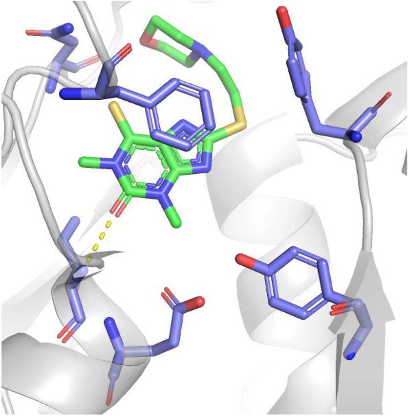
Downloaded from genesdev.cshlp.org on May 20, 2020 - Published by Cold Spring Harbor Laboratory Press

Slade

since 2009 have demonstrated PARP inhibitor efficacy in            pharmacodynamic properties (Menear et al. 2008; Fong
BRCA mutated ovarian and breast cancer, but also pros-             et al. 2009). All clinically relevant PARP1/2 inhibitors
tate, pancreatic cancer, and small cell lung carcinoma             have high catalytic activity with IC50 in the low nanomo-
(SCLC), irrespective of the BRCA status (Weaver and                lar range and inhibit PARP1 and PARP2 with similar effi-
Yang 2013; Sonnenblick et al. 2015; Mirza et al. 2018;             ciency (Fig. 1; Menear et al. 2008; Jones et al. 2009; Shen
Franzese et al. 2019; Keung et al. 2019; Mateo et al.              et al. 2013, 2015; Wang et al. 2016a).
2019; Pant et al. 2019; Pilie et al. 2019a). Inhibitors of            Despite improved selectivity, many PARP1/2 inhibitors
poly(ADP-ribose) glycohydrolase (PARG) joined the stage            are not highly selective over other family members (Wahl-
once structures of the PARG catalytic site became avail-           berg et al. 2012; Papeo et al. 2014; Thorsell et al. 2017).
able (Slade et al. 2011; Dunstan et al. 2012; Kim et al.           Among clinically relevant inhibitors, veliparib is the
2012; Barkauskaite et al. 2013). Rather than synergizing           most selective PARP1/2 inhibitor, followed by niraparib
with deficiencies in DNA repair pathways, PARG inhibi-             (Thorsell et al. 2017). Their selectivity is based on forma-
tors seem to exploit deficiencies in replication machinery         tion of a PARP1/2-unique water-mediated hydrogen-
and higher levels of replication stress in cancer cells (Pil-      bond interaction with a regulatory subdomain residue
lay et al. 2019).                                                  (D766 in PARP1), which is conserved in PARP1/2 but
   In general, cancers with high levels of replication stress      not in other PARPs (Fig. 1). Compared with veliparib,
and genomic instability due to DNA repair deficiency               which exhibits >100-fold higher selectivity for PARP1/2
and/or oncogene-induced increase in replication origin fir-        compared with other family members, olaparib and talazo-
ing are particularly responsive to PARP and PARG inhibi-           parib show only 15-fold to 20-fold higher selectivity (Thor-
tion. PARP and PARG inhibitors exploit and exacerbate              sell et al. 2017). Rucaparib is the least selective clinical
these tumor vulnerabilities by inducing further DNA                PARP1 inhibitor, which inhibits different PARPs
damage, preventing DNA repair and amassing unresolved              (PARP1, PARP2, PARP5A, and PARP5B) as well as mono
replication intermediates that instigate replication and           (ADP-ribosyl) transferases PARP3, PARP4, PARP10,
mitotic catastrophe.                                               PARP15, and PARP16 (Thomas et al. 2007; Wahlberg
                                                                   et al. 2012). Moreover, some PARP inhibitors such as ruca-
                                                                   parib and niraparib also inhibit non-PARP targets, albeit
Molecular mechanisms of PARP and PARG inhibitors                   with lower efficiency; rucaparib inhibits hexose-6-phos-
                                                                   phate dehydrogenase (H6PD), while niraparib inhibits
PARPs synthesize poly(ADP-ribose) (PAR) from NAD, re-              deoxycytidine kinase (DCK) (Knezevic et al. 2016). Such
leasing nicotinamide as the reaction product (Okayama              cross-inhibition may potentiate cancer cell death, as in
et al. 1977). PARP1, as the major producer of cellular             the case of rucaparib and PARP/H6PD inhibition, but
PAR, is activated by binding DNA lesions (Benjamin and             may also be detrimental for combination therapy with nir-
Gill 1980a,b). Catalytic activation of PARP1 is a multistep        aparib and nucleoside analogs such as gemcitabine due to
process of binding to DNA through N-terminal zinc fin-             cross-inhibition of DCK required for their activation (Kne-
gers (ZnF), unfolding of the helical domain (HD), binding          zevic et al. 2016).
of NAD to the catalytic pocket, and PAR catalysis (Lange-             In addition to inhibiting PARP catalytic activity, PARP
lier et al. 2012; Eustermann et al. 2015). The first PARP1         inhibitors also trap PARP1 and PARP2 on DNA (Murai
inhibitor was nicotinamide itself (Clark et al. 1971), fol-        et al. 2012, 2014a). PARP1 is the dominant target for
lowed by 3-aminobenzamide (3-AB) (Purnell and Whish                DNA trapping by PARP inhibitors, as depletion of
1980). All subsequently developed PARP1 inhibitors con-            PARP1—but not PARP2—reduces sensitivity to PARP in-
tain nicotinamide/benzamide pharmacophores and com-                hibitors (Murai et al. 2012). PARP1 being the relevant tar-
pete with NAD for the catalytic pocket of PARPs (Fig. 1;           get is consonant with its high nuclear abundance and its
Ferraris 2010; Steffen et al. 2013). PARP1 inhibitors dock         requirement for synthetic lethality with HR deficiency
into the catalytic site by forming hydrogen bonds with             (Amé et al. 1999; Ronson et al. 2018; Murai and Pommier
Gly, Ser, and Glu as well as hydrophobic stacking interac-         2019). Entrapment of PARP1 on DNA can be determined
tions with two Tyr residues within the nicotinamide-bind-          based on the shift in distribution of PARP1 from nuclear-
ing pocket (Fig. 1; Ferraris 2010). Given the high degree of       soluble to chromatin-bound fraction (Murai et al. 2012).
conservation of the catalytic pocket among different               PARP entrapment can occur on DNA-strand breaks as
PARPs, additional interactions are required for selective          well as topoisomerase I (TOP1)-processed ribonucleotides
inhibition (Steffen et al. 2013). A screen for more potent         and unligated Okazaki-fragment intermediates of DNA
and selective inhibitors identified different scaffolds            replication (Fig. 2; Strom et al. 2011; Hanzlikova et al.
from which new-generation PARP1 inhibitors evolved;                2018; Zimmermann et al. 2018). Once trapped, PARP1
phthalazinone and tetrahydropyridophthalazinone served             cannot dissociate from DNA due to inhibition of its cata-
as a scaffold for olaparib and talazoparib, benzimidazole          lytic activity, which is required for repulsion between
and indazole carboxamide for veliparib and niraparib, tri-         auto-PARylated PARP1 and DNA (Pommier et al. 2016).
cyclicindole lactam for rucaparib (Banasik et al. 1992;            Catalytic inhibition of PARP1 auto-PARylation is thus a
White et al. 2000; Canan Koch et al. 2002). Olaparib was           prerequisite for PARP1-DNA trapping (Hopkins et al.
the first PARP inhibitor that entered clinical trials due to       2015).
its selectivity for inhibiting PARP1/2 as well as its poten-          The catalytic inhibitory effects of the clinically relevant
cy, oral availability, and favorable pharmacokinetic and           PARP inhibitors olaparib, rucaparib, niraparib, and

2       GENES & DEVELOPMENT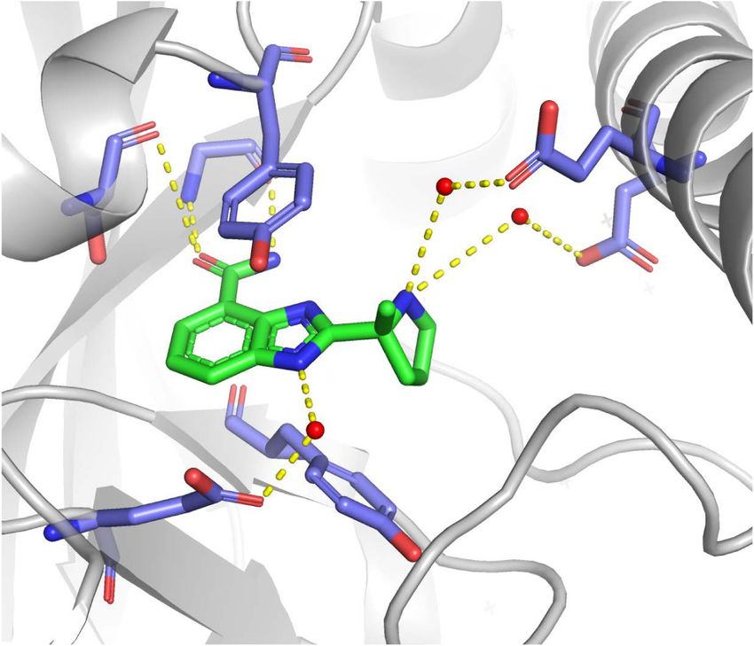
Downloaded from genesdev.cshlp.org on May 20, 2020 - Published by Cold Spring Harbor Laboratory Press

                                                                                                      PARP and PARG inhibitors in cancer

                A

                                                                                    B

                 C

                                                                                    D

Figure 1. Structures of PARP and PARG inhibitors. (A,C ) Chemical structures. IC50 denotes half-maximal inhibitory concentration
based on measurements of PARP/PARG activity in vitro. Cellular PAR synthesis EC50 denotes half-maximal effective concentration de-
termined by measuring PAR levels in cellular extracts treated with inhibitors. Cytotoxicity CC50 denotes half-maximal cytotoxic concen-
tration determined by measuring cell viability after PARP/PARG inhibitor treatment. (B,D) X-ray structures. (B) Veliparib bound to
PARP1 active site (PDB: 2RD6). (D) JA2131 bound to PARG active site (PDB: 6OA3). Inhibitors are labeled in green, PARP1/PARG residues
in the binding pocket are labeled in blue, water molecules are shown as red dots, and hydrogen bonds are represented by yellow dashes.

talazoparib are comparable; however, their potency in                 parib) and has the most rigid structure (Murai et al.
trapping PARP–DNA complexes varies considerably,                      2014a). Veliparib is one of the weakest PARP1/2 inhibitors
which is why PARP1 trapping was proposed to rely on al-               with low PARP trapping efficiency (Murai et al. 2012).
losteric changes in the PARP1 DNA-binding domain in-                    PARG hydrolyzes ribose–ribose bonds within PAR with
duced by the PARP inhibitor binding to the D-loop at                  high specific activity and processivity, particularly after
the outer border of the NAD site (Murai et al. 2012,                  DNA damage (Wielckens et al. 1982; Hatakeyama et al.
2014a). Talazoparib exhibits the highest trapping efficien-           1986; Alvarez-Gonzalez and Althaus 1989). PARG has a
cy (talazoparib >> niraparib > olaparib = rucaparib >> veli-          macro domain that binds ADP-ribose moiety and a

                                                                                                       GENES & DEVELOPMENT            3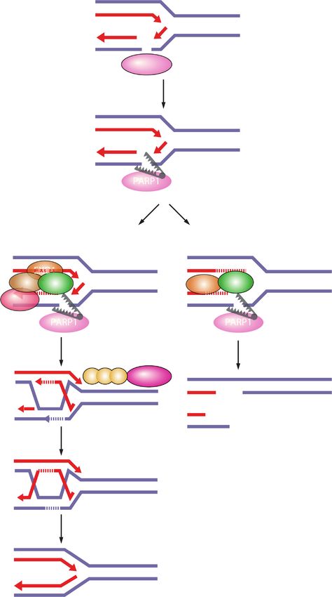
Downloaded from genesdev.cshlp.org on May 20, 2020 - Published by Cold Spring Harbor Laboratory Press

Slade

Figure 2. DNA lesions recognized by PARP1 as potential PARP-trapping sites. Unligated Okazaki fragments are DNA replication inter-
mediates. Single-strand DNA breaks (SSBs) are a frequent form of endogenous DNA damage and are particularly hazardous for replication
forks. Ribonucleotides incorporated into DNA need to be removed by RNase H2-mediated ribonucleotide excision repair. In RNase H2-
deficient cells these ribonucleotides are removed by topoisomerase I (TOP1)-mediated excision. TOP1 cleavage results in nicks, covalent
TOP1–DNA adducts, and single-strand DNA gaps that can engage PARP1.

PARG-specific loop with conserved glutamates that                     the integrity of replication forks under conditions that in-
cleave ribose–ribose bonds in an exoglycohydrolase                    duce replication stress (Hanzlikova and Caldecott 2019).
mode (Slade et al. 2011; Dunstan et al. 2012; Kim et al.              PARP1 or PARG depletion or inhibition exert the most
2012; Barkauskaite et al. 2013). The first PARG inhibitors            profound effects on SSB repair and replication fork
gallotannin and GPI-16552 showed low activity in vitro                stability.
and off-target effects in cells (Falsig et al. 2004; Erdèlyi             In SSB repair, PARP1 activity is important for the re-
et al. 2005). ADP-HPD and rhodanine-based PARG                        cruitment of the scaffold protein XRCC1 to the sites of
inhibitors (RBPIs) are potent and specific inhibitors, but            DNA damage, while PARG regulates XRCC1 dissociation
lack cell permeability (Slama et al. 1995; Finch et al.               (Fig. 3; El-Khamisy et al. 2003; Okano et al. 2003; Fisher
2012). The quinazolinedione-type PARG inhibitor                       et al. 2007; Chen and Yu 2019). PARP1/XRCC1-depen-
PDD00017273 inhibits PARG selectively and with high                   dent SSB repair was implicated as an alternative pathway
efficiency, is cell-permeable and cell-active, but has limit-         of Okazaki fragment processing (Fig. 2; Hanzlikova et al.
ed bioavailability, which makes it unsuitable for clinical            2018).
application (James et al. 2016). The naphthalen-type                     PARP1 interacts with DNA replication machinery and
PARG inhibitor COH34 is a potent, specific, and cell-per-             is active during S phase and in response to replication
meable inhibitor with a terminal half-life of 3.9 h, and              stress (Jump et al. 1979; Anachkova et al. 1989; Dantzer
may thus prove a good candidate for clinical studies                  et al. 1998; Simbulan-Rosenthal et al. 1998; Bryant et al.
(Chen and Yu 2019). Chemical library screening identified             2009). Replication stress leads to uncoupling between
thioxanthine/methylxanthine derivatives JA2–4 and                     DNA polymerase and helicase activities, which generates
JA2131 as potent, specific, cell-permeable, and cell-active           single-stranded DNA (ssDNA). RPA binds ssDNA and re-
PARG inhibitors, which are also likely to show good bio-              cruits the S/G2 checkpoint kinase ATR. ATR suppresses
availability given their structural similarity with caffeine          silent origin firing and activates the checkpoint kinase
(Houl et al. 2019). PARG inhibitors compete with PAR for              CHK1 to induce cell cycle arrest (Zeman and Cimprich
the PARG active site by occupying the subsite normally                2014). By preventing unscheduled origin firing, replication
occupied by the adenine moiety of ADP-ribose (Fig. 1;                 checkpoints prevent accumulation of ssDNA and exhaus-
James et al. 2016; Chen and Yu 2019; Houl et al. 2019).               tion of RPA, and thereby safeguard against fork breakage
                                                                      (Toledo et al. 2013). Replication stress generates SSBs
                                                                      and exposes unligated Okazaki fragments as DNA sub-
Cellular mechanisms of PARP and PARG inhibitors                       strates for PARP1 binding (Hanzlikova and Caldecott
                                                                      2019). In response to replication stress, PARP1 slows
Functions of PARP1 and PARG in DNA repair
                                                                      down replication forks to promote fork reversal by antago-
and replication fork protection
                                                                      nizing the RECQ1 helicase (Yang et al. 2004; Sugimura
Nuclear functions of PARP1 and PARG in DNA repair,                    et al. 2008; Bryant et al. 2009; Ray Chaudhuri et al. 2012;
replication fork protection, and transcription regulation             Berti et al. 2013), protects replication forks from degrada-
are critical for understanding the mechanism of action                tion by the MRE11 nuclease (Ying et al. 2012), stabilizes
of PARP inhibitors. PARP1 is involved in different path-              RAD51 nucleofilaments at stalled forks together with
ways of DNA repair, including single-strand DNA break                 PARP2 (Ronson et al. 2018), and activates the S-phase
(SSB) repair, nucleotide excision repair (NER), alternative           checkpoint kinase CHK1 (Fig. 3; Min et al. 2013). PARG lo-
nonhomologous end-joining (alt-NHEJ), and homologous                  calizes at replication forks by binding PCNA and promotes
recombination (HR) (Ray Chaudhuri and Nussenzweig                     recovery from prolonged replication stress (Mortusewicz
2017). PARP1 and PARG are also critical for preserving                et al. 2011; Illuzzi et al. 2014; Kaufmann et al. 2017).

4       GENES & DEVELOPMENT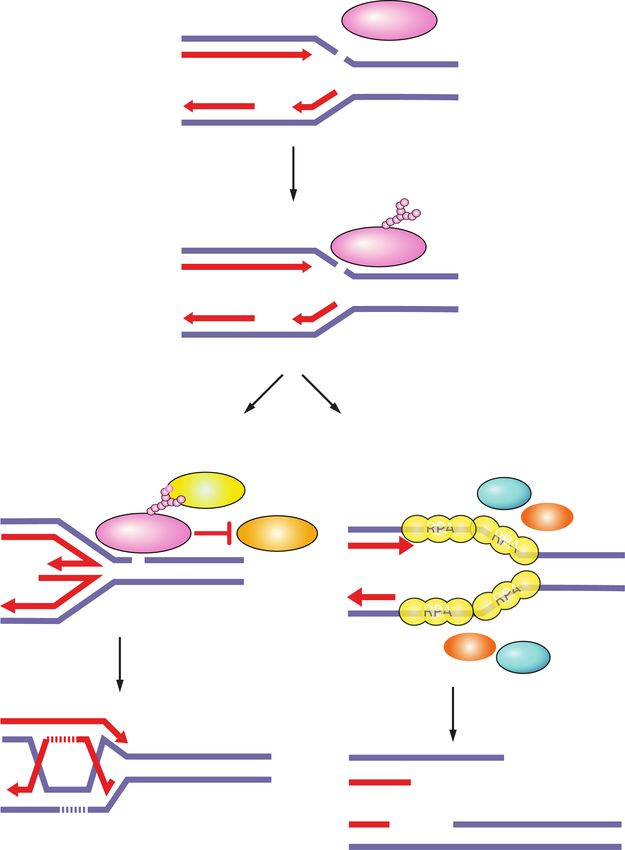
Downloaded from genesdev.cshlp.org on May 20, 2020 - Published by Cold Spring Harbor Laboratory Press
PARP and PARG inhibitors in cancer
SINGLE-STRAND BREAK REPAIR REPLICATION FORK STABILITY
Fork reversal Homologous recombination-mediated fork restart
XRCC1
3'
5' PARP1 3' Polε SSB
5'
3'
End processing Polδ 3'
ZRANB3 SSB DSB
APE1 TDP1 FANCM SMARCAL1 Limited resection
PNKP APTX Fork remodelers HLTF RAD54
5' 3'
Polβ XRCC1 5'
Fork reversal PARP1 MRE11 3'
Gap filling and SSB repair
ligation CHK1
3' EXO1 CtIP
LIG3 PARP1 RECQ1 Strand invasion
5'
BRCA1
Polβ XRCC1 5'
3' PARP1
RAD51 BRCA2
Fork protection 3'
3'
MRE11 3'
RAD51
3'
PARP1 Second end capture
BRCA2
BRCA1
3'
Fork restart
3'
3' RECQ1 HJ resolution or dissolution
Replication restart
3'
3'
3'
Figure 3. Single-strand break (SSB) repair and replication fork protection by PARP1. PARP1 acts as a sensor of SSBs and recruits XRCC1.
XRCC1 is a scaffold for the recruitment of proteins that process damaged termini, DNA polymerase β that fills the gap, and DNA ligase III
that seals the nick. PARP1 rescues damaged replication forks through fork reversal or homologous recombination (HR). SSBs on the lead-
ing strand trigger fork reversal by fork remodeling proteins. PARP1 promotes fork reversal by inhibiting the RECQ1 helicase involved in
fork restart. PARP1 stabilizes RAD51 filaments on reversed forks and together with BRCA1 and BRCA2 protects forks from degradation
by the MRE11 nuclease. If forks collapse when encountering an SSB on the lagging strand, PARP1 promotes HR-mediated fork repair and
restart by recruiting MRE11, EXO1, and BRCA1-CtIP for end resection, and BRCA2 for RAD51 filament formation.
PARP1 also regulates replication and DNA repair at the hibitors given that some of them exhibit weaker target
transcription level by stimulating activity of the transcrip- specificity.
tion factor E2F1, which regulates the expression of replica-
tion and HR genes (Simbulan-Rosenthal et al. 1998, 2003).
Synthetic lethality between PARP or PARG inhibitors
Replication forks are prone to breakage if they encoun-
and genomic instability in cancer cells
ter an SSB, which is why homologous recombination is a
critical pathway for repairing replication forks to prevent Genomic instability underlies the ability of cancer cells
fork collapse (Ait Saada et al. 2018). PARP1 contributes to to acquire different tumorigenic properties. Genomic in-
the homologous recombination pathway of DSB repair by stability entails chemical alterations in DNA known as
promoting rapid recruitment of MRE11, EXO1, BRCA1, mutations as well as changes in the chromosome number
and BRCA2 to DNA damage sites (Fig. 3; Haince et al. or structure defined as chromosomal instability. Genomic
2008; Li and Yu 2013; Zhang et al. 2015a,b). The MRE11 instability arises due to high levels of DNA damage
nuclease is responsible for the early processing of DNA le- caused by oxidative or replication stress, defects in DNA
sions, while EXO1 and BRCA1-CtIP contribute to exten- repair pathways, and/or dysfunctional surveillance mech-
sive end resection. BRCA2 is required for the loading of anisms that fail to trigger cellular senescence or apoptosis
RAD51 filaments onto ssDNA generated by end resec- (Tubbs and Nussenzweig 2017). Cancer cells experience
tion. PARP1 also counteracts nonhomologous end-joining high levels of oxidative and replication stress, resulting
(NHEJ) as the alternative pathway of DSB repair by pre- in high mutational rates (Bartkova et al. 2005; Gorgoulis
venting the binding of the NHEJ protein Ku to DNA et al. 2005; Dobbelstein and Sørensen 2015; Macheret
ends (Hochegger et al. 2006; Wang et al. 2006; Patel and Halazonetis 2015; Kotsantis et al. 2018). Activation
et al. 2011; Yang et al. 2018). of oncogenes such as MYC, RAS, and cyclin E1
In addition to PARP1 and PARG, other members of the (CCNE1) induces replication stress by promoting prema-
PARP family have also been implicated in DSB repair and ture entry into S phase, increasing replication origin fir-
replication fork stability, most notably PARP2, PARP3, ing, changing replication fork rates, causing nucleotide
PARP10, and PARP14 (Martin-Hernandez et al. 2017), depletion, and inducing replication–transcription con-
and may contribute to cellular phenotypes of PARP1/2 in- flicts (Bester et al. 2011; Kotsantis et al. 2016; Macheret
GENES & DEVELOPMENT 5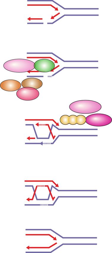
Downloaded from genesdev.cshlp.org on May 20, 2020 - Published by Cold Spring Harbor Laboratory Press

Slade

and Halazonetis 2018). Replication stress leads to accu-
mulation of replication errors and DNA lesions that com-
promise fork stability and require DNA repair pathways to
restore fork progression (Dobbelstein and Sørensen 2015;
Macheret and Halazonetis 2015; Kotsantis et al. 2018).
Many cancers have germline or somatic mutations in
DNA repair genes. Mutations in tumor suppressor genes
such as the cell cycle checkpoint gene TP53 are common
across different cancer types and allow cancer cells to es-
cape senescence or apoptosis and continue proliferating in
the presence of DNA damage (The Cancer Genome Atlas
Research Network 2011; Kandoth et al. 2013; Nik-Zainal
et al. 2016; Robinson et al. 2017; Hafner et al. 2019).
   Genotoxic agents have been used routinely in cancer
therapy in order to induce high levels of DNA damage
that render cancer cells particularly vulnerable due to
their high proliferation rates. These include ionizing radi-
ation and chemotherapeutic drugs that damage DNA by
inducing DSBs (e.g., bleomycin, doxorubicin, topoisomer-
ase inhibitors), intrastrand or interstrand DNA cross-links
(platinum compounds; e.g., cisplatin, carboplatin, and
oxaliplatin), DNA base alkylation (e.g., temozolomide),
or that interfere with DNA replication such as nucleoside
and base analogs (e.g., gemcitabine and 5-fluorouracil).
Mitotic drugs that inhibit cell division such as taxanes
(e.g., docetaxel and paclitaxel) are also used in chemother-
apy. Chemotherapy is often combined with radiotherapy.
Synergistic effects are additionally achieved in patients
with genetic deficiencies in DNA repair pathways. For ex-
ample, platinum drugs (carboplatin) improved response
rate in BRCA mutated advanced triple-negative breast
cancer (TNBC) patients and are more effective than tax-            Figure 4. Synthetic lethality between PARP inhibitors and ho-
                                                                   mologous recombination deficiency. PARP entrapment on
anes (Telli et al. 2016; Tutt et al. 2018). However, geno-
                                                                   DNA lesions blocks replication machinery and loss of PARP ac-
toxic agents also affect normal cells and have severe side         tivity prevents fork protection, fork reversal, and fork restart.
effects such as myelosuppression.                                  This results in DSBs that need to be repaired by homologous re-
   Precision medicine has revolutionized cancer therapy            combination. In the case of homologous recombination defi-
by putting forth the concept of selective targeting of cancer      ciency due to, for example, mutations in BRCA1/2, PARP1-
cells. PARP inhibitors represent a successful example of           trapping lesions elicit excessive fork degradation by the MRE11
precision medicine applied in the clinic. PARP inhibitors          nuclease, the activity of which is unrestrained in the absence of
act through synthetic lethality, whereby genetic DNA re-           BRCA1/2 and PARP1. This results in fork collapse.
pair defects are enhanced by drug-induced defects in a
compensatory pathway (Lord and Ashworth 2017). Two
seminal studies showed how PARP inhibitors specifically            CHEK1, CHEK2, FANC genes, ERCC1, POLB, FEN1,
kill HR-deficient cells mutated in BRCA1/2 (Fig. 4; Bryant         and CDK12 have shown synthetic lethality in combina-
et al. 2005; Farmer et al. 2005). Carriers of heterozygous         tion with PARP inhibitors (Bryant and Helleday 2006;
BRCA1/2 mutations are sensitive to PARP inhibitor                  McCabe et al. 2006; Murai et al. 2012; Postel-Vinay et al.
treatment as they lose the wild-type allele during tumori-         2013; Bajrami et al. 2014). Synthetic lethality between mu-
genesis and thereby become BRCA1/2-null. Since this first          tations in HR-related genes and PARP inhibition was con-
example of synthetic lethality between genetic defects and         firmed by CRISPR screens, which enable high-throughput
PARP inhibitors, it has become clear that oxidative stress         investigation of synthetic lethal interactions (Zimmer-
and genomic instability, manifested not just through mu-           mann et al. 2018).
tations in DNA repair proteins but also replication stress,           In contrast to PARP inhibitors, a clear correlation be-
sensitize cells to PARP and PARG inhibitors (Bryant et al.         tween HR deficiency and synthetic lethality with PARG
2005; Farmer et al. 2005; McCabe et al. 2006; Bunting et al.       inhibitors is lacking. Depletion of the HR proteins
2010; Lord and Ashworth 2012, 2017; McLellan et al. 2012;          BRCA1/2, PALB2, ABRAXAS, and BARD1 in MCF7 breast
Murai et al. 2012; Dréan et al. 2016; Gravells et al. 2017;        cancer cells was shown to elicit synthetic lethal interac-
Zimmermann et al. 2018; Chen and Yu 2019; Giovannini               tions with PARG depletion or PARG inhibition (with gal-
et al. 2019; Pillay et al. 2019). In addition to BRCA1/2,          lotannin or PDD00017273) (Fathers et al. 2012; Gravells
mutations in DNA damage response genes such as ATM,                et al. 2017). The PARG inhibitor COH34 efficiently kills
PRKDC, ATR, RPA1, DSS1, NBN, RAD51, RAD54,                         BRCA mutated or olaparib-resistant ovarian and breast

6       GENES & DEVELOPMENT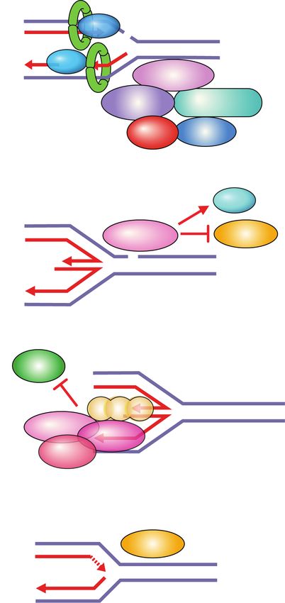
Downloaded from genesdev.cshlp.org on May 20, 2020 - Published by Cold Spring Harbor Laboratory Press

                                                                                                       PARP and PARG inhibitors in cancer

cancer cells (Chen and Yu 2019). However, PARG deple-                   2018). In PARP1-depleted or inhibited cells, DSBs arise
tion did not show synthetic lethality with BRCA1 muta-                  due to deprotection of stalled replication forks and their
tions in different cancer cell lines (Noll et al. 2016), the            degradation by the MRE11 nuclease, due to impaired
PARG inhibitor JA2131 efficiently killed BRCA-proficient                fork reversal, or from unligated Okazaki fragments en-
cancer cells (Houl et al. 2019), and only one out of six tested         countered by replication forks (Lonn and Lonn 1985;
ovarian cancer cells with BRCA1/2 mutations showed                      Ray Chaudhuri et al. 2012; Ying et al. 2012; Hanzlikova
sensitivity to PARG inhibition with PDD00017273 (Pillay                 et al. 2018).
et al. 2019). Instead, synthetic lethal interactions with the              In line with the opposing catalytic activities of PARP1
PARG inhibitor PDD00017273 involve replication-associ-                  and PARG, PARG depletion or inhibition show opposite
ated genes such as TIMELESS, HUS1, and RFC2 (Fig. 5; Pil-               effects. PARG depletion or inhibition slows down replica-
lay et al. 2019).                                                       tion forks, causes accumulation of reversed forks and
                                                                        ssDNA gaps, and prevents fork restart through suppres-
                                                                        sion of the RECQ1 helicase (Fig. 6; Illuzzi et al. 2014;
Amplifying genomic instability with PARP and PARG                       Ray Chaudhuri et al. 2015; Gravells et al. 2017; Houl
inhibitors                                                              et al. 2019; Pillay et al. 2019). This causes cell cycle stall-
                                                                        ing in the S/G2 phase, accumulation of ssDNA as judged
PARP1 depletion or inhibition increases replication fork
                                                                        by RPA foci, and accumulation of DNA damage, as indi-
speed, impairs replication fork reversal, and causes un-
                                                                        cated by pan-nuclear γH2AX staining (Illuzzi et al. 2014;
timely fork restart following replication stress (Fig. 6).
                                                                        Pillay et al. 2019). Prolonged exposure of PARG-depleted
This leads to an accumulation of DNA damage in S-phase
                                                                        cells to replication stress results in the loss of RPA foci, in-
cells, S-phase stalling, and G2 delay (Sugimura et al. 2008;
                                                                        dicating RPA exhaustion due to replication stress-induced
Bryant et al. 2009; Ray Chaudhuri et al. 2012; Berti et al.
                                                                        formation of ssDNA—a concept known as replication ca-
2013; Dale Rein et al. 2015; Farrés et al. 2015; Maya-Men-
                                                                        tastrophe (Illuzzi et al. 2014; Toledo et al. 2017). PARP1 or
doza et al. 2018; Michelena et al. 2018; Ronson et al.
                                                                        PARG depletion or inhibition sensitize cells to DNA-
                                                                        damaging agents and cause increased DNA damage levels
                                                                        due to decreased HR efficiency (Rottenberg et al. 2008;
                                                                        Ame et al. 2009; Min et al. 2010; Shirai et al. 2013; Murai
                                                                        et al. 2014b; Dréan et al. 2016; James et al. 2016; Gravells
                                                                        et al. 2018; Houl et al. 2019). Collectively, loss or inhibi-
                                                                        tion of PARP1 or PARG destabilize replication forks and
                                                                        cause fork breakage, particularly under conditions of HR
                                                                        deficiency, oxidative and replication stress, or exogenous
                                                                        DNA damage.
                                                                           Impaired fork progression and accumulation of DNA
                                                                        damage are more pronounced in PARP inhibitor-treated
                                                                        cells compared with PARP-depleted cells, which suggests
                                                                        that PARP inhibition does more than just inhibit PARP1
                                                                        catalytic activity. For example, PARP inhibition increases
                                                                        fork rate and reduces fork reversal following camptothe-
                                                                        cin exposure more than PARP depletion (Sugimura et al.
                                                                        2008; Ray Chaudhuri et al. 2012; Maya-Mendoza et al.
                                                                        2018). Furthermore, PARP inhibition results in more
                                                                        strand breaks (SSBs and DSBs), as judged by γH2AX foci
                                                                        (Murai et al. 2012) and delayed SSB repair after ionizing ra-
                                                                        diation or alkylation damage (Godon et al. 2008; Strom
                                                                        et al. 2011). Consequently, PARP inhibition exhibits
                                                                        stronger synthetic lethality with HR deficiency compared
                                                                        with PARP depletion (Bryant et al. 2005; Ronson et al.
                                                                        2018).
                                                                           In fact, PARP inhibitor cytotoxicity was found to corre-
                                                                        late with the strength of PARP–DNA entrapment rather
                                                                        than a reduction in PARP1 catalytic activity (Murai
Figure 5. Synthetic lethality between PARG inhibitors and rep-          et al. 2012; Pettitt et al. 2013). A combination of the
lication stress. PARG inhibition increases PARylation levels and        chemical inhibition of PARP1 catalytic activity and the
may prevent dissociation of PARP1 and PAR-binding repair pro-
                                                                        physical obstruction caused by PARP–DNA entrapment
teins (e.g., XRCC1) from DNA damage sites. Loss of PARG activ-
ity causes fork stalling and impairs restart of reversed forks. Forks
                                                                        seems to be responsible for the greater cytotoxicity of
can presumably restart by homologous recombination. Under               PARP inhibitors compared with PARP depletion. The
replication stress conditions, increased origin firing and pro-         physical obstruction caused by PARP–DNA entrapment
longed fork stalling generates excessive ssDNA and causes RPA           exacerbates replication problems caused by loss of PARP
exhaustion. Such replication catastrophe results in fork collapse.      activity and induces mitotic phenotypes of premature

                                                                                                        GENES & DEVELOPMENT            7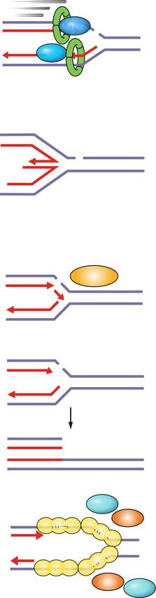
Downloaded from genesdev.cshlp.org on May 20, 2020 - Published by Cold Spring Harbor Laboratory Press

Slade

                                                                                    Figure 6. Cellular consequences of PARP and
                                                                                    PARG inhibition on replication and mitosis.
                                                                                    PARP inhibitors increase replication fork
                                                                                    rate, reduce fork reversal, and cause premature
                                                                                    fork restart. In HR-deficient cells, PARP inhib-
                                                                                    itors cause fork collapse and DSBs. PARG in-
                                                                                    hibitors reduce fork rate, increase fork
                                                                                    reversal, and impair fork restart. In the pres-
                                                                                    ence of replication stress, PARG inhibitors
                                                                                    cause replication catastrophe. Destabilization
                                                                                    of replication forks causes mitotic defects and
                                                                                    death by mitotic catastrophe. PARG inhibitors
                                                                                    cause mitotic defects in combination with
                                                                                    DNA-damaging agents.

loss of cohesion or anaphase DNA bridges, which are not            2018). A contingency pathway for DSB repair, NHEJ,
observed upon PARP depletion (Murai et al. 2012; Shen              which is functional throughout the cell cycle, is thought
et al. 2015; Kukolj et al. 2017; Schoonen et al. 2017).            to compensate for inactive HR in PARP inhibitor-treated
Therefore, the cytotoxicity of PARP inhibitors seems to            cells. NHEJ is an error-prone pathway that can lead to
arise from an accumulation of replication problems,                small mutations as well as chromatid fusions (Bunting
which are carried over into mitosis resulting in death by          et al. 2010). Fusion of two broken sister chromatids, chro-
mitotic catastrophe (Slade 2019).                                  mosomes, or telomeres during interphase can generate
   PARP and PARG inhibitors activate the S/G2 check-               dicentric chromosomes visible as radial fusions in meta-
point kinases ATR and CHK1 (Ray Chaudhuri et al.                   phase and as anaphase DNA bridges (Ganem and Pellman
2015; Colicchia et al. 2017; Kim et al. 2017; Maya-Mendo-          2012). Chromatid fusions also give rise to acentric chro-
za et al. 2018; Pillay et al. 2019), which generally halt the      mosomes, which cannot attach to the mitotic spindle
cell cycle to allow DNA repair and completion of DNA               and cannot segregate accurately during anaphase, thus ap-
replication before mitotic entry (Lecona and Fernandez-            pearing as lagging chromosomes. During telophase and
Capetillo 2018). In accordance with the activation of the          cytokinesis, acentric or lagging chromosomes obtain their
ATR checkpoint, PARG inhibitor PDD00017273-treated                 own nuclear envelope and form micronuclei (Fenech et
cells stall in the S/G2 phase without progressing into mi-         al. 2011). Chromosomes within micronuclei are prone to
tosis and assume a “fried egg” morphology (Pillay et al.           chromothripsis, whereby reduced and asynchronous
2019). Conversely, despite the activation of ATR and               DNA replication results in DNA damage and chromo-
CHK1, cells treated with PARP inhibitors progress into             some fragmentation (Crasta et al. 2012). DNA released
mitosis and exhibit different mitotic defects, which arise         from micronuclei triggers cGAS accumulation and activa-
from problems during S phase (Fig. 6; Colicchia et al.             tion of proinflammatory response (Harding et al. 2017;
2017; Kim et al. 2017; Maya-Mendoza et al. 2018).                  Mackenzie et al. 2017). Following chromothripsis, frag-
   As explained above, fork destabilization caused by              mented chromosomes can assemble randomly, resulting
PARP inhibition results in DSBs. DSBs cannot be repaired           in chromosome rearrangements (Luijten et al. 2018).
by the HR pathway in BRCA-deficient cells, which forms             Chromatid breaks, radial chromosomes, anaphase DNA
the basis for synthetic lethality approaches with PARP in-         bridges, lagging chromosomes, and micronuclei are all
hibitors. Moreover, PARP inhibition itself induces HR              common in BRCA1/2-deficient cells treated with PARP
deficiency by reducing the expression of the E2F1 target           inhibitors (Fig. 6; Bunting et al. 2010; Schoonen et al.
genes involved in DNA replication and cell cycle regula-           2017).
tion (e.g., PCNA, MCM7, and CCNA2) and HR factors                     Genomic regions called common fragile sites (CFSs) are
such as BRCA1/2 and RAD51, as shown in prostate and                particularly sensitive to impaired fork progression. CFS
small cell lung cancer (Byers et al. 2012; Schiewer et al.         are found within long genes and are prone to form

8       GENES & DEVELOPMENT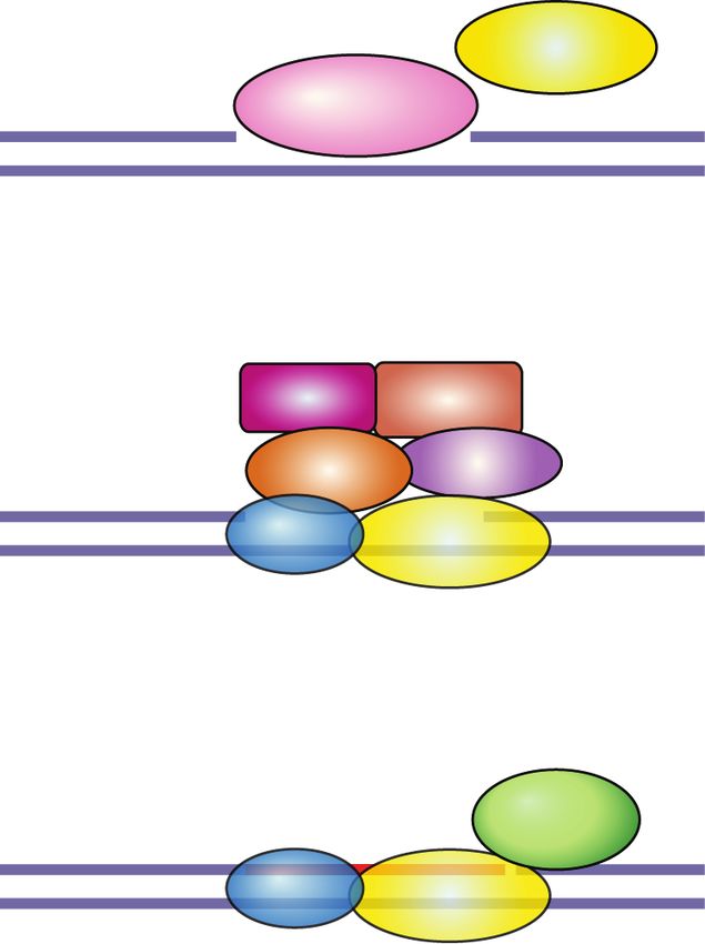
Downloaded from genesdev.cshlp.org on May 20, 2020 - Published by Cold Spring Harbor Laboratory Press

                                                                                                 PARP and PARG inhibitors in cancer

abnormal replication intermediates due to transcription–           2009; Min et al. 2010). PARG inhibition coupled with
replication conflicts (Helmrich et al. 2011). CFSs are late-       ionizing radiation also yields aberrant spindle formation
replicating and remain underreplicated at the G2/M tran-           and metaphase arrest (Fig. 6; Gravells et al. 2018).
sition (Le Beau et al. 1998). Underreplicated CFSs remain             In sum, cytotoxicity of PARP inhibitors is a multistage
connected through thin threads of DNA in mitosis known             process of the destabilization of replication forks through
as ultrafine anaphase DNA bridges (Le Beau et al. 1998).           PARP entrapment and loss of PARP activity, the genera-
Replication intermediates that remain unresolved during            tion of unresolved replication intermediates and DSBs,
mitosis are marked by 53BP1 in G1 cells (Lukas et al.              their transmission into mitosis, and the induction of mi-
2011). Replication stress induced by PARP inhibitors               totic defects (premature loss of cohesion, misalignment,
gives rise to ultrafine anaphase DNA bridges in mitotic            missegregation) that ultimately result in mitotic catastro-
cells and 53BP1-positive nuclear bodies in G1 cells (Gem-          phe. The cytostatic effects of PARG inhibitors involve
ble et al. 2015; Michelena et al. 2018). Furthermore, PARP         fork stalling, which results in replication catastrophe
inhibition during S phase causes weakening of sister chro-         and cell cycle arrest in S/G2.
matid cohesion, resulting in premature loss of cohesion
(“cohesion fatigue”) and chromosome alignment prob-
lems in metaphase (Fig. 6; Kukolj et al. 2017).                    Targeting transcription, RNA metabolism, and ribosome
   It is clear that mitotic defects in PARP-inhibitor treated      biogenesis through PAPR inhibition
cells arise from destabilization of replication forks and
                                                                   In addition to DNA repair and replication fork stability,
DNA damage acquired during S phase. Replication
                                                                   PARP1 is also implicated in gene expression regulation,
stress-induced mitotic defects result in death by mitotic
                                                                   RNA processing, and ribosome biogenesis, which may
catastrophe (Dale Rein et al. 2015; Gemble et al. 2015;
                                                                   contribute to cellular effects of PARP inhibition (Weaver
Majuelos-Melguizo et al. 2015; Colicchia et al. 2017;
                                                                   and Yang 2013; Feng et al. 2015) and give rise to synthet-
Kukolj et al. 2017; Schoonen et al. 2017; Maya-Mendoza
                                                                   ic lethal interactions with transcription and splicing fac-
et al. 2018; Michelena et al. 2018). Mitotic catastrophe
                                                                   tors identified in CRISPR screens (Zimmermann et al.
is a special type of cell death whereby cells die by apopto-
                                                                   2018). PARP1 modulates the activity of different tran-
sis or slip out of mitosis through multinucleation or mac-
                                                                   scription regulators implicated in cancer (e.g., p53, nucle-
ronucleation due to chromosome missegregation, as well
                                                                   ar receptors) or inflammation (e.g., NF-κB) (Schiewer and
as micronucleation that results from lagging or acentric
                                                                   Knudsen 2014; Bai 2015). Hence, transcriptional deregu-
chromosomes (Galluzzi et al. 2018).
                                                                   lation may sensitize cancer cells to PARP inhibitors, as
   While HR-deficient cancer cells were shown to respond
                                                                   shown for DNA-repair proficient HER2-positive breast
better to PARP inhibitors, PARP inhibitors are also effec-
                                                                   cancer cells whereby NF-κB overactivation is attenuated
tive in HR-proficient cells that experience high levels of
                                                                   through PARP inhibition (Nowsheen et al. 2012). PARP
oxidative and replication stress. Indeed, anaphase DNA
                                                                   inhibitors are also effective in Ewing’s sarcomas by
bridges, lagging chromosomes, micronuclei, ultrafine ana-
                                                                   blocking, on the one hand, PARP1-dependent transcrip-
phase DNA bridges, and premature loss of cohesion occur
                                                                   tional activation effects of ETS gene fusions such as
in HR-proficient cells exposed to PARP inhibitors (Majue-
                                                                   EWS-FLI-1, and by exacerbating DNA damage on the
los-Melguizo et al. 2015; Kukolj et al. 2017; Schoonen
                                                                   other (Brenner et al. 2012). Furthermore, PARP inhibitors
et al. 2017; Michelena et al. 2018). Moreover, PARP inhib-
                                                                   reduce rDNA transcription and ribosome biogenesis in
itor efficacy was shown to correlate with basal levels of
                                                                   BRCA1/2-proficient cancer cells by preventing DDX21
replication stress in cancer cells (Kukolj et al. 2017).
                                                                   ADP-ribosylation, and thereby reduce breast cancer
   Unlike PARP inhibitors, the PARG inhibitor
                                                                   growth (Kim et al. 2019).
PDD00017273 exhibits cytostatic rather than cytotoxic
effects by causing a replication catastrophe that is not
transferred into mitosis but remains contained in inter-
                                                                   Determinants of PARP inhibitor sensitivity in cancer
phase (Pillay et al. 2019). PARP trapping is most likely
                                                                   cells
the reason why PARP inhibitors lead to mitotic catastro-
phe and are more potent in killing cells compared with             Since the first example of synthetic lethality between
PARG inhibitors. However, the combination of PARG                  PARP inhibitors and BRCA1/2 mutations, it has become
inhibitors with cell cycle checkpoint inhibitors such as           clear that any form of HR deficiency in tumors that phe-
CHK1 or combination of PARG inhibition/depletion                   nocopies BRCA1/2 mutations, often referred to as BRCA-
with DNA-damaging agents allows cells to progress                  ness, may sensitize cells to PARP inhibitors (Lord and
into mitosis where they experience various mitotic ab-             Ashworth 2016). Different patient biomarkers have been
normalities (Koh et al. 2004; Ame et al. 2009; Min et al.          used to assess HR deficiency as a measure of sensitivity
2010; Gravells et al. 2018; Pillay et al. 2019; Slade              to PARP inhibitor treatment, such as mutations in DNA
2019). For example, PARG-depleted cells or PARG hypo-              repair genes, their expression levels, as well as mutational
morphic cells lacking nuclear and cytoplasmic PARG                 and genomic signatures of HR deficiency. Replication
isoforms show centrosome amplification, centrosome                 stress markers and transcriptome profiles are complemen-
fragmentation, multipolar spindles, chromosome mis-                tary means of evaluating PARP inhibitor response given
alignment, and missegregation, which are more pro-                 the importance of PARP1 for replication fork stability
nounced after exposure to ionizing radiation (Ame et al.           and gene expression regulation.

                                                                                                  GENES & DEVELOPMENT            9
Downloaded from genesdev.cshlp.org on May 20, 2020 - Published by Cold Spring Harbor Laboratory Press

Slade

Homologous recombination (HR) deficiency Germline                  HR pathway (see “Mechanisms of Resistance to PARP In-
or somatic mutations in DNA repair genes as well as their          hibitors”; Mateo et al. 2019).
transcriptional down-regulation are frequently used as                Genomic signatures of HR deficiency comprise loss
biomarkers of PARP inhibitor response. Ten percent to              of heterozygosity (LOH), telomeric allelic imbalances
15% of breast and ovarian cancer patients carry germline           (TAIs), and large-scale state transitions (LSTs). LOH re-
mutations in HR genes BRCA1 and BRCA2 (Gallagher                   sults in irreversible loss of one of the parental alleles in re-
et al. 2011; Mavaddat et al. 2012; Nik-Zainal et al. 2016).        gions >15 Mb (Abkevich et al. 2012). TAI refers to unequal
Strikingly, 75% of germline mutations in metastatic can-           contribution of maternal and paternal telomeric DNA se-
cers affect DNA repair genes such as MUTYH, BRCA2,                 quences (Birkbak et al. 2012). LSTs are defined as chromo-
CHEK2, and BRCA1 (Robinson et al. 2017). Moreover,                 somal breaks between adjacent regions of at least 10 Mb
BRCA1/2, ATM, and CHEK2 are the most frequently mu-                (Popova et al. 2012). LOH, TAIs, and LSTs were shown
tated DNA repair genes in somatic cancer cells (Heeke              to correlate well with mutations in HR genes BRCA1/2
et al. 2018). In addition to being directly inactivated by         in breast and ovarian cancer (Abkevich et al. 2012; Birk-
mutation, BRCA1 and RAD51C were also found to be                   bak et al. 2012; Popova et al. 2012). All these genomic sig-
down-regulated through promoter hypermethylation in                natures of HR deficiency have been combined in a
breast and ovarian cancer (Chiang et al. 2006; The Cancer          “homologous recombination deficiency” (HRD) score as
Genome Atlas Research Network 2011; Lips et al. 2013;              a measure of genomic instability (Timms et al. 2014; Telli
Timms et al. 2014; Polak et al. 2017; Bernards et al.              et al. 2016). HRDetect and HRD scores are both used to
2018; Castroviejo-Bermejo et al. 2018; Kondrashova et al.          predict PARP inhibitor sensitivity in clinical settings.
2018). BRCA1 promoter methylation confers the same de-                Examining transcriptional signatures (or RNA) of can-
gree of sensitivity to PARP inhibitors as BRCA1 muta-              cer cells rather than their mutational signatures (or
tions (Veeck et al. 2010). Furthermore, the expression             DNA) emerged as another means of predicting PARP in-
level of BRCA1 was shown to be reduced due to depletion            hibitor sensitivity. Gene expression profile derived from
of the CDK12 kinase, which sensitizes breast and ovarian           BRCA mutated ovarian cancers, termed “the BRCAness
cancer cells to PARP inhibition (Bajrami et al. 2014).             profile,” was found to correlate with platinum and
CDK12 regulates transcription of HR genes by suppressing           PARP inhibitor sensitivity and was efficient in predicting
intronic polyadenylation (Dubbury et al. 2018). CDK12 is           platinum sensitivity of non-BRCA mutated ovarian can-
often mutated in ovarian and prostate cancer and CDK12             cers (Konstantinopoulos et al. 2010). Furthermore, gene
deficiency may thus prove useful as a biomarker of                 expression profiles from cell lines depleted in different
PARP inhibitor response (The Cancer Genome Atlas Re-               HR proteins revealed an HRD transcriptome signature
search Network 2011; Bajrami et al. 2014; Wu et al.                that can predict HR deficiency and PARP inhibitor sensi-
2018). A recent CRISPR screen identified TP53-induced              tivity (Peng et al. 2014).
glycolysis and apoptosis regulator (TIGAR) as another                 Last, the cytological signature of HR deficiency is given
modulator of expression of HR genes (Fang et al. 2019a).           by the number of RAD51 foci. Reduced RAD51 foci for-
TIGAR is amplified in different cancer types and its               mation indicates HR deficiency and correlates with
down-regulation sensitizes cancer cells to PARP inhibi-            PARP inhibitor sensitivity, as shown in BRCA mutated
tors through inhibition of the pentose phosphate                   breast tumor samples 2 h after 5 Gy of IR (Naipal et al.
pathway, increase in ROS and DNA damage, down-regula-              2014), ovarian cancer cell lines 8 h after 4 Gy of IR (Shah
tion of BRCA1/2 and RAD51, and induction of cellular               et al. 2014), and breast cancer patient-derived xenografts
senescence (Fang et al. 2019a).                                    (PDXs) (Castroviejo-Bermejo et al. 2018; Cruz et al.
   HR deficiency can also be scored based on different mu-         2018). Unlike RAD51, γH2AX foci are not a reliable pre-
tational and genomic signatures. A mutational signature            dictor of sensitivity to PARP inhibition as γH2AX foci
of HR deficiency in BRCA mutated breast, ovarian, and              may correlate positively or negatively with HR deficiency
pancreatic cancers are large indels (3- to 50-bp insertions        and PARP inhibitor sensitivity (Fong et al. 2009; Mukho-
and deletions) with overlapping microhomology at break-            padhyay et al. 2010; Dale Rein et al. 2015; Michelena et al.
point junctions that result from NHEJ as the alternative           2018).
pathway of DSB repair (Alexandrov et al. 2013). NHEJ
joins two broken DNA ends, which may lead to small in-             Replication stress High levels of replication stress and
sertions or deletions (indels) (Chang et al. 2017). In addi-       depletion of replication-associated genes may render can-
tion to microhomology-mediated indels as the main                  cer cells sensitive to PARP inhibitors even in the absence
signature of BRCA1/2 deficiency, base substitutions and            of HR deficiency. For example, loss of TP53 and RB1 cou-
rearrangements also reflect an abrogation of DSB repair            pled with amplification of MYC generate replication
pathways (Nik-Zainal et al. 2016). Mutational signatures           stress and sensitize small cell lung cancer cells (SCLC)
identified from whole-genome sequencing of breast, ovar-           to PARP inhibitors (George et al. 2015; Sen et al. 2018).
ian, and pancreatic cancers were used to generate an               Schlafen 11 (SLFN11) is a recently identified biomarker
HRDetect tool that can predict HR deficiencies (Davies             of PARP inhibitor response in SCLC. SLFN11 binds to
et al. 2017). However, mutational signatures are not prog-         RPA in response to replication stress and blocks replica-
nostic of PARP inhibitor sensitivity in the case of tumors         tion fork progression by changing chromatin structure
with restored HR, which harbor mutational signatures but           (Murai et al. 2018). SLFN11 overexpression correlates
are resistant to PARP inhibitors due to restoration of the         with PARP inhibitor sensitivity, as shown in SCLC

10      GENES & DEVELOPMENT
Downloaded from genesdev.cshlp.org on May 20, 2020 - Published by Cold Spring Harbor Laboratory Press

                                                                                                 PARP and PARG inhibitors in cancer

PDXs treated with olaparib or talazoparib, as well as in           be used as a biomarker of PARP inhibitor sensitivity
SCLC patients treated with veliparib in combination                (Bian et al. 2019).
with temozolomide (Allison Stewart et al. 2017; Lok
et al. 2017).
   Overexpression of the cytidine deaminase APOBEC3,               Clinical studies with PARP inhibitors
which causes an increase in abasic sites at replication
forks, is frequently encountered in cancer and was shown           The very first clinical trials demonstrated the efficacy of
to sensitize cells to PARP inhibitors (Burns et al. 2013;          the PARP inhibitor olaparib in breast and ovarian cancer
Roberts et al. 2013; Nikkilä et al. 2017). Mutations in            patients carrying germline mutations in BRCA1/2, thus
genes required for Okazaki fragment processing such as             supporting the rationale for synthetic lethality (Fong
FEN1 also sensitize cells to PARP inhibition, but FEN1             et al. 2009; Audeh et al. 2010; Tutt et al. 2010). The phase
is rarely mutated in cancer (Murai et al. 2012). Depletion         1 trial showed the antitumor activity of olaparib at 400 mg
of replication-associated proteins such as cohesin (SMC1,          twice daily and acceptable adverse effects (nausea, fatigue,
SMC3, and RAD21), cohesin-associated factors (ESCO1                vomiting, taste alteration, anorexia) (Table 1; Fong et al.
and ESCO2), core replication machinery (MCM2/3/6 hel-              2009). The PARP inhibitor niraparib showed antitu-
icases), and topoisomerases (TOP2B and TOP3A) increas-             mor activity at 300 mg daily with more pronounced
es sensitivity to PARP inhibition, as shown in colon and           hematologic adverse effects compared with olaparib (ane-
breast cancer cell lines (McLellan et al. 2012; Bajrami            mia, thrombocytopenia, and neutropenia) (Sandhu et al.
et al. 2014).                                                      2013). Rucaparib showed partial or complete response in
   A recent CRISPR screen revealed synthetic lethality be-         ovarian, breast, and pancreatic cancer patients given 600
tween mutations in RNASEH2 and PARP inhibition due                 mg twice daily, with fatigue, nausea, anemia, and vomit-
to increased levels of replication-dependent DNA damage            ing as the most common adverse effects (Kristeleit et al.
(Zimmermann et al. 2018). RNase H2 deficiency results in           2017). Talazoparib administered at the recommended
impaired ribonucleotide excision repair and accumulation           dose of 1 mg/d demonstrated high antitumor activity in
of ribonucleotides that are cleaved by TOP1 (Zimmer-               BRCA mutated breast and ovarian cancer patients with fa-
mann et al. 2018). Cleavage of these ribonucleotides pro-          tigue, anemia, and thrombocytopenia as the most pro-
duces nicks, covalent TOP1–DNA adducts, and ssDNA                  nounced adverse effects (de Bono et al. 2017). Clinically
gaps that can act as PARP-trapping lesions, thus contrib-          recommended doses and the severity of side effects corre-
uting to PARP inhibitor efficacy (Zimmermann et al.                late with the PARP inhibitor trapping potency; talazo-
2018). RNASEH2B deletions are frequently found in                  parib as the strongest PARP trapper has the lowest
chronic lymphocytic leukemia and metastatic prostate               recommended dose and shows the highest occurrence of
cancer, which renders them more sensitive to PARP inhi-            anemia (de Bono et al. 2017; Litton et al. 2018; Pilié
bition (Zimmermann et al. 2018).                                   et al. 2019a).
                                                                      Olaparib treatment in advanced breast or ovarian can-
Transcriptome profiles, PARP1 expression levels, and               cer patients with BRCA1/2 germline mutations showed
PARP1 activity Given the important roles of PARP1 in               a 41% and a 33% objective response rate defined as the
transcription regulation (Kraus and Hottiger 2013), gene           proportion of patients with tumor size reduction of a pre-
expression profiles from ovarian and breast cancer cell            defined amount and for a minimum time period (Table 2;
lines with known sensitivity to olaparib and rucaparib             Audeh et al. 2010; Tutt et al. 2010). Olaparib administered
were used to derive a transcriptional algorithm that can           in high-grade serous and/or undifferentiated ovarian can-
predict sensitivity to PARP inhibitors (McGrail et al.             cer patients showed a 41% and 24% objective response
2017). The expression levels of PARP1 itself may also              rate with or without BRCA1/2 mutations; however, there
determine PARP inhibitor response. PARP1 expression                was no response in TNBC patients (Gelmon et al. 2011). A
is increased in different cancer types, particularly at ad-        study comparing olaparib with placebo in platinum-sensi-
vanced stages (Ossovskaya et al. 2010; Domagala et                 tive, relapsed, high-grade serous ovarian cancer patients
al. 2011; Mascolo et al. 2012; Bi et al. 2013; Bieche et al.       who had received two or more platinum-based regimens
2013; Gan et al. 2013; Salemi et al. 2013; Dziaman et al.          showed longer median progression-free survival from 4.3
2014; Park et al. 2015; Zhai et al. 2015; Li et al. 2016;          to 11.2 mo for BRCA mutated cancer and from 5.5 to 7.4
Hou et al. 2018), and in some cases correlates positively          mo for wild-type (WT) BRCA (Ledermann et al. 2012,
with the cytotoxic effects of PARP inhibition (Byers               2014). However, the overall survival of olaparib-treated
et al. 2012; Kukolj et al. 2017).                                  versus placebo patients was not significantly different af-
   PARP1 catalytic activity is enhanced by the receptor ty-        ter 5 yr (Ledermann et al. 2016). Long-term responders to
rosine kinase c-Met-mediated phosphorylation on Y907,              olaparib with progression-free survival >2 yr had a preva-
which in turn reduces PARP inhibitor binding. Blocking             lence of BRCA2 mutations and a high HRD score, con-
PARP1 phosphorylation with a c-Met inhibitor can in-               firming that mutations in HR genes such as BRCA2 and
crease the efficacy of PARP inhibitors (Du et al. 2016). En-       HRD score can be used as predictive biomarkers for
dogenous inhibition of PARP activity through increased             PARP inhibitor response (Lheureux et al. 2017b). A study
levels of NADP+ was shown to render ovarian cancer cells           with platinum-resistant ovarian and breast cancer pa-
hypersensitive to PARP inhibitors irrespective of the              tients with ≥ three chemotherapy regimens for metastatic
BRCA status, suggesting that NADP+ levels could also               disease, all carrying BRCA1/2 mutations, showed a 31.1%

                                                                                                 GENES & DEVELOPMENT            11
Downloaded from genesdev.cshlp.org on May 20, 2020 - Published by Cold Spring Harbor Laboratory Press
Slade
Table 1. Phase 1 clinical trials with PARP inhibitors
PARP inhibitor
Olaparib N = 60
Dose
90% at doses >60 mg twice daily
Response
Partial or complete radiologic responsea 15%; 47%
Radiologically stable diseasea 12%; 11%
Tumor marker responsea 12%; 37%
Radiologic or tumor marker responsea 17%; 53%
Reference Fong et al. 2009
Niraparib N = 100
Dose
a) 10 escalating doses from 30 to 400 mg in 21-d cycles N = 60
b) 300 mg/d N = 40
Cancer type
a) Advanced solid tumors N = 60
b) Sporadic platinum-resistant high-grade serous ovarian cancer N = 22
b) Sporadic prostate cancer N = 18
Germline mutations
BRCA1/2 N = 29
Pharmacokinetic and pharmacodynamic parameters
Peak plasma concentration 3–4 h after dosing cmax ∼3.1 µM for 300 mg/mL niraparib
Terminal elimination half-life 32.8–46.0 h
PARP inhibition >50% at doses >80 mg/d
Response
Partial response for BRCA1/2 mutation carriersb 50%; 33%
Partial response for noncarriers of BRCA1/2 mutationb 33%; 5%
Reference Sandhu et al. 2013
Rucaparib N = 56
Dose
40/80/160/300/500 mg/d in 21-d cycle N = 26
240/360/480/600/840 mg twice daily in 21-d cycles N = 30
Cancer type
Breast N = 27
Ovarian N = 20
Pancreatic N=2
Other N=7
Germline mutations
BRCA1/2 N = 36
Pharmacokinetic and pharmacodynamic parameters
Peak plasma concentration 1.5–6 h after dosing cmax ∼5.7 µM for 600 mg twice daily rucaparib
Terminal elimination half-life 17 h
Continued
12 GENES & DEVELOPMENTYou can also read