"Tomorrow Never Dies": Recent Advances in Diagnosis, Treatment, and Prevention Modalities against Coronavirus (COVID-19) amid Controversies - MDPI
←
→
Page content transcription
If your browser does not render page correctly, please read the page content below
diseases
Review
“Tomorrow Never Dies”: Recent Advances in
Diagnosis, Treatment, and Prevention Modalities
against Coronavirus (COVID-19) amid Controversies
Partha Laskar 1 , Murali M. Yallapu 2,3, * and Subhash C. Chauhan 2,3, *
1 Strathclyde Institute of Pharmacy and Biomedical Sciences, University of Strathclyde, 161 Cathedral Street,
Glasgow G4 0RE, UK; laskarpartha@gmail.com
2 Department of Immunology and Microbiology, School of Medicine, University of Texas Rio Grande Valley,
McAllen, TX 78504, USA
3 South Texas Center of Excellence in Cancer Research, School of Medicine, University of Texas Rio Grande Valley,
McAllen, TX 78504, USA
* Correspondence: murali.yallapu@utrgv.edu (M.M.Y.); subhash.chauhan@utrgv.edu (S.C.C.);
Tel.: +1-956-256-1734 (M.M.Y.); +1-956-296-5000 (S.C.C.)
Received: 6 July 2020; Accepted: 4 August 2020; Published: 6 August 2020
Abstract: The outbreak of novel coronavirus disease (2019-nCoV or COVID-19) is responsible for
severe health emergency throughout the world. The attack of severe acute respiratory syndrome
coronavirus 2 (SARS-CoV-2) is found to be responsible for COVID-19. The World Health Organization
has declared the ongoing global public health emergency as a pandemic. The whole world fights
against this invincible enemy in various capacities to restore economy, lifestyle, and safe life. Enormous
amount of scientific research work(s), administrative strategies, and economic measurements are
in place to create a successful step against COVID-19. Furthermore, differences in opinion, facts,
and implementation methods laid additional layers of complexities in this battle against survival.
Thus, a timely overview of the recent, important, and overall inclusive developments against this
pandemic is a pressing need for better understanding and dealing with COVID-19. In this review, we
have systematically summarized the epidemiological studies, clinical features, biological properties,
diagnostic methods, treatment modalities, and preventive measurements related to COVID-19.
Keywords: coronavirus; COVID-19; controversies; biology; diagnosis; treatment; prevention;
technology; data; alternatives
1. Introduction
The emergence and spread of 2019 novel coronavirus (2019-nCoV) or the severe acute respiratory
syndrome (SARS) coronavirus 2 (SARS-CoV-2) has threatened global public health. The coronavirus
disease 2019 (COVID-2019), which is a transmission of SARS-CoV-2 to humans, was reported first
in Wuhan, Hubei province, China in December 2019. Later, COVID-19 rapidly spread worldwide
creating a pandemic as there have been around 15,656,884 reported cases and 636,576 reported deaths
to date (24 July 2020) affecting 213 countries and territories around the world and two international
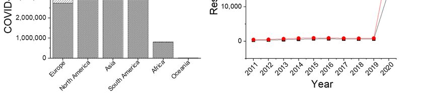
conveyances (https://www.worldometers.info/coronavirus/). North America, South America, and Asia
are the most affected (maximum number of cases) continents by this pandemic so far (https://
www.worldometers.info/coronavirus/, data accessed on 24 July 2020) (Figure 1A). In spite of rapid
development of knowledge, precautionary measurements, and clinical trials, researchers, regulatory
bodies, and government administrations are facing a great challenge globally in various aspect to
prevent COVID-19 pandemic. The present situation is leading us to a still unknown future and
conflicting paradox in the battle against SARS-CoV-2. Further, a plethora of documents have been
Diseases 2020, 8, 30; doi:10.3390/diseases8030030 www.mdpi.com/journal/diseases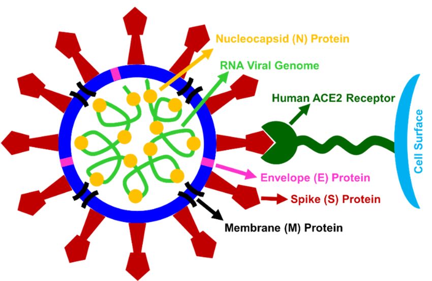
Diseases 2020, 8, 30 2 of 30
Diseases 2020, 8, x; doi: 2 of 31
published on “coronavirus”
still unknown future andand “coronavirus
conflicting paradoxdisease” (year-wise
in the battle againstkeyword search
SARS-CoV-2. for last
Further, 10 years
a plethora
in PubMed on 24 July
of documents have2020)
been research,
published where a maximum
on “coronavirus” andand a very large
“coronavirus number
disease” of results
(year-wise were
keyword
search for last 10 years in PubMed on 24 July 2020) research, where a maximum
obtained in 2020 on both the keywords in comparison to previous years (Figure 1B). Nature Index hasand a very large
number aofhugely
also reported resultsincreasing
were obtained in research
global 2020 on both the keywords
publishing in comparison
phenomenon to previous
on COVID-19 years
pandemic
(Figure by
as evidenced 1B).66,883
Nature Indexand
articles has19,420
also reported
preprintsa(https://www.natureindex.com/news-blog/the-
hugely increasing global research publishing
phenomenon on COVID-19 pandemic as A
top-coronavirus-research-articles-by-metrics). evidenced
thoroughbyand 66,883 articles and
cumulative 19,420knowledge
scientific preprints
(https://www.natureindex.com/news-blog/the-top-coronavirus-research-articles-by-metrics). A
about the progress against COVID-19 amid various conflicts and controversies is the need of the
thorough and cumulative scientific knowledge about the progress against COVID-19 amid various
time to set a clear directive in this battle. Through this review article, we provide an overview of
conflicts and controversies is the need of the time to set a clear directive in this battle. Through this
updated and rapidly evolving progress against COVID-19 pandemic including various conflicts to
review article, we provide an overview of updated and rapidly evolving progress against COVID-19
the readers in a comprehensive manner. Considering this, we have summarized diverse research
pandemic including various conflicts to the readers in a comprehensive manner. Considering this,
areas covering
we have the current known
summarized diversebiological properties
research areas of SARS-CoV-2,
covering diagnostic
the current known tools properties
biological for detection,
of
therapeutic measurements for possible treatment, and prevention techniques to stop further
SARS-CoV-2, diagnostic tools for detection, therapeutic measurements for possible treatment, spreading
and
of this prevention
pandemic.techniques to stop further spreading of this pandemic.
Figure 1. (A) Distribution of COVID-19 cases over various continents according to Worldometer (data
Figure 1. (A) Distribution of COVID-19 cases over various continents according to Worldometer (data
accessed on 24th July, 2020). (B) Year-wise results for research on “Coronavirus” and “Coronavirus
accessed on 24th July, 2020). (B) Year-wise results for research on “Coronavirus” and “Coronavirus
Disease” keyword for last 10 years (2020–2011) according to PubMed (data accessed on 24th July, 2020).
Disease” keyword for last 10 years (2020–2011) according to PubMed (data accessed on 24th July,
2020).
2. Origin, Transmission, and Symptoms
2. Origin, Transmission, and Symptoms
2.1. Origin
2.1. Origin
2.1.1. Genesis, Structure, and Features
Coronaviruses (subfamily:
2.1.1. Genesis, Structure, Coronavirinae, family: Coronaviridae, order: Nidovirales) are
and Features
enveloped single stranded positive sense RNA genomes that range in size from 26 to 32 kilobases [1,2].
Coronaviruses (subfamily: Coronavirinae, family: Coronaviridae, order: Nidovirales) are
Coronavirus
envelopedconsists
singleofstranded
four structural
positive proteins:
sense RNA the nucleocapsid,
genomes that rangeenvelope, membrane,
in size from 26 to 32 and spike
kilobases
forming a core-shell
[1,2]. Coronavirus morphology
consists of (Figure 2), whose
four structural diameter
proteins: is in the range
the nucleocapsid, from 60membrane,
envelope, nm to 140and nm
with spike like projections on its surface [2–4]. The name, coronavirus (Latin: Corona
spike forming a core-shell morphology (Figure 2), whose diameter is in the range from 60 nm to 140 = Crown) came
out duenmtowith
the spike
presence of a crownon
like projections orits
the sun’s [2–4].
surface corona-like
The name,spike (S) glycoprotein
coronavirus on viral
(Latin: Corona surface
= Crown)
forming club-shaped
came out due to protrusions,
the presence which is also
of a crown evidenced
or the through the
sun’s corona-like electron
spike microscope
(S) glycoprotein on [2–4].
viral
This transmembrane
surface formingspike (S) glycoprotein
club-shaped protrusions,on which
viral surface
is also mediates
evidencedthe entry the
through intoelectron
host cells, forming
microscope
[2–4]. This
homotrimers transmembrane
protruding from the spike (S)surface
viral glycoprotein on viral
[5]. The surface
receptor mediates
binding domainthe entry
(RBD)into
inhost cells,
the spike
forming homotrimers
(S) glycoprotein is the mostprotruding
mutable part fromofthe viral
the surface [5].genome
coronavirus The receptor binding
leading domain (RBD)
to generation in
of new
the spike
properties (S) glycoprotein
and ability of virus to is infect
the mostnewmutable partor
cell types of even
the coronavirus
new species genome leading
[6]. Based on to generation
phylogenetic
of new and
relationships properties
genomic and ability of the
structures, virus to infect Coronavirinae
subfamily new cell types isordivided
even new intospecies [6]. Based
four genera on
(α-CoV,
phylogenetic relationships and genomic structures, the subfamily Coronavirinae is divided into four
β-CoV, γ-CoV, and δ-CoV). α-CoV and β-CoV only infect mammals, whereas γ-CoV and δ-CoV infect
genera (α-CoV, β-CoV, γ-CoV, and δ-CoV). α-CoV and β-CoV only infect mammals, whereas γ-CoV
generally birds and sometimes even infect mammals. β-CoV and γ-CoV are responsible for respiratory
and δ-CoV infect generally birds and sometimes even infect mammals. β-CoV and γ-CoV are
diseases in humans and gastroenteritis in animals [2,7]. Presence of four corona viruses (HKU1, NL63,
responsible for respiratory diseases in humans and gastroenteritis in animals [2,7]. Presence of four
229E and OC43) have been found in human circulation which are generally cause mild respiratory
disease [8].
Diseases 2020, 8, x; doi:                                                                                                                      3 of 31

 corona viruses (HKU1, NL63, 229E and OC43) have been found in human circulation which are
Diseases 2020, 8, 30                                                                 3 of 30
 generally cause mild respiratory disease [8].

        Figure 2.2. Hypothetical
       Figure       Hypothetical cartoon
                                 cartoon presentation
                                          presentation of
                                                        of morphology,
                                                           morphology, structural
                                                                          structural constituents,
                                                                                      constituents, and
                                                                                                     and possible
                                                                                                          possible
        receptor   mediated cellular  binding of SARS-CoV-2.    Size, shape,  and  integral  components
       receptor mediated cellular binding of SARS-CoV-2. Size, shape, and integral components are          are for
                                                                                                                for
        visualizationpurpose
       visualization  purposeonly
                               onlyand
                                     anddo
                                         donot
                                            notexactly
                                                exactlyreplicate
                                                        replicatethe
                                                                  theultra-structural
                                                                      ultra-structuralmorphology
                                                                                       morphologyofofvirus.
                                                                                                       virus.

2.1.2.
2.1.2. SARS-CoV-2
       SARS-CoV-2
      The
       The 2019-nCoV
             2019-nCoV isisphylogenetically
                                   phylogenetically closely  closely related
                                                                         related to   tobat
                                                                                          batSARS-like
                                                                                               SARS-likecoronaviruses,
                                                                                                               coronaviruses, hence    hence namename
SARS-CoV-2       and    belongs      to  β-CoV      genus      lineage    B   [9].   Regarding
 SARS-CoV-2 and belongs to β-CoV genus lineage B [9]. Regarding pathogenicity and transmissibility, pathogenicity         and    transmissibility,
SARS-CoV-2
 SARS-CoV-2 may    may differ
                            differ from
                                      from otherother known
                                                         known SARS-CoV
                                                                      SARS-CoV due        due to to aa significant
                                                                                                        significant change change in   in itsits spike
                                                                                                                                                  spike
glycoproteins
 glycoproteins (ORF8, and ORF3b) [10]. Doremalen et al. [11] showed that the SARS-CoV-2 virus
                   (ORF8,       and     ORF3b)      [10].     Doremalen          et   al. [11]   showed      that    the    SARS-CoV-2            virus
remained
 remainedviable
              viablein  inaerosols
                           aerosols(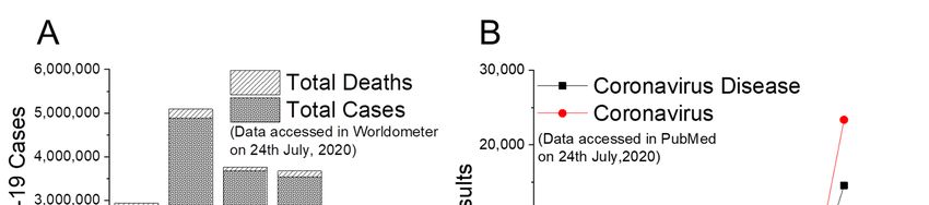
Diseases 2020, 8, 30                                                                                    4 of 30

COVID-19) also warned further possible coronavirus outbreaks from bats and with a high probability
that outbreak will occur in China [17].

2.3. Transmission

2.3.1. Unknown Intermediary Host
     Both SARS-CoV and MERS-CoV bat β-coronaviruses crossed over to humans through
an intermediary host, which was palm civet cats in the Guangdong province of China (SARS-CoV,
in 2002–2003, mortality rate 11%) and dromedary camels in Saudi Arabia (MERS-CoV, in 2012, fatality
rate 34%), respectively [18]. In the case of COVID-19, the virus was transmitted to human from bats,
but the intermediary host animal(s) are not yet known. A study claimed that the intermediary host
animal is pangolin due to the following findings on Pangolin-CoV, SARS-CoV-2, and BatCoV RaTG13
viruses: (i) At the whole-genome level, both Pangolin-CoV and SARS-CoV-2 share 91.02% similarity
among them, (ii) Pangolin-CoV and SARS-CoV-2 are reported to be the second closest relative to each
other than to BatCoV RaTG13, (iii) In the receptor binding domain (RBD) of spike glycoprotein, five key
amino acid residues involved in the interaction with human angiotensin converting enzyme 2 (ACE2)
of Pangolin-CoV and SARS-CoV-2 are consistent, and (iv) Only SARS-CoV-2 contains a potential
cleavage site for furin proteases unlike both Pangolin-CoV and RaTG13 [19]. It was also concluded
that the transmission of human SARS-CoV-2 virus from bat may include more than one intermediary
host including pangolins [20].

2.3.2. Human to Human Transmission
      Similar to SARS-CoV, the 2019-nCoV is reported to have the capability to transmit efficiently
among humans due to familial cluster of pneumonia [9]. Several cases were reported person-to-person
transmission of this virus not only through family settings, but were also in hospital and infected
travelers [9,21]. Person-to-person transmission of the SARS-CoV-2 infection is occurred via airborne
droplets to the nasal mucosa in closed environments, close contact between people, unwashed hands,
and touching contaminated surfaces with less possibilities. Within the incubation period ranges from 2
to 14 days in general, SARS-CoV-2 may replicate locally in cells of the ciliated epithelium resulting
cell damage and inflammation. Primarily, respiratory secretions of any infected person are used to
diagnose the presence of virus by special molecular tests including normal/low white cell counts with
elevated C-reactive protein (CRP) [18]. Additionally, abnormal computerized tomographic (CT) chest
scan is also proved to be helpful to diagnose any infected person even for those with no symptoms or
mild disease [18]. The SARS-Cov-2 showed lower mortality but faster spreading than SARS-CoV and
MERS-CoV. Isolation of SARS-CoV-2 from oral swabs, bronchoalveolar lavage fluid, and stool proved
them to be highly contagious [22,23].

2.3.3. Receptor-Mediated Cellular Entry
     SARS-CoV-2 infects human by interacting with a functional receptor, metallopeptidase named
angiotensin converting enzyme 2 (ACE2), for its successful cellular entry (Figure 2) [22,24]. Crystal
structure of the C-terminal domain of spike protein in complex with human ACE2 (hACE2) revealed an
overall similar binding mode as that of SARS-CoV with hACE2 [24]. It was determined that 2019-nCoV
uses ACE2 as a cellular entry receptor in human, Chinese horseshoe bats, civets, and pigs but not for
mice and cells without ACE2 protein expression capability [22]. Other coronavirus receptors, such as
aminopeptidase N and dipeptidyl peptidase 4 do not play any role for cellular entry of 2019-nCoV [22].
Previous studies revealed that almost all human organs are known to have ACE2 mRNA, though the
protein expression of ACE2 mRNA was largely unknown. Such ACE2 receptor is found to be present
in arterial and venous endothelial cells, arterial smooth muscle cells in the lungs, stomach, small
intestine, liver bile ducts, colon, skin, kidney parietal epithelial cells, lymph nodes, and in the brain [25].
The surface of lung alveolar epithelial cells and enterocytes of the small intestine also express ACE2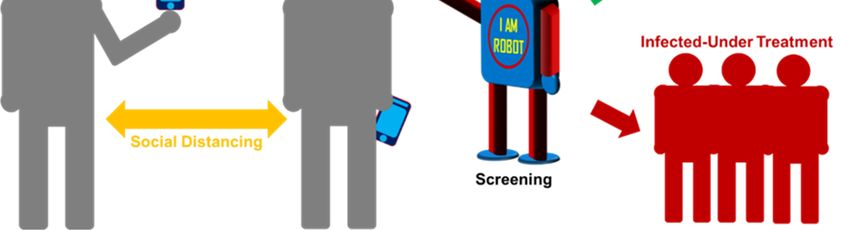
Diseases 2020, 8, 30                                                                                  5 of 30

protein allowing them to be infected by SARS-CoV-2 [25]. The tissues of the upper respiratory tract
are not the primary site of entrance for SARS-CoV, as oral and nasal mucosa and nasopharynx did
not show ACE2 expression on the surface of epithelial cells, rather upper respiratory tract might be
susceptible to secondary infections from the infected lower respiratory tract. Lower lungs may show
higher opacity in the CT scans due to its more ACE2 expression [26]. Higher viral loads have been
recorded in the nose than the in throat, with similar viral loads seen in asymptomatic and symptomatic
patients [27].

2.4. Symptoms and Impact
      Coronaviruses generally are found to cause acute and chronic respiratory, enteric, and central
nervous system diseases in humans as well as in other animals. The symptoms of a COVID-19 patient
are usually fever, cough, sore throat, breathlessness, fatigue, and feeling of discomfort. For most of the
people, it is found mild. For elderly and the patient with comorbidities may develop pneumonia, acute
respiratory distress syndrome (ARDS), and multi-organ dysfunction leading to death. Many infected
people are found to be asymptomatic causing a problem for early detection and controlling the spread
of disease. Mortality rate estimated by the World Health Organization (WHO) (as of 3 March 2020)
is 3.4% (https://www.worldometers.info/coronavirus/coronavirus-death-rate/). Further, speculation
about the association of human coronaviruses with more serious human diseases (such as multiple
sclerosis, hepatitis, or enteric disease in infants) are still under question due to no proper evidence [8].

3. Diagnostic Modalities
     In absence of any approved therapeutics or vaccines for the treatment of COVID-19, WHO has
promoted “test, isolate, and trace” method as a preventive measure. Thus, early, rapid, and accurate
diagnosis of COVID-19 patients is becoming very crucial to control the sources of infection and
to prevent further community spread. With a gradual understanding of biological properties of
SARS-CoV-2, various diagnostic methods and device strategy with point of care facilities have been
developed for COVID-19 detection worldwide. A summary of various diagnostic methods (Table 1)
are presented for the COVID-19 detection. Various countries approved and implied different testing
methods according to the regulation of their own health agencies based on situation and availabilities.
Below subsections are summarized the recent developments on diagnostic methods based on (i) nucleic
acid, (ii) protein, (iii) chest scan and (iv) autopsy.

                       Table 1. Various detection methods implied for COVID-19 diagnosis.

               Test                           Method                             Sample/Organ
                                    1.Polymerase chain reaction         1. Nasal (Nasopharyngeal)/Throat
      Nucleic acid-based             2. Isothermal nucleic acid              (Oropharyngeal) Swab
          detection                         amplification                   2. Blood (Serological test)
                                           3. CRISPR-Cas                           3. Fecal swab
                                                                            1. Blood (Serological test)
   Protein-based detection              Viral antibody levels                  2. Respiratory swab
                                                                                      3. Fecal
                                                1. X-ray
   Chest scan (auxiliary test)                                                        Lung
                                     2. Computed tomography
            Autopsy              Surgical (Post-mortem examination)                  Corpse

3.1. Nucleic Acid-Based Detection
     Nucleic acid-based detection strategy has been widely used against detection of various diseases,
including coronavirus and recent COVID-19. In this section, we review some nucleic acid-based
detection methods that are commonly being employed for the diagnosis of COVID-19.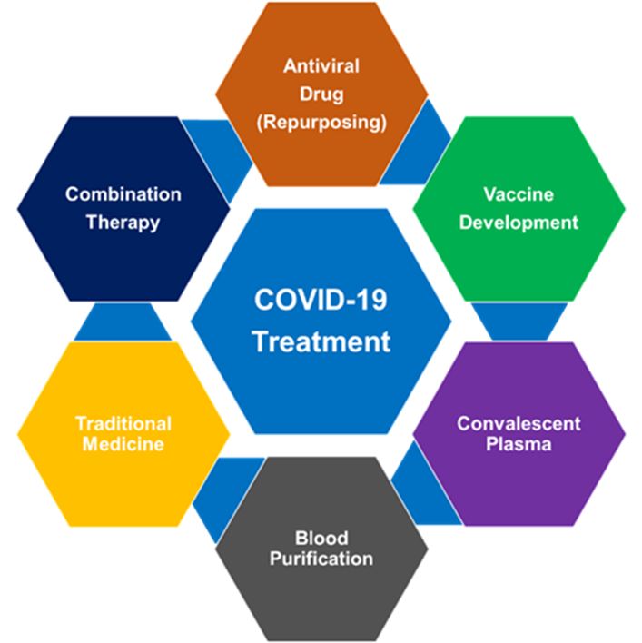
Diseases 2020, 8, 30                                                                                   6 of 30

3.1.1. Polymerase Chain Reaction
      Polymerase chain reaction (PCR) is an enzymatic method widely used in molecular biology to
make millions to billions of copies of a specific DNA sample [28]. This method involves following
steps in a series or cycles of temperature changes: (i) Denaturation: separating the two strands of the
DNA containing the gene segment with the application of heat, (ii) Annealing-marking gene segment
of each strand of DNA with a primer, (iii) Primer extension: using a DNA polymerase to assemble
a copy alongside each segment, and (iv) Repeat: continuously copy the copies [28]. Various PCR-based
methods are an indispensable, common, and rapid techniques for scientist to amplify a minute nucleic
acid sample to a large enough amount for a number of applications [29]. Due to high sensitivity and
high sequence specificity, the PCR-based method has been used as a routine and reliable technique for
detecting coronaviruses. Coronavirus is a RNA virus, so in general reverse transcriptase-PCR (RT-PCR)
method is implied as follows: coronavirus RNA is transcribed into cDNA by reverse transcription,
then the PCR is performed on cDNA, and finally detection of PCR product through specific detection
method(s) (gel visualization and sequencing) [30,31].
      Real-time: Real-time reverse transcriptase-PCR (RT-PCR) detection method is evolved as a
common platform for detection of all kinds of coronaviruses due to its low cost per test, less
time-consuming process and more sensitive than the conventional RT-PCR assay [32,33]. The whole
genome sequence of SARS-CoV-2 enabled to develop PCR-based kits to diagnose COVID-19 in
laboratory and clinical settings [34–39]. Corman et al. [34] developed a robust diagnostic methodology
considering the SARS-related virus sequences available in GenBank. A close genetic relatedness to
the 2003 SARS-CoV and synthetic nucleic acid technology helped this process to design and validate
such strategy without using any virus isolates and samples from infected person. Such a technique
can successfully discriminate 2019-nCoV from SARS-CoV. This approach provided the first version
of the diagnostic protocol to the WHO from exclusivity testing on 75 clinical samples (13 January
2020). A real-time RT-PCR based test is found to be more sensitive than radiological test for pediatric
patients [36]. Pediatric patients with milder symptoms, showed no clear clinical signs or chest X-ray
findings but their real-time RT-PCR exhibited positive results. Further, in this report, real-time RT–PCR
showed positive results in rectal swab-testing but negative results in nasopharyngeal swab-testing for
eight out of ten pediatric patients suggesting shedding of virus in the gastrointestinal tract and a possible
fecal-oral transmission. Wang’s group [37] reported that the RT-PCR based findings using different
types of clinical specimens collected from 82 infected individuals. In their study, it was found that
viral loads were significantly correlated among 30 pairs of throat swab and sputum samples. Overall,
real-time RT-PCR based method enables developing a high-throughput testing for rapid, on-demand,
low-cost, reliable, quantitative detection technique against COVID-19 in clinical settings [39].
      Probe free: A team of Indian Institute of Technology, Delhi, India reported first probe-free real
time PCR assay for COVID-19 detection (http://www.iitd.ac.in/content/icmr-approves-probe-free-
covid-19-detection-assay-developed-iit-delhi-0). They have used comparative sequence analyses to
identify unique regions (short stretches of RNA sequences) in the SARS COV-2 genome, which are not
present in other human coronaviruses. In this highly sensitive assay, primers can specifically target
unique regions (conserved in over 400 fully sequenced) of COVID-19 genomes, which was reported
after extensive optimization using synthetic DNA constructs followed by in vitro generated RNA
fragments. Indian Council of Medical Research has approved this technique as it does not require any
fluorescent probes (thus low-price) but still useful for high throughput testing.

3.1.2. Isothermal Nucleic Acid Amplification
     Isothermal amplification of nucleic acids is a rapid, efficient, and alternative amplification technique
than PCR. This process can be applied at a constant temperature without any thermos-cycling apparatus,
unlike in the case of PCR [40,41]. The isothermal amplification technique can be performed in water bath,
on the cell surface, or even inside living cells, making it a superior technique over PCR [40,41]. Based on
reaction kinetics of isothermal nucleic acid amplification, it is divided to exponential amplification,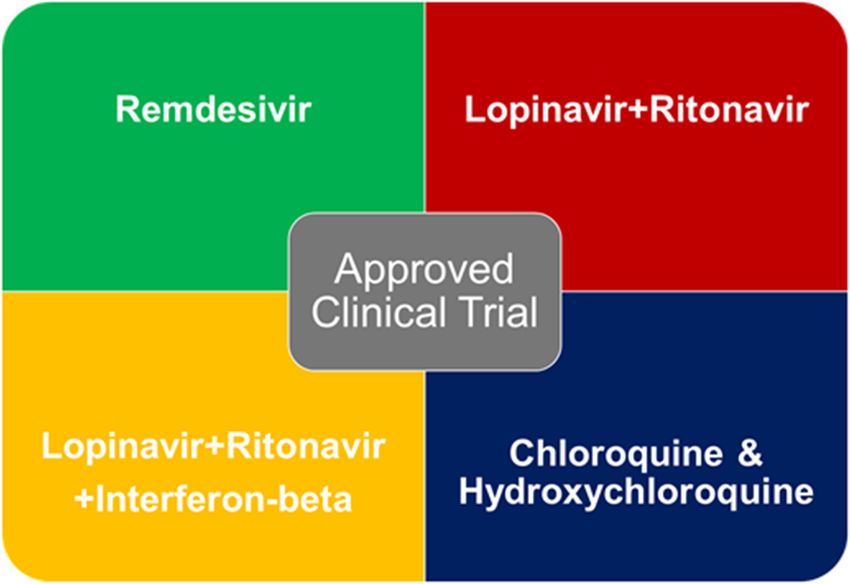
Diseases 2020, 8, 30                                                                            7 of 30

linear amplification, and cascade amplification. These are further sub-divided into transcription
mediated amplification, nucleic acid sequence-based amplification, signal mediated amplification
of RNA technology, strand displacement amplification, rolling circle amplification, loop-mediated
isothermal amplification of DNA, isothermal multiple displacement amplification, helicase-dependent
amplification, single primer isothermal amplification, and circular helicase-dependent amplification,
based on the developments in molecular biology of DNA/RNA synthesis [40,41]. Furthermore, the use
of microfluidic chips, capillary platforms, and test paper with isothermal amplification technique has
been developed for single-cell or single-molecule analysis. Among these, loop-mediated isothermal
amplification (LAMP) has been implied successfully for coronavirus detection [42–45]. LAMP technique
can amplify target nucleic acid sequence using two or three sets of primers and a polymerase at a
constant temperature (~60–65 ◦ C) [46–48]. In comparison to PCR-based technique, LAMP can produce
considerably higher amount of DNA with high strand displacement and replication activity due to the
use of additional pair of “loop primers”.
      Park et al. [46] developed reverse transcription LAMP (RT-LAMP) assay(s) to detect genomic
RNA of SARS-CoV-2. These RT-LAMP assays (in combination with leuco crystal violet colorimetric
detection method) can detect as low as 100 copies of SARS-CoV-2 RNA within 30 min. These RT-LAMP
assays were highly specific towards SARS-CoV-2 compared to other human coronaviruses (hCoV-229E,
hCoVOC43, MERS-CoV, and SARS-CoV). Yu et al. [49] also developed a rapid and sensitive isothermal
LAMP based method (iLACO) for the detection of COVID-19 virus RNA or cDNA samples. In this
method, iLACO was used to amplify a fragment of the ORF1ab gene using 6 primers, which was
proved to be specific for SARS-COV-2 species (i.e., low chance for false positives) in comparison
to the sequences of 11 related viruses by the help of online tool Primer-BLAST (including 7 similar
coronaviruses, 2 influenza viruses and 2 normal coronaviruses). iLACO can detect synthesized RNA
equivalent to 10 copies of 2019-nCoV (performance is comparable to Taqman based qPCR detection
method), where reaction time varied from 15–40 min based on virus load in the collected samples.
Another LAMP-based colorimetric detection method was reported to identify SARS-CoV-2 virus
RNA from purified RNA or cell lysis (without an RNA purification step) [48]. The sensitivity of this
portable method is equivalent to a commercial RT-qPCR test with only heating and visual inspection.
Zhu et al. [47] demonstrated a successful and accurate diagnosis of COVID-19 using one-step RT-LAMP
coupled with nanoparticles-based biosensor (NBS) assay (RT-LAMP-NBS) within approximately 1 h
(from sample collection to result interpretation). They have employed two designed LAMP primer
sets (F1ab-RT-LAMP and np-RT-LAMP), heating block (to maintain a constant temperature at 63 ◦ C),
a real-time turbidity (LA-320C) and visual detection reagents (VDR) in addition to NBS interpretation
to simultaneously amplify and detect genes of SARS-CoV-2 in a “one-step” and “single-tube” reaction.
The sensitivity of SARS-CoV-2 RT-LAMP-NBS was 12 copies (each of detection target) per reaction,
whereas no cross-reactivity was observed for all pathogens of non-SARS-CoV-2 (virus, bacteria,
and fungi). The RT-LAMP-NBS assay showed 100% the analytical sensitivity of SARS-CoV-2 for
oropharynx swab samples of clinically diagnosed COVID-19 patients and 100% specificity for clinical
samples collected from non-COVID-19 patients.

3.1.3. CRISPR Diagnostics
     CRISPR-Cas (clustered regularly interspaced short palindromic repeats-CRISPR associated) is an
adaptive immune system, which was discovered first in Escherichia coli in 1987 and later also in other
bacteria species. These are found predominantly in archaea (87% of genomes) than in bacteria (50% of
genomes) [50,51]. Being an immune system of archaea and bacteria, CRISPR and CRISPR-associated
proteins deliver protection against invasive nucleic acids (such as DNA, or RNA from phages, plasmids,
and other exogenous DNA elements) [50,51]. Scientists later exploited this immune responsive system
by reengineering to target parts of genetic material for precise genetic alterations of any particular
cellular type, which is the basis of CRISPR therapeutic and diagnostic platforms for human [52,53].
This adaptive immune system is also widely used as a tool for SARS-CoV-2 detection. CRISPR
Diseases 2020, 8, 30                                                                                 8 of 30

associated enzyme Cas13 has already been utilized for rapid and portable sensing for successful
RNA-targeting [54]. A Specific High-sensitivity Enzymatic Reporter unLOCKing (SHERLOCK)
platform was developed by combining isothermal preamplification with Cas13 to detect single molecules
of RNA or DNA for Dengue or Zika virus [55]. An updated SHERLOCK protocol has been reported for
multiplexable, portable, rapid, and quantitative COVID-19 detection (https://broad.io/sherlockprotocol),
which can target sequences in a range between 20 and 200 aM (10–100 copies per microliter of input).
Another development of accurate CRISPR-Cas12-based lateral flow assay able to detect SARS-CoV-2
with 95% positive predictive agreement and 100% negative predictive agreement from respiratory
swab RNA extracts (less than 40 min) [56]. Another newly developed method, SARS-CoV-2 DNA
Endonuclease-Targeted CRISPR Trans Reporter (DETECTR), was found to perform simultaneous reverse
transcription and isothermal amplification by (i) RT-LAMP for RNA extracted (for nasopharyngeal or
oropharyngeal swabs), (ii) Cas12 detection of predefined coronavirus sequences, and (iii) cleavage of a
reporter molecule confirms, which detects the virus [56]. A FnCas9 Editor Linked Uniform Detection
Assay (FELUDA) was developed for detecting nucleotide sequences, classifying nucleobase identity,
and inferring zygosity [57]. FELUDA is able to distinguish clear signatures of SARS-CoV-2 sequence in
synthetic DNA within one hour using a specific ribonucleoprotein (RNP) from non-specific RNP (such
as H1N1 or HBB). FELUDA can also clearly distinguish between two SARS-CoV-2 and SARS-CoV-1
sequences. This approach further can be developed as lateral flow assay on a paper strip to distinguish
SARS-CoV-2 synthetic DNA using SARS-CoV-2 specific RNP.

3.2. Protein-Based Detection
     Protein-based testing has become as an alternative and additive detection strategy in addition
to nucleic-acid based testing methods for coronavirus [58]. In response to any infected viral protein
antigens, antibodies (i.e., a blood protein produced in response to and counteracting a specific
antigen) are generated in patient’s body resulting a very specific antigen-antibody (Ag-Ab) serological
interaction. Detection of this specific antibody level(s) due to the SARS-CoV-2 infection can be useful for
surveillance of COVID-19 pandemic. This indirect serological test opens up wide range of possibilities,
such as, (i) successful detection of asymptomatic patients, (ii) creating large a window of testing
time even with a gradual decrease of viral load, (iii) protect community transmission due to false
negative results by other methods, and (iv) proper guidance for individual quarantine period [59–66].
Kwok-Yung Yuen’s group [61] successfully detected antibodies generated in response to SARS-CoV-2
viral proteins at the time when the detection of the viral proteins become difficult due to gradual
declining trend of viral load(s). Serological test using enzyme-linked immunosorbent assay (ELISA)
for antibodies (immunoglobulin M, IgM and immunoglobulin G, IgG) is more confirmatory and
unreliable results from oral swabs for 2019-nCoV detection [62]. This test can be applied for respiratory,
blood, or fecal samples. Guo et al. [63] conducted a COVID-19 profiling study on early humoral
response based on IgA, IgM, and IgG response. This study found that IgM and IgA antibody were
detected 5 days, while IgG was detected 14 days after symptom onset, with a positive rate of 85.4%,
92.7%, and 77.9%, respectively [63]. The detection of IgM by ELISA was found more efficient than
that of qPCR after 5.5 days of symptom onset [63]. A successful immunological field-effect transistor
(FET)-based biosensing device was developed for detecting SARS-CoV-2 in clinical samples, where
the sensor was developed by conjugating a specific antibody against SARS-CoV-2 spike protein to
graphene sheet coated FET [64]. This rapid diagnostic device for SARS-CoV-2 antigen requires no
sample pre-treatment or labelling. This is a highly-sensitive detection method for the SARS-CoV-2 spike
protein at concentrations of 1 fg/mL in phosphate-buffered saline and 100 fg/mL in clinical transport
medium, 1.6 × 101 pfu/mL in culture medium and 2.42 × 10² copies/mL in clinical samples. The false
positive results in serological tests for COVID-19 is a concern due to the presence antibodies generated
against other coronaviruses (such as for common cold) irrespective of the presence of SARS-CoV-2
antibodies. Recently, researchers have got a high frequency of cross-reactivity in plasma samples
from 15 COVID-19 patients against the S protein of SARS-CoV-2 and SARS-CoV [65]. However, more
Diseases 2020, 8, 30 9 of 30
accurate antibody-based detection method can be made with additional features of infected patient in
serological test, such as (i) combined (IgM and IgG) antibody assay rather than a single antibody test,
(ii) more number of testing, (iii) report of elevated levels of C-reactive protein, D-dimer, lymphocytes,
leukocytes, or blood platelets [66].
3.3. Chest Scan-Auxiliary Test
There are growing concerns regarding the COVID-19 testing despite of huge efforts in this direction.
Due to sudden outbreak and a huge increase of COVID-19 cases, a sufficient number of COVID-19 test
kits are unavailable in hospitals and healthcare centers. Further, an automatic detection system with a
quick diagnostic capability could be an alternative or auxiliary method to prevent community spreading
of COVID-19. In this situation, most countries have recommended the RT-PCR-based methods as the
standard technique for COVID-19 diagnosis. Serological tests are also considered to be the primary
technique for COVID-19 detection. However, small hospitals, health centers in sub-urban and village
areas, even private hospitals in sub-urban area may not have an approved RT-PCR testing center
or PCR testing infrastructure facilities. On the other hand, chest-scan, a routine technique implied
for prominent pneumonia pattern, has been evolved as useful non-invasive technique for COVID-19
detection [67]. Both chest X-ray and computed tomography (CT)-scan were successful to distinguish
the manifestations of typical pneumonia in the case of MERS-CoV and SARS-CoV infection [68,69].
These scans have helped to diagnoses suspected person to isolate and treat more quickly, even when the
RT-PCR based test did not respond properly. Such tests have proven lung histology (lung damage or
holes/honeycomb-like appearance) of COVID-19 patients [70]. Thus, chest-scan is useful for suspected
COVID-19 patients with negative RT-PCR result.
3.3.1. X-ray
X-rays, a form of high-energy electromagnetic radiation, are shorter wavelengths than UV rays and
longer wavelengths than gamma rays. X-ray machines are widely available sophisticated diagnostic
imaging technique for body, bone and other dense objects that can block the radiation through a
limited exposure to radiation. In addition, X-ray scans can be used for lung infections, pneumonia and
tumors. An automatic prediction of COVID-19 was successfully reported using Chest X-ray images
and a deep convolution neural network based pre-trained transfer models (ResNet50, InceptionV3
and Inception-ResNetV) [71]. These pre-trained transfer models helped to obtain a higher prediction
accuracy for small X-ray dataset. This model has end-to-end structure without manual feature
extraction and selection methods, where the ResNet50 is an effective one among all pre-trained models
in the small dataset (50 COVID-19 vs. 50 Normal).
3.3.2. Computed Tomography
Computed tomography (CT) scan is a computer-assisted medical imaging device which combines
cross-sectional (tomographic) scanned images of specific areas or virtual slices of any organ taken from
different angles producing a 3D view of that particular organ. CT-scan is one of the methods used to
diagnose various abnormalities of the chest (such as pneumonia, lung cancer etc.) [72,73]. Thus, chest
CT-scan is also being used as a fast, painless, non-invasive and accurate auxiliary diagnostic method
in addition to the RT-PCR test for the suspected COVID-19 patient [74–76]. The National Health
Commission of China included the chest CT findings as evidence of clinical diagnosis of COVID-19
for patients in Hubei province at the fifth edition of the Diagnosis and Treatment Program of 2019
New Coronavirus Pneumonia due to the false-negative rate of RT-PCR test for COVID-19 patient [74].
Several groups found chest CT scan more sensitive and better diagnostic tool in comparison to RT-PCR
for COVID-19 detection [75,76]. Recent investigations demonstrated that the CT-scan of COVID-19
patient(s) clearly showed bilateral pulmonary parenchymal ground-glass and consolidative pulmonary
opacities, with a rounded morphology, crazy-paving pattern, linear opacities, and peripheral lung
distribution [77,78]. In contrast, other study did not find any lung cavitation, discrete pulmonaryimportant tool to help clinicians to diagnose quickly and accurately the effected lung disease [79].
Artificial Intelligence (AI) and deep learning-based automated CT image analysis of lung have been
also developed to distinguish COVID-19 pneumonia and Influenza-A viral pneumonia [80–82]. These
automated deep learning based methods can produce graphical pattern of a particular COVID-19
Diseases 2020, 8, 30 10 of 30
patient which is helpful for clinician to diagnose prior to pathogenic testing [83]. In spite of such
clinical diagnostic values, CT scan still fails to come at the forefront of COVID-19 diagnosis due to
nodules,
the following pleural effusions,
reasons: and lymphadenopathy
(i) it is expensive, [77]. Further,
(ii) it requires high-resolution
technical expertise, CTand(HRCT)-scan for
(iii) it is incapable of
the chest is reported to be important tool to help clinicians to diagnose quickly and accurately the
distinguishing SARS-CoV-2 pneumonia from other viral pneumonia and hysteresis.
effected lung disease [79]. Artificial Intelligence (AI) and deep learning-based automated CT image
analysis of lung have been also developed to distinguish COVID-19 pneumonia and Influenza-A viral
3.4. Autopsy-On
pneumonia Demand
[80–82]. These automated deep learning based methods can produce graphical pattern of a
particular COVID-19 patient which is helpful for clinician to diagnose prior to pathogenic testing [83].
An autopsy report, through examination of a corpse by dissection, is an important source of
In spite of such clinical diagnostic values, CT scan still fails to come at the forefront of COVID-19
information for research
diagnosis due to the purposes to evaluate
following reasons: (i) it is any disease
expensive, (ii) itcausing
requires death.
technicalAutopsy
expertise, has been
and (iii) it proved
to be hugely beneficial
is incapable to diagnose
of distinguishing emergingpneumonia
SARS-CoV-2 and reemerging
from other infectious diseases,
viral pneumonia andlike COVID-19 [84–
hysteresis.
86]. In contrary, several groups raised their concern that the few autopsies have been performed on
3.4. Autopsy-On Demand
patients who died with suspected or confirmed COVID-19 infection especially in the primary
epicenters ofAnpandemic
autopsy report, through examination of a corpse by dissection, is an important source of
(such as China and Italy) [85,87]. Despite the suggestion by WHO on
information for research purposes to evaluate any disease causing death. Autopsy has been proved to
performing post-mortem examinations for COVID-19 deaths with following recommended safety
be hugely beneficial to diagnose emerging and reemerging infectious diseases, like COVID-19 [84–86].
procedures, manyseveral
In contrary, Governments
groups raised including
their concernItaly discouraged
that the few autopsiesthehavepractice of autopsy
been performed during the
on patients
period ofwhoincreasing
died with number
suspected of death andCOVID-19
or confirmed even some scientific
infection report
especially in thehighlighted that the post-
primary epicenters
mortem of pandemic (such
examination does as not
China andany
have Italy)primary
[85,87]. diagnostic
Despite the suggestion
role, whereas by WHO on performing
autopsy may still have a
post-mortem examinations for COVID-19 deaths with following recommended
clinical role in selected cases [88,89]. Though based on autopsies, physicians can determine a safety procedures,
many Governments including Italy discouraged the practice of autopsy during the period of increasing
profound change of the view of COVID-19 disease not as a pneumonia but a systemic, vascular
number of death and even some scientific report highlighted that the post-mortem examination
disease, does
putatively
not havegenerated
any primaryby autoimmunity
diagnostic [85].autopsy
role, whereas Thus,maya strong recommendation
still have was urged to
a clinical role in selected
performcases
full autopsies on patients
[88,89]. Though who diedphysicians
based on autopsies, with suspected or confirmed
can determine a profoundCOVID-19
change of theinfection
view with
of COVID-19
recommended disease not
exceptional as a pneumonia
biosafety but a systemic,
guidelines to reduce vascular disease, spread
the further putatively ofgenerated
potentialbyinfection
autoimmunity
from any corpse. [85]. Thus, a strong recommendation was urged to perform full autopsies on patients
who died with suspected or confirmed COVID-19 infection with recommended exceptional biosafety
guidelines to reduce the further spread of potential infection from any corpse.
4. Treatment Modalities
4. Treatment Modalities
There is currently no clinically proven therapeutic regimen to prevent and eradicate SARS-CoV-
2 infection There
[90]. isCOVID-19
currently no clinically proven therapeutic regimen to prevent and eradicate SARS-CoV-2
is being managed by the supportive treatment (oxygenation and
infection [90]. COVID-19 is being managed by the supportive treatment (oxygenation and ventilation,
ventilation, conservation fluid management). However, the use of broad-spectrum antibiotics [91].
conservation fluid management). However, the use of broad-spectrum antibiotics [91]. This section
This section summarizes
summarizes various various
treatmenttreatment modalities
modalities for COVID-19for COVID-19
(Figure 3). (Figure 3).
Figure 3. Possible treatment modalities against COVID-19.
Figure 3. Possible treatment modalities against COVID-19.Diseases 2020, 8, 30 11 of 30
Diseases 2020, 8, x; doi: 11 of 31
4.1. Antiviral
4.1. Antiviral Drugs
Drugs
Viral infection
Viral infection isis always
always aa majormajor concern
concern forfor morbidity
morbidity and and mortality
mortality in in animals
animals and and humans
humans
worldwide. Development
worldwide. Developmentofofantiviral antiviral drugs
drugshavehavebeen always
been alwaysa pressing
a pressingneedneedto treat such such
to treat viral
infections. Since the approval of first antiviral drug, idoxuridine in 1963,
viral infections. Since the approval of first antiviral drug, idoxuridine in 1963, 90 drugs were 90 drugs were clinically
approvedapproved
clinically to treat nine human
to treat nineinfectious diseases (human
human infectious diseases immunodeficiency
(human immunodeficiencyvirus, HIV;virus,
hepatitis
HIV;B
virus, HBV;
hepatitis hepatitis
B virus, HBV;Chepatitis
virus, HCV; herpesvirus;
C virus, influenza virus;
HCV; herpesvirus; human
influenza cytomegalovirus;
virus; varicella-
human cytomegalovirus;
varicella-zoster virus; respiratory syncytial virus; and human papillomavirus) [92]. The antiviralmostly
zoster virus; respiratory syncytial virus; and human papillomavirus) [92]. The antiviral drugs drugs
inhibit inhibit
mostly the viralthedevelopment
viral development ratherrather
than destroying
than destroyingthe target pathogen
the target unlike
pathogen most
unlike antibiotics.
most antibiotics.A
broad-spectrum
A broad-spectrum antiviral
antiviral is isfound
foundtotobebeeffective
effectiveagainst
againstaawidewide range
range ofof viruses
viruses based
based on on drug
drug
repurposing strategy
repurposing strategy [93]. [93].
Drug repurposing
Drug repurposing or or drug
drug repositioning
repositioning is is aa cost-effective
cost-effective and
and time-efficient
time-efficient alternative
alternative strategy,
strategy,
which involves the recycle or re-use of clinically approved drugs for new disease
which involves the recycle or re-use of clinically approved drugs for new disease instead of searching of instead of searching
of new
new drugsdrugs
[94,95].[94,95]. In contrary
In contrary to in vitro to in vitroscreening
phenotypic phenotypic screening
of known drugs, inof silico/computational
known drugs, in
silico/computational
drug repurposing strategy drug repurposing strategy is aapproach
is a hypothesis-driven hypothesis-driven
to identify approach
the drugs to identify the drugs
for the treatment
of any disease using big data analysis [96,97]. The drug-repurposing has been implied forhas
for the treatment of any disease using big data analysis [96,97]. The drug-repurposing been
several
implied for several human diseases including antiviral drug development
human diseases including antiviral drug development against coronavirus [95]. Various groups have against coronavirus [95].
Various groups
proposed number have proposed
of drug candidatesnumber of drug
through candidates through
drug-repurposing drug-repurposing
(in vitro and in silico) for (inCOVID-19
vitro and
in silico) for COVID-19 treatment [98–101]. WHO focused and initiated
treatment [98–101]. WHO focused and initiated the “SOLIDARITY Trial” (announced on 18 March the "SOLIDARITY Trial"
(announced
2020) of four on 18 March
existing 2020)
antiviral of four existing antiviral(Figure
compounds/formulations compounds/formulations (Figure
4) to assess their clinical 4) toagainst
benefit assess
their clinical benefit
COVID-19 [102,103]. against COVID-19 [102,103].
Figure 4.4.Four
Figure Fourmost
mostpromising drugdrug
promising candidates or combinations
candidates approved
or combinations for clinical
approved fortrial for COVID-19
clinical trial for
treatment by WHO (announced on 18 March 2020).
COVID-19 treatment by WHO (announced on 18 March 2020).
Remdesivir, an antiviral compound, which showed activity against multiple variants of
Remdesivir, an antiviral compound, which showed activity against multiple variants of Ebola
Ebola virus in cell-based assays and rhesus monkey model. Chloroquine (CQ) and its derivative
virus in cell-based assays and rhesus monkey model. Chloroquine (CQ) and its derivative
hydroxychloroquine (HCQ), antiviral compound(s) have been used to treat malaria and amebiasis.
hydroxychloroquine (HCQ), antiviral compound(s) have been used to treat malaria and amebiasis.
A combination of lopinavir and ritonavir, is co-formulated for HIV-1 treatment. Another combination
A combination of lopinavir and ritonavir, is co-formulated for HIV-1 treatment. Another combination
of lopinavir and ritonavir plus interferon-beta (LPV/RTV-IFNb) has been approved for the treatment of
of lopinavir and ritonavir plus interferon-beta (LPV/RTV-IFNb) has been approved for the treatment
relapsing–remitting multiple sclerosis and secondary progressive multiple sclerosis. About 115 clinical
of relapsing–remitting multiple sclerosis and secondary progressive multiple sclerosis. About 115
trials were identified by Belhadi et.al., which includes open-label studies (46%), double-blind (13%),
clinical trials were identified by Belhadi et.al., which includes open-label studies (46%), double-blind
and single blind studies (10%) [104]. They also classified the number of trials (n) and total numbers
(13%), and single blind studies (10%) [104]. They also classified the number of trials (n) and total
of planned inclusions (N) for lopinavir/ritonavir (n = 15, N = 2606), chloroquine (n = 11, N = 1102),
numbers of planned inclusions (N) for lopinavir/ritonavir (n = 15, N = 2606), chloroquine (n = 11, N =
hydroxychloroquine (n = 7, N = 1048), and remdesivir (n = 5, N = 2155).
1102), hydroxychloroquine (n = 7, N = 1048), and remdesivir (n = 5, N = 2155).
In contrary, several controversial reports including toxic side effects on these promising
In contrary, several controversial reports including toxic side effects on these promising
candidates under clinical trial(s) raised some challenging questions for researchers. Those have
candidates under clinical trial(s) raised some challenging questions for researchers. Those have been
been presented below:
presented below:Diseases 2020, 8, 30 12 of 30
1. A report presents that patients (with symptom duration of 10 days or less) receiving remdesivir
showed clinical improvement than those receiving placebo, but it did not make any statistically
significant clinical benefits [105].
2. An open-label non-randomized clinical trial study demonstrated significant decrease in viral
load and carriage duration in COVID-19 patients receiving hydroxychloroquine (600 mg/day
during ten days). This treatment showed enhanced effects in combination with azithromycin,
but it identified serious methodological flaws [106,107]. Another randomized clinical study did
not make any difference in recovery rates upon hydroxychloroquine treatment in 30 COVID
patients [108]. However, a hype on CQ and HCQ has created drug shortages and affected other
potential treatments (such as for patients with Lupus).
3. There was no significant benefit (clinical improvement) observed with lopinavir–ritonavir
treatment [108]. Mortality and percentages of patients with detectable viral RNA at various time
points were similar in the lopinavir–ritonavir group and the standard-care group. It was also
reported median time to clinical improvement was shorter by one day for lopinavir–ritonavir
group than that observed with standard care.
4.2. Vaccine Development
Vaccination is one of the most effective and preventive medications against various diseases caused
by pathogens (such as virus or bacteria). Currently there are about 25 approved vaccinations available
against various life-threatening diseases, including measles, polio, tetanus, diphtheria, meningitis,
influenza, typhoid, and cervical cancer (https://www.who.int/topics/vaccines/en/). A vaccine typically
contains an agent (weakened or killed forms of any microbe, its toxins, or one of its surface proteins),
which though resembles a disease-causing microorganism, but provides active acquired immunity to
that particular infectious disease [109,110]. An antiviral vaccine helps to boost our natural immune
response to an invading virus by priming it to recognize viral antigens. In general, antiviral vaccines
can be classified as follows: (i) inactive or live-attenuated viruses, (ii) virus-like particle (VLP), (iii) viral
vectors, (iv) protein-based, (v) DNA-based, and (vi) mRNA-based vaccines [110,111]. Like many
other diseases, the vaccine development is not successful and conclusive for coronavirus disease.
Until now, there is no proper vaccine is developed or approved for the treatment of human coronavirus
diseases (such as SARS-CoV and MERS-CoV) [111–113]. Most big pharmaceutical companies also in
the race to develop effective vaccines for CoV infection [112,113]. The existing knowledge on previous
strategies for CoV vaccine developments can benefit the ongoing research as sequence analysis of the
SARS-CoV-2 genome showed close relation to SARS (80%) and to one bat RaTG13 SARS-like CoV (96%)
than to MERS CoV (54%) [112]. Liu et al. [111] reported that 188 patents (mentioned in CAS content
collection) are directly associated with anti-SARS and anti-MERS vaccines (15 patents on inactive
and live-attenuated virus vaccines, 28 patents on DNA vaccines, 21 patents on viral vector vaccines,
13 patents on VLP vaccines, and three patents on mRNA vaccines) with a demonstrated immune
response, which could be a huge boost for COVID-19 vaccine developers. An accelerated evaluation
of next-generation vaccine for COVID-19 has been triggered as soon as the genetic sequence of
SARS-CoV-2 is published on 11 January 2020 [112–115]. As of 28 July 2020, 164 vaccine candidates have
been identified (https://www.who.int/who-documents-detail/draft-landscape-of-covid-19-candidate-
vaccines), of which 25 candidate vaccines are in clinical evaluation (Table 2) and 139 candidate vaccines
are in preclinical stages.
Associated problems and pitfalls are surely a major concern resulting huge roadblock for vaccine
discovery against COVID-19. The pitfalls are many folded, which as follows: (i) antibody-dependent
enhancement (ADE) of viral replication for vaccinated people in future recurrence due to immune
backfiring, (ii) rouge immunization resulting a huge damage on someone’s own immune cells (such as
neutrophil and basophil), (iii) in addition to immune response malfunctioning, three imperatives
of vaccine effort: speed, manufacture and deployment at scale, and (iv) global access for a newly
developed vaccine [116,117].Diseases 2020, 8, 30 13 of 30
Table 2. List of candidate vaccines presently in clinical evaluation for COVID-19 (Source: WHO official
website, data accessed on 28 July 2020).
Platform Type of Vaccine Developer Status
Phase 1/2: PACTR202006922165132
Non-Replicating University of 2020-001072-15
ChAdOx1-S
Viral Vector Oxford/AstraZeneca Phase 2: 2020-001228-32
Phase 3: ISRCTN89951424
Phase 1/2:
NCT04383574
Inactivated Inactivated Sinovac
NCT04352608
Phase 3: NCT04456595
Wuhan Institute of Biological Phase 1/2: ChiCTR2000031809
Inactivated Inactivated
Products/Sinopharm Phase 3: ChiCTR2000034780
Beijing Institute of Biological Phase 1/2: ChiCTR2000032459
Inactivated Inactivated
Products/Sinopharm Phase 3: ChiCTR2000034780
Phase 1: NCT04283461
RNA LNP-encapsulated mRNA Moderna/NIAID Phase 2: NCT04405076
Phase 3: NCT04470427
Phase 1/2: 2020-001038-36
BioNTech/Fosun
RNA 3 LNP-mRNAs ChiCTR2000034825
Pharma/Pfizer
Phase 3: NCT04368728
Non-Replicating CanSino Biological Inc./Beijing Phase 1: ChiCTR2000030906
Adenovirus Type 5 Vector
Viral Vector Institute of Biotechnology Phase 2: ChiCTR2000031781
Anhui Zhifei Longcom
Adjuvanted recombinant Biopharmaceutical/Institute of Phase 1: NCT04445194
Protein Subunit
protein (RBD-Dimer) Microbiology, Chinese Phase 2: NCT04466085
Academy of Sciences
Institute of Medical Biology,
Phase 1: NCT04412538
Inactivated Inactivated Chinese Academy of Medical
Phase 1/2: NCT04470609
Sciences
Inovio
DNA plasmid vaccine with Phase 1/2: NCT04447781
DNA Pharmaceuticals/International
electroporation NCT04336410
Vaccine Institute
DNA plasmid vaccine + Osaka
DNA Phase 1/2: NCT04463472
Adjuvant University/AnGes/Takara Bio
DNA DNA plasmid vaccine Cadila Healthcare Limited Phase 1/2: CTRI/2020/07/026352
DNA DNA Vaccine (GX-19) Genexine Consortium Phase 1/2: NCT04445389
Inactivated Whole-Virion Inactivated Bharat Biotech NA
Full length recombinant
SARS CoV-2 glycoprotein
Protein Subunit Novavax Phase 1/2: NCT04368988
NPs vaccine adjuvanted
with Matrix M
Protein Subunit RBD-based Kentucky Bioprocessing, Inc Phase 1/2: NCT04473690
RNA mRNA Arcturus/Duke-NUS Phase 1/2: NCT04480957
Non-Replicating Phase 1: NCT04436471
Adeno-based Gamaleya Research Institute
Viral Vector NCT04437875
Native like Trimeric
Clover Biopharmaceuticals
Protein Subunit subunit Spike Protein Phase 1: NCT04405908
Inc./GSK/Dynavax
vaccine
Recombinant spike protein
Protein Subunit Vaxine Pty Ltd./Medytox Phase 1: NCT04453852
with Advax™ adjuvant
Molecular clamp stabilized
University of
Protein Subunit Spike protein with MF59 Phase 1: ACTRN12620000674932p
Queensland/CSL/Seqirus
adjuvant
RNA LNP-nCoVsaRNA Imperial College London Phase 1: ISRCTN17072692
RNA mRNA Curevac Phase 1: NCT04449276
People’s Liberation Army
RNA mRNA (PLA) Academy of Military Phase 1: ChiCTR2000034112
Sciences/Walvax Biotech.
Plant-derived VLP
VLP adjuvanted with GSK or Medicago Inc. Phase 1: NCT04450004
Dynavax adjs.
Medigen Vaccine Biologics
Protein Subunit S-2P protein + CpG 1018 Phase 1: NCT04487210
Corporation/NIAID/DynavaxYou can also read