International Vertebral Subluxation Summit International Chiropractors Association Cleveland University Twelve Studies 2018 - Dan Murphy, DC ...
←
→
Page content transcription
If your browser does not render page correctly, please read the page content below
2018
  International Vertebral
   Subluxation Summit

International Chiropractors
        Association

   Cleveland University

      Twelve Studies

          Dan Murphy, DC
1
The Official History Of Chiropractic in Texas
By Walter R Rhodes, DC
Published by the Texas Chiropractic Association
1978
CHAPTER VI:
THE THREE GREAT SURVIVAL FACTORS
[Excerpts by Dan Murphy, DC]
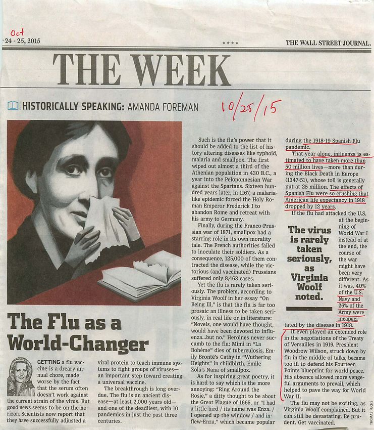
“The 1917 - 1918 influenza epidemic swept silently across the world bringing
death and fear to homes in every land. Disease and pestilence, especially the
epidemics, are little understood even now and many of the factors that spread them
are still mysterious shadows, but in 1917-1918 almost nothing was known about
prevention, protection, treatment or cure of influenza. The whole world stood at its
mercy, or lack of it.”
“But out of that particular epidemic, the young science of chiropractic grew
into a new measure of safety. While many struggles would lie ahead this successful
passage of the profession into early maturity assured its immediate survival and
made the eventual outcome of chiropractic a matter for optimism. If there had been
any lack of enthusiasm among the doctors of chiropractic, or a depleting of the
sources of students then the epidemic took care of them too. These chiropractic
survivors of the flu epidemic were sure, assured, determined, and ready to fight
any battle that came up. The effect of the epidemic becomes evident in interviews
made with old-timers practicing in those years. The refrain comes repeatedly,”
‘I was about to go out of business when the flu epidemic came - but when
it was over, I was firmly established in practice.’
“Why?
The answer is reasonably simple. Chiropractors got fantastic results from
influenza patients while those under medical care died like flies all around.”
“Statistics reflect a most amazing, almost miraculous state of affairs. The
medical profession was practically helpless with the flu victims but chiropractors
seemed able to do no wrong.”
“In Davenport, Iowa, 50 medical doctors treated 4,953 cases, with 274
deaths. In the same city, 150 chiropractors including students and faculty of the
Palmer School of Chiropractic, treated 1,635 cases with only one death.”
“In the state of Iowa, medical doctors treated 93,590 patients, with 6,116
deaths - a loss of one patient out of every 15. In the same state, excluding
Davenport, 4,735 patients were treated by chiropractors with a loss of only 6 cases
- a loss of one patient out of every 789.”2
“National figures show that 1,142 chiropractors treated 46,394 patients for
influenza during 1918, with a loss of 54 patients - one out of every 886.”
“Reports show that in New York City, during the influenza epidemic of 1918,
out of every 10,000 cases medically treated, 950 died; and in every 10,000
pneumonia cases medically treated 6,400 died. These figures are exact, for in that
city these are reportable diseases.”
“In the same epidemic, under drugless methods, only 25 patients died of
influenza out of every 10,000 cases; and only 100 patients died of pneumonia out
of every 10,000 cases. This comparison is made more striking by the following
table:”
Influenza
Cases Deaths
Under medical methods 10,000 950
Under drugless methods 10,000 25
Pneumonia
Cases Deaths
Under medical methods 10,000 6,400
Under drugless methods 10,000 100
“In the same epidemic reports show that chiropractors in Oklahoma treated
3,490 cases of influenza with only 7 deaths. But the best part of this is, in
Oklahoma there is a clear record showing that chiropractors were called in 233
cases where medical doctors had cared for the patients, and finally gave them up as
lost. The chiropractors saved all these lost cases but 25.”
“Statistics alone, however, don't put in that little human element needed to
spark the material properly. Dr. S. T. McMurrain [DC] had a makeshift table
installed in the influenza ward in Base Hospital No. 84 unit stationed in Perigau, in
Southwestern France, about 85 kilometers from Bordeaux [during WWI]. The
medical officer in charge sent all influenza patients in for chiropractic adjustments
from Dr. McMurrain [DC] for the several months the epidemic raged in that area. Lt.
Col. McNaughton, the detachment commander, was so impressed he requested to
have Dr. McMurrain [DC] commissioned in the Sanitary Corps.”3
“Dr. Paul Myers [DC] of Wichita Falls was pressed into service by the County
Health Officer and authorized to write prescriptions for the duration of the epidemic
there - but Dr. Myers [DC] said he never wrote any, getting better results without
medication.”
Dr. Helen B. Mason [DC], whose “son, when only a year old, became very ill
with bronchitis. My husband and I took him to several medical specialists without
any worthwhile results. We called a chiropractor, as a last resort, and were amazed
at the rapidity of his recovery. We discussed this amazing cure at length and came
to the decision that if chiropractic could do as much for the health of other
individuals as it had done for our son we wanted to become chiropractors.”
Dr. M. L. Stanphill [DC] recounts his experiences:
“I had quite a bit of practice in 1918 when the flu broke out. I stayed (in Van
Alstyne) until the flu was over and had the greatest success, taking many
cases that had been given up and restoring them back to health. During the
flu we didn't have the automobile. I went horseback and drove a buggy day
and night. I stayed overnight when the patients were real bad. When the rain
and snow came I just stayed it out. There wasn't a member of my family that
had the flu.”
When he came to Denison he said:
“I had a lot of trouble with pneumonia when I first came. Once again took all
the cases that had been given up. C. R. Crabetree, who lived about 18 miles
west of Denison, had double pneumonia and I went and stayed all night with
him and until he came to the next morning. He is still living today. That gave
me a boost on the west side of town.”
“And when interviews of the old timers are made it is evident that each still
vividly remembers the 1917-1918 influenza epidemic. We now know about 20
million persons [recent estimates are as high as 100 million deaths] around the
world died of the flu with about 500,000 Americans among that number. But most
chiropractors and their patients were miraculously spared and we repeatedly hear
about those decisions to become a chiropractor after a remarkable recovery or
when a close family member given up for dead suddenly came back to vibrant
health.”
“Some of these men and women were to become the major characters thrust
upon the profession's stage in the 20's and 30's and they had the courage, the
background and the conviction to withstand all that would shortly be thrown against
them” [including being thrown in jail for practicing medicine without a license].
“The publicity and reputation of such effectiveness in handling flu cases also
brought new patients and much acclaim from people who knew nothing of
chiropractic before 1918.”“The Innate Immune System”
Chapter 2
How The immune System Works
By
Lauren Sompayrac, PhD
Department of Molecular, Cellular, and
Developmental Biology
University of Colorado, Boulder
Blackwell Science
1999
“Until recently, most immunologists didn’t pay
much attention to the innate system, perhaps
because the adaptive system seemed more
exciting.
However, studies of the adaptive immune system
have led to a new appreciation of the role that the
innate system plays, not only as a second line of
defense (if we count physical barriers as our first
defense), but also as an activator and a controller
of the adaptive response.” p. 170031-6997/00/5204-0595$03.00/0
PHARMACOLOGICAL REVIEWS Vol. 52, No. 4
U.S. Government work not protected by U.S. copyright 41/865371
Pharmacol Rev 52:595–638, 2000 Printed in U.S.A
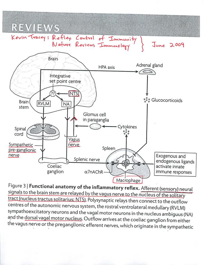
The Sympathetic Nerve—An Integrative Interface
between Two Supersystems: The Brain and the
Immune System
ILIA J. ELENKOV, RONALD L. WILDER, GEORGE P. CHROUSOS, AND E. SYLVESTER VIZI1
Inflammatory Joint Diseases Section, Arthritis and Rheumatism Branch, National Institute of Arthritis and Musculoskeletal and Skin
Diseases, National Institutes of Health, Bethesda, Maryland (I.J.E., R.L.W.); Pediatric Endocrinology Section, Developmental
Endocrinology Branch, National Institute of Child Health and Human Development, National Institutes of Health, Bethesda, Maryland
(I.J.E., G.P.C.); Department of Pharmacology, Institute of Experimental Medicine, Hungarian Academy of Sciences, Budapest, Hungary
(E.S.V.); and Department of Pharmacology and Pharmacotherapy, Semmelweis University, Budapest, Hungary (E.S.V.)
This paper is available online at http://www.pharmrev.org
Abstract . . . . . . . . . . . . . . . . . . . . . . . . . . . . . . . . . . . . . . . . . . . . . . . . . . . . . . . . . . . . . . . . . . . . . . . . . . . . . . 596
I. Introduction . . . . . . . . . . . . . . . . . . . . . . . . . . . . . . . . . . . . . . . . . . . . . . . . . . . . . . . . . . . . . . . . . . . . . . . . . . . 597
A. Overview . . . . . . . . . . . . . . . . . . . . . . . . . . . . . . . . . . . . . . . . . . . . . . . . . . . . . . . . . . . . . . . . . . . . . . . . . . 597
B. Historical perspectives . . . . . . . . . . . . . . . . . . . . . . . . . . . . . . . . . . . . . . . . . . . . . . . . . . . . . . . . . . . . . . 597
II. Anatomy and physiology of the autonomic nervous system . . . . . . . . . . . . . . . . . . . . . . . . . . . . . . . . . 598
A. Organization of the autonomic/sympathetic nervous system . . . . . . . . . . . . . . . . . . . . . . . . . . . . . 598
B. Role of sympathetic nervous system and hypothalamo-pituitary-adrenal axis in maintaining
basal and stress-related homeostasis . . . . . . . . . . . . . . . . . . . . . . . . . . . . . . . . . . . . . . . . . . . . . . . . . 599
III. Autonomic/sympathetic innervation of lymphoid organs: nonsynaptic communication . . . . . . . . . 599
A. Innervation of the thymus . . . . . . . . . . . . . . . . . . . . . . . . . . . . . . . . . . . . . . . . . . . . . . . . . . . . . . . . . . . 601
B. Innervation of the spleen . . . . . . . . . . . . . . . . . . . . . . . . . . . . . . . . . . . . . . . . . . . . . . . . . . . . . . . . . . . . 601
C. Innervation of lymph nodes and tonsils . . . . . . . . . . . . . . . . . . . . . . . . . . . . . . . . . . . . . . . . . . . . . . . 601
D. Innervation of the bone marrow . . . . . . . . . . . . . . . . . . . . . . . . . . . . . . . . . . . . . . . . . . . . . . . . . . . . . . 601
E. Innervation of mucosa-associated lymphoid tissues . . . . . . . . . . . . . . . . . . . . . . . . . . . . . . . . . . . . . 602
F. Coexistence patterns . . . . . . . . . . . . . . . . . . . . . . . . . . . . . . . . . . . . . . . . . . . . . . . . . . . . . . . . . . . . . . . . 602
G. General pattern of the autonomic/sympathetic innervation of lymphoid organs. . . . . . . . . . . . 602
H. Spatial relationships with peptidergic innervation . . . . . . . . . . . . . . . . . . . . . . . . . . . . . . . . . . . . . 603
I. Neuroimmune connection in nonorganized lymphoid compartments . . . . . . . . . . . . . . . . . . . . . . 603
IV. Nonsynaptic release of norepinephrine in lymphoid organs: presynaptic modulation and effect of
drugs . . . . . . . . . . . . . . . . . . . . . . . . . . . . . . . . . . . . . . . . . . . . . . . . . . . . . . . . . . . . . . . . . . . . . . . . . . . . . . . . . 603
A. Evidence for neural release of norepinephrine (and dopamine) in lymphoid organs . . . . . . . . 603
B. Norepinephrine is released and affects immune cells nonsynaptically . . . . . . . . . . . . . . . . . . . . 604
C. Presynaptic modulation of norepinephrine release in lymphoid organs: effect of drugs . . . . . 605
D. Release of neuropeptide Y and its action on immune cells. . . . . . . . . . . . . . . . . . . . . . . . . . . . . . . 606
V. Systemic and local effects of cytokines on sympathetic nervous system activity. . . . . . . . . . . . . . . 606
A. Systemic effects: long feedback loop between the immune system and the brain. . . . . . . . . . . 606
B. Local effects of tumor necrosis factor-! and interleukin-1 . . . . . . . . . . . . . . . . . . . . . . . . . . . . . . . 607
VI. Expression of adrenoreceptors on lymphoid cells: signal transduction . . . . . . . . . . . . . . . . . . . . . . . 608
A. Expression and distribution of adrenoreceptors on lymphoid cells. . . . . . . . . . . . . . . . . . . . . . . . 608
B. Signal pathways and molecular aspects of catecholamines actions . . . . . . . . . . . . . . . . . . . . . . . 609
1. Cyclic adenosine 5!-monophosphate . . . . . . . . . . . . . . . . . . . . . . . . . . . . . . . . . . . . . . . . . . . . . . . 609
2. Intracellular Ca2" . . . . . . . . . . . . . . . . . . . . . . . . . . . . . . . . . . . . . . . . . . . . . . . . . . . . . . . . . . . . . . . 610
VII. Role of sympathetic innervation in immune system development and hematopoiesis . . . . . . . . . . 611
A. Immune system development . . . . . . . . . . . . . . . . . . . . . . . . . . . . . . . . . . . . . . . . . . . . . . . . . . . . . . . . 611
B. Hematopoiesis. . . . . . . . . . . . . . . . . . . . . . . . . . . . . . . . . . . . . . . . . . . . . . . . . . . . . . . . . . . . . . . . . . . . . . 611
C. Thymocyte development . . . . . . . . . . . . . . . . . . . . . . . . . . . . . . . . . . . . . . . . . . . . . . . . . . . . . . . . . . . . . 612
VIII. Sympathetic control of lymphocyte traffic and circulation . . . . . . . . . . . . . . . . . . . . . . . . . . . . . . . . . . 612
1
Address for correspondence: Dr. E. Sylvester Vizi, Department of Pharmacology, Institute of Experimental Medicine, Hungarian
Academy of Sciences, H-1450 Budapest, P.O. Box 67, Hungary. E-mail: esvizi@koki.hu
595Autonomic innervation and regulation of th... [Brain Behav Immun. 2007] - PubMed - NCBI 11/29/13 3:22 PM PubMed Display Settings: Abstract Brain Behav Immun. 2007 Aug;21(6):736-45. Epub 2007 Apr 27. Autonomic innervation and regulation of the immune system (1987-2007). Nance DM, Sanders VM. Susan Samueli Center for Integrative Medicine, University of California Irvine, Orange, CA 92868-4283, USA. dnance@uci.edu Abstract Since 1987, only a few neuroanatomical studies have been conducted to identify the origin of innervation for the immune system. These studies demonstrated that all primary and secondary immune organs receive a substantial sympathetic innervation from sympathetic postganglionic neurons. Neither the thymus nor spleen receive any sensory neural innervation; however, there is evidence that lymph nodes and bone marrow may be innervated by sensory neurons located in dorsal root ganglia. There is no neuroanatomical evidence for a parasympathetic or vagal nerve supply to any immune organ. Thus, the primary pathway for the neural regulation of immune function is provided by the sympathetic nervous system (SNS) and its main neurotransmitter, norepinephrine (NE). Activation of the SNS primarily inhibits the activity of cells associated with the innate immune system, while it either enhances or inhibits the activity of cells associated with the acquired/adaptive immune system. Innate immune cells express both alpha and beta-adrenergic receptor subtypes, while T and B lymphocytes express adrenergic receptors of the beta2 subtype exclusively, except for murine Th2 cells that lack expression of any subtype. Via these adrenergic receptors, NE is able to regulate the level of immune cell activity by initiating a change in the level of cellular activity, which often involves a change in the level of gene expression for cytokines and antibodies. PMID: 17467231 [PubMed - indexed for MEDLINE] PMCID: PMC1986730 Free PMC Article Images from this publication. See all images (1) Free text Publication Types, MeSH Terms, Grant Support http://www.ncbi.nlm.nih.gov/pubmed/17467231 Page 1 of 2
PubMed Central, FIGURE 1: Brain Behav Immun. 2007 August; 21(6): 736–745. Published online 2007 April 27. doi: 10.1016/j.bbi.2007.03.008   5/28/13 4:15 PM

    FIGURE 1

    All primary and secondary immune organs receive a substantial sympathetic innervation from sympathetic
    postganglionic neurons. There is no neuroanatomical evidence for a parasympathetic or vagal nerve supply to any
    immune organ. Input to the brain comes from sensory, e.g., dorsal root ganglia, or immune stimuli, e.g., cytokines.
    The primary pathway for the neural regulation of immune function is provided by the sympathetic nervous system
    and its main neurotransmitter, norepinephrine. Activation of the SNS primarily inhibits the activity of cells
    associated with the innate immune system, while it either enhances or inhibits the activity of cells associated with

http://www.ncbi.nlm.nih.gov/pmc/articles/PMC1986730/figure/F1/?report=objectonly                                                                Page 1 of 2
1
Sympathetic Segmental Disturbances
The Evidences of the Association, in Dissected Cadavers, of Visceral
Disease with Vertebral Deformities of the Same Sympathetic Segments
Medical Times, November 1921, pp. 1-7
Henry Winsor, MD
THIS AUTHOR NOTES:
“The object of these necropsies was to determine whether any connection existed
between minor curvatures of the spine, on the one hand, and diseased organs on
the other.”
This author used 50 cadavers from the University of Pennsylvania.
49 of the 50 cadavers displayed minor curvatures of the spine, and 1 cadaver
displayed the normal “slight smooth lateral curve in the thoracic spine.”
This 1 cadaver still showed “very minor visceral pathology in the segments
immediately above and below the reported curve,” at “segments which should form
compensatory curves.”
“All [other] curves and deformities of the spine were rigid, apparently of long
duration; irreducible by ordinary manual force: extension, counter-extension,
rotation, even strong lateral movements failed to remove them or even cause them
to change their relative positions.”
Importantly, minor spinal curvatures “their association with disease of organs
belonging to the same sympathetic segment is more frequent than with gross
curves.”
Also importantly, in the 4 spines with gross curvatures “diseased organs were not
found to belong to the same sympathetic segments as the gross curves, but were
[found at] the same sympathetic segments as the minor compensatory curvatures
above and below the greater curves.”2
Visceral Disturbance Vertebral Curvatures OfSympathetic
The Same Sympathetic Connections Between
Segment As Visceral Vertebrae And Diseased
Trouble Organs
Diseased Thymus #2 C7, T1 #1Inferior Cervical
T-2-3-4 #1Sympathetic Ganglia
Adhered Pleurae #21 Upper Thoracics #19 Upper Thoracic Ganglia
Lower Thoracics #2Lower Thoracic Ganglia
Lung Diseases #26 Upper Thoracics #26 Upper Thoracic Ganglia
Heart & Pericardium T1-2-3-4-5 #18 Upper Thoracic Ganglia
Diseases #20 C7, T1 #2Inferior Cervical Ganglia
Stomach Diseases #9 T5-6-7-8-9 #8Greater Splanchnic From
An Adjacent Segment #1 Thoracics 5-9
Liver Diseases #13 T5-6-7-8-9 #12 Greater Splanchnic From
An Adjacent Segment #1 Thoracics 5-9
Gall Bladder Disease #5 T5-6-7-8-9 #5Greater Splanchnic From
Thoracics 5-9
Pancreas Disease #3 T5-6-7-8-9 #3 Greater Splanchnic From
Thoracics 5-9
Spleen Diseases #11 T5-6-7-8-9 #10 Greater Splanchnic From
Thoracics 5-9
T10-11-12 #1 Lesser Splanchnic Nerves
Inguinal Diseases #2 T12 #2 Ilio-inguinal Nerve
Kidney Disease #17 T10-11-12 #14 Least, Lesser & Greater
T5-6-7-8-9 #1 Splanchnic Nerves
L1-2 #2 Upper Lumbar Ganglia
Prostate & Bladder L1-2-3 #7 Upper Lumbar Ganglia
Disease #8 T12 #1 Last Thoracic Ganglia
Uterus Diseases #2 Lumbar Lordosis #2 Lumbar & Sacral Ganglia
Total Visceral Diseases Vertebral Curve Of Vertebral Curve Of
#139 Same Sympathetic Adjacent Segment #10
Segment As Disease
Site #128
“Therefore, in 50 cadavers with disease in 139 organs, there was found curve of the
vertebrae, belonging to the same sympathetic segments as the diseased organs
128 times, leaving an apparent discrepancy of 10, in which the vertebrae in curve
belonged to an adjacent segment to that which should supply the diseased organs
with sympathetic filaments.” [VERY IMPORTANT!]
The author then notes that the ten “apparent discrepancies from adjacent
segments” can be accounted for by “nerve filaments leaving the spinal cord and
traveling for a few segments.” [IMPORTANT]3 The author then states that if he included the cadaver with “faint curve and slight visceral pathology” that the correlation was 139 out of 139 for 100%. [WOW!] Importantly, the types documented include: Larynx cancer, fatty degeneration of the thymus, pleural adhesions, pleural effusions, pneumonia, tuberculosis, pulmonary edema, pulmonary congestion, lung fibrosis, bronchitis, enlarged lymph nodes, influenza, heart endocarditis, heart dilatation, heart muscle degeneration, pericarditis, aortic aneurysm, liver cirrhosis, liver swelling, liver tumors, enlarged spleen, atrophied spleen, inflamed spleen, pancreas degeneration, cystic kidneys, appendicitis, uterine adhesions, prostate hypertrophy, prostate atrophy, cystitis, hydrocele, osteomyelitis of the tibia, etc. “In general, we found the ordinary diseases of adult life.” In a separate evaluation, these authors found: 221 diseased organs; “Of these, 212 were observed to belong to the same sympathetic segment as the vertebrae in curvature.” “Nine diseased organs belonged to different sympathetic segments from the vertebrae out of line.” “These figures cannot be expected to exactly coincide, for an organ may receive sympathetic filaments from several spinal segments, and several organs may be supplied with sympathetic filaments from the same spinal segments.” “In no instance was a complete sympathetic block observed.” “Sympathetic disturbances are just as likely to cause functional or organic disease in viscera, by altering the blood-supply of viscera, through vaso-motor spasm.” [This is very important because vaso-motor spasm is subsequent to increased sympathetic tone. Sympathetic nerve compression would reduce sympathetic tone. Consequently the nerve interference resulting in visceral pathology in this study is not compression, but rather an irritation that causes increased sympathetic tone, vaso-motor spasm, and reduced blood flow]. In other research, this author has found that: 1) “Irritation of the sympathetic system and disease in the organs supplied by the same sympathetic nerves as the vertebrae affected.” 2) “That it was rare to find an organ diseased which was not supplied by the same sympathetic nerves as the vertebrae in curvature.” 3) “The sympathetic nerves were stretched over bony exudates [bone spurs] which angulated the nerves.” 4) “That even where no bony exudates was found, there was intense rigidity of the segments [sound much like subluxation complex], showing that fibrous or callous exudates could irritate the sympathetic nerves.” [Fibrosis of Repair]
4
5)   “The organs were in many instances affected by acute disease, while the
deformed vertebrae proved that the curvatures preceded the organic diseases…”
[EXTREMELY IMPORTANT]

6)    “…though theoretically, reflexes through muscle spasm may reverse the order
of precedence.” [WOW!]

The author notes that spondylosis is a process, “the last stage being fixation of
segments, immobilization of painful joints being one of nature’s later efforts to
check disease.”
“The disease [process then] going to the point of least resistance, in this instance to
the minor curvatures of the spine.”

The author describe the spondylosis process as follows:
A “sacro-iliac subluxation, an apparent shortening of the leg, comparative elevation
of the posterior superior iliac spine of the ilium, combined with lateral curve in the
lumbar region, lumbar curve and sacro-iliac subluxation (rotation of the
innominate) appear to be interdependent.”
[He even uses subluxation in the same context as a chiropractor].

“The stages of the process appears to be:
1)    Minor curves, or so-called sacroiliac subluxations;
2)    The muscles are converted into ligaments, ligaments to bone.
3)    Finally true bony ankylosis occurs.”
[This perfectly describes the phases of subluxation degeneration from
Renaissance from the 1970s by Feleesia and Riekeman].

“The disease appears to precede old age and to cause it. The spine becomes stiff
first and old age follows. Therefore, we may say a man is as old as his spine, the
arteries becoming hardened later from constant vaso-motor spasm, following
sympathetic irritation.” [Wow, can you believe this?]

The author notes that the sympathetic nerves can become entrapped extraspinally,
peripherally. “When the lungs were pulled out of the cadavers [of pleurisy patients
with pleural adhesions], the adhesions were sufficiently strong to pull the
intercostals vessels and nerves” including the sympathetic nerves. This “irritation
of the sympathetic nerves causes reflex spasm of the vaso-motors deranging the
blood-supply of the organs supplied by the sympathetic segment in curve.” The
results are an increase in lung disease, heart disease, and pneumonia [infection].

“Of three cadavers with inguinal disturbances (bilateral hernia, hydrocele, idiopathic
bubo or cancer, which had been excised in an old woman), all showed rotation of
the twelfth dorsal vertebrae; the connection links being the ilio-inguinal and genito-
crural nerves.” [WOW!]

“Skin diseases: two cadavers with warts exhibited minor curvatures in the region
from which the affected skin derived its nerve supply.” [WOW!]
7
KEY POINTS FROM DAN MURPHY

1)    Curvatures of the spine adversely affect the sympathetic nervous system.

2      The sympathetic nervous system controls the blood supply to the viscera, and
is therefore related to all manner of visceral diseases and pathology, and
specifically, “the ordinary diseases of adult life.”

3)  Visceral diseases and pathology can be traced back to the segmental levels of
sympathetic involvement with nearly 100% correlation.

4)     Prolonged abnormal spinal posture stretches the sympathetic nervous system,
firing the sympathetics, causing reduced blood supply to visceral organs, and
resulting in visceral pathology.

5)    Abnormal spinal curvatures precede organic visceral diseases.

6)     The author perfectly describes pelvic-lumbar subluxations, fibrosis, reduced
motion, and sympathetic nerve interference adversely influencing blood flow and
resulting in visceral pathology.

7)    Spinal disease precedes old age and causes old age.

8)   Stiff distorted spines cause sympathetic irritation, vascular spasm, arterial
hardening, and old age follows.

9)    A person is as old as his spine.

10) Postural distortions causing sympathetic dysfunction can be treated with
fulcrum-assisted reversal of the postural distortion. [Incredible]

11) This author reverence both osteopathic and chiropractic literature in his
bibliography.

COMMENT FROM DAN MURPHY

I originally saw this article at Renaissance Seminars from Joe Feleesia and Guy
Riekeman in the 1970s. Riekeman is now the President of Life University in Georgia.

Why don’t chiropractic colleges do more of this type of research?
The spinal cord as organizer of disease processes: III. Hyperactivi... - PubMed - NCBI                     11/21/14, 1:45 PM

 PubMed

Display Settings:                   Abstract

J Am Osteopath Assoc. 1979 Dec;79(4):232-7.

The spinal cord as organizer of disease processes: III. Hyperactivity of
sympathetic innervation as a common factor in disease.
Korr IM.

PMID: 583147 [PubMed - indexed for MEDLINE]

MeSH Terms

LinkOut - more resources

PubMed Commons                                                                               PubMed Commons home

     0 comments
                                                                                         How to join PubMed Commons

http://www.ncbi.nlm.nih.gov/pubmed/583147                                                                         Page 1 of 1
The modulation of visceral functions by somatic afferent activity. -... https://www.ncbi.nlm.nih.gov/pubmed/3302431
PubMed
Format: Abstract
Jpn J Physiol. 1987;37(1):1-17.
The modulation of visceral functions by somatic afferent activity.
Sato A, Schmidt RF.
Abstract
We began by briefly reviewing the historical background of neurophysiological studies of the
somato-autonomic reflexes and then discussed recent studies on somatic-visceral reflexes in
combination with autonomic efferent nerve activity and effector organ responses. Most of the
studies that have advanced our knowledge in this area have been carried out on anesthetized
animals, thus eliminating emotional factors. We would like to emphasize again that the
functions of many, or perhaps all visceral organs can be modulated by somato-sympathetic
or somato-parasympathetic reflex activity induced by a appropriate somatic afferent
stimulation in anesthetized animals. As mentioned previously, some autonomic nervous
outflow, e.g. the adrenal sympathetic nerve activity, is involved in the control of hormonal
secretion. John F. Fulton wrote in his famous textbook "Physiology of the Nervous System"
(1949) that the posterior pituitary neurosecretion system (i.e. for oxytocin and vasopressin)
could be considered a part of the parasympathetic nervous system. In the study of body
homeostasis and environmental adaptation it would seem very important to further analyze
the contribution of somatic afferent input to the autonomic nervous and hormonal regulation of
visceral organ activity. Also, some immunological functions have been found to be influenced
by autonomic nerves or hormones (e.g. adrenal cortical hormone and catecholamines).
Finally, we must take into account, as we have briefly discussed, that visceral functions can
be modulated by somatic afferent input via various degrees of integration of autonomic
nerves, hormones, and immunological processes. We trust that such research will be
expanded to higher species of mammals, and that ultimately this knowledge of somato-
visceral reflexes obtained in the physiological laboratory will become clinically useful in
influencing visceral functions.
PMID: 3302431
[Indexed for MEDLINE]
1 of 2 7/3/18, 2:57 PMSomatovisceral reflexes. - PubMed - NCBI https://www.ncbi.nlm.nih.gov/pubmed/8775021
PubMed
Format: Abstract
J Manipulative Physiol Ther. 1995 Nov-Dec;18(9):597-602.
Somatovisceral reflexes.
Sato A1.
Author information
1 Tokyo Metropolitan Institute of Gerontology, Japan.
Abstract
In experimental animals, both noxious and innocuous stimulation of somatic afferents have
been shown to evoke reflex changes in sympathetic efferent activity and, thereby, effector
organ function. These phenomena have been demonstrated in such sites as the
gastrointestinal tract, urinary bladder, adrenal medulla, lymphatic tissues, heart and vessels of
the brain and peripheral nerves. Most often, reflexes have been elicited experimentally by
stimulation of cutaneous afferents, although some work has also been conducted on muscle
and articular afferents, including those of spinal tissues. The ultimate responses may
represent the integration of multiple tonic and reflex influences and may exhibit laterality and
segmental tendencies as well as variable excitability according to the afferents involved.
Given the complexity and multiplicity of mechanisms involved in the final expression of the
reflex response, attempts to extrapolate to clinical situations should probably be eschewed in
favor of further systematic physiological studies.
Comment in
Manual healing diversity and other challenges to chiropractic integration. [J Manipulative Physiol
Ther. 2000]
PMID: 8775021
[Indexed for MEDLINE]
MeSH terms
1 of 2 7/3/18, 2:59 PMReflex effects of subluxation: the autonomic nervous system. - Pu... https://www.ncbi.nlm.nih.gov/pubmed/10714536
PubMed
Format: Abstract Full text links
J Manipulative Physiol Ther. 2000 Feb;23(2):104-6.
Reflex effects of subluxation: the autonomic nervous system.
Budgell BS1.
Author information
1 RMIT University-Japan, Tokyo.
Abstract
BACKGROUND: The collective experience of the chiropractic profession is that aberrant
stimulation at a particular level of the spine may elicit a segmentally organized response,
which may manifest itself in dysfunction within organs receiving autonomic innervation at that
level. This experience is at odds with classic views of neuroscientists about the potential for
somatic stimulation of spinal structures to affect visceral function.
OBJECTIVE: To review recent findings from basic physiologic research about the effects of
somatic stimulation of spinal structures on autonomic nervous system activity and the function
of dependent organs.
DATA SOURCE: Findings were drawn from a major recent review of the literature on the
influences of somatic stimulation on autonomic function and from recent original physiologic
studies concerning somatoautonomic and spinovisceral reflexes.
CONCLUSIONS: Recent neuroscience research supports a neurophysiologic rationale for the
concept that aberrant stimulation of spinal or paraspinal structures may lead to segmentally
organized reflex responses of the autonomic nervous system, which in turn may alter visceral
function.
PMID: 10714536
[Indexed for MEDLINE]
Publication types, MeSH terms
1 of 2 7/3/18, 3:05 PMInnocuous mechanical stimulation of the neck and alterations in hea... https://www.ncbi.nlm.nih.gov/pubmed/11515806
PubMed
Format: Abstract Full text links
Auton Neurosci. 2001 Aug 13;91(1-2):96-9.
Innocuous mechanical stimulation of the neck and alterations in
heart-rate variability in healthy young adults.
Budgell B1, Hirano F.
Author information
1 College of Medical Technology, Kyoto University, Japan. budgell@itan.kyoto-u.ac.jp
Abstract
The present study examined the effects of cervical spinal manipulation, a widely applied form
of physical therapy, which involves innocuous mechanical stimulation, on heart rate and heart-
rate variability, in a cohort of healthy young adults. Using a cross-over treatment design, with
a one-week washout period and, in contrast to a sham procedure, the authentic manipulation
produced significant alterations in both heart rate and measures of heart-rate variability
calculated from power spectrum analysis. In particular, there was an increase in the ratio of
low-frequency (LF)-to-high-frequency (HF) components of the power spectrum of heart-rate
variability, which may reflect a shift in balance between sympathetic and parasympathetic
output to the heart.
PMID: 11515806 DOI: 10.1016/S1566-0702(01)00306-X
[Indexed for MEDLINE]
Publication types, MeSH terms
LinkOut - more resources
1 of 1 7/3/18, 3:07 PM3 KEY POINTS FROM DAN MURPHY 1) The spinal adjustments used in this study were to C1-C2 and involved traditional supine rotary maneuver that achieved audible cavitation of the joint. 2) The adjustments were done by a chiropractor. 3) The ECG showed a significant reduction in heart rate as compared to the sham adjustment group, which supports inhibition of the sympathetic nervous system. 4) The results also support that upper cervical spinal adjustments alter the balance between sympathetic and parasympathetic output to the heart. 5) The leading explanation for the observed sympathetic inhibition of heart rate is that it is subsequent to mechanical afferent input from receptors in cervical tissues. 6) Other studies have also shown that innocuous mechanical stimulation of the neck via spinal manipulation is capable of eliciting changes in heart rate and blood pressure.
8324 • The Journal of Neuroscience, August 1, 2007 • 27(31):8324 – 8333

Behavioral/Systems/Cognitive

The Neurochemically Diverse Intermedius Nucleus of the
Medulla as a Source of Excitatory and Inhibitory Synaptic
Input to the Nucleus Tractus Solitarii
Ian J. Edwards,1* Mark L. Dallas,1* Sarah L. Poole,1 Carol J. Milligan,1 Yuchio Yanagawa,2 Gábor Szabó,3
Ferenc Erdélyi,3 Susan A. Deuchars,1 and Jim Deuchars1
1Institute of Membrane and Systems Biology, University of Leeds, Leeds LS2 9JT, United Kingdom, 2Department of Genetic and Behavioral Neuroscience,

Gunma University Graduate School of Medicine, and Solution Oriented Research for Science and Technology, Japan Science and Technology Agency,
Maebashi 371-8511, Japan, and 3Department of Gene Technology and Developmental Neurobiology, Institute of Experimental Medicine, H-1450 Budapest,
Hungary

Sensory afferent signals from neck muscles have been postulated to influence central cardiorespiratory control as components of
postural reflexes, but neuronal pathways for this action have not been identified. The intermedius nucleus of the medulla (InM) is a target
of neck muscle spindle afferents and is ideally located to influence such reflexes but is poorly investigated. To aid identification of the
nucleus, we initially produced three-dimensional reconstructions of the InM in both mouse and rat. Neurochemical analysis including
transgenic reporter mice expressing green fluorescent protein in GABA-synthesizing neurons, immunohistochemistry, and in situ hy-
bridization revealed that the InM is neurochemically diverse, containing GABAegric and glutamatergic neurons with some degree of
colocalization with parvalbumin, neuronal nitric oxide synthase, and calretinin. Projections from the InM to the nucleus tractus solitarius
(NTS) were studied electrophysiologically in rat brainstem slices. Electrical stimulation of the NTS resulted in antidromically activated
action potentials within InM neurons. In addition, electrical stimulation of the InM resulted in EPSPs that were mediated by excitatory
amino acids and IPSPs mediated solely by GABAA receptors or by GABAA and glycine receptors. Chemical stimulation of the InM resulted
in (1) a depolarization of NTS neurons that were blocked by NBQX (2,3-dioxo-6-nitro-1,2,3,4-tetrahydrobenzo[f ]quinoxaline-7-
sulfonoamide) or kynurenic acid and (2) a hyperpolarization of NTS neurons that were blocked by bicuculline. Thus, the InM contains
neurochemically diverse neurons and sends both excitatory and inhibitory projections to the NTS. These data provide a novel pathway
that may underlie possible reflex changes in autonomic variables after neck muscle spindle afferent activation.
Key words: posture; neck; cardiovascular; respiration; medulla oblongata; autonomic

Introduction                                                                                                           site for cardiorespiratory integration (Potts et al., 2003). Cardiore-
Reflex changes in cardiorespiratory variables during body move-                                                        spiratory changes can also be evoked by stimulation of neck muscle
ments rely on interactions between the somatic and autonomic                                                           afferents (Bolton et al., 1998; Bolton and Ray, 2000), proposed to
nervous systems. A prime example of such interaction is the so-                                                        contribute to alterations in cardiorespiratory outflow in preparation
matosympathetic reflex, in which stimulation of thinly myelin-                                                         for a change in posture (Bolton and Ray, 2000). In contrast to limb
ated group III (A!) and unmyelinated group IV (C-fiber) limb                                                           afferents, the sensory signals from these muscles appear to be medi-
muscle afferent fibers can reflexly increase cardiorespiratory out-                                                    ated by group IA muscle spindle afferents (Bolton et al., 1998). How-
put (Potts et al., 2000, 2003; Wilson, 2000). These reflexes are                                                       ever, the neural pathways that link these afferent signals to cardiore-
mediated via sensory afferent input to the spinal cord, which is                                                       spiratory control are completely unknown.
then relayed to the nucleus tractus solitarius (NTS), a brainstem                                                          One target for sensory information from neck muscles is the
                                                                                                                       cervical spinal cord where terminations can be found in the dor-
                                                                                                                       sal horn (although sparse) and the central cervical nucleus (CCN)
Received Feb. 13, 2007; revised May 25, 2007; accepted June 20, 2007.                                                  (Bakker et al., 1984; Pfaller and Arvidsson, 1988; Prihoda et al.,
   This work was supported in part by the Wellcome Trust (C.J.M. and J.D.) and Grants-in-Aid for Scientific Research   1991). The CCN projection is generally considered to underlie
from the Ministry of Education, Culture, Sports, Science and Technology and the Ministry of Health, Labor, and
Welfare, Japan (Y.Y.). I.J.E. was supported by the Biotechnology and Biological Sciences Research Council. We
                                                                                                                       spinal somatic reflex circuits, such as those for the tonic neck
acknowledge the contribution of Gareth Dobson, who was an undergraduate project student, to this work.                 reflex involved in postural control (Wilson et al., 1984; Brink et
   *I.J.E. and M.L.D. contributed equally and significantly to this work.                                              al., 1985; Hongo et al., 1988; Popova et al., 1995). There is also a
   Correspondence should be addressed to either Jim Deuchars or Susan A. Deuchars, Institute of Membrane and           strong direct neck muscle afferent projection to the medulla ob-
Systems Biology, Garstang Building, University of Leeds, Leeds LS2 9JT, UK. E-mail: J.Deuchars@leeds.ac.uk or
S.A.Deuchars@leeds.ac.uk.
                                                                                                                       longata where fibers terminate in the external cuneate nucleus
   DOI:10.1523/JNEUROSCI.0638-07.2007                                                                                  and a nucleus located at the lateral edges of the dorsal aspect of
Copyright © 2007 Society for Neuroscience 0270-6474/07/278324-10$15.00/0                                               the hypoglossal motor nucleus (XII), referred to either as the
The Neurochemically Diverse Intermedius Nucleus of the Medulla as a Source of Excitatory and
Inhibitory Synaptic Input to the Nucleus Tractus Solitarii
The Journal of Neuroscience
August 1, 2007
Cerebellum
External Upper
Cuneate Cervical
Nucleus Mechanoreceptors
Dorsal Motor Nucleus Nucleus From
Nucleus Tractus Intermedius Chiropractic
of the Solitarius Upper
Vagus Central Cervical
Cervical Adjustments
Nucleus
Parasympathetic Integrated
Efferents Autonomic
And
Cardiorespiratory
Circuits
Heart Tonic
Parasympathetic Postural
Lungs Afferents Reflexes
From
Stomach Thoracic
And
Intestines Abdominal
Viscera
Etc.Journal of Chemical Neuroanatomy 38 (2009) 166–175
Contents lists available at ScienceDirect
Journal of Chemical Neuroanatomy
journal homepage: www.elsevier.com/locate/jchemneu
Review
The intermedius nucleus of the medulla: A potential site for the integration of
cervical information and the generation of autonomic responses
Ian J. Edwards, Susan A. Deuchars, Jim Deuchars *
Institute of Membrane and Systems Biology, Garstang Building, University of Leeds, Leeds, LS2 9JT, United Kingdom
A R T I C L E I N F O A B S T R A C T
Article history: The intermedius nucleus of the medulla (InM) is a small perihypoglossal brainstem nucleus, which
Received 24 September 2008 receives afferent information from the neck musculature and also descending inputs from the vestibular
Received in revised form 6 January 2009 nuclei, the gustatory portion of the nucleus of the solitary tract (NTS) and cortical areas involved in
Accepted 6 January 2009
movements of the tongue. The InM sends monosynaptic projections to both the NTS and the hypoglossal
Available online 14 January 2009
nucleus. It is likely that the InM acts to integrate information from the head and neck and relays this
information on to the NTS where suitable autonomic responses can be generated, and also to the
Keywords:
hypoglossal nucleus to influence movements of the tongue and upper airways.
Autonomic
Central to the integratory role of the InM is its neurochemical diversity. Neurones within the InM
Proprioception
Perihypoglossal utilise the amino acid transmitters glutamate, GABA and glycine. A proportion of these excitatory and
Brainstem inhibitory neurones also use nitric oxide as a neurotransmitter. Peptidergic transmitters have also been
found within InM neurones, although as yet the extent of the pattern of co-localisation between
peptidergic and amino acid transmitters in neurones has not been established.
The calcium binding proteins calretinin and parvalbumin are found within the InM in partially
overlapping populations. Parvalbumin and calretinin appear to have complementary distributions
within the InM, with parvalbumin being predominantly found within GABAergic neurones and calretinin
being predominantly found within glutamatergic neurones.
Neurones in the InM receive inputs from glutamatergic sensory afferents. This glutamatergic
transmission is conducted through both NMDA and AMPA ionotropic glutamate receptors.
In summary the InM contains a mixed pool of neurones including glutamatergic and GABAergic in
addition to peptidergic neurones. Neurones within the InM receive inputs from the upper cervical region,
descending inputs from brain regions involved in tongue movements and those involved in the co-
ordination of the autonomic nervous system. Outputs from the InM to the NTS and hypoglossal nucleus
suggest a possible role in the co-ordination of tongue movements and autonomic responses to changes in
posture.
! 2009 Elsevier B.V. All rights reserved.
Contents
1. Introduction . . . . . . . . . . . . . . . . . . . . . . . . . . . . . . . . . . . . . . . . . . . . . . . . ....... . . . . . . . . . . . . . . . . . . . . . . . . . . . . . . . . . . . . . . . . . . . . . . 167
1.1. Nomenclature . . . . . . . . . . . . . . . . . . . . . . . . . . . . . . . . . . . . . . . . . ....... . . . . . . . . . . . . . . . . . . . . . . . . . . . . . . . . . . . . . . . . . . . . . . 167
1.2. Insights into function . . . . . . . . . . . . . . . . . . . . . . . . . . . . . . . . . . . ....... . . . . . . . . . . . . . . . . . . . . . . . . . . . . . . . . . . . . . . . . . . . . . . 167
2. Neurotransmitters . . . . . . . . . . . . . . . . . . . . . . . . . . . . . . . . . . . . . . . . . . . ....... . . . . . . . . . . . . . . . . . . . . . . . . . . . . . . . . . . . . . . . . . . . . . . 168
2.1. Amino acids . . . . . . . . . . . . . . . . . . . . . . . . . . . . . . . . . . . . . . . . . . ....... . . . . . . . . . . . . . . . . . . . . . . . . . . . . . . . . . . . . . . . . . . . . . . 168
2.2. NOS . . . . . . . . . . . . . . . . . . . . . . . . . . . . . . . . . . . . . . . . . . . . . . . . . ....... . . . . . . . . . . . . . . . . . . . . . . . . . . . . . . . . . . . . . . . . . . . . . . 170
2.3. Peptide transmitters. . . . . . . . . . . . . . . . . . . . . . . . . . . . . . . . . . . . ....... . . . . . . . . . . . . . . . . . . . . . . . . . . . . . . . . . . . . . . . . . . . . . . 170
3. Calcium binding proteins . . . . . . . . . . . . . . . . . . . . . . . . . . . . . . . . . . . . . ....... . . . . . . . . . . . . . . . . . . . . . . . . . . . . . . . . . . . . . . . . . . . . . . 171
3.1. Parvalbumin is predominantly found in inhibitory neurones . . . ....... . . . . . . . . . . . . . . . . . . . . . . . . . . . . . . . . . . . . . . . . . . . . . . 171
3.2. Calretinin is found within inhibitory and excitatory neurones in the InM . . . . . . . . . . . . . . . . . . . . . . . . . . . . . . . . . . . . . . . . . . . . . . 171
4. Receptors . . . . . . . . . . . . . . . . . . . . . . . . . . . . . . . . . . . . . . . . . . . . . . . . . . ....... . . . . . . . . . . . . . . . . . . . . . . . . . . . . . . . . . . . . . . . . . . . . . . 171
4.1. Glutamate receptors. . . . . . . . . . . . . . . . . . . . . . . . . . . . . . . . . . . . ....... . . . . . . . . . . . . . . . . . . . . . . . . . . . . . . . . . . . . . . . . . . . . . . 171
* Corresponding author. Tel.: +44 113 343 4249.
E-mail address: J.Deuchars@leeds.ac.uk (J. Deuchars).
0891-0618/$ – see front matter ! 2009 Elsevier B.V. All rights reserved.
doi:10.1016/j.jchemneu.2009.01.001Brain Struct Funct
DOI 10.1007/s00429-014-0734-8
ORIGINAL ARTICLE
Neck muscle afferents influence oromotor and cardiorespiratory
brainstem neural circuits
I. J. Edwards • V. K. Lall • J. F. Paton •
Y. Yanagawa • G. Szabo • S. A. Deuchars •
J. Deuchars
Received: 9 August 2013 / Accepted: 11 February 2014
! The Author(s) 2014. This article is published with open access at Springerlink.com
Abstract Sensory information arising from the upper labelled afferents co-localised with parvalbumin and
neck is important in the reflex control of posture and eye vesicular glutamate transporter 1 indicating that they are
position. It has also been linked to the autonomic control of proprioceptive. Anterograde tracing from the InM identi-
the cardiovascular and respiratory systems. Whiplash fied projections to brain regions involved in respiratory,
associated disorders (WAD) and cervical dystonia, which cardiovascular, postural and oro-facial behaviours—the
involve disturbance to the neck region, can often present neighbouring hypoglossal nucleus, facial and motor tri-
with abnormalities to the oromotor, respiratory and car- geminal nuclei, parabrachial nuclei, rostral and caudal
diovascular systems. We investigated the potential neural ventrolateral medulla and nucleus ambiguus. In brain sli-
pathways underlying such symptoms. Simulating neck ces, electrical stimulation of afferent fibre tracts lateral to
afferent activity by electrical stimulation of the second the cuneate nucleus monosynaptically excited InM neuro-
cervical nerve in a working heart brainstem preparation nes. Direct stimulation of the InM in the WHBP mimicked
(WHBP) altered the pattern of central respiratory drive and the response of second cervical nerve stimulation. These
increased perfusion pressure. Tracing central targets of results provide evidence of pathways linking upper cervical
these sensory afferents revealed projections to the inter- sensory afferents with CNS areas involved in autonomic
medius nucleus of the medulla (InM). These anterogradely and oromotor control, via the InM. Disruption of these
neuronal pathways could, therefore, explain the dysphagic
and cardiorespiratory abnormalities which may accompany
I. J. Edwards (&) ! V. K. Lall ! S. A. Deuchars ! cervical dystonia and WAD.
J. Deuchars (&)
School of Biomedical Sciences, University of Leeds,
Leeds LS2 9JT, UK Keywords Proprioception ! Autonomic !
e-mail: i.j.edwards@leeds.ac.uk Immunohistochemistry ! Electrophysiology
J. Deuchars
e-mail: J.Deuchars@leeds.ac.uk
Introduction
J. F. Paton
School of Physiology and Pharmacology, Bristol Heart Institute,
University of Bristol, Medical Sciences Building, Bristol, The intermedius nucleus of the medulla (InM) is a neuro-
BS8 1TD, UK chemically diverse perihypoglossal nucleus (Edwards et al.
2007, 2009) with no known function. Furthermore, very
Y. Yanagawa
little is known regarding the anatomical connectivity of the
Department of Genetic and Behavioral Neuroscience, Gunma
University Graduate School of Medicine JST, CREST, nucleus. We have previously identified a monosynaptic
Maebashi 371-8511, Japan projection from the InM into the neighbouring nucleus of
the solitary tract (NTS) using electrophysiology (Edwards
G. Szabo
et al. 2007), indicating a possible role in autonomic and/or
Department of Gene Technology and Developmental
Neurobiology, Institute of Experimental Medicine, respiratory control. Direct primary afferent input to the
Budapest 1450, Hungary InM arises from upper cervical levels in a number of
1235
[an important Oromotor
role in Control
cardiorespiratory
control]
[a pontine Orofacial To Phrenic [fovea, clarity
viscerosensory Control Nerve for of vision]
relay] Inspiratory
Activity
Pontine CN V C4—C5—C6 Eye Position
Parabrachial Motor
Nucleus CN VII Neurons
Hypoglossal NUCLEUS C1—C3 Vestibular
Nucleus INTERMEDIUS MECHANOS Nucleus
Tongue, Splanchnic
Swallowing, Sympathetic
Airway Patency Nerves
Nucleus Caudal Nucleus Autonomic Posture
Ambiguus Ventrolateral Tractus Innervation to
Medulla Solitarius and From the
Viscera
Muscles of the Inhibits The Most of the
Soft Palate, Sympathetic Integratory Sympathetic
Pharynx, Larynx Tone and Blood Center Nerves in the
Pressure Body are
Splanchnic
Regulation of Respiratory
Reflex and
Cardiovascular Cardiovascular
Activity and Behaviors
Modulate
Respiratory
FunctionsAtlas vertebra realignment and achievement of arterial pressure goa... https://www.ncbi.nlm.nih.gov/pubmed/?term=bakris+g+and+dickholtz
PubMed bakris g and dickholtz
Format: Abstract Full text links
J Hum Hypertens. 2007 May;21(5):347-52. Epub 2007 Mar 2.
Atlas vertebra realignment and achievement of arterial pressure
goal in hypertensive patients: a pilot study.
Bakris G1, Dickholtz M Sr, Meyer PM, Kravitz G, Avery E, Miller M, Brown J, Woodfield C, Bell B.
Author information
1 Department of Preventive Medicine, Rush University Hypertension Center, Chicago, IL, USA.
gbakris@earthlink.net
Abstract
Anatomical abnormalities of the cervical spine at the level of the Atlas vertebra are associated
with relative ischaemia of the brainstem circulation and increased blood pressure (BP).
Manual correction of this mal-alignment has been associated with reduced arterial pressure.
This pilot study tests the hypothesis that correcting mal-alignment of the Atlas vertebra
reduces and maintains a lower BP. Using a double blind, placebo-controlled design at a single
center, 50 drug naïve (n=26) or washed out (n=24) patients with Stage 1 hypertension were
randomized to receive a National Upper Cervical Chiropractic (NUCCA) procedure or a sham
procedure. Patients received no antihypertensive meds during the 8-week study duration. The
primary end point was changed in systolic and diastolic BP comparing baseline and week 8,
with a 90% power to detect an 8/5 mm Hg difference at week 8 over the placebo group. The
study cohort had a mean age 52.7+/-9.6 years, consisted of 70% males. At week 8, there
were differences in systolic BP (-17+/-9 mm Hg, NUCCA versus -3+/-11 mm Hg, placebo;
PCerebral metabolic changes in men after chiropractic spinal manipu... https://www.ncbi.nlm.nih.gov/pubmed/22314714
PubMed
Format: Abstract
Altern Ther Health Med. 2011 Nov-Dec;17(6):12-7.
Cerebral metabolic changes in men after chiropractic spinal
manipulation for neck pain.
Ogura T1, Tashiro M, Masud M, Watanuki S, Shibuya K, Yamaguchi K, Itoh M, Fukuda H, Yanai K.
Author information
1
Division of Cyclotron Nuclear Medicine, Tohoku University, Sendai, Japan.
Abstract
BACKGROUND: Chiropractic spinal manipulation (CSM) is an alternative treatment for back
pain. The autonomic nervous system is often involved in spinal dysfunction. Although studies
on the effects of CSM have been performed, no chiropractic study has examined regional
cerebral metabolism using positron emission tomography (PET).
OBJECTIVE: The aim of the present study was to investigate the effects of CSM on brain
responses in terms of cerebral glucose metabolic changes measured by
[18F]fluorodeoxyglucose positron emission tomography (FDG-PET).
METHODS: Twelve male volunteers were recruited. Brain PET scanning was performed twice
on each participant, at resting and after CSM. Questionnaires were used for subjective
evaluations. A visual analogue scale (VAS) was rated by participants before and after
chiropractic treatment, and muscle tone and salivary amylase were measured.
RESULTS: Increased glucose metabolism was observed in the inferior prefrontal cortex,
anterior cingulated cortex, and middle temporal gyrus, and decreased glucose metabolism
was found in the cerebellar vermis and visual association cortex, in the treatment condition (P
< .001). Comparisons of questionnaires indicated a lower stress level and better quality of life
in the treatment condition. A significantly lower VAS was noted after CSM. Cervical muscle
tone and salivary amylase were decreased after CSM. Conclusion The results of this study
suggest that CSM affects regional cerebral glucose metabolism related to sympathetic
relaxation and pain reduction.
PMID: 22314714
[PubMed - indexed for MEDLINE]
1 of 2 2/28/17, 11:33 AMGlucose Metabolic Changes in the Brain and Muscles of Patients w... https://www.ncbi.nlm.nih.gov/pubmed/28167971
PubMed
Format: Abstract Full text links
Evid Based Complement Alternat Med. 2017;2017:4345703. doi: 10.1155/2017/4345703. Epub 2017 Jan 12.
Glucose Metabolic Changes in the Brain and Muscles of Patients with
Nonspecific Neck Pain Treated by Spinal Manipulation Therapy: A
[18F]FDG PET Study.
Inami A1, Ogura T2, Watanuki S1, Masud MM3, Shibuya K4, Miyake M1, Matsuda R1, Hiraoka K1, Itoh
M4, Fuhr AW5, Yanai K6, Tashiro M1.
Author information
Abstract
Objective. The aim of this study was to investigate changes in brain and muscle glucose
metabolism that are not yet known, using positron emission tomography with
[18F]fluorodeoxyglucose ([18F]FDG PET). Methods. Twenty-one male volunteers were
recruited for the present study. [18F]FDG PET scanning was performed twice on each subject:
once after the spinal manipulation therapy (SMT) intervention (treatment condition) and once
after resting (control condition). We performed the SMT intervention using an adjustment
device. Glucose metabolism of the brain and skeletal muscles was measured and compared
between the two conditions. In addition, we measured salivary amylase level as an index of
autonomic nervous system (ANS) activity, as well as muscle tension and subjective pain
intensity in each subject. Results. Changes in brain activity after SMT included activation of
the dorsal anterior cingulate cortex, cerebellar vermis, and somatosensory association cortex
and deactivation of the prefrontal cortex and temporal sites. Glucose uptake in skeletal
muscles showed a trend toward decreased metabolism after SMT, although the difference
was not significant. Other measurements indicated relaxation of cervical muscle tension,
decrease in salivary amylase level (suppression of sympathetic nerve activity), and pain relief
after SMT. Conclusion. Brain processing after SMT may lead to physiological relaxation via a
decrease in sympathetic nerve activity.
PMID: 28167971 PMCID: PMC5267084 DOI: 10.1155/2017/4345703
[PubMed - in process] Free PMC Article
Images from this publication. See all images (7) Free text
1 of 2 2/28/17, 11:39 AMMeasureable changes in the neuro-endocrinal mechanism followin... https://www.ncbi.nlm.nih.gov/pubmed/26464145
PubMed
Format: Abstract Full text links
Med Hypotheses. 2015 Dec;85(6):819-24. doi: 10.1016/j.mehy.2015.10.003. Epub 2015 Oct 14.
Measureable changes in the neuro-endocrinal mechanism
following spinal manipulation.
Kovanur Sampath K1, Mani R2, Cotter JD3, Tumilty S2.
Author information
1 Centre for Health, Activity, and Rehabilitation Research, School of Physiotherapy, University
of Otago, New Zealand. Electronic address: kesava.kovanur-sampath@otago.ac.nz.
2 Centre for Health, Activity, and Rehabilitation Research, School of Physiotherapy, University
of Otago, New Zealand.
3 School of Physical Education, Sport and Exercise Sciences, University of Otago, New
Zealand.
Abstract
The autonomic nervous system and the hypothalamic-pituitary-adrenal axis have been shown
to be dysfunctional in a number of chronic pain disorders. Spinal manipulation is a therapeutic
technique used by manual therapists, which may have widespread neuro-physiological
effects. The autonomic nervous system has been implicated to modulate these effects. A
theory is proposed that spinal manipulation has the potential to be used as a tool in restoring
the autonomic nervous system balance. Further, it is also hypothesised that through its
anatomical and physiological connections, the autonomic nervous system activity following a
thoracic spinal manipulation may have an effect on the hypothalamic-pituitary-adrenal axis
and therefore pain and healing via modulation of endocrine and physiological processes. To
substantiate our hypothesis we provide evidence from manual therapy studies, basic science
and animal studies. According to the proposed theory, there will be measurable changes in
the neuro-endocrinal mechanisms following a thoracic spinal manipulation. This has far-
reaching implications for manual therapy practice and research and in the integration of spinal
manipulation in the treatment of a wide array of disorders.
Copyright © 2015 Elsevier Ltd. All rights reserved.
PMID: 26464145 DOI: 10.1016/j.mehy.2015.10.003
[Indexed for MEDLINE]
1 of 2 7/2/17, 12:37 PMYou can also read