ACTA ORTHOPAEDICA ET TRAUMATOLOGICA - EEXOT
←
→
Page content transcription
If your browser does not render page correctly, please read the page content below

VOLUME 69 | ISSUE 4 | OCTOBER - DECEMBER 2018                    ISSN 2241-4347

    Acta
 Orthopaedica et
Traumatologica
 He l l eni c a

BASIC SCIENCE
 Muscle
        activity during locomotion in various inclination surfaces
 and different running speeds

ORIGINAL PAPER
 Arthroscopic
               debridement of minor meniscal lesions: Clinical
 outcome of three years follow up based on questionnaire and search
 for causes of failure

review ARTICLE
 The
     role of zoledronic acid in the treatment of post-menopausal
 osteoporosis

Case report
 Alveolar
          rhabdomyosarcoma of the thenar eminence in a 7-year-old
 child. A case report.

                   Hellenic Association
                   of Orthopaedic Surgery
                   and Traumatology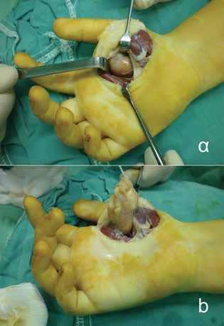
Acta      Orthopaedica et Traumatologica Hellenica

             HAOST Administrative Board
                         President:   Stamatios A. Papadakis
                    President 2017:   Pantelis K. Nikolaou
                 A’ Vice-President:   Panayiotis I. Papaggelopoulos
                 B’ Vice President:   Athanasios Th. Kostakos
                 General Secretary:   Odysseas A. Paxinos
                         Treasurer:   Konstantinos Th. Kateros
                 Special Secretary:   Alexandros A. Eleftheropoulos
                  Simple Member:      Zinon Th. Kokkalis
               Member’s Delegate:     Panayiotis D. Megaloikonomos

        HAOST College Administrative Board
                         President:   Stefanos M. Proveleggios
                    Vice-President:    Aristides B. Zoubos
                        Ex Officio:    Panayiotis I. Papaggelopoulos
                 General Secretary:   Nikolaos Markeas
                          Member:     Anastasios Mourikis
               Residents’ Delegate:   Panayiotis Megaloikonomos

             Presidents of HAOST Sections
            Reconstructive Surgery:   Efstathios Chronopoulos
                         Oncology:    Theodoros Kormas
                     Foot & Ankle:    Iason Petroutsas
                            Spine:    Vasileios Lykomitros
                Shoulder & Elbow:     Pericles Papadopoulos
                         Research:    Anastasios Christodoulou
                        Infections:   Konstantinos Malizos
            Intramedullary nailing:   Christine Arnaoutoglou
           Paediatric Orthopaedics:   Dimitrios Metaxiotis
              Primary Health Care:    Athanasios Badekas

Hellenic Association            Published by: ZITA MEDICAL MANAGEMENT S.A.
of Orthopaedic Surgery          1st klm Peanias - Markopoulou Avenue, Peania, Attica,
and Traumatology                Greece, P.O BOX 155, 190 02, tel.: +30 211 1001 777, fax:
                                +30 210 6642116, E-mail: info@zita-management.gr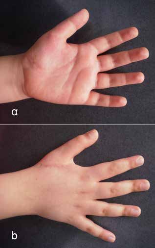
Acta  Orthopaedica et Traumatologica Hellenica

                Editor in chief
                  Nikolaos Papaioannou

              Assistant Editors
                    Theodoros Grivas
                    Nikolaos Markeas
                   Stamatios Papadakis
                Ioannis Triantafyllopoulos

               Editorial Board
            Dimitrios-Sergios Evaggelopoulos
                 Dimitrios Economopoulos
                 Efstathios Chronopoulos
                   Konstantinos Kateros
            Kalliopi Lampropoulou-Adamidou
                   Andreas Mavrogenis

 Scientific Committee and Reviewers

            Georgios Babis     Khaldi Lubna
        Athanasios Badekas     Georgios Machairas
            Alexia Balanika    Evaggelos Magnisalis
             Christos Baltas   Konstantinos Malizos
    Hippocrates Chatzokos      Panayiotis Megas
  Anastasios Christodoulou     Dionysios Mouzakis
   Konstantinos Demetzos       Pantelis Nikolaou
        Ioannis Dionysiotis    Elias Panayiotopoulos
        Ismini-Niki Dontas     Georgios Panayiotakopoulos
               Eleni Douni
                               Andreas Panagopoulos
      Panayiotis Efstathiou
                               Panayiotis Papaggelopoulos
          Ioannis Feroussis
                               Apostolos Papalois
           Antonis Galanos
                               Georgios Papanikolaou
             Ioannis Gliatis
                               Athanasios Papavassiliou
            Michael Hantes
  Anastasios Kanellopoulos     Georgios Petsatodis
     Theofilos Karachalios     Spyridon Pnevmatikos
        Aikaterini Katsalira   Georgios Sapkas
     Konstantinos Kazakos      Symeon Tournis
         Georgios Kontakis     Georgios Trovas
         Theodoros Kormas      Eleftherios Tsiridis
     Anastasios Korobilias     Minos Tyllianakis
           Dimitrios Korres    Eleni Vavouraki
      Irene Lambrinoudaki      Theodoros Xenakis
Acta
 instructions
 to authors
1. Scope                                                    uscript and submit their recommendation is three
“Acta Orthopaedica Et Traumatologica” is the official       weeks. The Editor-in-chief makes the final decision
journal of the Hellenic Association of Orthopaedic          for publication. The Editorial office will communi-
Surgery and Traumatology, first published in 1948.          cate the reviewer’s comments and the decision to the
This revived edition of Acta Orthopaedica Et Trau-          authors.
matologica, published in English, aspires to promote
scientific knowledge in Orthopaedics and Traumatol-         4. Manuscript originality and copyright
ogy worldwide. It is a peer-reviewed Journal, aim-          The submitted manuscript should be original,
ing at raising the profile of current evidence-based        should not contain previously published material
Orthopaedic practice and at improving the scientif-         and should not be under consideration for publica-
ic multidisciplinary dialogue. Acta Orthopaedic Et          tion in another journal. The submission needs to be
Traumatologica Hellenica presents clinically perti-         approved by all co-authors and in case of original
nent, original research and timely review articles. It      research a ‘guarantor’ of the study is required. As
is open to International authors and readers and of-        ‘guarantor’ may be considered a senior author that is
fers a compact forum of communication to Orthopae-          deemed to take overall responsibility for all aspects
dic Surgeons and related science specialists.               of the study (ethics, originality, consent, data han-
                                                            dling, and all aspects of Good Medical Practice). The
2. Language                                                 ‘guarantor’ of the study does not necessarily need
English is the official language of the journal. All sub-   to be the corresponding author. The journal will not
mitted manuscripts should be written in English.            hold legal responsibility should there be any claim
                                                            for compensation.
3. How to submit a paper                                       All authors need to sign the copyright transfer form
All submissions for peer-review should be performed         (link) and must have made substantial contributions
online through the journal.                                 as established by the ICMJE (http://www.icmje.org).
  The Editorial office and the Editor-in-chief will per-
form the initial assessment of the manuscript and if        5. Conflict of interest disclosure
the manuscript is suitable for the journal and the sub-     Each author needs to disclose any type of financial
mission is complete, it will be sent to the relative re-    interest that is related to the study and might create
viewers. The reviewing process that is followed is          a potential conflict. Funding of the study needs to be
double blinded. During on-line submission, authors          disclosed.
can enter the name/s of non-preferred reviewers.              If there is no conflict of interest, this should be
  The time allocated for reviewers to assess the man-       stated in the manuscript before the Reference sec-

acta Orthopaedica et Traumatologica Hellenica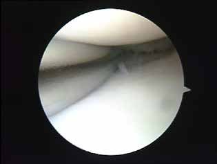
instructions to authors
VOLUME 69 | ISSUE 4 | OCTOBER - DECEMBER 2018      review    Acta
tion as follows: “The authors declared no conflicts           stract of 200 words, 3-5 keywords, text of no more
of interest”.                                                 than 6,000 words, figures (up to eight figures), a
                                                              maximum of six tables, a maximum of a hundred
6. Research ethics and compliance                             references and a maximum of three authors are re-
The journal follows the guidelines of the Internation-        quired for review articles.
al Committee of Medical Journal Editors (www.icm-              asic Science. Basic science manuscripts could be
                                                              B
je.org). For all original articles a statement in the text    either original or review articles on recent research
of approval from the local ethics committee, a state-         achievements. Authors should follow the corre-
ment that research was performed according to the             sponding insturctions according to the type of man-
ethical standards as described by the Declaration of          uscript (original or review).
Helsinki and a statement that informed consent for             onographs. Highly detailed and thoroughly doc-
                                                              M
participation in the study was obtained from all sub-         umented studies or reviews written about a limited
jects, are required. In case of study with animals the        area of a subject or field of inquiry. Monographies
following statement needs to be added in the text:            will be published on special issues.
“All applicable international, national, and/ or in-           ictorial Essays: The purpose of pictorial essays is
                                                              P
stitutional guidelines for the care and use of animals        to provide a teaching message through high qual-
were followed”.                                               ity images. A brief text is required to accompany
                                                              figures. An unstructured abstract of 200 words, 3-5
7. Permissions and plagiarism                                 keywords, text of no more than 6,000 words, a max-
For the use of any figures already published else-            imum of 15 figures, a maxi-mum of 6 tables, a max-
where the authors are required to obtain written per-         imum of a 100 references and a maximum of 4 au-
mission from the copyright owner(s) and to submit             thors are required for pictorial essays.
the evidence in the submission process. Plagiarism             ase Reports: Reports on new or very rare clini-
                                                              C
will not be accepted in any case. Dedicated software          cal cases on Orthopaedics, Orthopaedic Patholog-
will be used on this purpose; manuscripts with pla-           yand Trauma, new diagnostic criteria, new thera-
giarism will be returned to the corresponding author          peutic methods with proven result. Maximum 1,500
without consideration for peer review.                        words, 10 references and 6 figures. Abstract up to
                                                              100 words.
8. Types of manuscript                                         Letters  to the editor: Communication to the edi-

The journal accepts the following types of articles:           tor is welcomed and will be published if they offer
   riginal articles: The paper needs to offer new
  O                                                            pertinent and/ or constructive comment on arti-
  knowledge on Orthopaedics ant Traumatology. The              cles published in the Acta Orthopaedica Et Trau-
  conclusions need to be sound and supported by sta-           matologica Hellenica. Letters are published at the
  tistical analysis. When the accuracy of a diagnostic         discretion of the Editorial team and should be re-
  test is assessed, following the Standards for Report-        ceived within three months after on-line publi-
  ing of Diagnostic Accuracy (STARD) flow diagram              cation of an article. Following acceptance, letters
  (http://www. stard-statement. org) is suggested. A           will be sent to authors for response. Letter com-
  structured abstract of 250 words, 3-5 keywords, text         munications should include text of no more than
  up to 4,500 words, figures (up to four figures or eight      500 words, up to 2 figures and 10 references, with-
  figure parts), a maximum of six tables, a maximum of         out any abstract or keywords and a maximum of
  fifty references and a maximum of seven authors are          3 authors.
  required for original articles.
   eview Articles: The journal may accept system-
  R                                                          9. Manuscript organization
  atic reviews, meta-analyses, literature reviews and        A manuscript must contain the following parts for
  historical reviews of a subject. An unstructured ab-       submission:

                                                       acta Orthopaedica et Traumatologica Hellenica
Acta
review                                                       VOLUME 69 | ISSUE 4 | OCTOBER - DECEMBER 2018

 C
  over letter: Each manuscript needs to be accompa-           cal companies, biomedical device manufacturers or
 nied by a cover letter signed by the corresponding            other corporations whose products or services have
 author on behalf of the rest of the authors stating           been used needs to be included in the Conflicts of
 that the article is not under consideration in anoth-         Interest Form and also mentioned in acknowledge-
 er journal. In case of article resubmission a point-          ments section.
 by-point answer to the reviewer’s comments needs              M
                                                                easurement Units: All measurements should be
 to be submitted with the cover letter.                        mentioned in international units (SI). The full stop
 T
  itle page: It includes the title of the manuscript, the     should be used as a decimal (i.e. 3.5 cm). Spac-
 names, affiliations and e-mail addresses of all au-           es should be added around the plus/minus sym-
 thors and the affiliation, address, e-mail address,           bol (i.e. 13.6 ± 1.2). There should not be any spaces
 telephone and fax number of the corresponding au-             around range indicators (i.e. 15-20) or equality/in-
 thor. The name and affiliation of the ‘guarantor’ of          equality symbols (i.e. r=0.37, p
instructions to authors
VOLUME 69 | ISSUE 4 | OCTOBER - DECEMBER 2018 review Acta
11. References Surg 2017; doi: 10.1016/j.jse.2017.05.032 Epub 2017
The accuracy of references is the responsibility of the Jul 21.
authors. or
References need to be cited in the text in the order Papaioannou NA, Triantafyllopoulos IK, Khaldi L,
in which they appear. The numbering needs to be in et al. Effect of calcitonin in early and late stages of
Arabic numbers and placed in the respective areas of experimentally induced osteoarthritis. A histomor-
text into square brackets i.e [1]. phometric study. Osteoarthritis Cartilage 2007; 15(4):
References that have not been published at the 386-95.
point of submission need to cited with the respective
DOI (digital object identifier) number given for on- Book chapters:
line first articles. Triantafyllopoulos IK, Papaioannou NA. The Effect
All authors (surnames and initials of first name) of Pharmacological Agents on the Bone-Implant In-
should be listed when they are three or fewer. If au- terface. In: Karachalios Th. (ed). Bone-Implant Inter-
thors are more than three, the first three authors face in Orthopaedic Surgery. Springer – Verlag, Lon-
should be listed, then ‘et al.’ needs to follow the name don 2014, pp 221-237.
of the third author.
When a book chapter is cited, the authors and ti- Online document:
tle of the chapter, editors, book title, edition, city and National Institute for Health and Care Excellence.
country, publisher, year and specific chapter pages Fractures (Complex): Assessment and Management.
should be mentioned. Available via www.nice.org.uk/guidance/ng37.
For Online Document, the following should be Published Feb 2016. Updated Sept 2017. Accessed
mentioned: authors (if any), title of page, name of in- January 2014.
stitution or owner of Web site; URL; dates of publi-
cation, update, and access. 12. Review of manuscripts
Acceptance of manuscripts for publication is decided
Reference examples: by the Editor, based on the results of peer review. Au-
thors need to make proof corrections within 72 hours
Journal article: upon pdf supplied, check the integrity of the text, ac-
Trianafyllopoulos IK, Lampropoulou-Adamidou K, cept any grammar or spelling changes and check if
Schizas NP, et al. Surgical treatment of acute type V all the Tables and Figures are included and proper-
acromioclavicular joint dislocations in professional ly numbered. Once the publication is online, no fur-
athletes: An anatomic ligament reconstruction with ther changes can be made. Further changes can only
synthetic implant augmentation. J Shoulder Elbow be published in form of Erratum.
acta Orthopaedica et Traumatologica HellenicaActa Orthopaedica et Traumatologica Hellenica Contents BASIC SCIENCE Muscle activity during locomotion in various inclination surfaces and different running speeds Theodoros V. Roussos, Athanasia Smirniotou, Flora N. Panteli, Ioannis K. Triantafyllopoulos 154-163 ORIGINAL PAPER Arthroscopic debridement of minor meniscal lesions: Clinical outcome of three years follow up based on questionnaire and search for causes of failure Nick Sekouris, Evans Glyn, Antonios Aggoules 164-171 review ARTICLE The role of zoledronic acid in the treatment of post-menopausal osteoporosis S. Dellis, I.K. Triantafyllopoulos 172-184 Case report Alveolar rhabdomyosarcoma of the thenar eminence in a 7-year-old child. A case report. Dimitrios Begkas, Nikolaos G. Markeas, Panagiotis Touzopoulos, Leonardos Benakis 185-191
Acta
BASIC SCIENCE                                              VOLUME 69 | ISSUE 4 | OCTOBER - DECEMBER 2018

              Muscle activity during locomotion
              in various inclination surfaces and
                   different running speeds.
       Theodoros V. Roussos1, Athanasia Smirniotou2, Flora N. Panteli3, Ioannis K. Triantafyllopoulos4

                       1
                         Laboratory for the Research of Musculoskeletal Disorders Medical School,
                                 National and Kapodistrian University of Athens, Greece
                           2
                             Associate Professor, School of Physical Education and Sport Science,
                                 National and Kapodistrian University of Athens, Greece
      4
        MSc, School of Physical Education and Sport Science National and Kapodistrian University of Athens, Greece
                                            4
                                              Assistant Professor of Orthopaedics
                         Medical School, National and Kapodistrian University of Athens, Greece

Abstract
During dynamic activities – walking, jogging and running, muscular function is affected by running techniques
and foot strike patterns, inclined surfaces and running speed. In order to assess muscle function during
these activities, most studies examine certain muscles such as tibialis anterior, gastrocnemius (lateral and
medial), soleus, rectus femoris, vastus (medialis and lateralis), hamstrings (biceps femoris, semimembranosus,
semitendinosus), and gluteus. These muscles are commonly selected because they provide supportive and
propulsive forces during running. Results of these studies may conclude to special training programs for
runners in order to improve their performance.

KEYWORDS: Running; muscle activation; running surfaces; running speeds

Introduction                                                 reaction forces magnitude influences mechanical
Running is a popular physical activity and a key             function of the musculoskeletal system and muscle
element in most conditioning programs. At each               activation patterns.
running step, when the foot strikes the supporting             During dynamic activities – walking and running,
ground, a ground reaction force (GRF) of two- or             muscular function is affected by running techniques
three-times body weight is generated [6] inducing            and foot strike patterns, inclined surfaces and run-
shock waves that propagate throughout the lo-                ning speed. Inclined support surfaces affect the con-
comotor system. The load resulting from ground               trol of movement in terms of the maintenance of an

Corresponding                I.K.Triantafyllopoulos
                             MD, MSci, PhD, FEBOT
author,
                             Tel. 210-6124007,
guarantor
                             Email: i.triantafillopoulos@med.uoa.gr

154       acta Orthopaedica et Traumatologica Hellenica
Roussos VT, et al. Muscle activity during locomotion
in various inclination surfaces and different running speeds
VOLUME 69 | ISSUE 4 | OCTOBER - DECEMBER 2018
upright posture [22], the foot strike patterns used but not during the support phase. It has also been
and the related centre of pressure in anterior – pos- observed that in high – mileage runners the muscu-
terior direction during stance, and muscles activity lar activity of the gastrocnemius lateralis during the
[24]. Sasagawa [40] assessed the active stabilization support phase was reduced compared to asympto-
mechanisms on an inclined surface during quiet matic controls [4]. Probably, the pre-activation of
standing and found that muscle activity changed as the gastrocnemius lateralis is in fact necessary in
a function of support surface conditions. midfoot strike running technique since the plantar
In order to assess muscle function during running, flexors need to counteract the dorsiflexor moment
most studies examine the following muscle groups: created during the midfoot strike pattern [16].
tibialis anterior, gastrocnemius (lateral and medial), An earlier, longer and greater plantar flexors (PF)
soleus, rectus femoris, vastus (medialis and later- activity, lower dorsiflexor activity, and greater bi-
alis), hamstrings (biceps femoris, semimembrano- ceps femoris activity have been observed when run-
sus, semitendinosus), and gluteus. These muscles ning with a forefoot strike (FFS) pattern [1,16,50].
are selected because they provide supportive and Runners adopting a forefoot strike pattern activated
propulsive forces during running [21]. their plantar flexors muscles 11% earlier and 10%
longer than runners with a rearfoot strike pattern.
Effects of foot strike pattern and inclined surfaces Specifically, the activation phase of medial gastroc-
on muscle activity nemius (MG) occurred 7.7-16.3% of the gait cycle
The work performed by muscle groups is partial- earlier and lasted on average 9.7% longer for the
ly affected by the foot strike pattern adopted dur- forefoot strike runners compared to rearfoot strike
ing locomotion [1,16,50]. According to the heel and runners, at all speeds (2.5, 2.8, 3.2 and 3.5m/sec).
metatarsal positioning at landing, three foot strike A similar trend was observed for the activation
patterns have been identified: rearfoot strike (RFS) phase of lateral gastrocnemius (LG) as well. Fore-
in which the heel lands before the ball of foot, mid- foot strike runners activated their lateral gastroc-
foot strike (MFS) in which the heel and the ball of nemius muscles 7.7-13.1% of the gait cycle earlier
foot lands almost simultaneously, and forefoot and 6.3-14.3% longer than rearfoot strike runners
strike (FFS) in which the ball of foot lands before at all speeds. However, calf muscles deactivation
the heel [17]. time was not influenced by running technique. This
Muscle activity differs depending on foot strike earlier and longer relative activation of the plantar
pattern. During level running, anterior patterns flexors is likely associated with an improved capac-
(MFS and FFS) are associated with greater plantar ity for elastic energy storage [1].
flexion and knee flexion at initial contact and with Differences in muscle activity between rearfoot
higher gastrocnemius lateralis activity and lower and forefoot strike running patterns were also iden-
tibialis anterior and vastus lateralis activity com- tified while running on a treadmill at a speed of
pared to posterior patterns (RFS) [1,16,42,47,50]. 4m/sec [50]. Muscle activity was assessed just prior
When adopting a forefoot strike running technique, to and after foot contact – an instant with signifi-
a more compliant ankle and stiffer knee were ob- cant kinematic differences between strike patterns
served during the stance phase, resulting in a great- [3,29]. In accordance with other studies, results
er negative work at the ankle and a lower negative revealed that forefoot strike running pattern was
work at the knee in forefoot strike patterns com- associated with lower tibialis anterior and higher
pared to rearfoot strike patterns [20]. Giandolini gastrocnemius (MG and LG) muscle activity dur-
[16] reported that adopting a midfoot strike pattern, ing late swing phase, compared to rearfoot strike
in order to reduce loading rate during running, re- patterns. Additionally, the muscle activity of vas-
sulted in a higher muscular activity of the gastroc- tus medialis and lateral hamstrings, during late
nemius lateralis during the pre-activation phase swing phase, was lower in forefoot strike runners
acta Orthopaedica et Traumatologica Hellenica 155Roussos VT, et al. Muscle activity during locomotion in various inclination surfaces and different running speeds VOLUME 69 | ISSUE 4 | OCTOBER - DECEMBER 2018 compared to rearfoot strike runners. Muscle activ- square (RMS) values from raw electromyography ity recorded during early stance phase presented (EMG) signals, recorded during the 6.5km downhill no significant differences between forefoot and run, were 28.2 ± 14.5% of RMSmax for vastus later- rearfoot strike patterns. The muscle activity of so- alis, 23.5 ± 10.3% for biceps femoris, 28.1 ± 12.0% for leus – during the early stance phase – was lower in gastrocnemius lateralis and 35.9 ± 18.0% for tibialis forefoot strike runners; however this difference was anterior [17]. not significant. Although forefoot strike pattern is The lower vastus lateralis activity observed with related to a greater knee flexion angle at foot contact anterior patterns may be associated with less pro- compared to rearfoot strike pattern, rectus femoris nounced knee extension at initial contact which activity during either the late swing or early stance may decrease vastus lateralis pre-activation [42] phase presented no significant differences between and /or with a negative work developed by knee foot strike patterns [50]. This finding is in contrast extensor muscles during the braking phase [20]. In with the results of Shih [42] who reported that rear- contrast, the higher vastus lateralis activity when foot strike runners had greater muscle activity in rearfoot striking may be related to further altera- the rectus femoris during swing phase when adopt- tions in sarcolemma excitability at knee extensors ing a forefoot strike running pattern. during downhill running [17]. Similar results about foot strike patterns and re- Adopting a forefoot strike pattern during down- lated muscle activation patterns are reported dur- hill running could induce greater plantar flexors fa- ing running at inclined surfaces. Running at in- tigue and damage by increasing their recruitment, clined surfaces influences lower limb joint function and alternatively reduce knee extensors fatigue and and muscle activity. Hill running at different slopes damage by decreasing their contribution during the and varied surfaces is a commonly used method in energy absorption phase. Increasing plantar flexors training programs for distance runners. fatigue or damage in downhill sections could af- Downhill running is characterized by eccentric fect performance in the subsequent uphill sections, contractions with the associated mechanical stress where the work performed at the ankle is substan- and consequently causes damage within the muscle tial [38]. Trail running, which is characterized by fiber cytoskeleton, delayed-onset muscle soreness large positive and negative inclined surfaces, may and decreased muscle function [30,35]. Downhill mainly cause greater alterations of muscle function running also influences running economy and run- in plantar flexors than in dorsiflexors, as has been ning kinematics. Chen [8] reported that running observed after a 5h hilly run [13]. patterns were modified (step frequency was in- Changing foot strike pattern could modulate the creased, ankle and knee joints range of motion was eccentric work done by knee extensors and plantar decreased) up to three days after a downhill run. flexors during downhill running, affecting this way Kinematic changes observed after downhill run- the severity of muscle fatigue and damage observed ning might be due to reduced stretch reflex sensi- in these muscle groups after downhill sections [17]. tivity and contractile failure resulting from tissue It is speculated that altering muscle activation pat- damage. terns by switching between running techniques and During downhill trail run, the more posterior the foot strike patterns could better distribute the me- foot strike (rearfoot strike – RFS), the higher the tibi- chanical load and the muscular work done to the alis anterior (TA) and vastus lateralis (VL) activities lower-limb muscles [1,16,42,47]. but the lower the gastrocnemius lateralis (GL) ac- While during level running - at a constant speed tivity. Conversely, anterior patterns (MFS and RFS) - the mechanical work required by limb muscles is are associated with higher gastrocnemius lateralis negligible, uphill running is characterized by in- (GL) activity and lower tibialis anterior (TA) and creased demands for muscle mechanical work / vastus lateralis (VL) activities [16,17]. Root mean muscle function in order to increase the body po- 156 acta Orthopaedica et Traumatologica Hellenica
Roussos VT, et al. Muscle activity during locomotion
                                                  in various inclination surfaces and different running speeds
                                                                  VOLUME 69 | ISSUE 4 | OCTOBER - DECEMBER 2018

tential energy [38]. It is suggested that the most of     mined by the timing (in relation to the gait cycle)
the work necessary to perform uphill running is           and amplitude of activation.
produced at the hip joint, while the knee and ankle          Results revealed that during running at speeds
joints performed similar functions at all inclines (0º,   from 2.25m/sec to 4.5m/sec, the EMG activity for
6º, 12º). Mechanical work produced at the hip joint       the quadriceps group started before foot contact
increased significantly with increasing running           (80% of the gait cycle) and ended at about midstance
incline, as a result of either an increase in the mo-     (115%). Although the profiles were very similar,
ment of muscle force developed by hip extensors           small differentiations were observed with speed.
or through power transfer by knee extensors to the        For the vastii muscles (VM, VL), the EMG ampli-
hip via the hamstrings [38]. Sloniger [43,44] also re-    tude increases for walking and jogging (speeds:
ported an increased muscle activity (based on MRI)        1.25-2.25m/sec), while during running at higher
in knee extensors with increasing running incline.        speeds (2.5-4.5m/sec) it presents a more constant
                                                          form with higher peaks. The amplitude of activa-
Muscle function during locomotion at different            tion in jogging and running is always higher than
running speeds                                            in walking. Rectus femoris (RF) presents an earli-
Assessing muscle activation profiles during loco-         er onset of activation at about 40 - 70% before foot
motion at different speeds, it appears that many          contact. As speed increases, the onset of activation
muscles show a similar profile in running as in           occurs from 47% at a speed of 2.25m/sec to 37% at
walking. During running, basic patterns of EMG            4.5m/sec and EMG amplitude increases as well.
activity presents an almost simultaneous activation          During running the EMG profiles of the ham-
of leg extensors. The onset of activation occurs be-      string group (BF, ST, SM) present two peaks. The
fore foot contact with the quadriceps activation be-      first peak was recorded in the second half of swing,
ing observed first, followed by the calf muscles, as a    70-100% of the gait cycle, while the second peak
function of joint kinematics (maximum knee flexion        was recorded in stance, 6-30% of the gait cycle. Ac-
occurs earlier than maximum ankle dorsiflexion).          tivation profiles of the three hamstring muscles pre-
This part of the extensor activation goes along with      sented differentiations with speed dependence. In
a co-contraction of the hamstrings for the knee and       SM both peaks appeared to be constant, while in ST
of tibialis anterior for the ankle. Muscles activation    both peaks increased. In BF the first peak increased,
(burst) end before toe-off, however muscle force          while the second peak showed maximum activity
continues for sufficient time after the end of activa-    at 3m/sec and decreased at higher speeds. During
tion to cover the complete stance phase [15].             walking, the same two-peaked activation pattern
  Specifically, Gazendam & Hof [15] assessed aver-        was recorded, with a 10% later onset of activation.
aged EMG patterns during locomotion at different          The jogging profile presents the same timing pat-
speeds (1.25-2.25m/sec: walking and jogging, 2.5-         tern of walking, but with higher amplitude.
4.5m/sec: running). EMG profiles were recorded               The EMG profile of the calf group (SO, GM, GL,
separately for tibialis anterior (TA) and adductor        PL) showed a single activation peak, similar to the
magnus (AM) muscles and for the following mus-            quadriceps peak but with 10% later onset of activa-
cle groups: 1) a quadriceps group: vastus medialis        tion. Muscles activity started shortly before stance
(VM), vastus lateralis (VL), and rectus femoris (RF),     (86%) and ended before toe-off (125%). It seems
2) a hamstring group: biceps femoris (BF), semi-          that during running, an almost simultaneous ac-
tendinosus (ST) and semimembranosus (SM), 3)              tivation of quadriceps and calf group is observed
a calf group: soleus (SO), gastrocnemius medialis         which is associated with an energy absorption and
(GM), gastrocnemius lateralis (GL) and peroneus           production process. In contrast, during walking the
longus (PL), 4) a gluteal group: gluteus maximus          activation peak was recorded at the end of stance
(GX) and medius (GD). EMG profiles were deter-            (26-55%) as such impact absorption and push-

                                           acta Orthopaedica et Traumatologica Hellenica                   157
Roussos VT, et al. Muscle activity during locomotion in various inclination surfaces and different running speeds VOLUME 69 | ISSUE 4 | OCTOBER - DECEMBER 2018 off are separated in time and done separately by supporting the idea of not being a hip flexor. The quadriceps and calf. With increasing running speed activation amplitude of iliacus and psoas sharply from 2.25-4.5m/sec, the activation amplitude of so- increased with running speed. leus and peroneus longus remained constant, while Running speed appears to “interact” with leg gastrocnemius medialis and lateralis amplitude in- muscles contribution to joint and body segment ac- creased at about 40%. celerations during dynamic locomotion [9]. Activa- The gluteus muscles (GX, GD) profile, recorded tion patterns of calf muscles (medial gastrocnemius during running, showed two peaks. The first peak and lateral gastrocnemius) were affected by run- is similar for both gluteus maximus and medius, ning speed. When running on a motorized tread- and its timing occurs from 88% to 118% of the gait mill, runners activated and deactivated both medial cycle. A constant amplitude for GD is appeared, (MG) and lateral (LG) gastrocnemius muscles earli- while the amplitude of activation linearly increas- er in the step as they run faster (running speed: 2.5, es with speed in GX. The second peak is observed 2.8, 3.2 and 3.5m/sec). Additionally, the activation at mid-wing (60-84% of the gait cycle) for the GX, amplitudes of medial and lateral gastrocnemius in- and at the transition from stance to swing (30-50%) creased with increasing running speed (Ahn et al., for the GD. Both muscles activation amplitude in- 2014). creased with speed. Walking patterns appeared to Kyrolainen [26] assessed electromyographic be similar with those of running, with the exception (EMG) activity of the leg muscles (gluteus maxi- of GX second peak which was lower and the ampli- mus, vastus lateralis, biceps femoris, gastrocnemius tude of GD which was lower as well. and tibialis anterior) and the ground reaction forc- The EMG activity of the tibialis anterior (TA) es, in 17 elite male middle–distance runners, dur- extended over the complete swing phase. During ing running at different speeds. The results showed running, it started before toe-off (27%) and ended that the averaged EMG activities of all the muscles abruptly at heel contact (100%), with a peak in final increased with increasing running speed, especially swing at 90%. In walking, TA activity started later in the pre-contact and braking phases. and extended into stance, with a peak at heel con- As running speed increased from 3.5-7 m/sec, tact. the ankle plantarflexors (soleus and gastrocnemius) During running at speeds higher than 3m/sec, were mainly responsible for generating higher ver- the EMG activity for the adductor magnus (AM) tical support forces during ground contact, contrib- shows three peaks: in midstance (18%), in mid- uting this way in step length increment. At higher swing (68%) and in final swing (90%). At lower running speeds –above 7m/sec, peak forces devel- running speeds, EMG activity is low and irregular. oped by soleus and gastrocnemius decreased, while The walking profile is different from running, pre- hip muscles – iliacus and psoas combined (ILPSO), senting peaks at foot contact (0%) and toe-off (57%). gluteus maximus, hamstrings and rectus femoris A study [2] for the hip flexors (iliacus, psoas, sar- – generated increased forces and contributed in a torius, rectus femoris and tensor fasciae latae) ac- vigorous acceleration of hip and knee joints during tivity during running revealed that all hip flexors swing phase, increasing this way step frequency [9]. were active from about 30-65% of the gait cycle. The During level running at moderate speed, hip mus- rectus femoris activation recorded slightly later (45- cles generate low forces which might reflect a strat- 65%) which is in accordance with the results of Ga- egy for minimizing metabolic energy cost [38] on zendam and Hof [15], suggesting that RF function the basis of the design of the musculoskeletal sys- is more as a hip flexor than as part of quadriceps tem which has been shaped by the need to produce (knee extensor). Psoas showed a second peak in late force economically [39,45]. However, during very swing, 80-100%. Tensor fasciae latae activity was fast level running (at an exercise intensity equiva- recorded during stance and early swing (0-50%), lent to 115% of peak oxygen uptake), a very high 158 acta Orthopaedica et Traumatologica Hellenica
Roussos VT, et al. Muscle activity during locomotion
                                                  in various inclination surfaces and different running speeds
                                                                  VOLUME 69 | ISSUE 4 | OCTOBER - DECEMBER 2018

level of activity of all of the hamstrings, gluteal and   (3.84m/sec). The cadence (number of steps / min)
adductor muscles was observed [43]. During uphill         was significantly higher and the step time and step
running at high speed, the vastus medialis and lat-       length were significantly shorter for the instrument-
eralis and the rectus femoris muscles found to be         ed treadmill running condition. Concerning the
more active compared to level slow running [49].          angular kinematics, peak knee angles were signifi-
  Liebenberg [28] investigated how lower extremi-         cantly different between treadmill and over-ground
ty muscles are influenced by body weight support          running [36]. The above findings are similar with
during running at different speeds. Muscle activity       the results from previous studies [11,41,46]. Elliott
from the biceps femoris, rectus femoris, tibialis an-     & Blanksby [11] reported a shorter unsupported
terior and gastrocnemius was recorded during run-         (flight) phase, decreased step length and increased
ning on a treadmill, which provided body weight           cadence in moderate speeds (3.3-4.8 m/sec) when
support, at different speed and body weight con-          running on a treadmill compared to over-ground
ditions. Results revealed that muscle activity (aver-     running. Frishberg [14], comparing over-ground
age EMG and root mean square EMG) decreased               (mean velocity 8.54 ±0.09 m/sec) and treadmill
as body weight decreased for all muscles, without         (mean velocity 8.46 ±0.13 m/sec) sprinting, found
however changing muscle activity patterns, and in-        no significant differences in step parameters (fre-
creased across speed for all muscles.                     quency, length, support time, flight time) between
                                                          the two conditions, however, he reported differ-
Comparison between treadmill and over-ground              ences in segmental kinematics. When sprinting on
running                                                   a treadmill, the thigh of the support leg was more
Treadmills have often been used to investigate hu-        erect at contact and moved with a slower angular
man locomotion (walking and running) and to eval-         velocity, whereas the shank of the support leg was
uate performance parameters. Treadmill running            less erect at contact and moved with a greater range
is a popular training method for distance runners,        of motion and angular velocity. It has also been re-
as it is characterized by decreased ground reaction       ported that when running on a treadmill the foot
forces [36] and less stress / load propagated to their    position at landing is flatter than when running
bodies compared to over-ground running. When              over-ground [34]. McKenna & Riches [31], assess-
running on a treadmill, the supporting ground (the        ing sprinting kinematics, reported no fundamental
treadmill belt) is moving relatively to subjects cen-     differences between field and treadmill conditions.
tre of mass (CM), which is opposite to real world            In contrast, Morin et al. [33] reported that 100m
bipedal locomotion where subjects centre of mass          sprint performance parameters were different be-
moves relatively to the supporting ground [33]. As        tween treadmill and field conditions, resulting in
such, many studies have investigated the differenc-       a lower performance on the treadmill compared
es between over-ground / field and treadmill con-         to field sprint running. Specifically, the maximal
ditions, attempting to answer the question whether        running speed variable was significantly lower on
over-ground locomotion could be interpreted and           treadmill (Smax = 6.90 ± 0.39 m/sec) compared to
related in light of the measurements performed on         the running speed obtained on the track (Smax =
treadmill.                                                8.84 ± 0.51 m/sec). Nevertheless, the value of tread-
   Comparing over-ground and treadmill running,           mill maximal running speed is comparable with the
it was found that in both conditions running step         values recorded in previous studies (ranging from
was quite similar. However, differences concern-          6.10m/sec, [7]), to 11.1m/sec, [48]). Additionally,
ing the kinematic and kinetic parameters were ob-         the variables assessed determining 100m sprint per-
served [36]. The average speed for instrumented           formance – the 100m time and the corresponding
treadmill running (3.80m/sec) was similar com-            mean 100m speed, and the time required for accel-
pared to the average over-ground running speed            eration – are associated with a significantly lower

                                           acta Orthopaedica et Traumatologica Hellenica                  159
Roussos VT, et al. Muscle activity during locomotion in various inclination surfaces and different running speeds VOLUME 69 | ISSUE 4 | OCTOBER - DECEMBER 2018 performance when running on a treadmill than tribute to lower intensity’s activation for certain on the track. However, the time to reach maximal muscles during stance, during the swing phase this running speed and deceleration time presented no decreased activation is not observed for all muscle significant differences between field and treadmill. groups. When using positive – pressure treadmill, Differences in kinetic parameters were also ob- compared to a traditional treadmill, some muscle served, comparing treadmill and over-ground activation patterns may not be altered during the running. In treadmill running, the ground reaction swing phase. During this part of gait cycle, the ac- forces (GRF) components (peak propulsive force tivity of hip adductors appeared to be relatively and peak medial force) were significantly reduced, unchanged as different amounts of body weight which is associated with the reduced knee moments were supported [23], which could be explained by recorded. Nevertheless, the higher ankle moments the fact that during the swing phase the function of and preserved power recorded support the preser- hip adductors is to keep the swing leg moving in vation of push-off during treadmill running [36], the forward direction [15]. During early stance, the finding which has been observed in treadmill walk- medial and lateral hamstrings remained unchanged ing as well [37]. as well - independently of body weight condition. However, Kram [25], attempting to measure the Although this phase is related to supporting body vertical and anterior – posterior ground reaction weight, it appears that the hamstrings are less in- forces in a treadmill running condition, reported volved in body support than expected. However, that when running either on a treadmill or over- high muscle activation is necessary in order to pro- ground at the same speed the GRF components duce the appropriate horizontal forces required in were very similar, suggesting that the underlying running, which were not decreased by the positive biomechanics are identical. – pressure treadmill [23]. It is suggested that familiarity with treadmill run- It is suggested that when using a treadmill and al- ning tend to influence biomechanical characteristics lowing subjects to accelerate the belt voluntarily, it of running [27], however, adaptations to treadmill is possible to interpret – not to reproduce – running locomotion differ between individuals [34]. performance and evaluate inter-subject differences As ground reaction forces are decreased while [33]. running either on an instrumented treadmill [36] or on a positive - pressure treadmill [23], it is expected Conclusion some muscles to require less intensities of activation During dynamic locomotion, muscular function is since metabolic cost is reduced [18,19]. According affected by running techniques and foot strike pat- to Hunter’s [23] findings, who investigated changes terns, inclined surfaces and running speed. The foot in muscle activation for various lower limb muscles positioning at landing influences running technique while running on a positive – pressure treadmill at and muscle activation patterns. Running at varied different amounts of body weight support, most of inclined surfaces affect lower limb joint function the lower limb muscles showed decreases in activa- and the corresponding muscle activity. Additional- tion as more body weight was supported. Specifi- ly, it is reported that running speed “interacts” with cally, the two vastii muscles (medialis and lateralis) leg muscles contribution to joint and body segment and rectus femoris activities decreased dramatical- accelerations during dynamic locomotion, affecting ly as more body weight was supported. Peroneus this way muscle activation patterns. Taking into longus activity presented a significantly descend- consideration these determinants of running per- ing trend with body weight support; however, the formance and the fact that training adaptation dif- amount of this decrease was lower compared to fers between individuals; the above-mentioned pa- other muscles. rameters should be combined effectively in order to While reduced ground reaction forces may con- design suitable and beneficial training programs for 160 acta Orthopaedica et Traumatologica Hellenica
Roussos VT, et al. Muscle activity during locomotion
                                                          in various inclination surfaces and different running speeds
                                                                            VOLUME 69 | ISSUE 4 | OCTOBER - DECEMBER 2018

professional and recreational athletes. Many train-                and is speculated that this training method could
ing programs include running on a treadmill which                  provide an over-distance running benefit. A
is characterized by decreased ground reaction forc-
es and less stress / mechanical load propagated to                 Conflict of interest:
athletes’ bodies compared to over-ground running,                  The authors declared no conflicts of interest.

References
  1.   Ahn AN, Brayton C, Bhatia T, Martin P. Muscle ac-               and acceleration. Journal of Strength and Conditioning
       tivity and kinematics of forefoot and rearfoot strike           Research. 2008; 22 (3): 898-902.
       runners. Journal of Sport and Health Science. 2014; 3:      11. Elliot BC, Blanksby BA. A cinematographic analysis
       102-112.                                                        of over-ground and treadmill running by males and
  2.   Andersson EA, Nilsson J, Thorstensson A. Intramus-              females. Med. Sci. Sports. 1976; 8: 84-87.
       cular EMG from the hip flexor muscles during human          12. Eston RG, Mickleborough J, Baltzopoulos V. Eccen-
       locomotion. Acta Physiol Scand. 1997; 161: 361-370.             tric activation and muscle damage: biomechanical
  3.   Arendse RE, Noakes TD, Azevedo LB, Romanov N,                   and physiological considerations during downhill
       Schwellnus MP, Fletcher G. Reduced eccentric load-              running. British Journal of Sports Medicine. 1995; 29 (2):
       ing of the knee with the pose running method. Med.              89-94.
       Sci. Sports Exerc. 2004; 36: 272-277.                       13. Fourchet F, Millet GP, Tomazin K, Guex K, Nosaka
  4.   Baur H, Hirschmuller A, Muller S, Cassel M, Mayer F.            K, Edouard P, Degache F, Millet GY. Effects of a 5-h
       Is EMG of the lower leg dependent on weekly running             hilly running on ankle plantar and dorsal flexor force
       mileage? International Journal of Sports Medicine. 2011a;       and fatigability. European Journal of Applied Physiology.
       doi: 10.1055/s-0031-1286250.                                    2011; 112: 2645-2652.
  5.   Baur H, Muller S, Hirschmuller A, Cassel M, Weber           14. Frishberg BA. An analysis of over-ground and tread-
       J, Mayer F. Comparison in lower leg neuromuscular               mill sprinting. Medicine and Science in Sports and Exer-
       activity between runners with unilateral mid-portion            cise. 1983; 15 (6): 478-485.
       Achilles tendinopathy and healthy individuals. J Elec-      15. Gazendam MGJ, Hof AL. Averaged EMG profiles in
       tromyogr Kinesiol. 2011b; 21 (3): 499-505.                      jogging and running at different speeds. Gait Posture.
  6.   Cavanagh PR, Lafortune MA. Ground reaction forces               2007; 25: 604-614.
       in distance running. Journal of Biomechanics. 1980; 13      16. Giandolini M, Arnal PJ, Millet GY, Peyrot N, Samoz-
       (5): 397-406.                                                   ino P, Dubois B, Morin JB. Impact reduction during
  7.   Chelly SM, Denis, C. Leg power and hopping stiff-               running: efficiency of simple acute interventions in
       ness: relationship with sprint running performance.             recreational runners. European Journal of Applied
       Med. Sci. Sports Exerc. 2001; 33: 326-333.                      Physiology. 2013; 113: 599-609.
  8.   Chen TC, Nosaka K, Tu JH. Changes in running econ-          17. Giandolini M, Horvais N, Rossi J, Millet GY, Morin
       omy following downhill running. Journal of sports               JB, Samozino P. Effects of the foot strike pattern on
       Sciences. 2007; 25: 55-63.                                      muscle activity and neuromuscular fatigue in down-
  9.   Dorn TW, Schache AG, Pandy MG. Muscular strategy                hill trail running. Scandinavian Journal of Medicine &
       shift in human running: dependence of running speed             Science in Sports. 2016; doi: 10.1111/sms.12692.
       on hip and ankle muscle performance. The Journal of         18. Grabowski AM. Metabolic and biomechanical effects
       Experimental Biology. 2012; 215: 1944-1956.                     of velocity and weight support using a lower body
  10. Ebben WP, Davies JA, Clewien RW. Effect of the de-               positive – pressure device during walking. Arch Phys
       gree of hill slope on acute downhill running velocity           Med Rehab. 2010; 91: 951-957.

                                                    acta Orthopaedica et Traumatologica Hellenica                              161
Roussos VT, et al. Muscle activity during locomotion
in various inclination surfaces and different running speeds
VOLUME 69 | ISSUE 4 | OCTOBER - DECEMBER 2018
19. Grabowski AM, Kram R. Effects of velocity and cle and blood after uphill or downhill running. Journal
weight support on ground reaction forces and meta- of Physiology. 2004; 556 (3): 983-1000.
bolic power during running. Journal of Applied Biome- 31. McKenna MJ, Riches PE. A comparison of sprint-
chanics. 2008; 24: 288-297. ing kinematics on two types of treadmill and over-
20. Hamill J, Gruber AH, Derrick TR. Lower extremity ground. Scandinavian Journal of Medicine & Science in
joint stiffness characteristics during running with dif- Sports. 2007; 17: 649-655.
ferent footfall patterns. European Journal of Sport Sci- 32. Morin JB, Tomazin K, Edouard P, Millet GY. Changes
ence. 2014; 14: 130-136. in running mechanics and spring-mass behavior in-
21. Hamner SR, Seth A, Delp SL. Muscle contributions to duced by a mountain ultra-marathon race. Journal of
propulsion and support during running. Journal of Bi- Biomechanics. 2011; 44 (6): 1104-1107.
omechanics. 2010; 43: 2709-2716. 33. Morin JB, Seve P. Sprint running performance: com-
22. Horak FB. Central programming of postural move- parison between treadmill and field conditions. Euro-
ments: adaptation to altered support-surface configu- pean Journal of Applied Physiology. 2011; 111: 1695-1703.
rations. Journal of Neurophysiology. 1986; 55: 1369-1381. 34. Nigg BM, DeBoer RW, Fischer V. A kinematic com-
23. Hunter I, Seeley MK, Hopkins JT, Carr C, Franson parison of over-ground and treadmill running. Med-
JJ. EMG activity during positive – pressure treadmill icine and Science in Sports and Exercise. 1995; 27 (1): 98-
running. Journal of Electromyography and Kinesiology. 105.
2014; 24(3): 348-352. 35. Peake J, Nosaka K, Suzuki K. Characterization of in-
24. Ishizawa M, Yakamoto S, Forner-Cordero A. Control flammatory responses to eccentric exercise in humans.
of the lower leg muscles during stance on inclined Exerc. Immunol. Rev. 2005; 11: 64-85.
moving surfaces. Proceedings of the XXIV Congress of 36. Riley PO, Dicharry J, Franz J, Della Croce U, Wilder
the International Society of Biomechanics. 2013; Brazil. RP, Kerrigan CD. A kinematic and kinetic compari-
25. Kram R, Griffin TM, Donelan JM, Chang YH. Force son of over-ground and treadmill running. Medicine
treadmill for measuring vertical and horizontal and Science in Sports and Exercise. 2008; 40(6):1093-
ground reaction forces. Journal of Applied Physiology. 1100.
1998; 85: 764-769. 37. Riley PO, Paolini G, Della Croce U, Paylo KW, Kerri-
26. Kyrolainen H, Avela J, Komi PV. Changes in mus- gan CD. A kinematic and kinetic comparison of over-
cle activity with increasing running speed. Journal of ground and treadmill walking in healthy subjects.
Sports Sciences. 2005; 23 (10):1101-1109. Gait Posture. 2007; 26: 17-24.
27. Lavanska V, Taylor NF, Schache AG. Familiarization 38. Roberts TJ, Belliveau RA. Sources of mechanical pow-
to treadmill running in young unimpaired adults. Hu- er for uphill running in humans. The Journal of Experi-
man Movement Science. 2005; 24: 544-557. mental Biology. 2005; 208: 1963-1970.
28. Liebenberg J, Scharf J, Forrest D, Dufek JS, Masumoto 39. Roberts TJ, Marsh RL, Weyand PG, Taylor CR. Mus-
K, Mercer JA. Determination of muscle activity dur- cular force in running turkeys: the economy of mini-
ing running at reduced body weight. Journal of Sports mizing work. Science. 1997; 275: 1113-1115.
Sciences.2011; 29 (2): 207-214. 40. Sasagawa S. Balance control under different passive
29. Lieberman DE, Venkadesan M, Werbel WA, Daoud contributions of the ankle extensors: quiet standing
AI, D’Andrea S, Davis IS, Mang’Eni RO, Pitsiladis Y. on inclined surfaces. Experimental Brain Research. 2009;
Foot strike patterns and collision forces in habitually 196: 537-544.
barefoot versus shod runners. Nature. 2010; 463: 531- 41. Schache AG, Blanch PD, Rath DA, Wrigley TV, Starr
535. R, Bennell KL. A comparison of over-ground and
30. Malm C, Sjodin TL, Sjoberg B, Lenkei R, Renstrom treadmill running for measuring the three – dimen-
P, Lundberg IE, Ekblom B. Leukocytes, cytokines, sional kinematics of the lumbo – pelvic – hip complex.
growth factors and hormones in human skeletal mus- Clinical Biomechanics. 2001; 16: 667-680.
162 acta Orthopaedica et Traumatologica HellenicaRoussos VT, et al. Muscle activity during locomotion
in various inclination surfaces and different running speeds
VOLUME 69 | ISSUE 4 | OCTOBER - DECEMBER 2018
42. Shih Y, Lin KL, Shiang TY. Is the foot striking pattern ground and treadmill running. International Journal of
more important than barefoot or shod conditions in Sports Medicine. 1998; 19: 455-461.
running? Gait Posture. 2013; 38 (3): 490-494. 47. Warne JP, Warrington GD. Four-week habituation to
43. Sloniger MA, Cureton KJ, Prior BM, Evans EM. An- simulated barefoot running improves running econo-
aerobic capacity and muscle activation during hori- my when compared with shod running. Scandinavian
zontal and uphill running. Journal of Applied Physiolo- Journal of Medicine & Science in Sports. 2012; 24: 563-568.
gy. 1997a; 83: 262-269. 48. Weyand PG, Sternlight DB, Bellizzi MJ, Wright S.
44. Sloniger MA, Cureton KJ, Prior BM, Evans EM. Low- Faster top running speeds are achieved with greater
er extremity muscle activation during horizontal and ground forces not more rapid leg movements. Journal
uphill running. Journal of Applied Physiology. 1997b; 83: of Applied Physiology. 2000; 89: 1991-1999.
2073-2079. 49. Yokozawa T, Fujii N, Ae M. Muscle activities of the
45. Taylor CR. Relating mechanics and energetics during lower limb during level and uphill running. Journal of
exercise. In Advances in Veterinary Science and Compar- Biomechanics. 2007; 40: 3467-3475.
ative Medicine. (ed. J.H. Jones) 1994; 38A: 181-215. San 50. Yong JR, Silder A, Delp SL. Differences in muscle ac-
Diego: Academic Press. tivity between natural forefoot and rearfoot strikers
46. Wank V, Frick U, Schmidtbleicher, D. Kinematics and during running. Journal of Biomechanics. 2014; 47: 3593-
electromyography of lower limb muscles in over- 3597.
Ready - Made Roussos VT, Smirniotou A, Panteli NF, Triantafyllopoulos KI. Muscle activity
Citation during locomotion in various inclination surfaces and different running speeds.
Acta Orthop Trauma Hell 2018; 69(4): 154-163.
ΠΕΡΙΛΗΨΗ
Κατά τη διάρκεια δυναμικών αθλημάτων τρεξίματος, μικρών ή μεγάλων αποστάσεων, η μυϊκή λειτουργία εξαρ-
τάται από την τεχνική τρεξίματος, το είδος βάδισης του αθλητή, την κλίση των επιφανειών τρεξίματος αλλά
και την αναπτυσσόμενη ταχύτητα. Οι σύγχρονες εργομετρικές μελέτες εξετάζουν συγκεκριμένες μυϊκές ομάδες
και πως αυτές ανταποκρίνονται στις ανωτέρω μεταβλητές. Σκοπός είναι ο σχεδιασμός εξατομικευμένων προ-
πονητικών τεχνικών βελτίωσης της αθλητικής απόδοσης άρα και επίδοσης ανάλογα με το είδος του τρεξίματος.
ΛΈΞΕΙΣ ΚΛΕΙΔΙΆ: τρέξιμο, μυϊκή δραστηριότητα, επιφάνειες κλίσης, ταχύτητα
acta Orthopaedica et Traumatologica Hellenica 163You can also read