CPG Management of Dengue Infection In Adults (Third Edition)
←
→
Page content transcription
If your browser does not render page correctly, please read the page content below
CPG Management of Dengue Infection In Adults (Third Edition)   2015

                                 1
CPG Management of Dengue Infection In Adults (Third Edition)   2015

Published by:
Malaysia Health Technology Assessment Section (MaHTAS)
Medical Development Division, Ministry of Health Malaysia
Level 4, Block E1, Precinct 1
Federal Government Administrative Centre
62590, Putrajaya, Malaysia

Copyright
The copyright owner of this publication is MaHTAS. Content may be
reproduced in any number of copies and in any format or medium
provided that a copyright acknowledgement to MaHTAS is included
and the content is not changed, not sold, nor used to promote or
endorse any product or service, and not used in an inappropriate or
misleading context.

ISBN: 978-967-0769-29-5

Available on the following websites:
http://www.moh.gov.my
http://www.acadmed.org.my

Also available as an app for Android and IOS platform:
MyMaHTAS

STATEMENT OF INTENT

These clinical practice guidelines (CPG) are meant to be guides for
clinical practice, based on the best available evidence at the time of
development. Adherence to these guidelines may not necessarily
guarantee the best outcome in every case. Every healthcare provider is
responsible for the management of his/her unique patient based on the
clinical picture presented by the patient and the management options
available locally.

These guidelines were issued in 2015 and subject to be reviewed in
minimum four (4) years time or in the advent of any significant change
in management of patient. When it is due for updating, the Chairman
of the CPG or National Advisor of the related specialty will be informed
about it. A discussion will be done on the need for a revision including
the scope of the revised CPG. A multidisciplinary team will be formed
and the latest systematic review methodology used by MaHTAS will be
employed.

                                        2
CPG Management of Dengue Infection In Adults (Third Edition)   2015

                             TABLE OF CONTENTS
 No. Title                                                               Page

      Levels of Evidence & Formulation of Recommendation                     i
      Guidelines Development and Objectives                                 ii
      Guidelines Development Group                                          v
      Review Committee                                                     vi
      External Reviewers                                                   vii

1.    EPIDEMIOLOGY                                                          1
2.    DENGUE VIRUS AND SEROTYPE TRENDS IN MALAYSIA                          2
3.    CLINICAL MANIFESTATIONS AND PATHOPHYSIOLOGY                           3
      3.1 Spectrum of Dengue Infection                                      3
      3.2 Clinical Course of Dengue Infection                               3
      3.3 Pathophysiology of Plasma Leakage in Severe Dengue Infection      5
      3.4 WHO Dengue Classification                                         7
      3.5 Diagnostic Challenges                                             8
4.    DISEASE NOTIFICATION                                                  8
5.    INVESTIGATIONS                                                        9
      5.1 Disease Monitoring Tests                                          9
      5.2 Diagnostic Tests                                                  9
6.    INVESTIGATION OF POST MORTEM CASE                                    12
7.    MANAGEMENT OF DENGUE INFECTION                                       13
      7.1 Outpatient Management                                            13
      7.2 Patient Triaging at Emergency and Outpatient Department          16
      7.3 Criteria for Hospital Referral / Admission                       16
      7.4 Disease Monitoring                                               18
      7.5 Fluid Management                                                 21
           ALGORITHM A : Fluid Management in Compensated Shock             26
           ALGORITHM B : Fluid Management in Decompensated Shock           27
           ALGORITHM C : Fluid Management in Decompensated Shock           28
                         (With Presence of Bleeding & Leaking/ Other
                         Causes of Shock)
      7.6 Management of Complications in Dengue Infection                  30
      7.7 Intensive Care Management of Dengue Infection                    37
8.    DENGUE INFECTION IN PREGNANCY                                        40
9.    DISCHARGE CRITERIA                                                   42

                                       3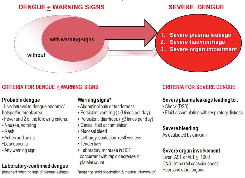
CPG Management of Dengue Infection In Adults (Third Edition)    2015

No. Title                                                            Page

10. PREVENTION OF DENGUE TRANSMISSION IN HOSPITALS                        43

11. VACCINATION                                                           43

12. FOOD AND SUPPLEMENTS                                                  43

13. IMPLEMENTING THE GUIDELINES                                           44

14. REFERENCES                                                            46

    Appendix 1 Search Strategy                                            56

    Appendix 2 Clinical Questions                                         57

    Appendix 3 World Health Organization (WHO) Classification of          58
               DF and DHF (1997)

    Appendix 4 Methods of Sample Collection                               60

    Appendix 5 Type of Tests for Dengue Diagnosis and                     61
               Recommended Use

    Appendix 6 Type of Dengue Tests Recommended Based                     62
               On Clinical History

    Appendix 7 Outpatient Dengue Monitoring Record                        63

    Appendix 8 Dengue Assessment Checklist                                64

    Appendix 9 Home Care Advice Leaflet for Dengue Patients               65

    Appendix 10 Inpatient Dengue Monitoring Chart                         66

    List of Abbreviations                                                 67
    Acknowledgement                                                       68
    Disclosure Statement                                                  68
    Source of Funding                                                     68

                                        4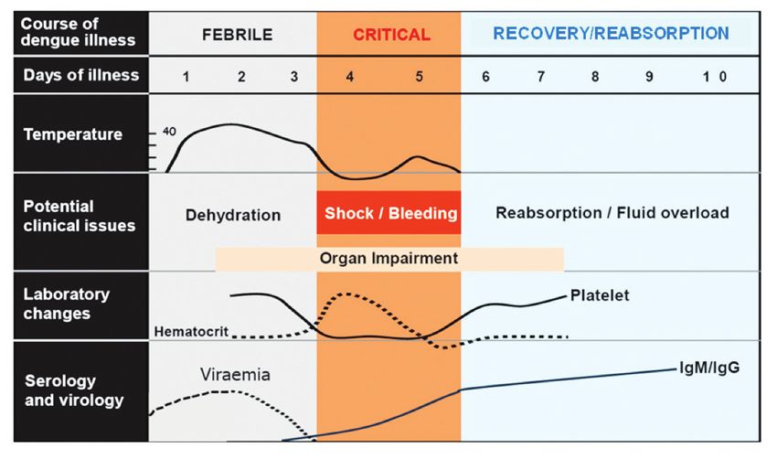
CPG Management of Dengue Infection In Adults (Third Edition)   2015

                        LEVELS OF EVIDENCE
  Level      Study design
     I       Evidence obtained from at least one properly
             randomised controlled trial
    II-1     Evidence obtained from well-designed controlled trials
             without randomisation
    II-2     Evidence obtained from well-designed cohort or case-
             control analytic studies, preferably from more
             than one centre or research group
    II-3     Evidence obtained from multiple time series with or
             without the intervention. Dramatic results in uncontrolled
             experiments could also be regarded as this type of
             evidence
    III      Opinions of respected authorities based on clinical
             experience; descriptive studies and case reports; or
             reports of expert committees

SOURCE: US / CANADIAN PREVENTIVE SERVICES TASK FORCE 2001

In line with the current development in CPG methodology, the
CPG Unit of MaHTAS is in the process of incorporating Grading
Recommendations, Assessment, Development and Evaluation
(GRADE) in its work process. The quality of each retrieved evidence
and its effect size are carefully assessed/reviewed by the CPG
Development Group. In formulating the recommendations, overall
balances of the following aspects are considered in determining the
strength of the recommendations:-
   • Overall quality of evidence
   • Balance of benefits vs harms
   • Values and preferences
   • Resource implications
   • Equity, feasibility and acceptability

                                   i
                                   5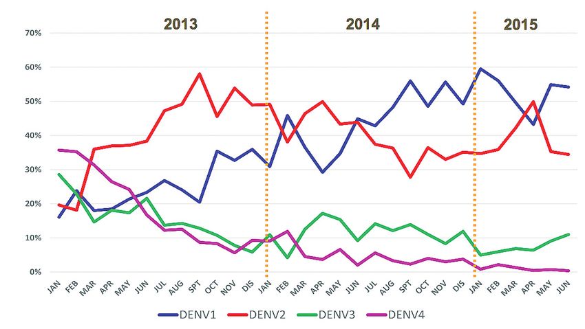
CPG Management of Dengue Infection In Adults (Third Edition)    2015

GUIDELINES DEVELOPMENT AND OBJECTIVES

GUIDELINES DEVELOPMENT

The members of the Development Group (DG) for these Clinical
Practice Guidelines (CPG) were from the Ministry of Health (MoH).
There was also active involvement of a multidisciplinary Review
Committee (RC) during the process of the CPG development.

A literature search was carried out using the following electronic
databases: Guidelines International Network (G-I-N); Medline via Ovid,
Pubmed and Cochrane Database of Systemic Reviews (CDSR) (refer
to Appendix 1 for Search Strategy). The inclusion criteria are all
literature on dengue infection regardless of study design. The search
was limited to literature published in the last five years, on humans and
in English. In addition, the reference lists of all retrieved literature and
guidelines were searched to further identify relevant studies. Experts
in the field were also contacted to identify relevant studies. In certain
situations, pivotal papers beyond the scope of search were used in the
CPG. All searches were conducted from 26 May 2015 to 24 July 2015.
Future CPG updates will consider evidence published after this cut-off
date. The details of the search strategy can be obtained upon request
from the CPG Secretariat.

The previous edition of CPG (2010) was used as the basis in updating
these present guidelines. Reference was also made to other guidelines
on dengue infection in adults as well as handbooks from WHO such as
Dengue Guidelines for Diagnosis, Treatment, Prevention and Control
(2009) and Handbook for Clinical Management of Dengue (2012).

A total of 18 clinical questions were developed under different
sections. Members of the DG were assigned individual questions
within these sections. (Refer to Appendix 2 for Clinical Questions)
The DG members met 7 times throughout the development of these
guidelines. The literature retrieved was appraised by at least two DG
members using Critical Appraisal Skill Programme checklist, presented
in evidence tables and further discussed in each DG meetings. All
statements and recommendations formulated after that were agreed
upon by both the DG and RC. Where evidence was insufficient, the
recommendations were made by consensus of the DG and RC. Any
differences in opinion were resolved consensually. The CPG was
based largely on the findings of systematic reviews, meta-analyses and
clinical trials, with local practices taken into consideration.

                                    ii
                                         6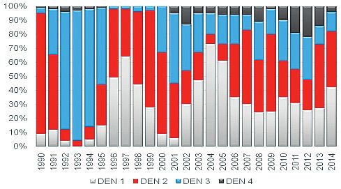
CPG Management of Dengue Infection In Adults (Third Edition)   2015

The literature used in these guidelines was graded using the US/
Canadian Preventive Services Task Force Level of Evidence (2001)
while the grading of recommendation was done using the principles of
GRADE (refer to the preceding page).

On completion, the draft of the CPG was reviewed by external
reviewers. It was also posted on the MoH Malaysia official website for
feedback from any interested parties. The draft was finally presented
to the Technical Advisory Committee for CPG, and the HTA and
CPG Council MoH Malaysia for review and approval. Details on
the CPG development by MaHTAS can be obtained from Manual on
Development and Implementation of Evidence-based Clinical Practice
Guidelines published in 2015 (available at http://www.moh.gov.my/
index.php/pages/view/117).

                                    iii
                                    7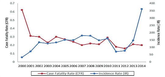
CPG Management of Dengue Infection In Adults (Third Edition)   2015

OBJECTIVES

GENERAL OBJECTIVES

To provide evidence-based guidance in the management of dengue
infection in adult patients.

SPECIFIC OBJECTIVES

•    To improve recognition and diagnosis of dengue cases and provide
     appropriate care to the patients.
•    To improve on early and accurate notification of dengue cases for
     prompt public health intervention.
•    To identify severe dengue and carry out more focused close
     monitoring and prompt appropriate management.
•    To provide guidance on appropriate and timely fluid management
     and the use of blood and blood products.
•    To create awareness on early detection of dengue infection with
     complications.

CLINICAL QUESTIONS

Refer to Appendix 2

TARGET POPULATION

Adult patients with dengue fever and severe dengue.

TARGET GROUP/USER

This CPG is intended to guide those involved in the management of
dengue infection in adults particularly healthcare professionals in
primary and secondary/tertiary care namely:-
  a. Physicians and specialists from related disciplines
  b. Family Health Specialists
  c. Medical officers and general practitioners
  d. Allied health professionals
  e. Pharmacists
  f. Students (medical postgraduates and undergraduates, and allied
     health students)
  g. Patients and carers

HEALTHCARE SETTINGS

Outpatient, inpatient and community settings inclusive of private
healthcare facilities.

                                    iv
                                         8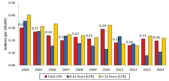
CPG Management of Dengue Infection In Adults (Third Edition)                            2015

CHAIRPERSON

Dato’ Dr. Mahiran Mustafa
Senior Consultant Infectious Disease Physician
Hospital Raja Perempuan Zainab II

Members (alphabetical order)
Dr. Ahmad Tajuddin Mohamad Nor                    Dr. Rose Nani Mudin
Consultant Emergency Medicine Specialist          Head of Vector - Borne Diseases
Hospital Tengku Ampuan Rahimah                    Disease Control Division, MoH

Dr. Anilawati Mat Jelani                          Dr. Saiful Safuan Md. Sani
Infectious Disease Physician                      Internal Medicine Physician
Hospital Raja Perempuan Zainab II                 Hospital Kuala Lumpur

Dr. Chow Ting Soo                                 Datin Dr. Salbiah Hj. Nawi
Consultant Infectious Disease Physician           Consultant Pathologist
Hospital Pulau Pinang                             Hospital Kuala Lumpur

Dr. Faisal Salikin                                Dr. Salmah Idris
Consultant Emergency Medicine Specialist          Consultant Pathologist
Hospital Kuala Lumpur                             Hospital Sungai Buloh

Dr. Faridah Mohd. Amin                            Dato’ Dr. Santha Kumari
Head of Epidemiology Division                     Senior Consultant Physician
National Public Health Laboratory Sg. Buloh       Hospital Tengku Ampuan Rahimah, Klang

Dr. Hanin Farhana Kamaruzaman                     Dr. Shahanizan Mohd. Zain
Principal Assistant Director, CPG Unit            Principal Assistant Director
Health Technology Assessment Section, MoH         Medical Development Division, MoH

Dr. Haniza Omar                                   Dr. Shanthi Ratnam
Consultant Hepatologist                           Consultant Intensivist & Physician
Hospital Selayang                                 Hospital Sungai Buloh

Dr. Intan Iliana Iliassa                          Dr. Shari Mohd. Nor
Transfusion Medicine Specialist                   Consultant Obstaetric & Gynaecology
Hospital Serdang                                  Sabah Women & Children Hospital, Likas

Dr. Izzuna Mudla Ghazali                          Dato’ Dr. K. Sree Raman
Senior Principal Assistant Director               Senior Consultant Physician
Health Technology Assessment Section, MoH         Hospital Tuanku Ja’afar, Seremban

Dr. Kan Fong Kee                                  Dr. Suresh Kumar Chidambaram
Consultant Infectious Disease Physician           Consultant Infectious Disease Physician
Hospital Sultanah Aminah                          Hospital Sugai Buloh

Dr. Nahla Irtiza Ismail                           Dr. Siti Zulfa Zulkifli
Intensivist & Anaesthesiologist                   Internal Medicine Physician
Hospital Melaka                                   Hospital Kuala Lumpur

Dr. Nor Azilah Abu Bakar @ Mansor                 Dr. Tai Li Ling
Assistant Director                                Senior Consultant Intensivist & Anaesthesiologist
Medical Development Division, MoH                 Hospital Kuala Lumpur

Dr. Nor Hafizah Ahmad                             Mrs. Yu Kie a/p Chem
Transfusion Medicine Specialist                   Science Officer (Microbiology)
National Blood Bank                               National Public Health Laboratory Sg. Buloh

Dr. Norhayati Shaharudin                          Dr. Zailiza Suli
Infectious Disease Physician                      Senior Principal Assistant Director
Hospital Melaka                                   Disease Control Division, MoH

Dr. Ravindran Thayan
Research Officer, Virology Unit
Institute for Medical Research

                                              v
                                              9
CPG Management of Dengue Infection In Adults (Third Edition) 2015
REVIEW COMMITTEE
The draft guideline was reviewed by a panel of experts from both
public and private sectors. They were asked to comment primarily on
the comprehensiveness and accuracy of the interpretation of evidence
supporting the recommendations in the guidelines.
CHAIRPERSON
Datuk Dr. Jeyaindran Tan Sri Sinnadurai
Deputy Director General (Medical)
Ministry of Health Malaysia
Members (alphabetical order)
Datuk Dr. Christopher Lee Kwok Chong
Senior Consultant Infectious Disease
Hospital Sungai Buloh
Dato’ Dr. Faraizah Dato’ Abdul Karim
Deputy Director
National Blood Bank
Dr. J. Ravichandran Jeganathan
Senior Consultant Obstetric & Gynaecology
Hospital Sultanah Aminah
Datin Dr. Rugayah Bakri
Deputy Director
Health Technology Assessment Section, MoH
Dr. Sabariah Faizah Jamaluddin
Senior Consultant Emergency & Trauma
Hospital Sungai Buloh
Datin Dr. Sivasakthi Velayuthapillai
Senior Consultant Anaesthesiology
Hospital Kuala Lumpur
vi
10CPG Management of Dengue Infection In Adults (Third Edition) 2015
EXTERNAL REVIEWERS (in alphabetical order)
The following external reviewers provided feedback on the draft:-
Dr. Hari Dass
Private General Practitioner
Klinik Seremban
Dr. Jameela Sathar
Senior Consultant Haematologist
Hospital Ampang
Dr. Petrick Periyasamy
Lecturer and Infectious Disease Physician
Universiti Kebangsaan Malaysia Medical Centre
Dr. Tan Lian Huat
Consultant Infectious Disease Physician
Sunway Medical Centre
vii
11CPG Management of Dengue Infection In Adults (Third Edition) 2015
1. EPIDEMIOLOGY
Dengue is one of the most important arthropod-borne viral diseases in
terms of public health problem with high morbidity and mortality. It affects
tropical and subtropical regions around the world, predominantly in urban
areas.
The global increase of dengue incidence is also experienced by Malaysia.
Since the year 2000, the dengue incidence in Malaysia continues to
increase from 32 cases per 100,000 population to 361 cases per 100,000
population in 2014 (Figure 1). The dengue incidence rate is higher in the
age group of 15 and above. Most of the dengue cases reported were from
urban areas (70%–80%) where factors such as high density population and
rapid development favour dengue transmission.
With regards to case fatality rate, the national target is less than 0.2%. The
case fatality rate has been reduced from 0.6% in year 2000 to 0.2% in year
2014 (Figure 1). Most of the dengue death has been observed to be higher
in the age group of 15 years and above (Figure 2) and the highest has
been observed in 2004.
Figure 1: Dengue Incidence Rate and Case Fatality Rate, Malaysia 2000-2014
Figure 2: Dengue Case Fatality Rate (CFR) By Age Group In Malaysia, 2004-2014
1CPG Management of Dengue Infection In Adults (Third Edition) 2015
2. DENGUE VIRUS AND SEROTYPE TRENDS IN MALAYSIA
Dengue infection is caused by dengue virus which is a mosquito-borne
flavivirus. It is transmitted by Aedes aegypti and Aedes albopictus. There
are four distinct serotypes, DENV-1,2,3 and 4. Each episode of infection
induces a life-long protective immunity to the homologous serotype but
confers only partial and transient protection against other serotypes.
Secondary infection is a major risk factor for severe dengue due to
antibody-dependent enhancement. Other important contributing factors are
viral virulence, host genetic background, T-cell activation, viral load and
auto-antibodies.
In Malaysia, all four serotypes can be isolated at any one time. However,
a particular dengue virus serotype can predominate for at least two years
before it is replaced by another serotype (Figure 3). In year 2013-2014,
the predominant serotype had switched twice from DENV-2 to DENV-1 in
February and June 2014 (Figure 4).
Figure 3: Dengue Serotypes in Malaysia (1990-2014)
Figure 4: Dengue Serotypes in Year 2013, 2014 and 2015 (Jan-June)
2CPG Management of Dengue Infection In Adults (Third Edition) 2015
3. CLINICAL MANIFESTATIONS AND PATHOPHYSIOLOGY
3.1 SPECTRUM OF DENGUE INFECTION
The incubation period for dengue infection is 4-7 days (range 3-14).1 It
may be asymptomatic or may result in a spectrum of illness ranging from
undifferentiated mild febrile illness to severe disease, with or without
plasma leakage and organ impairment. Symptomatic dengue infection
is a systemic and dynamic disease with clinical, haematological and
serological profiles changing from day to day. These changes accelerate
within hours or even minutes during the critical phase, particularly in those
with plasma leakage (refer to subchapter 3.3).
Understanding the systemic and dynamic nature of dengue disease as well
as its pathophysiological changes during each phase of the disease will
produce a rational approach in the management of dengue infection.
3.2 CLINICAL COURSE OF DENGUE INFECTION
After the incubation period, the illness begins abruptly and will be followed
by three phases: febrile, critical and recovery phase (refer to Figure 5).2,3
i. Febrile Phase
Patients develop high grade fever suddenly and usually last 2-7 days. It
is often accompanied by facial flushing, rash, generalised body ache,
vomiting and headache. 2,3 Some patients may have sore throat, injected
pharynx and conjunctival injection.
Mild haemorrhagic manifestations like petechiae and mucosal membrane
bleeding may be seen during the illness.4,5 Per vaginal bleeding may occur
in females but rarely massive. Gastrointestinal bleeding is not uncommon.5,6
The findings of tender liver are warning signs of dengue infection.
The earliest abnormality in the full blood count is a progressive decrease
in total white cell count followed by platelet reduction. This should alert the
physician to a high index of suspicion of dengue infection. This disease
should be notified as early as possible for preventive measures.
ii. Critical Phase
The critical phase often occurs after third day of fever (may occur earlier)
or around defervescence indicated by a rapid drop in temperature. This
coincides with an increase in capillary permeability in some patients. In
other viral infections, the patient’s condition improves as the temperature
subsides, but the contrary happens in severe dengue infection wherein the
patient may deteriorate and manifest third space plasma leakage or organ
dysfunction.2,3,7,8
3CPG Management of Dengue Infection In Adults (Third Edition) 2015
The critical phase lasts about 24-48 hours (refer to Figure 5). Varying
circulatory disturbances (refer to Table 1) can develop. In less severe
cases, these changes are minimal and transient. Many of these patients
either recover spontaneously or after a short period of fluid or electrolyte
therapy. In more severe forms of plasma leakage, the patients may
develop compensated or decompensated shock (Table 1). Abdominal pain,
persistent vomiting and/or diarrhoea, restlessness, altered conscious level,
clinical fluid accumulation, mucosal bleed or tender liver are the clinical
warning signs of dengue infection with high possibility of complications.8-10
Organ dysfunctions such as hepatitis, encephalitis and myocarditis usually
but not exclusively occur during this phase.
It is important to note that thrombocytopaenia and haemoconcentration are
usually detectable in this phase. The haematocrit (HCT) level correlates
well with plasma volume loss and disease severity. However, interpretation
of HCT may be difficult when there are confounding factors such as
haemorrhage, excessive fluid replacement or in haemodilutional state.
Leucopaenia with relative lymphocytosis, clotting abnormalities, elevation
of transaminases [typically the level of aspartate aminotransaminase
(AST) is higher than the level of alanine aminotransaminase (ALT)],
hypoproteinaemia and hypoalbuminaemia are usually observed.2-4
iii. Recovery/Reabsorption Phase
After 24-48 hours of critical phase, usually plasma leakage stops followed
by reabsorption of extravascular fluid. Patient’s general well being improves,
appetite returns, gastrointestinal symptoms improve, haemodynamic status
stabilises and diuresis ensues. Some patient may have a classical rash of
“isles of white in the sea of red” with generalised pruritus.2 It is important to
note that during this phase, HCT level stabilises and drops further due to
haemodilution following reabsorption of extravascular fluid. The recovery of
platelet count is typically preceded by recovery of white cell count (WCC).
In some instances, organ dysfunctions may worsen (hepatitis, encephalitis
and intracranial bleed) as the patient enters reabsorption phase.
Figure 5 : Clinical Course of DHF 11-13
4CPG Management of Dengue Infection In Adults (Third Edition) 2015
• The critical phase often occurs after third day of fever (may occur
earlier) or around defervescence with a rapid drop in temperature.
• Clinical deterioration often occurs during the critical phase with
plasma leakage or organ dysfunction.
• Evidence of plasma leakage includes raised HCT, haemodynamic
instability, fluid accumulation in extravascular space or
hypoproteinaemia.
• Abdominal pain, persistent vomiting and/or diarrhoea, restlessness,
altered conscious level, clinical fluid accumulation, tender liver or
mucosal bleed are the clinical warning signs of dengue infection with
high possibility of rapid progression to severe dengue.
3.3 PATHOPHYSIOLOGY OF PLASMA LEAKAGE IN SEVERE
DENGUE INFECTION
The primary pathophysiological abnormality seen in dengue infection is
an acute increase in vascular permeability that leads to leakage of plasma
into the extravascular compartment, resulting in haemoconcentration and
hypovolaemia or shock.2,3,14 Hypovolaemia leads to reflex tachycardia and
generalised vasoconstriction due to increased sympathetic output.15,16
Clinical manifestations of vasoconstriction in various systems are as follows:
• Skin - coolness, pallor and delayed capillary refill time
• Cardiovascular system - raised diastolic blood pressure and a
narrowing of pulse pressure
• Renal system - reducing urine output
• Gastrointestinal system - persistent vomiting, persistent diarrhoea and
abdominal pain
• Central nervous system – lethargy, restlessness, apprehension,
reduced level of consciousness
• Respiratory system – tachypnoea (respiratory rate >20/min)
Inadequate perfusion of the tissue leads to increased anaerobic glycolysis
and lactic acidosis. If the hypovolaemia is not corrected promptly, the
patient will progress to a refractory shock state. By then, the tissue
perfusion would not respond to vasopressor drugs, even if the blood
pressure and intravascular volume were to be restored and cardiac output
would remain depressed. The resultant lactic acidosis further depresses
the myocardium and worsens the hypotension.16
The common late complications of prolonged shock are massive bleeding,
disseminated intravascular coagulopathy (DIC) and multi-organ failure
which are often fatal. The pathophysiology of organ dysfunction will be
described in subchapter 7.6.
The following Table 1 is the summary of the continuum of various
pathophysiological changes in a patient who progresses from normal
circulatory state to hypovolaemic shock.
5CPG Management of Dengue Infection In Adults (Third Edition) 2015
Table 1: A continuum of pathophysiological changes from normal circulation to
compensated and decompensated/hypotensive shock
Normal Circulation Compensated Shock Decompensated /
Hypotensive Shock
• Clear consciousness • Clear consciousness • Change of mental
- shock can be state - restless,
• Brisk capillary refill missed if you do not combative or lethargy
time (2 sec) refill time
• Good volume • Cool extremities • Cold, clammy
peripheral pulses extremities
• Weak & thready
• Normal heart rate peripheral pulses • Feeble or absent
for age peripheral pulses
• Tachycardia
• Normal blood • Severe tachycardia
pressure for age • Normal systlic with bradycardia in
pressure with raised late shock
• Normal pulse diastolic pressure
pressure for age • Hypotension /
• Postural hypotension unrecordable BP
• Normal respiratory
rate for age • Narrowing pulse • Narrowed pulse
pressure pressure (CPG Management of Dengue Infection In Adults (Third Edition) 2015
3.4 WHO DENGUE CLASSIFICATION
Based on 1997 WHO dengue classification scheme (refer to Appendix 3),
the key differentiating feature between DF and DHF is the presence of plasma
leakage in DHF. However, in the early febrile phase of dengue infection, the
symptoms can overlap and one cannot differentiate DF and DHF.
3.4.1 Limitations of 1997 WHO classification 23, level I
It has been observed that the 1997 WHO classification scheme has several
limitations.
i. Dengue with shock without fulfilling all the four criteria for DHF
ii. Severe organ impairment without shock
iii. The 1997 classification scheme does not address the entire spectrum
of the disease.
3.4.2 WHO Classification 2009
This classification addresses the levels of severity of dengue infection which
is more practical to be used in the management decision of the patient
(Figure 6).
Figure 6: 2009 WHO Dengue Classification and Level of Severity 24
Adapted: World Health Organization. Dengue Guidelines for Diagnosis, Treatment,
Prevention and Control - New Edition 2009. WHO: Geneva; 2009
7CPG Management of Dengue Infection In Adults (Third Edition) 2015
3.5 DIAGNOSTIC CHALLENGES
Clinical features of dengue infection are rather non-specific and mimic
many other diseases, therefore can be easily misinterpreted. A high index
of suspicion and appropriate history taking, particularly with regards to a
recent stay in dengue hotspots or clusters of fever in neighbourhood, are
useful for early and accurate diagnosis of dengue infection.
In addition, a dengue patient may have a co-infection with another
pathogen. Diseases that may mimic dengue infection includes other viral
illnesses. Organ dysfunction can occur in other diseases apart from dengue
infection and should be investigated.
4. DISEASE NOTIFICATION
All suspected dengue cases from private and public health facilities must
be notified by telephone/fax/e-notification to the nearest health office within
24 hours of diagnosis, followed by written notification using the standard
notification format. Failure to notify is liable to be compounded under the
Prevention and Control of Infectious Diseases Act, 1988 (Act 342).25
Any delay in notification will increase the risk of dengue transmission in
the locality of the residence. It is also important to update the notification if
there is a change in severity of dengue infection or when there is dengue
death.
Notified cases will be followed up by the health authorities for the
verification of case definition and preventive measures. Since 2014,
the Ministry of Health Malaysia has set up new criteria for dengue cases
registration, whereby all registered dengue cases must be confirmed by
laboratory investigations.
All dengue deaths need to be notified as soon as possible by the treating
doctor in the hospital to the district health office and/or State Health
Director and must be investigated by District Health Officer or Epidemiology
Officer.26
Dengue mortality audit should be conducted within seven days after
the dengue death has occurred at hospital level followed by state level,
chaired by Hospital Director and State Health Director respectively. The
complete dengue death report should be sent to Communicable Diseases
Division, Ministry of Health within one week via State Vector Officer. The
implementation of dengue case management and death should comply to
the Director General of Health Malaysia Circular No.15/2010 dated 24 May
2010.26
8CPG Management of Dengue Infection In Adults (Third Edition) 2015
5. INVESTIGATIONS
5.1 DISEASE MONITORING TESTS
i. White cell count (WCC) and Platelet count:
In the early febrile phase WCC and platelet count are usually normal
but will decrease rapidly as the disease progresses.4, level III The
decrease in WCC is accompanied by platelet reduction.
There is no correlation between disease severity and platelet count
2,level III;25,level III
and it is not predictive of bleeding.28,level I;29,level II-2;30-32,level III In
recovery phase, the WCC normalises followed by platelet.
ii. Haematocrit (HCT):
A rising HCT is a marker of plasma leakage in dengue infection. The median
values of normal HCT level among Malaysian populations are: 33,level II-2
• male < 60 years – 46%
• male > 60 years – 42%
• female (all age groups) – 40%
Other important blood tests in disease monitoring are Liver Function Test
(LFT), Renal profile (RP), coagulation profile, lactate and blood gases
(Table 6). Special tests such as Troponin and Creatine Kinase (CK) should
be discussed with the Specialists/Registrars before performing.
Recommendation 1
• Baseline haematocrit (HCT) and white cell count should be established
during the first visit in all patients with suspected dengue infection.
• Serial full blood count and HCT must be monitored as dengue infection
progresses.
5.2 DIAGNOSTIC TESTS
Diagnostic tests include point of care testing such as dengue NS1 antigen
test and rapid combo tests (NS1 antigen and dengue IgM/IgG antibodies).
Other laboratory tests include NS1 antigen test, dengue antibody detection
tests including IgM and IgG ELISA, dengue genome detection assay (real
time RT-PCR) and dengue viral isolation assay. Specifications for an ideal
dengue test include the ability to differentiate between dengue and other
diseases with similar clinical presentation, to detect during the acute stage
of infection, provides rapid results, inexpensive and easy to use. However,
the interpretation of diagnostic results should be done with the clinical
context.
5.2.1Rapid Combo Test (RCT)
Rapid combo tests are assays that can detect the presence of virus as well
as antibodies simultaneously.34-35,level II-2 Generally RCT tests can be read
within 15-20 minutes. However, it is important that the tests have to be
read according to the manufacturer’s recommendation in the product insert.
Reading too late gives false results.
9CPG Management of Dengue Infection In Adults (Third Edition) 2015
Interpretation of the results is through the presence or absence of bands
for dengue NS1 antigen and dengue IgM and IgG antibodies.34-35,level II-2
These tests have a longer detection window as they can detect both the
virus and antibodies, thus reducing the possibility of false negative results.
Hence these tests are useful during the early phase of onset when there is
viraemia as well as at a later stage when antibodies against dengue begin
to rise. Suitable samples that can be used for testing include whole blood,
serum and plasma. The sensitivity is 93.9% and specificity is 92%.36,level II-2
5.2.2 Dengue Antigen and Serology Tests by ELISA
Both antigen and serological tests are more commonly used to diagnose
dengue infections. The tests include antigen detection (NS1) or antibody
detection. Usually different patterns of antibody response are seen in primary
dengue infection as compared to secondary dengue infection. 37,level II-2
i. Non-Structural Protein-1 (NS1 Antigen)
NS1 antigen is a highly conserved glycoprotein that seems to be essential
for virus viability. Secretion of the NS1 protein is a hallmark of flavivirus
infecting mammalian cells and can be found in dengue infection as well as
in yellow fever and West Nile virus infection. False positive results have
been reported in chronic diseases and haematological malignancies.38,level III
The detection rate is much better in acute sera of primary infection
(75%-97%) when compared to the acute sera of secondary infection
(60%-70%).39-41,level III The sensitivity of NS1 antigen detection drops from
day 4-5 of illness onwards and usually becomes undetectable in the
convalescence phase.40-42,level III
The presence of NS1 detection after day five may predict severe dengue.44,level II-2
ii. Dengue IgM test
The IgM capture enzyme-linked immunosorbent assay (ELISA) is the
most widely used serological test. The antibody titre is significantly higher
in primary infections compared to secondary infections. Once the IgM is
detectable, it rises quickly and peaks at about two weeks after the onset of
symptoms, and it wanes to undetectable levels by 90 days.
In primary dengue infection, anti-dengue IgM can be detected after five
days of illness in approximately 80% of the cases.37,level II-2 Almost 93%-99%
of cases will have detectable IgM from day 6 through day 10. In the event
of a negative IgM result, a repeat serum should be collected after five days.
However, in secondary dengue infections, IgM was detected in among
78% of patients after day seven.45,level II-2 IgM appears earlier or at the same
time frame but usually at lower titres compared to primary dengue. This
is possibly due to appearance of high levels of anti-dengue IgG before
or sometimes simultaneously with the IgM response. Thus, 28% of all
secondary dengue infections were undiagnosed when only IgM was the
only assay performed. 46-48,level III
10CPG Management of Dengue Infection In Adults (Third Edition) 2015
iii. Dengue IgG test
In primary and secondary dengue infection, dengue IgG was detected in
100% of patients after day seven of onset of fever. Therefore, a repeat
dengue IgG is recommended if dengue IgM is still negative after day seven
with the negative IgG in the initial test sample. 45,level II-2; 47, level III
iv. Differentiation between primary and secondary dengue infection
Detection of significant elevation of IgG antibodies to dengue virus by
ELISA is very useful for identification of secondary dengue infections.
49-50,level II-2
There are commercially available ELISA kits which can be used
to differentiate between primary and secondary dengue infections as the
kits have incorporated in-house threshold levels of IgG. For example,
Panbio has incorporated a cut off of more than 22 Panbio Units which is
equivalent to HAI level of 1:2560, and thus indicative of secondary dengue
infections.51
Note: False positive dengue serology
Serological tests for dengue have been shown to cross-react with:
• other flavivirus – Japanese Encephalitis 48,level III; 52, level III
• non-flavivirus – malaria, leptospirosis, toxoplasmosis, syphilis 53-54, level III
• connective tissue diseases – rheumatoid arthritis 55, level III
5.2.3 Dengue Viral RNA Detection (Real time RT-PCR)
Dengue viral RNA detection is a molecular method that utilises reagents
that will target specific genome of the virus to enable amplification and
detection of the target. The method is useful only during the viraemic stage
of the disease and can detect the viral RNA target up to five days after
onset of the symptoms. 56-57,level II-2 The test is useful for determination of
circulating dengue serotypes in the country.
Limitations of RT-PCR are:
a) This test is only available in a few centres with facilities and trained
personnel (e.g.IMR, National Public Health Laboratory, State Public
Health Laboratories and University Malaya Medical Centre).
b) The test is expensive.
c) The specimen requires special storage temperatures and short
transportation time between collection and extraction (refer to Appendix 4).
5.2.4 Virus Isolation
Dengue virus isolation is carried out in Institute for Medical Research (IMR),
National Public Health Laboratory and University Malaya Medical Centre
for research, surveillance and genotyping purposes.
Please refer to Appendix 4 on details of sample collection for diagnostic tests.
As clinical diagnosis of dengue lacks specificity, a definitive diagnosis of
dengue infections requires laboratory confirmation. There are many targets
for laboratory confirmation of dengue which have been listed in Appendix 5
with information on sensitivity and specificity of each target and when to do
after the onset of symptoms.
11CPG Management of Dengue Infection In Adults (Third Edition) 2015
Appendix 6 summarises the type of tests recommended for patients
presented with clinical history and their interpretation. This serves as
laboratory guidelines for the clinicians who are managing dengue patients.
• Dengue IgM is usually positive after day 5-7 of illness. Therefore a
negative IgM taken before day 5-7 of illness does not exclude dengue
infection.
Recommendation 2
• Dengue rapid combo test or non-structural protein 1 antigen (NS1 Ag)
should be taken as soon as dengue infection is suspected.
• If dengue IgM is negative before day seven, a repeat sample must be
taken in recovery phase.
• If dengue IgM is still negative after day seven, dengue IgG should be
done for diagnostic confirmation of secondary dengue infection.
Caution: Massive blood transfusion may affect the test results mentioned above.
6. INVESTIGATION OF POST MORTEM CASE
Suitable samples for virus isolation and PCR can be obtained from most of
the organs however, the best tissue sample for investigation is
the liver.58-59,level III If patient had encephalitis, CSF should also be included
as samples for investigation. The liver should be placed in sterile containers
and moistened with viral transport media or sterile normal saline. CSF
should be submitted in sterile bijou bottle. Both should be refrigerated if
there is delay in transportation. Specimen for viral investigation should be
transported in ice to IMR and National Public Health Laboratory.
Recommendation 3
• A repeat dengue serology should be obtained at the time of dengue
death.
• Suitable specimens for viral isolation and/or real time reverse
transcriptase polymerase chain reaction (RT-PCR) and/or non-
structural protein 1 (NS1) antigen detection should be done for
confirmation of dengue infection.
12CPG Management of Dengue Infection In Adults (Third Edition) 2015
7. MANAGEMENT OF DENGUE INFECTION
7.1 OUTPATIENT MANAGEMENT
The management of dengue infection is symptomatic and supportive. An
approach to outpatient evaluation is as suggested in Table 2.
Management issues vary according to the three phases of the clinical
course (refer to section 7.4). It is crucial to recognise plasma leakage, early
shock and severe organ dysfunction. This can be achieved by frequent
clinical and laboratory monitoring during the early febrile phase of dengue
infection.
Dengue patients who are managed in the outpatient setting should be
provided with an outpatient dengue monitoring record (refer to Appendix 7)
to ensure that relevant informations are available for continuity of care by
health care providers.
• Primary care providers with no immediate haematocrit facilities and/or
point of care tests should refer patient to the nearest health facility for
further management.
• Point of care tests (RCT or NS1 antigen) should be done when dengue
infection is suspected.
13CPG Management of Dengue Infection In Adults (Third Edition) 2015
Table 2: Approach to Outpatient Evaluation of Dengue Infection 1-3
It is important to evaluate every patient for the following:
Overall assessment
1. History
• Date of onset of fever/ illness
• Oral intake
• Assess for warning signs – Refer to Table 3
• Change in mental state/seizure/dizziness
• Urine output (frequency, volume and time of last voiding)
• Other important relevant histories:
-- Family or neighbourhood history of dengue
-- Jungle trekking and swimming in waterfall (consider leptospirosis,
typhus, malaria)
-- Travelling
-- Recent unprotected sexual or intravenous drug user (consider acute
HIV seroconversion illness)
-- Co-morbidities (consider sepsis particularly in patients with diabetes
mellitus)
2. Physical examination
i. Assess mental state and GCS score
ii. Assess hydration status
iii. Assess haemodynamic status
-- Skin colour (C), capillary refill time (normalCPG Management of Dengue Infection In Adults (Third Edition) 2015
Table 3: Warning Signs
• Any abdominal pain/tenderness
• Persistent vomiting ( >3 times over 24 hours)
• Persistent diarrhoea ( >3 times over 24 hours)
• Third space fluid accumulation (such as ascites, pleural and
pericardial effusion)
• Spontaneous bleeding tendency
• Lethargy/restlessness/confusion
• Tender liver
• Raised HCT with rapid drop in platelet:
o HCT male 60 years – 42%
o HCT female (all age groups) – 40%
*median values of normal HCT in Malaysian population33,level II-2.
Adapted : World Health Organization. Dengue Guidelines for Diagnosis, Treatment,
Prevention and Control - New Edition 2009. WHO: Geneva; 2009
Table 4 : Clinical and Laboratory Criteria for Patients Who Can Be Treated at Home60-62
1. Able to tolerate orally well, good urine output and no history of
bleeding
2. Absence of warning signs (refer to Table 3)
3. Physical examination:
• Haemodynamically stable
• No tachypnoea or acidotic breathing
• No tender liver or abdominal tenderness
• No bleeding manifestation
• No sign of third space fluid accumulation
• No alterations in mental state
4. Investigation:
• Stable serial HCT
5. No other criteria for admission (i.e. co-morbidities, pregnancy, social
factors)
15CPG Management of Dengue Infection In Adults (Third Edition) 2015
7.2 PATIENT TRIAGING AT EMERGENCY AND OUTPATIENT DEPARTMENT
The purpose of triaging patients is to determine whether they require
urgent attention. This is to avoid critically ill patients being missed upon
arrival.60,level III; 63-64,level III
Triage Checklist at Registration Counter:
1. History of fever
2. Abdominal Pain
3. Vomiting
4. Dizziness/ fainting
5. Bleeding
Vital parameters to be taken:
Mental state, blood pressure, CCTVR and respiratory rate
7.3 CRITERIA FOR HOSPITAL REFERRAL / ADMISSION
7.3.1 Referral from Primary Care Providers to Hospital
The decision for referral and admission must not be based on a single
clinical parameter but should depend on the Total Assessment of the
patient.
Referral from primary care providers to hospital
1. Symptoms:
• Warning signs (refer to Table 3)
• Bleeding manifestations
• Inability to tolerate oral fluids
• Reduced urine output
• Seizure
2. Signs:
• Dehydration
• Shock (refer to Table 1)
• Bleeding
• Any organ failure
3. Special situations:
• Patients with co-morbidity e.g. Diabetes, Hypertension, Ischaemic
Heart Disease, Coagulopathy, Morbid Obesity, Renal failure,
Chronic Liver disease, COPD
• Elderly more than 65 years old
• Patients who are on anti-platelet and/or anticoagulants
• Pregnancy
• Social factors that limit follow-up e.g. living far from health facility,
no transport, patient living alone, etc.
4. Laboratory criteria: Rising HCT accompanied by reducing platelet count
16CPG Management of Dengue Infection In Adults (Third Edition) 2015
7.3.2 Referral from Hospitals Without Specialist To Hospitals
With Specialists
Nearest physician should be consulted for all cases of severe dengue,
those who are pregnant and patients with comorbidities.
Prerequisites for transfer
1. All efforts must be taken to optimise the patient’s condition before and
during transfer.
2. The Emergency Department, ICU and Medical Department of the
receiving hospital must be informed prior to transfer.
3. Adequate and essential information must be sent together with the
patient that includes fluid chart, monitoring chart and investigation
results.
7.3.3 Intervention in Emergency Department
Proactive ongoing clinical reassessment of airway, breathing and circulatory
status must be done.
7.3.4 Laboratory tests in the Emergency Department
These tests must be done immediately in cases of suspected dengue
infection :
• FBC and HCT
• Point of care testing e.g. Rapid combo test (RCT) or NS1 antigen
In suspected severe dengue, the following investigations should be done:
• Blood gases and serum lactate
• LFT/AST and RP
• Creatine kinase (CK) to detect myocarditis and rhabdomyolysis
• GXM
7.3.5 Imaging Investigations in the Emergency Department
for Patients Requiring Admission
Chest x-ray and ultrasound (where available) are required in patients
suspected to have vascular leakage. However, it should not delay
admission.
Ultrasonography may be performed by trained personal to look for evidence of:
i. Third space fluid loss such as the presence of pleural effusion,
pericardial effusion, gallbladder wall oedema and intraperitoneal
fluid collection.
ii. Collapsibility of the inferior vena cava (IVC) as an indirect indicator
of adequacy of the intravascular fluid compartment & response to
intravenous fluids.
17CPG Management of Dengue Infection In Adults (Third Edition) 2015
Recommendation 4
• Dengue assessment checklist should be filled by the attending doctor
for all patients with suspected dengue infection (refer to Appendix 8).
• All dengue patients requiring admission should be immediately
started on an appropriate fluid therapy [oral or intravenous (IV)].
o When indicated, IV fluid therapy should be initiated and adjusted
accordingly.
• In monitoring of dengue patients, the following must be done and
documented:
o serial monitoring of vital signs
o strict monitoring of ongoing fluid losses and hourly fluid
input/output charting
• Dengue patients with deteriorating vital signs must be uptriaged
accordingly.
7.4 DISEASE MONITORING
7.4.1 Principles of Disease Monitoring
i. During critical phase, monitoring of patients need to be intensified
and frequent adjustments in the fluid regime may be required.
ii. Recognition of onset of reabsorption phase is also important
because intravenous fluid regime needs to be progressively
reduced/discontinued at this stage.
7.4.2 Inpatient Disease Monitoring
Immediately after admission every patient with suspected dengue infection
should be reviewed thoroughly using the dengue assessment checklist
by the attending doctor (Appendix 8). The plan of management and
monitoring should be based on the phase and severity of the disease.
The clinical findings must also be documented in the Inpatient Dengue
Monitoring Chart (Appendix 10).
Table 5 and 6 summarise the issues, parameters and frequency of
monitoring according to the different phases of the illness.
18CPG Management of Dengue Infection In Adults (Third Edition) 2015
Table 5: Issues of Monitoring According to Different Phases of Dengue Illness
Phases of Issues
illness
Febrile -- Differentiation of dengue illness from other febrile
illnesses.
-- Plasma leakage occurs as patient progresses to late
febrile phase or as temperature begins to defervesce
(TCPG Management of Dengue Infection In Adults (Third Edition) 2015
Table 6 : Parameters and Frequency of Monitoring According to Different Phases
of Dengue Illness
Parameters for Frequency of monitoring
monitoring Febrile phase Critical phase Reabsorption
phase
Clinical Parameters
General well being Daily or more At least twice a day Daily or more
Appetite / oral intake frequently towards late and more frequently as frequently as indicated
Warning signs febrile phase indicated
Haemodynamic
status
• CCTVR
• BP
• Pulse pressure 2-4 hourly depending
on clinical status
Respiratory status 4-6 hourly depending
• RR on clinical status In shock 4-6 hourly
• SpO2 Every 15-30 minutes till
stable then 1-2 hourly
Neurological Status
• conscious level
• restlessness
• seizures
Urine output 4 hourly 2-4 hourly 4-6 hourly
In shock : Hourly
Parameters for Frequency of monitoring
monitoring Febrile phase Critical phase Reabsorption
phase
Laboratory Parameters and Imaging
All investigations done at ED must be reviewed immediately upon ward admission
4-12 hourly depending
on clinical status
FBC Daily or more In shock Daily
frequently as indicated Repeated before and
after each fluid
resuscitation and as
indicated
At least daily or more
BUSE/Creatinine frequently as indicated
LFT + AST As clinically indicated As clinically
RBS In shock indicated
Creatine Kinase Crucial to monitor ABG
and lactate closely
ABG, lactate As clinically indicated As clinically indicated As clinically indicated
Coagulation profile
CRP
Troponin or CKMB
Fibrinogen, LDH,
ferritin,
triglyceride
ECG
Echocardiogram
Ultrasound
Adapted : World Health Organization. Dengue Guidelines for Diagnosis, Treatment,
Prevention and Control - New Edition 2009. WHO: Geneva; 2009
20CPG Management of Dengue Infection In Adults (Third Edition) 2015
7.5 FLUID MANAGEMENT
7.5.1 Non-Shock Patients (DF with Warning Signs)
Common pitfalls in fluid therapy:
• Treating patients with unnecessary fluid boluses based on raised
HCT or warning signs as the sole parameter without considering other
clinical parameters.
• Excessive and prolonged fixed fluid regime in stable patients.
• Infrequent monitoring and adjustment of infusion rate.
• Continuation of intravenous fluid during the recovery phase.
• Excessive fluid therapy in patients with co-morbidities (such as heart
disease and renal disease).
In non shock dengue patients, increased oral fluid intake may be sufficient
in those who are haemodynamically stable and not vomiting. Inappropriate
intravenous fluid therapy had been shown to prolong hospitalisation with
a tendency to develop more fluid accumulation.65,level II-3 However IV fluid
(0.9% saline is recommended) is indicated in patients with increasing HCT
with evidence of ongoing plasma leakage, despite increased oral intake.
IV fluid therapy should also be considered in patients who are vomiting,
severe diarrhoea and not tolerating orally.60,level III; 66,level III
The normal maintenance requirement for IV fluid therapy in such patients
could be calculated based on the formula in Table 7. Frequent adjustment
of maintenance fluid regime is needed during the critical phase. In dengue
patients without co-morbidities who can tolerate orally, adequate oral fluid
intake of two to three litres daily should be encouraged (often 1.2-1.5 times
the normal maintenance will be required during the critical phase). Patients
may be able to take oral fluids after a few hours of IV therapy. If the fluid
infusion rate exceeds more than the maintenance requirement, the infusion
rate should be reviewed within 2-4 hours.
In patients with persistent warning signs with increasing or persistently high
HCT, the graded fluid bolus may be initiated with caution (refer to Table 8).
Frequent monitoring of clinical and laboratory parameters must be carried
out every 2-4 hours until patients improve. Aim for urine output of 0.5-1.0
ml/kg/hr.
A rising HCT indicates on-going plasma leakage and will require an
increase in the IV fluid infusion rate. If patients deteriorate and progress to
shock, refer to the section on fluid resuscitation in DSS (subchapter 7.5.2).
Reduce or consider discontinuation of IV fluid therapy when patients begin
to show signs of recovery (usually after 24-48 hours of defervescence, or
the HCT drops in a stable patient).
21CPG Management of Dengue Infection In Adults (Third Edition) 2015
Table 7 : Calculations for maintenance of intravenous fluid infusion
Non-obese patients
• 1.2 to 1.5 ml/kg/hour
Adapted : National Clinical Guideline Centre (UK). Intravenous Fluid
Therapy: Intravenous Fluid Therapy in Adults in Hospital
[Internet]. London: Royal College of Physicians (UK); 2013 Dec.
Available from http://www.ncbi.nlm.nih.gov/books/NBK247761/
Obese patients (BMI >27.5 kg/m2)*
Maintenance fluid can be calculated based on adjusted body weight
• Adjusted bodyweight (ABW) can be calculated using the
formula.
o ABW = IBW + 0.4 (actual weight - IBW)**
o Ideal bodyweight (IBW) can be estimated based on the
following formula. 72, level III
Female: 45.5 kg + 0.91(height in cm -152)
Male: 50.0 kg + 0.91(height in cm -152)
Adapted : * Malaysian Society for the Study of Obesity. Clinical Practice
Guidelines: Management of Obesity. Ministry of Health,
Malaysia;2004
* * GlobalRPH 2015, calculator adjusted body weight
(available at http://www.globalrph.com/ibw_calc.htm)
CAUTION : Fluid intake and urine output must be reviewed and
adjusted according to clinical response. Use of volumetric
pumps is encouraged, especially in patients requiring close
fluid monitoring.
Table 8 : Graded Fluid Bolus Regime
• Obtain a baseline HCT before fluid therapy.
• Give crystalloids solution (such as 0.9% saline).
• Start with 5 ml/kg/hour for 1–2 hours, then reduce to 3 ml/kg/hr for 2–4
hours, and then reduce to 2 ml/kg/hr or less according to the clinical
response.
• If the clinical parameters are worsening and HCT is rising, increase the
rate of infusion.
• Reassess the clinical status, repeat the HCT and review fluid infusion
rates accordingly.
22CPG Management of Dengue Infection In Adults (Third Edition) 2015
Recommendation 5
• In dengue patients without co-morbidities who can tolerate orally,
adequate oral fluid intake of two to three litres daily should be
encouraged. These patients may not require intravenous (IV) fluid
therapy.
• IV fluid should be instituted in dengue patients with:
o vomiting, unable to tolerate oral fluids or severe diarrhoea
o increasing haematocrit (with other signs of ongoing plasma
leakage) despite increased oral intake
• In patients with persistent warning signs with increasing or persistently
high HCT, the graded fluid bolus may be initiated with caution.
• Crystalloids solution should be the fluid of choice for non-shock dengue
patients.
7.5.2 Dengue Shock Syndrome (DSS)
Dengue shock syndrome is a medical emergency. Recognition of shock
in its early stage (compensated shock) and prompt fluid resuscitation will
give a good clinical outcome.67,level I Refer to Table 1 for details. However,
failure to recognise the phase of compensated shock will ultimately lead
to decompensated (hypotensive) shock with a more complicated disease
course and organ failures.
Pulse pressure ofCPG Management of Dengue Infection In Adults (Third Edition) 2015
Albumin as resuscitation fluid in DSS has not been studied, however from
extensive use in critically ill patients, 4%-5% albumin is comparable to
crystalloid and may be better in subgroup of septic patients.70,level I;72,level I
Hypertonic sodium lactate in DSS has shown positive results in only one
study.73,level I However, there is lack of clear evidence to support the use of
this solution and furthermore, the product is not available in this country.
7.5.3 Principles for Fluid Resuscitation
The volume of initial and subsequent fluid resuscitation depends on the
degree of shock and can vary from 10-20 ml/kg adjusted body weight.
The volume and rate of fluid replacement should be carefully titrated to the
clinical response to maintain an effective circulation while avoiding an over-
replacement.
Adequate and effective fluid resuscitation will leads to improvement in the
following parameters in Table 9.
Table 9 : Fluid Responsiveness Parameters
A. Clinical response parameters
Improvement of general well being/mental state
Warm peripheries
Capillary refill time 40% collapse will be fluid responsive.72
Adapted : Goldflam K, Saul T and Lewiss R. Focus on: inferior vena cava
ultrasound. ACEP News 6. 2011. pp 24-25. (available at : http://www.acep.org/
Content.aspx?id=80791)
CAUTION : IVC assessment should be performed by trained personnel and not
to be interpreted in isolation.
24You can also read