THE NEUTRALIZING ANTIBODY, LY-COV555, PROTECTS AGAINST SARS-COV-2 INFECTION IN NON-HUMAN PRIMATES
←
→
Page content transcription
If your browser does not render page correctly, please read the page content below
RESEARCH ARTICLES

                                                                                                                      Cite as: B. E. Jones et al., Sci. Transl. Med.
                                                                                                                           10.1126/scitranslmed.abf1906 (2021).

CORONAVIRUS

The neutralizing antibody, LY-CoV555, protects against
SARS-CoV-2 infection in non-human primates
Bryan E. Jones1*, Patricia L. Brown-Augsburger2, Kizzmekia S. Corbett3, Kathryn Westendorf4, Julian Davies1,
Thomas P. Cujec1, Christopher M. Wiethoff2, Jamie L. Blackbourne2, Beverly A. Heinz2, Denisa Foster1, Richard
E. Higgs2, Deepa Balasubramaniam1, Lingshu Wang3, Yi Zhang3, Eun Sung Yang3, Roza Bidshahri4, Lucas
Kraft4, Yuri Hwang4, Stefanie Žentelis4, Kevin R. Jepson4, Rodrigo Goya4, Maia A. Smith4, David W. Collins4,
Samuel J. Hinshaw4, Sean A. Tycho4, Davide Pellacani4, Ping Xiang4, Krithika Muthuraman4, Solmaz
Sobhanifar4, Marissa H. Piper1, Franz J. Triana1, Jorg Hendle1, Anna Pustilnik1, Andrew C. Adams2, Shawn J.

                                                                                                                                                                               Downloaded from http://stm.sciencemag.org/ by guest on April 26, 2021
Berens2, Ralph S. Baric5, David R. Martinez5, Robert W. Cross6, Thomas W. Geisbert6, Viktoriya Borisevich6,
Olubukola Abiona3, Hayley M. Belli7, Maren de Vries8, Adil Mohamed8, Meike Dittmann8, Marie I. Samanovic9,
Mark J. Mulligan9, Jory A. Goldsmith10, Ching-Lin Hsieh10, Nicole V. Johnson10, Daniel Wrapp10, Jason S.
McLellan10, Bryan C. Barnhart4, Barney S. Graham3, John R. Mascola3, Carl L. Hansen4, & Ester Falconer4*
1LillyBiotechnology Center, Eli Lilly and Company, San Diego, CA, 92121, USA. 2Eli Lilly and Company, Indianapolis, IN, 46225, USA. 3Vaccine Research Center, National
Institute of Allergy and Infectious Diseases, National Institutes of Health, Bethesda, MD, 20892, USA. 4AbCellera Biologics Inc., Vancouver, BC, V5Y0A1, Canada. 5University
of North Carolina at Chapel Hill, Chapel Hill, NC, 27599, USA. 6Galveston National Laboratory, University of Texas Medical Branch, Galveston, TX, 77555, USA; Department
of Microbiology and Immunology, University of Texas Medical Branch, Galveston, TX, 77555, USA. 7Department of Population Health, Division of Biostatistics, New York
University Grossman School of Medicine, New York, NY, 10016, USA. 8Department of Microbiology, New York University Grossman School of Medicine, New York, NY, 10016,
USA. 9NYU Langone Vaccine Center, Department of Medicine, Division of Infectious Diseases and Immunology, New York University Grossman School of Medicine, New
York, NY, 10016, USA. 10Department of Molecular Biosciences, The University of Texas at Austin, Austin, TX, 78712, USA.
*Corresponding author. Email: jones_bryan_edward@lilly.com and ester.falconer@abcellera.com

Severe acute respiratory syndrome coronavirus-2 (SARS-CoV-2) poses a public health threat for which
preventive and therapeutic agents are urgently needed. Neutralizing antibodies are a key class of
therapeutics which may bridge widespread vaccination campaigns and offer a treatment solution in
populations less responsive to vaccination. Herein, we report that high-throughput microfluidic screening
of antigen-specific B-cells led to the identification of LY-CoV555 (also known as bamlanivimab), a potent
anti-spike neutralizing antibody from a hospitalized, convalescent patient with coronavirus disease 2019
(COVID-19). Biochemical, structural, and functional characterization of LY-CoV555 revealed high-affinity
binding to the receptor-binding domain, angiotensin converting enzyme 2 binding inhibition, and potent
neutralizing activity. A pharmacokinetic study of LY-CoV555 conducted in cynomolgus monkeys
demonstrated a mean half-life of 13 days, and clearance of 0.22 mL/hr/kg, consistent with a typical human
therapeutic antibody. In a rhesus macaque challenge model, prophylactic doses as low as 2.5 mg/kg
reduced viral replication in the upper and lower respiratory tract in samples collected through study Day 6
following viral inoculation. This antibody has entered clinical testing and is being evaluated across a
spectrum of COVID-19 indications, including prevention and treatment.

INTRODUCTION                                                                             be an alternative to vaccination in populations where vac-
    The global COVID-19 pandemic continues to spread rap-                                cines have been found to be less efficacious (3, 4). The capa-
idly with substantial health, economic, and societal impact                              bilities required to rapidly identify, test, and ultimately
(1). Severe acute respiratory syndrome coronavirus-2 (SARS-                              manufacture antibodies have been established (5–7), which
CoV-2), the coronavirus responsible for coronavirus disease                              provide a path to make the most of individuals who have been
2019 (COVID-19), can induce acute respiratory distress syn-                              infected in the early stages of a pandemic as a source of neu-
drome and a wide spectrum of symptoms leading to substan-                                tralizing antibodies that could be deployed rapidly for pre-
tial morbidity and mortality (2). Neutralizing antibodies                                vention and treatment of viral infection.
represent an important class of therapeutics that could pro-                                 SARS-CoV-2 neutralizing antibody discovery efforts, in-
vide immediate benefit in treatment or as passive prophylaxis                            cluding this study, have focused on targeting the multi-do-
until vaccines are widely available. Passive prophylaxis could                           main surface spike protein, a trimeric class I fusion protein

First release: 5 April 2021                                               stm.sciencemag.org                (Page numbers not final at time of first release) 1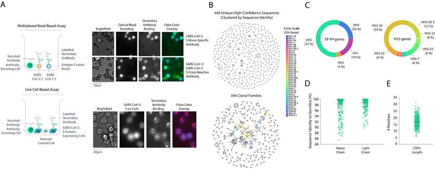
that mediates viral entry. Spike protein-dependent viral entry cell-based assay using mammalian cells that transiently ex-
is initiated by upward movement of the receptor-binding do- pressed full-length membrane-anchored SARS-CoV-2 spike
main (RBD) at the apex of the protein allowing access to bind protein (Fig. 1A). In total, 5.8 million PBMCs were screened
the angiotensin converting enzyme 2 (ACE2) cellular receptor and machine learning (ML)-based analysis pipelines were
(8–11). Upon receptor engagement, coordinated proteolytic used to automatically select and rank > 4,500 antibody “hits”
cleavage, shedding of the S1 subunit, and conformational re- (0.08% frequency), of which 2,238 single antibody-secreting
arrangement of the S2 subunit, leads to viral fusion with the cells were chosen for recovery. Next-generation sequencing
cell and transfer of genetic material. Given the critical nature (NGS) libraries of antibody genes from selected single B cells
of the RBD interaction with ACE2 for viral entry, antibodies were generated and sequenced, and a custom bioinformatics
that bind the RBD and interfere with ACE2 binding can have pipeline with machine learning (ML)-based sequence cura-
potent neutralizing activity (7, 12, 13) some of which have pro- tion was used to recover paired-chain antibody sequences, re-
gressed to clinical study (14). sulting in 440 unique high-confidence paired heavy and light
To test the potential for neutralizing monoclonal antibod- chain sequences (Fig. 1B). The sequences belonged to 394
ies (mAbs) to prevent SARS-CoV-2 infection in vivo, we used clonal families and used a diverse set of 39 heavy-chain-vari-
Downloaded from http://stm.sciencemag.org/ by guest on April 26, 2021
the rhesus macaque challenge model. Although rhesus ma- able (VH) genes, with the VH3 family of genes representing
caques do not exhibit the severe pulmonary symptoms some- 57% of total diversity (Fig. 1C), similar to other reports (23).
times associated with human COVID-19 disease, the model Among these, the VH3-30 gene was the most common (39%).
allows for assessment of viral replication in the upper and Of the 440 unique antibodies identified, 4% were cross-reac-
lower airways (15–19). Of particular interest, recent studies in tive to both full-length SARS-CoV-2 and SARS-CoV-1 spike
this model have shown that prior exposure to SARS-CoV-2 or proteins. The mean sequence identity to germline was high
administration of a SARS-CoV-2 vaccine are sufficient to pre- (98% and 99% for heavy and light chains respectively) (Fig.
vent infection upon subsequent challenge (18, 20). Protecting 1D) with a broad distribution of complementarity-determin-
non-human primates (NHPs) from SARS-CoV-2 infection ing region 3 (CDR3) lengths (Fig. 1E), likely due to sample
may inform the clinical development of medical counter- collection early in the immune response.
measures for patients with COVID-19 (17, 21). Down-selection and binding characterization of SARS-
In this study, we report a strategy for high-throughput CoV-2 antibodies.
screening, which allowed for the rapid identification and sub- From the set of 440 antibodies, we used an internally de-
sequent characterization of anti-spike neutralizing antibod- veloped informatics and data visualization software package,
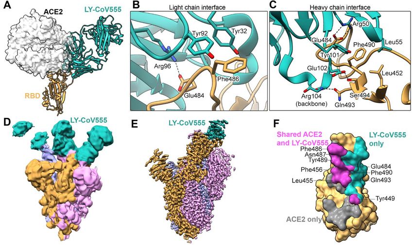
ies. An RBD-specific antibody (LY-CoV555) was discovered Celium, to select 187 antibodies for rapid cloning and recom-
that binds to the RBD in the up (active) or down (resting) binant expression. Preference was given to antibodies ob-
conformation and demonstrated substantially greater neu- served at high frequency across the dataset, especially those
tralization potency of SARS-CoV-2 in vitro relative to all other discovered in both multiplexed soluble protein and live-cell
antibodies analyzed from this patient. Passive immunization assays. The selection also maximized the diversity of VH
by infusion of LY-CoV555 protected both lower and upper air- genes and CDR3 sequences and limited CDR3 sequence lia-
ways from SARS-CoV-2 infection in a rhesus macaque model. bilities. A total of 175 sequences were successfully cloned into
These data supported the rapid progression of LY-CoV555 expression vectors to generate recombinant antibodies with
into clinical evaluation, where single antibody efficacy in the immunoglobulin G1 (IgG1) backbones for more detailed char-
treatment of SARS-CoV-2 infection was subsequently demon- acterization. Subsequent characterization included high-
strated (22). throughput biophysical analysis (fig. S1A), validation of solu-
RESULTS ble and cell-associated spike protein binding, cross reactivity
to other coronavirus spike proteins and three circulating
Identification of convalescent patient-derived SARS-
SARS-CoV-2 spike variants (fig. S1B), apparent binding affin-
CoV-2 antibodies.
ity to soluble spike by surface plasmon resonance (SPR) (Fig.
To identify potential therapeutic antibodies from a conva-
2A, fig. S1C), and functional screening in a high-throughput
lescent patient following diagnosis with COVID-19, a high-
pseudotyped lentivirus reporter neutralization assay.
throughput screening approach was used to identify relevant
Of the 175 selected antibodies, 92% of antibodies validated
anti-spike mAbs (Fig. 1). Peripheral blood mononuclear cells
as SARS-CoV-2 binders, 34% as bat SARS-like coronavirus
(PBMCs) were obtained approximately 20 days post-symp-
WIV1 binders, 31% as SARS-CoV-1 binders, 3% as Human
tom onset. Two screening assays were utilized: (1) a multi-
coronavirus HKU1, 2% as Middle Eastern respiratory syn-
plexed bead-based assay using optically-encoded microbeads,
drome coronavirus (MERS-CoV) binders, and 2% as cross-
each conjugated to either soluble prefusion-stabilized tri-
binders to all spike proteins (Fig. S1B). Furthermore, 51% of
meric SARS-CoV-2 or SARS-CoV-1 spike protein and (2) a live
antibodies validated as SARS-CoV-2 S1 subunit-specific
First release: 5 April 2021 stm.sciencemag.org (Page numbers not final at time of first release) 2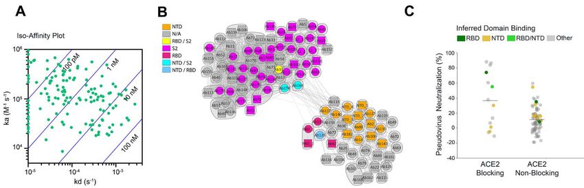
binders, with 8% cross-binding to full length WIV1 and 6%            tested (10, 1, 0.1, or 0.01 μg/mL), (3) dose-dependent neutral-
cross-binding to full-length SARS-CoV-1, suggesting that, as         ization profile, (4) RBD competition, (5) ACE2 blocking activ-
expected, most cross-binders are S2 subunit-specific. Anti-          ity, and (6) acceptable biophysical profile (melting
body binding to cell-expressed, full-length SARS-CoV-2 wild-         temperature, solubility, and polydispersity). The selected an-
type spike and known circulating variants (V367F, V483A,             tibodies were then produced at larger scale for further test-
D614G) was validated via automated high-throughput flow              ing. The binding properties of these selected antibodies,
cytometry (fig. S1B). In this assay format, 77% of antibodies        specifically binding to which domain of the spike protein, ap-
were validated as wild-type spike protein binders. Of that           parent antibody affinity to the trimeric spike protein, and
subgroup, 93% also validated for binding to two RBD muta-            monomeric Fab binding affinities are summarized in table
tions (V378F and V483A) and the very common D614G non-               S1; interestingly in spite of a relatively narrow range of fully
RBD mutation. In addition, 76% of antibodies were validated          avid antibody binding to the spike protein with nearly all ap-
in both multiplexed bead-based and live cell-based assays            parent affinities falling within a 100-fold window, monomeric
(fig. S1B) indicating the robustness of the single-cell screen-      Fab binding was much more variable and substantially
ing assays with integrated ML-based hit-detection for identi-        weaker (fig. S3). As expected from the diverse nature of these

                                                                                                                                          Downloaded from http://stm.sciencemag.org/ by guest on April 26, 2021
fying SARS-CoV-2-specific antibodies. Consistent with the            properties, these antibodies exhibit a range of competition
bead and cell-based binding studies, these antibodies exhib-         behavior with each other leading to a number of epitope com-
ited high affinity binding to the soluble spike protein in SPR       munities (fig. S4).
capture kinetic experiments using a Carterra LSA instrument          Binding epitope characterization of SARS-CoV-2 anti-
(Fig. 2A, fig. S1C). Of these, 53% of the selected antibodies had    bodies.
apparent binding affinity constant (KD) values in the picomo-            Using negative-stain electron microscopy (nsEM), we
lar range and the remaining 47% in the nanomolar range,              were able to further characterize structurally the binding of
with a mean KD value of 5.3 nM. Due to the trimeric nature           a subset of these antibodies (fig. S5A). Images of sufficient
of the soluble spike protein and the potential bi-valent bind-       quality to enable three dimensional reconstructions of
ing by the coupled antibodies, these affinities are substan-         Fab:spike protein complexes for 5 of the Fabs were collected
tially greater than true monomeric binding affinities (table         for 3 RBD binders (Ab104, Ab138, and Ab169) and two NTD
S1), but likely are more representative of the pharmacological       binders (Ab89 and Ab130). Although the individual antibod-
setting.                                                             ies have unique epitopes exhibiting different orientations of
    High-throughput SPR experiments were used to charac-             the Fab relative to the spike protein, similarities and overlaps
terize the epitope coverage of the 175 antibodies. These exper-      were observed between them (fig. S5A). We also employed hy-
iments included antibody pairing, isolated domain binding,           drogen-deuterium exchange (HDX) followed by mass spec-
and binding competition with ACE2 (Fig. 2). Benchmark an-            trometry (table S3) to obtain epitope information for
tibodies with known binding to the S1 subunit, N-terminal            antibodies not observable by nsEM, and to gain finer epitope
domain (NTD), RBD, and S2 subunit epitopes of the SARS-              sequence detail for several antibodies. Consistent with nsEM
CoV spike protein and cross-reactivity to SARS-CoV-2 spike           experiments for antibodies characterized by both methods,
protein were included to mark epitope identity. Antibody             peptides exhibiting protection from exchange resided within
cross-blocking results are summarized in a competition plot          the expected structural regions. Epitope information was also
(Fig. 2B), as well as in a heat map (fig. S2). In total, 95 unique   obtained for an additional five RBD binders, three NTD bind-
bins (including controls) were identified, and a clear divide        ers, and three antibodies where protection from HDX was not
between S1- and S2-specific antibodies, as inferred by bench-        localized to a single domain (Ab82) or S2 binders (Ab127 and
mark competition, was observed (fig. S2), suggesting that            Ab164).
these antibodies possessed a broad epitope diversity. Only ap-
                                                                     Neutralization activities of SARS-CoV-2 antibodies.
proximately 10% of the antibodies tested exhibited ACE2
                                                                         The selected antibodies had a broad range of neutralizing
competition. Antibodies with ACE2 binding inhibition prop-
                                                                     activity in multiple in vitro assays including pseudovirus (ta-
erties had the greatest neutralizing activity based on pseudo-
                                                                     ble S4) and various live virus assay formats. Using a replica-
typed lentivirus reporter neutralization (Fig. 2C), although
                                                                     tion-competent SARS-CoV-2 molecular clone in which a non-
antibodies to other domains also had detectable neutralizing
                                                                     essential gene (ORRF7) has been replaced with a nano-lucif-
activity.
                                                                     erase reporter (Fig. 3A), neutralizing activity values spanning
    A lead panel of 24 antibodies (table S2) was selected using
                                                                     nearly three orders of magnitude were observed (table S4).
the Celium software, based on the following criteria: (1) bind-
                                                                     For a smaller number of antibodies, viral neutralization was
ing to SARS-CoV-2 spike protein in either the multiplexed
                                                                     further characterized in a Plaque Reduction Neutralization
bead-based or the live cell-based validation assay, (2) >30%
                                                                     Test (PRNT) format against two different clinical SARS-CoV-
pseudovirus neutralizing activity at any of the concentrations
                                                                     2 isolates, the INMI-1 isolate (clade 19A, Fig. 3B) and the

First release: 5 April 2021                              stm.sciencemag.org       (Page numbers not final at time of first release)   3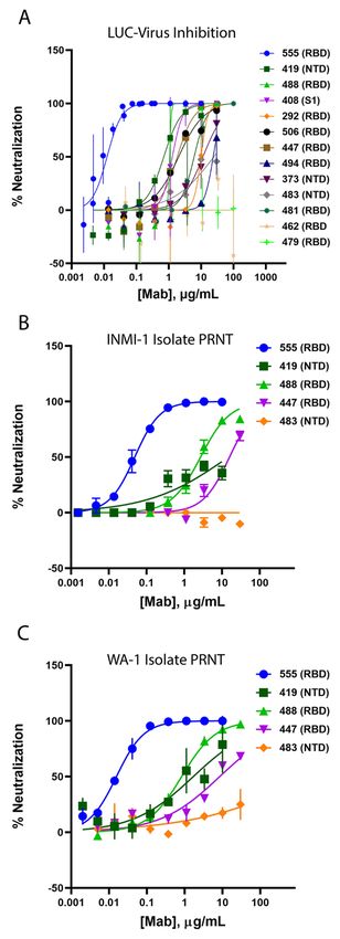
USA/WA-1/2020 isolate (clade 19B, Fig. 3C), representing two      intravenously (IV) to rhesus macaques at a dose of 1, 2.5, 15
major clades of SARS-CoV-2 (www.gisaid.com). The spike            or 50 mg/kg 24 hours prior to virus challenge. Control ani-
protein sequences for both isolates are identical to the Wu-      mals received 50 mg/kg of a control IgG1 antibody IV. The
han-Hu-1 isolate sequence (NCBI reference sequence entry          LY-CoV555 doses were chosen to provide a range of serum
NC_045512.2). Interestingly, it was observed that some non-       antibody concentrations and inform subsequent clinical dos-
RBD binding antibodies, for example Ab82, Ab89, and Ab130,        ing. Following inoculation, respiratory and clinical signs of
exhibited greater neutralizing activity in some of the live vi-   disease in the macaques were limited. Mild lobar congestion
rus SARS-CoV-2 assays compared to pseudovirus assays (ta-         and hyperemia were observed macroscopically across control
ble S4). Notably, the neutralization potency of one mAb,          and treated groups, suggestive of either interstitial or bron-
Ab169 (designated LY-CoV555), an RBD binder and ACE2              chopneumonia (table S5). Subgenomic RNA (sgRNA) and vi-
blocker, was consistently and substantially greater than the      ral genomes (gRNA), indicative of active viral replication (15),
rest and was selected for further development.                    were detectable in bronchoalveolar lavage fluid (BALF),
    LY-CoV555 possessed substantially (>10-fold) greater neu-     throat swabs, and nasal swabs for all control animals follow-
tralization potency relative to other identified RBD-binding      ing intranasal and intratracheal inoculation with SARS-CoV-

                                                                                                                                       Downloaded from http://stm.sciencemag.org/ by guest on April 26, 2021
and ACE2-blocking antibodies, such as Ab128 and Ab133, de-        2 (Fig. 5, Fig. 6).
spite similar apparent binding affinities (table S2), suggest-        Prophylactic administration of LY-CoV555 resulted in de-
ing a distinct binding mode of recognition. Structural            creases in viral replication and viral load as evaluated by
analysis using X-ray crystallography and cryo-electron mi-        sgRNA and gRNA, respectively, in the BALF and lung tissue
croscopy (cryo-EM) demonstrated that two of the RBD-              from the lower respiratory tract following SARS-CoV-2 inoc-
binding mAbs (Ab128 and Ab133) bind in a nearly identical         ulation (Fig. 5, table S6). In the BALF, reductions of 102 to 105
fashion to one another (fig. S5B), differing from LY-CoV555       copies per ml in viral replication and load were observed
and yet nearly identical in site and orientation to the previ-    compared to controls across Days 1, 3 and 6, with significant
ously described mAb CB6 (also known as etesevimab) (13).          reductions in viral replication (Fig. 5A; 1, 2.5, and 15 mg/kg
The epitope recognized by Ab128, Ab133, and CB6 only be-          doses) and load (Fig. 5B; 15 mg/kg dose) on Day 1 and at all
comes exposed on the RBD following its transition from the        doses on Day 3 relative to control IgG1-treated animals
down to the up and active state of the RBD. LY-CoV555 was         (q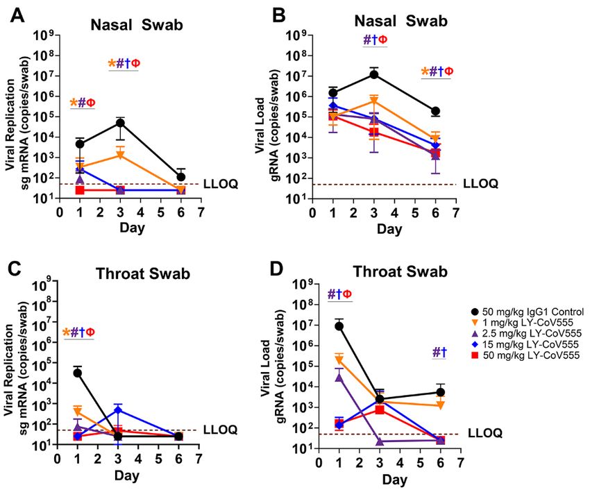
CoV555 dosed by the intravenous (IV) route. LY-CoV555 ad-         treatment may reduce virus replication in the upper airway,
ministration resulted in sustained serum concentrations af-       thus decreasing viral shedding and transmission following
ter IV dosing, with a half-life of elimination of 13 days, and    treatment. Overall, we show dose-related reductions in gRNA
clearance of 0.22 mL/hr/kg, consistent with expected phar-        and sgRNA in the upper and lower respiratory tracts with
macokinetics for human IgG1 in an NHP model (Fig. 7) (28,         maximal protection observed at doses of 2.5 mg/kg and
29). Serum concentrations of LY-CoV555 were evaluated dur-        above. Given the robust nature and route of administration
ing rhesus macaque prophylactic SARS-CoV-2 challenge ex-          of the viral inoculum in this model, we hypothesized that
periments. Serum LY-CoV555 in the rhesus macaques was             modest doses of LY-CoV555 could provide substantial clinical
dose proportional with the cynomolgus monkey PK (Fig. 7).         efficacy.
Mean serum concentrations of LY-CoV555 on the day of viral            Serum LY-CoV555 concentrations in the rhesus macaque
challenge were 15 ± 3, 38 ± 14, 276 ± 37 and 679 ± 101 mg/mL      model were dose-responsive and demonstrated sustained ex-
at doses of 1, 2.5, 15 and 50 mg/kg, respectively (table S7).     posure as expected for a human IgG1 antibody in a non-hu-
Based on the maximal infection protection provided at doses       man primate model. Maximal inhibition of viral replication
of 2.5 mg/mL and above, the 38 mg/mL serum concentration          across the upper and lower respiratory tract was observed at

                                                                                                                                       Downloaded from http://stm.sciencemag.org/ by guest on April 26, 2021
at the time of viral challenge provides a target for protective   doses of 2.5 mg/kg and above, associated with a mean serum
drug concentrations in this model. Given the substantial viral    concentration of 37.5 mg/mL at the time of infection. Median
inoculum, this value may overestimate serum concentrations        LY-CoV555 concentrations estimated in ELF fluid were 2-24%
needed to provide protection in community-acquired infec-         of serum concentrations, which was in general agreement
tions.                                                            with literature reports of antibody distribution to ELF fluid
    In addition, BALF concentrations of LY-CoV555 were de-        (31, 33). These data were also in the range of a literature-
termined in rhesus macaque prophylactic SARS-CoV-2 chal-          based physiologically based PK model-derived value of 6.5%
lenge experiments (table S8). BALF concentrations of LY-          used in clinical modeling and simulation to support study de-
CoV555, along with BALF urea concentrations, were used to         sign. At all of the LY-CoV555 doses tested in this study, me-
estimate lung ELF concentrations of LY-CoV555 using a pre-        dian lung ELF concentrations exceeded the effective
viously described method (30). Median BALF concentrations         concentration for 90% inhibition (EC90) for SARS-CoV-2 virus
and estimated ELF concentrations generally increased with         neutralization, which is consistent with the observed reduc-
increasing dose when comparing the 1, 2.5, and 15 mg/kg           tions in viral replication in the BALF and lung tissue across
doses of LY-CoV555. However, BALF and ELF concentrations          the dose range tested, even at substantially higher (10-fold)
in the 50 mg/kg groups (treatment and controls) did not           viral challenge doses relative to vaccine studies in this rhesus
show dose related increases compared to the 15 mg/kg dose.        macaque model (18). ELF fluid concentrations were not eval-
Median estimated ELF concentrations as a percentage of se-        uated in the nose and throat. As compared to the lung and
rum concentration ranged from 2% to 24% (table S8). These         throat, the delayed impact on viral loads in nasal swabs could
values are in the range of nasal ELF concentrations previ-        reflect differential distribution of antibody into the nasal
ously reported for another therapeutic antibody (31).             ELF.
                                                                      The PBMC sample, from which LY-CoV555 was derived,
DISCUSSION
                                                                  was collected approximately 20 days following symptom on-
    This study describes the rapid identification and charac-
                                                                  set. This is an early time point in the disease course and in
terization of a potent anti-spike neutralizing antibody, LY-
                                                                  the immune response to viral infection. In spite of the lack of
CoV555, derived from PBMCs isolated from a patient after re-
                                                                  substantial somatic mutation of antibodies, as evidenced by
covery from COVID-19. Following antibody screening, LY-
                                                                  the high sequence similarity to germline, we were able to
CoV555 demonstrated greater neutralization potency of
                                                                  identify several antibodies to the spike protein capable of
SARS-CoV-2 compared to the other antibodies discovered
                                                                  neutralizing viral infection in ACE2-bearing cells, including
from this patient (32). LY-CoV555 was found to possess high
                                                                  some that did not directly block ACE2 engagement. Compar-
affinity RBD binding and ACE2 blocking properties, which
                                                                  ison of similar discovery approaches using samples from con-
translated to high neutralization potency due to its SARS-
                                                                  valescent patients suggest that the collection of antibodies
CoV-2 spike protein-binding properties. In both in vitro as-
                                                                  derived from this patient may have had relatively few mAbs
says with full virus and an NHP model of SARS-CoV-2 infec-
                                                                  with potent neutralizing activity (7, 12). Several factors might
tion, LY-CoV555 displayed high protection potency,
                                                                  be responsible for these differences, including the patient’s
supporting its clinical development and testing as a thera-
                                                                  immune status and disease severity, the relatively early col-
peutic for the treatment and prevention of COVID-19.
                                                                  lection of the sample used for antibody discovery, the depth
    Our NHP challenge study also provides evidence that neu-
                                                                  of screening and robust assays afforded by our microfluidic
tralizing antibodies have potential as an important counter-
                                                                  platform, the availability of structurally-defined protein
measure for preventing COVID-19 disease. Antibody

First release: 5 April 2021                            stm.sciencemag.org      (Page numbers not final at time of first release)   5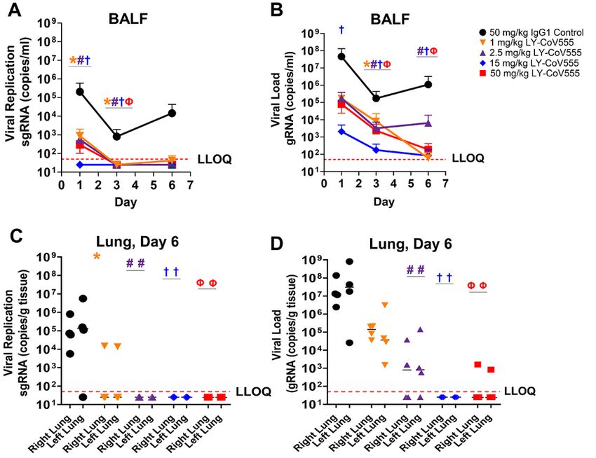
probes, or the very broad approach taken with respect to an-      eliminate binding and function, whereas mutations at V367,
tigenic diversity. Nonetheless, our approach identified a         K417, S477, and N501 have no effect (40). Importantly, alt-
highly potent neutralizing antibody from a patient sample         hough this paper focuses on the rapid identification and pre-
that had been characterized as possessing limited antibody        clinical characterization of one mAb, human studies are
response and neutralizing capacity (32).                          evaluating both single and mAb combinations using LY-
    The importance of prototype pathogen preparedness was         CoV555, and led to the Emergency Use Authorizations of both
demonstrated by the ability to rapidly design and produce         bamlanivimab alone and together with etesevimab (41, 42).
protein for B cell probes based on prior work defining the            This study focuses on the identification and characteriza-
structure and stabilization strategy for the betacoronavirus      tion of a single mAb that binds to the RBD of the SARS-CoV-
spike protein (34). The resulting speed at which this drug dis-   2 spike protein. This was a consequence, in part, of the very
covery and development effort proceeded (fig. S7), with pro-      limited number of the discovered antibodies exhibiting po-
gression to human treatment only 90 days after the initiation     tent neutralization. These results indicate that one limitation
of antibody screening, was due to advanced discovery and          to the approach taken was due to the timing of the patient
characterization platforms and pre-established public-pri-        sample relative to infection; at this early point in the evolu-

                                                                                                                                       Downloaded from http://stm.sciencemag.org/ by guest on April 26, 2021
vate partnership.                                                 tion of the patient’s immune response, very deep screening
    It is important to note that both monotherapy and anti-       was required to identify potent neutralizing antibodies. Fu-
body combinations are being explored clinically (14). Mono-       ture pandemic response efforts might take into consideration
therapy with a single potent antibody represents a pragmatic      this aspect and use an approach that balances timing with
option to combat an ongoing pandemic with a virus that            the ability to identify greater numbers of highly potent anti-
causes an acute, self-limited infection. For both respiratory     bodies to enable a rapid discovery of multiple antibodies for
syncytial virus (RSV) and Ebola virus, there are clinical prec-   use in cocktails.
edents for monotherapy prophylaxis or treatment, respec-              A limitation of the animal model studies is the focus on
tively, with potent neutralizing monoclonal antibodies (35).      testing LY-CoV555 in a prophylactic setting in a NHP model
Specifically, in the case of RSV, infants have been effectively   that does not recapitulate the full disease physiology of
treated with palivizumab since its introduction in 1996 (36).     COVID-19 in humans. In addition, we did not study the ther-
As therapy, neutralizing mAbs, such as LY-CoV555, could sup-      apeutic effect of LY-CoV555 in this animal or other animal
plement an ongoing endogenous adaptive immune response            models. We focused on prophylaxis for two primary reasons:
to the virus, with its own diverse polyclonal antibodies in ad-   first, the NHP model is better suited to test prevention of dis-
dition to other responses such as expansion of specific CD4+      ease; and second, due to the rapid speed of development of
and CD8+ T cell populations (37). This endogenous polyclonal      LY-CoV555, efficacy in the therapeutic setting was already be-
antibody repertoire, which will possess neutralizing activity     ing explored clinically at the time of these experiments. Sub-
against diverse epitopes, supplemented with virus-specific T      sequent clinical trial results with LY-CoV555 administered as
cell responses, should minimize the likelihood that escape        a treatment to patients infected with SARS-CoV-2-and with
mutants will arise during acute infection.                        mild to moderate disease demonstrated reduction in viral
    Not surprisingly, the spike protein, and the RBD in par-      load, reduction in COVID-19 symptoms, and an approximate
ticular, has been susceptible to mutations due to its pivotal     3-fold decreased rates of hospitalization in the 700, 2800, and
role in the infection process. There have been a number of        7000 mg dose groups relative to placebo, indicating activity
variants emerging recently that contain an N501Y mutation,        as a treatment and the potential for efficacy at lower doses
which is associated with increased transmissibility. This mu-     (22). In addition, we felt that it was important to understand
tation is found in the lineages B.1.1.7 and B.1.351, which were   the efficacy dose-response, especially with respect to blood
discovered in the United Kingdom and South Africa, respec-        and BALF concentrations of LY-CoV555, as they relate to pre-
tively (38, 39). Based on the structure of the LY-CoV555:RBD      ventive efficacy. This could be studied in this animal model.
complex, N501Y does not reside within the epitope for this        This study also informs subsequent use in a post-exposure
antibody. However, B.1.351 includes two other mutations at        prophylactic setting as is being explored clinically
important residues in the RBD, K417N and E484K, of which          (NCT04497987). Finally, other efforts had already demon-
only E484 falls within the epitope of LY-CoV555 (39). As          strated that antibodies effective in prophylaxis were also ef-
would be expected, mutations at residues within the epitope       fective in treatment in multiple animal models, albeit with
of LY-CoV555 have the potential to impact the binding and         different potency (13, 43).
function, whereas residues outside the epitope do not. For ex-        LY-CoV555 was developed as therapeutic antibody specif-
ample, low frequency mutations that have been observed in         ically to treat COVID-19. The treatment quickly entered clin-
GISAID (Global initiative on sharing all influenza data) at po-   ical testing (44, 45) and demonstrated clinical efficacy (22),
sitions V483, E484, F490, and S494 either decrease or             and gain Emergency Use Authorization (42). LY-CoV555 is

First release: 5 April 2021                            stm.sciencemag.org      (Page numbers not final at time of first release)   6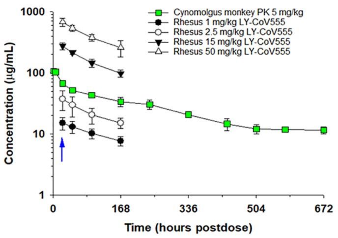
presently under clinical evaluation for the treatment and pre-   microfluidic screening devices with either 91,000 or 153,000
vention of COVID-19 (NCT04411628; NCT04427501;                   individual nanoliter-volume reaction chambers (46–54). Sin-
NCT04497987; NCT04501978; NCT04518410; NCT04634409)              gle cells secreting target-specific antibodies were identified
in various clinical settings. Overall, the identification and    and isolated using two assay types (55): a multiplexed bead
characterization of LY-CoV555 points to the feasibility of       assay using multiple optically-encoded beads, each conju-
strategies to rapidly identify neutralizing human mAbs as        gated to the soluble pre-fusion stabilized spike of either
part of an initial response to an evolving pandemic that can     SARS-CoV-1 or SARS-CoV-2 spike with T4-foldon domain, 3C
complement population-scale vaccination, provide immedi-         protease cleavage site, 6x His-tags, and twin-strep tags (34)
ate passive immunity, and provide protection for vulnerable      or negative controls (bovine serum albumin [BSA] His-tag
populations.                                                     and T4 FoldOn trimerization domain), and a live cell assay
                                                                 using passively dyed suspension-adapted Chinese hamster
MATERIALS AND METHODS
                                                                 ovary (CHO) cells transiently transfected to surface-express
Study Design                                                     full-length SARS-CoV-2 spike protein (GenBank ID
    This study was designed to identify SARS-CoV-2 neutral-      MN908947.3) with a green fluorescent protein (GFP) re-
izing antibodies from a convalescent patient with COVID-19.

                                                                                                                                      Downloaded from http://stm.sciencemag.org/ by guest on April 26, 2021
                                                                 porter, and non-transfected cells as a negative control. Beads
This objective was addressed by first conducting a detailed      or cells were flowed onto microfluidic screening devices and
screening of antibodies produced from patient-derived            incubated with single antibody-secreting cells, and mAb
PBMCs to identify high affinity SARS-CoV-2 spike protein         binding to cognate antigens was detected via a fluorescently
binding and was followed by a variety of high-throughput         labeled anti-human IgG secondary antibody. Positive hits
binding characterization experiments to identify SARS-CoV-       were identified using machine vision and recovered using au-
2 neutralizing antibodies. All in vitro characterization of      tomated robotics-based protocols.
binding properties and viral infection neutralization were
                                                                 Single-cell sequencing, bioinformatic analysis, and
carried out in a screening fashion, with n=1, and the number
                                                                 cloning
of technical replicates as described in the associated figure
                                                                     Single cell polymerase chain reaction (PCR) and NGS
legends. For in vivo characterization of the ability of LY-
                                                                 (MiSeq, Illumina) were performed using automated work-
CoV555 to provide protection from SARS-CoV-2 infection, an-
                                                                 stations (Bravo, Agilent) and custom molecular biology pro-
imals were randomized to dose groups to achieve a similar
                                                                 tocols for the recovery of paired heavy and light chain
average age for each group. The number of animals in each
                                                                 sequences. Sequencing data were analyzed using a custom bi-
dose group, timing of drug administration and virus inocula-
                                                                 oinformatics pipeline to yield paired heavy and light chain
tion was informed by available data regarding the rhesus ma-
                                                                 sequences for each recovered antibody-secreting cell (56).
caque model of SARS-CoV-2 infection (15). In the cynomolgus
                                                                 Each sequence was annotated with the closest germline
monkey PK study, naïve monkeys were selected from the PK
                                                                 (V(D)J) genes, degree of somatic hypermutation, and poten-
colony to minimize potential impact of anti-human antibod-
                                                                 tial sequence liabilities. Antibodies were considered members
ies on the PK profile. With respect to the number of animals
                                                                 of the same clonal family if they shared the same inferred
per group and duration of sample collection, the PK study
                                                                 heavy and light V and J genes and had the same CDR3 length.
leveraged a standard study design . Researchers were blinded
                                                                 The variable (V(D)J) region of each antibody chain was PCR
to the identity of antibodies where possible. All data points
                                                                 amplified and inserted into expression plasmids using a cus-
were included in the analyses, and no outliers were excluded.
                                                                 tom, automated high-throughput cloning pipeline. Plasmids
Single-cell screening and recovery                               were verified by Sanger sequencing to confirm the original
   A blood sample from a 35-year-old individual hospitalized     sequence previously identified by NGS. Antibodies were re-
with severe COVID-19 disease was obtained mid-February           combinantly produced by transient transfection in either hu-
2020, approximately 20 days following the onset of symp-         man-embryonic kidney (HEK)293 or CHO cells as described
toms. PBMC samples were collected under institutional re-        in Supplemental Methods.
view board (IRB)-approved protocols as part of the
                                                                 Binding validation and analysis
Hospitalized and Ambulatory Adults with Respiratory Viral
                                                                    Recombinant antibodies were confirmed to bind screen-
Infections (HAARVI) study at the University of Washington
                                                                 ing targets using two assay types via high-throughput flow
(protocol #STUDY00000959) and Vaccine Research Center
                                                                 cytometry. In a multiplexed bead-based assay, optically en-
(VRC), National Institute of Allergy and Infectious Diseases
                                                                 coded beads were conjugated to one of the following unique
(NIAID) and National Institutes of Health (NIH)(protocol-
                                                                 antigens: spike proteins of SARS-CoV-2, Middle Eastern res-
VRC400, NIH-07IN194). Cells were thawed, activated in cul-
                                                                 piratory syndrome coronavirus (MERS-CoV), Severe acute
ture to generate memory B cells, and enriched for antibody-
                                                                 respiratory syndrome (SARS-CoV-1), human coronavirus
secreting B cells prior to injection into AbCellera’s
                                                                 (HKU1-CoV), bat SARS-like WIV1 coronavirus, or the S1

First release: 5 April 2021                           stm.sciencemag.org      (Page numbers not final at time of first release)   7
subunit of SARS-CoV-2 spike protein. Purified antibodies followed by running buffer injection for 15 min (dissociation
were incubated with target-conjugated and negative control phase). Two regeneration cycles of 15 s were performed be-
BSA His-tag and T4 FoldOn-conjugated beads at either 50 tween each dilution series by injecting Pierce IgG elution
nM, 10 nM or 2 nM antibody concentration for 30 min at buffer (Thermo Fisher) + 1 M NaCl on the chip surface. The
room temperature (RT). In a live cell-based assay, full-length data were analyzed using the Carterra Kinetics analysis soft-
spike protein sequences of either the wild-type or mutants ware using a 1:1 Langmuir binding model to determine ap-
V367F, V483A, and D614G of SARS-CoV-2 with GFP inserts parent association (ka) and dissociation (kd) kinetic rate
were transiently transfected into CHO cells (MaxCyte STX constants and binding affinity constants (KD).
Scalable Transfection System). Full-length native confor- For epitope binning experiments, antibodies coupled to
mation spike protein expression was confirmed via GFP de- the chip surface were exposed to various antibody:antigen
tection, flow cytometry-detected binding to S1 and S2 complexes. Samples were prepared by mixing each antibody
subunit-specific benchmark antibodies, and by Western blot. in 10 to 20-fold molar excess with antigen (1:1 freshly pre-
Purified antibodies were incubated with target-expressing pared mix of 400 nM antibody and 40 nM antigen, both di-
cells and non-transfected control cells at 50 nM, 10 nM, or 2 luted in 1X HBSTE + 0.1% BSA running buffer). Each antigen-
Downloaded from http://stm.sciencemag.org/ by guest on April 26, 2021
nM antibody concentration for 30 min at 4°C. Beads or cells antibody premix was injected sequentially over the chip sur-
were washed, and binding was detected using a fluorescently face for 4 min (association phase to ligand printed onto chip
labeled anti-human IgG secondary antibody. Fluorescence previously), followed by a running buffer injection for 2 min
was measured using high-throughput plate-based flow cy- (dissociation phase). Two regeneration cycles of 15 s were per-
tometry. Benchmark antibodies previously identified from formed between each premix sample by injecting 10 mM gly-
SARS-CoV-1 convalescent patient samples and cross-reactive cine pH 2.0 onto the chip surface. An antigen-only injection
to SARS-CoV-2 spike protein were used as positive controls; (20 nM concentration in running buffer) was performed
human IgG isotype and an irrelevant antibody specific to hu- every 8 cycles. The data were analyzed using the Carterra
man immunodeficiency virus (HIV), VRC01, were used as Epitope analysis software (version 1.2.0.1960) for heat map
negative controls. Median fluorescence intensity of each an- and competition network generation. Analyte binding signals
tibody was normalized over the median fluorescence inten- were normalized to the antigen-only binding signal, such that
sity of the human isotype, with signals greater than 5-fold the antigen-only signal average is equivalent to one relative
over isotype control (and less than 2.5-fold binding to nega- unit (RU). A threshold window ranging from 0.9 RU to 1.1 RU
tive controls) considered as specific binding. was used to classify analytes into 3 categories: blockers (bind-
Surface-plasmon resonance binding experiments ing signal under the lower limit threshold), sandwichers
All high-throughput surface plasmon resonance (SPR) (binding signal over the higher limit threshold) and ambigu-
binding, epitope binning and ACE2 competition experiments ous (binding signal between limit thresholds). Antibodies
were performed on a Carterra LSA instrument equipped with with low coupling to the chip, poor regeneration or with ab-
an HC-30M chip type (Carterra-bio) using a 384-ligand array sence of self-blocking were excluded from the binning analy-
format. For all experiments, antibodies were coupled to the sis. Like-behaved antibodies were automatically clustered to
HC-30M chip: the chip surface was first activated by flowing form a heat map and competition plot.
a freshly prepared 1:1:1 activation mix of 100 mM MES pH For ACE2 competition experiments, antibodies coupled to
5.5, 100 mM S-NHS, and 400 mM EDC for 7 min, and anti- the chip were exposed to spike protein:ACE2 complex; 20
bodies diluted to either 10 μg/mL or 1 μg/mL in 10 mM nM of SARS-CoV-2 spike protein was premixed with 200
NaOAc pH 4.25 buffer + 0.01% Tween were injected and nM of the His-tagged ACE2 (ACE2-His) diluted
printed simultaneously onto the chip surface for 10 min by in HBSEP+ with 0.5 M NaCl, 1% BSA, 1x dextran, and 2
direct coupling. The chip surface was quenched by flowing 1 mg/mL heparin, and incubated for about 12 hours. The com-
M EtOHamine for 7 min, followed by two wash steps of 15 s plex of spike protein/ACE2-His was then tested for binding
each in 25 mM MES pH 5.5 buffer. Relevant benchmarks and to immobilized antibodies on the prepared HC30M chip, with
negative control antibodies (HIV VRC01, mouse FoldOn association for 5 min and dissociation for 1 min. Regenera-
8203-C1, and rabbit His-tag PA1-983) were also printed on the tion was performed in 20mM Glycine pH 2.0 with 1 M NaCl
chip surface. for 30 s twice.
For binding kinetics and affinity measurements, a 3-fold Negative-stain electron microscopy
dilution series of the antigen of interest, starting at 300 nM SARS-CoV-2 spike ectodomain was diluted to 0.04 mg/mL
in HEPES-buffered saline containing 0.00% Tween-20 and 3 in 2 mM Tris pH 8.0, 200 mM NaCl, 0.02% NaN3 (dilution
mM EDTA (HBSTE) + 0.1% BSA running buffer was sequen- buffer) in the presence of 10-fold excess Fab and incubated
tially injected onto the chip surface. For each concentration, on ice for 10 s. CF400-Cu grids (Electron Microscopy Sci-
the antigen was injected for 5 min (association phase), ences) were plasma cleaned for 30 s in a Solarus 950 plasma
First release: 5 April 2021 stm.sciencemag.org (Page numbers not final at time of first release) 8cleaner (Gatan) with a 4:1 ratio of O2/H2. A volume of 4.8 μL of (approximately 12 mg/mL) were crystallized by vapor diffu-
the protein sample was applied to the grid and allowed to sion sitting drops. Crystals of complexes formed within 1 to 2
incubate for 30 s. The grid was then washed twice with dilu- days and were harvested on the third day. Crystals were flash-
tion buffer prior to staining with methylamine tungstate frozen in liquid nitrogen following 1-min incubation in cryo-
(NANO-W, Nanoprobes). Grids were imaged us- protectant solution containing 25% glycerol in mother liquor:
ing a FEI Talos TEM (Thermo Fisher Scientific) and LY-CoV555 Fab-RBD complex crystallized using 100 mM so-
a Ceta 16M detector. Micrographs were collected manually dium acetate pH 4.6 and 20% PEG 10K; the 481CK Fab-RBD
using TIA v4.14 software at a magnification of ×92,000, cor- complex crystallized using 100 mM Tri-Sodium Citrate
responding to a pixel size of 1.63 Å/pixel. Contrast transfer pH=5.8, and 14% PEG 4K, and 10% 2-Propanol; and the 488
function (CTF) estimation and particle picking were per- CK Fab-RBD complex crystallized using 100 mM HEPES
formed in cisTEM. A 2D classification was performed in ei- pH=7.7, and 8% PEG 3350, and 200 mM L-Proline.
ther cisTEM (57) or cryoSPARC v2.15.10 (58), and antibody Diffraction data were collected at Lilly Research Labora-
initio reconstruction and refinement of 3D maps were per- tories Collaborative Access Team and beamline at Sector 31
formed in cryoSPARC. of the Advanced Photon Source at Argonne National Labora-
Downloaded from http://stm.sciencemag.org/ by guest on April 26, 2021
tory. Crystals stored in liquid nitrogen were mounted on a
Cryo-electron microscopy
goniometer equipped with an Oxford Cryosystems cry-
A purified, prefusion stabilized SARS-CoV-2 spike variant,
ostream maintained at a temperature of 100 K. The wave-
HexaPro (59) at 0.2 mg/mL was complexed with 1.3-fold mo-
length used was 0.9793 Å, collecting 900 diffraction images
lar excess of LY-CoV555 Fab in 2 mM Tris pH 8, 200 mM
at a 0.2-degree oscillation angle and 0.12 s exposure time on
NaCl, 0.02% NaN3 for 5 min on ice. Three microliters of pro-
a Pilatus3 S 6M detector at a distance of 392 mm.
tein complex were deposited on an UltrAuFoil 1.2/1.3 grid
The diffraction data were indexed and integrated using
(Electron Microscopy Sciences) which had been plasma
autoPROC (66) / XDS (67) and merged and scaled in
cleaned for 2 min using a Gatan Solarus 950 with a 4:1 O2:H2
AIMLESS (68) from the CCP4 suite (69). Non-isomorphous
ratio. The grid was then plunge-frozen in liquid ethane using
data readily yielded initial structures by Molecular Replace-
a Vitrobot Mark IV (Thermo Scientific) set to 100% humidity
ment using for the Fab portion crystal structures from the
and 22°C, with a blot time of 5 s and a blot force of -4. Data
proprietary Eli Lilly structure database and for the SARS-
were collected on a Titan Krios operating at 300kV and
CoV-2 spike RBD from the public domain structure with the
equipped with a K3 detector using a magnification of
access code 6yla (70). The initial structure coordinates for
22,500x, resulting in a pixel size of 1.045 Å. A total of 30
each dataset were further refined using Refmac5 (CCP4) ap-
frames were collected for each micrograph, with defocus val-
plying isotropic temperature factors. Model building was per-
ues ranging from -0.8 μm to -2.8 μm, a total exposure time of
formed with Coot (CCP4) and final structure validation with
4.5 s, and a total electron dose of ~32.7 e−/Å2. A full descrip-
MolProbity (71) and CCP4 validation tools. Table S10 presents
tion of the data collection parameters can be found in table
the crystallographic data statistics. Protein coordinates and
S9 and fig. S6. Motion correction, CTF estimation, and parti-
structure factors have been deposited with the Protein Data
cle picking were performed in Warp (60). Particles were sub-
Bank under the access codes 7KMG, 7KMH and 7KMI for
sequently transferred to cryoSPARC v2.15.10 (58) for 2D
Ab169 (LY-CoV555), Ab133 and Ab128, respectively.
classification and 3D reconstruction. The refined map was
then subjected to local B-factor sharpening using Lo- Pseudotyped neutralization assay for monoclonal anti-
calDeBlur (61). Model building and refinement were subse- body screen
quently performed using Coot, Phenix and ISOLDE (62–64). SARS-CoV-2 spike pseudotyped lentiviruses that harbor a
luciferase reporter gene were produced and neutralization as-
Protein crystallography
say was performed as described previously (72, 73). Pseudo-
For protein crystallography, an isolated RBD (using resi-
virus was produced by co-transfection of 293T cells with
dues 329 to 527), was fused to a 6-His tag at the C terminus,
plasmids encoding the lentiviral packaging and luciferase re-
expressed in CHO cells, enzymatically deglycosylated using
porter, a human transmembrane protease serine 2
endoglycosidase-H (Endo-Hf, New England Biolabs), and pu-
(TMPRSS2), and SARS-CoV-2 S (Wuhan-1, GenBank #:
rified by cation exchange chromatography. The Fab portions
MN908947.3) genes. Forty-eight hours after transfection, su-
of selected antibodies, containing mutations in the constant
pernatants were harvested, filtered and frozen. For initial
region known to encourage crystallization (65), were ex-
screening neutralization assay 4 dilutions of monoclonal an-
pressed in CHO cells, and purified. The Fab:RBD complexes
tibodies (10, 1, 0.1, and 0.01ug/mL) were mixed with titrated
were prepared by mixing the components with a 20% excess
pseudovirus, incubated for 45 min at 37°C and added to pre-
of the RBD, and then the complex purified from the excess
seeded ACE2-transfected 293T cells (either transiently or sta-
RBD by size-exclusion chromatography. Fab:RBD complexes
bly transfected) in 96-well white/black Isoplates (Perkin
First release: 5 April 2021 stm.sciencemag.org (Page numbers not final at time of first release) 9Elmer). Following 2 hours of incubation, wells were replen- 1 hour at RT. Background staining was quenched by adding
ished with 150 μL of fresh medium. Cells were lysed 72 hours 50 mM NH4Cl to cells and rocking for 10 min at RT, followed
later and luciferase activity (relative light unit, RLU) was by washing. Cells were permeabilized with 0.1% Triton-X 100
measured. Percent neutralization was calculated relative to (by rocking at RT for 10 min), washed three times with Dul-
pseudovirus-only wells. becco’s Phosphate Buffered Saline (DPBS), and nonspecific
Neutralization activity of antibodies against authentic antibody binding was blocked with 1% BSA. Mouse anti-
SARS-CoV-2 SARS-CoV-2 nucleoprotein antibody (1 C7C7, a kind gift from
Authentic SARS-CoV-2 neutralization activity of the dis- Thomas Moran, Icahn School of Medicine at Mount Sinai),
covered antibodies was measured by detecting the neutrali- diluted at 1:1000 in DPBS with 1% BSA, was added to each
zation of infectious virus in cultured Vero E6 cells (African well and incubated overnight at 4°C. After washing, cells
Green Monkey Kidney; American Type Culture Collection were stained with goat anti-mouse Alexa Fluor plus 647 anti-
(ATCC) #CRL-1586). These cells are known to be highly sus- body (Thermo Fisher # A32728; green dye) and DAPI (4’,6-
ceptible to infection by SARS-CoV-2. Cells were maintained diamidino-2-phenylindole, dihydrochloride; Thermo Fisher #
according to standard ATCC protocols. Briefly, Vero E6 cells 62247; blue dye) by incubating for 1 hour at 37°C. Images
Downloaded from http://stm.sciencemag.org/ by guest on April 26, 2021
were grown in Minimal Essential Medium (MEM) supple- were collected using a CellInsight CX7 with the 4× objective
mented with 10% heat-inactivated fetal bovine serum (FBS), covering the entire well. The percentage of infected cells per
2mM L-glutamine, and 1% of MEM Nonessential Amino Acid well relative to the uninfected and no-antibody controls was
(NEAA) Solution (Fisher #MT25025CI). Cell cultures were analyzed using the instrument’s “Target Activation” analysis
grown in 75 or 150 cm2 flasks at 37°C with 5% CO2 and pas- protocol.
saged 2 to 3 times per week using trypsin-EDTA. Cell cultures Virus neutralization detected by luciferase reporter
used for virus testing were prepared as subconfluent mono- Luciferase assays were performed using a molecular com-
layers. All incubations containing cells were performed at plementary DNA clone of a SARS CoV-2 isolate (USA/WA-
37°C with 5% CO2. 1/2020) in which a non-essential gene (ORF7) was replaced
Production of virus inocula by the NanoLuc luciferase reporter gene (Promega), as previ-
Immunofluorescent and plaque reduction assays were ously described for SARS-CoV-1 and MERS-CoV (74). Virus
conducted using virus produced by infecting cultured Vero infectivity assays were conducted in 96-well tissue culture
E6 cells with the SARS-CoV-2 clinical isolate USA/WA-1/2020 plates. Vero E6 cells were seeded at a density of 2x10⁴ cells
(BEI resources number NR52281) or the INMI-1 isolate (Eu- per well in DMEM medium supplemented with 10% FBS
ropean Virus Archive – Global, ref #008V-03893) and incu- (DMEM/FBS) and incubated for 15 to 24 hours. The next day,
bating at 37°C until cytopathology is evident (typically 48 to serial dilutions of antibodies or human IgG1 isotype control
72 hours). Expansion was limited to 1 to 2 passages in cell were prepared in DMEM/FBS. The SARS-CoV-2-NanoLuc in-
culture to retain integrity of the original viral sequence. The oculum was diluted in DMEM/FBS, mixed with an equal vol-
virus stock was quantified by standard plaque assay, and ali- ume of diluted antibody to produce a final virus titer of 140
quots were stored at -80°C. A freshly thawed aliquot was used plaque-forming units (PFU) per well, and incubated for 1
for each neutralization experiment. hour. After removing the culture medium from the plated
Vero E6 cells, the virus-antibody solution was inoculated onto
Virus neutralization detected by immunofluorescence
duplicate wells and incubated for 48 hours. Following stand-
Virus infectivity assays were conducted in 96-well tissue
ard protocols as recommended by the vendor, NanoGlo rea-
culture plates. Vero E6 cells were seeded at a density of 8x10⁴
gent (Promega #N1110) was added and luciferase activity was
cells/cm2 and incubated overnight to a confluency of approx-
quantified on a SpectraMax plate reader (Molecular Devices).
imately 95%. Serial dilutions of antibodies or positive control
polyclonal serum from a convalescent SARS-CoV-2 patient, Virus neutralization detected by plaque reduction
were prepared in DMEM (Dulbecco’s Modified Essential Me- Plaque reduction assays were performed in 6-well
dium, Gibco # 11965-092) supplemented with 1% NEAA and plates. Vero E6 cells were seeded at a concentration of ap-
10 mM HEPES. Virus stock (prepared for a final concentra- proximately 106 cells/well and grown overnight to reach 95%
tion of 18 to 20 TCID50 per well) was added to each dilution confluency. The next day, serial three-fold dilutions of anti-
of antibody and incubated for 1 hour. Virus with no antibody body were prepared in Eagle’s MEM, mixed with approxi-
and no-virus wells served as controls. Incubated samples mately 100 PFU of SARS-CoV-2, and incubated for 1 to
were inoculated onto Vero E6 cell at a final volume of 100 μl, 2 hours. The antibody/virus mixtures were inoculated di-
and plates were incubated for 24 hours. To detect virus repli- rectly onto the cells and allowed to adsorb for 1 hour, with
cation, the inoculum was removed, and monolayers were rocking at 15-min intervals. An overlay media composed of
fixed in 10% formalin solution (4% active formaldehyde) for 1.25% Avicel RC-581 (FMC BioPolymer) in Eagle’s MEM with
First release: 5 April 2021 stm.sciencemag.org (Page numbers not final at time of first release) 105% FBS was added, and plates were incubated for 48 (INMI- intervention or euthanasia. COVID-19 observations were
1 isolate) or 72 hours (USA/WA-1 isolate) for virus plaques to scored on a scale of 0 to 10 and included measures of respir-
develop. After incubation, overlays were removed by aspira- atory rate and dyspnea, overall appearance, activity, and re-
tion and the cells were fixed with 10% buffered formalin-con- sponsiveness. Clinical observations were assessed cage side
taining crystal violet stain for 1 hour. Plaques were twice daily and included evaluations of overall animal ap-
counted manually, and plaque forming units were deter- pearance, fecal consistency, and appetence. Body weights and
mined by averaging technical replicates per sample. Percent rectal body temperatures were measured daily in anesthe-
neutralization was determined relative to IgG isotype anti- tized animals. Macroscopic observations in the lung were
body control-treated wells. evaluated at termination on study Day 6.
Serum pharmacokinetics BALF, nasal and oral swabs were collected on Days 1, 3
Study procedures complied with Animal Welfare Act Reg- and 6, and lung tissue samples were collected at necropsy on
ulations (9 CFR 3) and were approved by the IACUC of Co- Day 6 to assess sgRNA and gRNA via quantitative real-time
vance, Inc. Serum pharmacokinetics of LY-CoV555 were polymerase chain reaction (qRT-PCR), conducted as previ-
determined in naïve cynomolgus monkeys, N=3 animals, fol- ously reported (15, 18). The lower limit of detection for ge-
Downloaded from http://stm.sciencemag.org/ by guest on April 26, 2021
lowing administration of 5 mg/kg LY-CoV555 (in 5 mM histi- nomic and sub-genomic RNA copies was 50. In cases where
dine, 150 mM NaCl, 0.05% polysorbate 80, pH 6) by the IV the values were below the lower limit of detection in the as-
route. At each timepoint after dosing (predose and 1, 6, 24, say, a value of 25 (1/2 the limit of quantitation) was used for
48, 96, 168, 240, 336, 432, 504 576, and 672 hours), 2 mL of calculations. This is a common approach for analytical data
whole blood was collected and processed as serum. Samples below the limit of quantification (75), and was adopted to pro-
were analyzed with an immunocapture/mass spectroscopy vide a conservative estimate. Serum and BALF samples were
assay and human IgG ELISA assay. Serum PK parameters also assayed for determination of LY-CoV555 concentrations
were determined using a non-compartmental model (Wat- by total human IgG ELISA assay.
son, version 7.5 SP1). Immunocapture Liquid Chromatography/Mass Spec-
Non-human primate challenge trometry assay for LY-CoV555 in cynomolgus monkey
The rhesus macaque model of SARS-CoV-2 infection was serum
conducted according to the method of Chandrashekar et al. The bioanalytical assay for determination of LY-CoV555 in
(15). This study was approved by the Institutional Animal cynomolgus monkey serum is based on a hybrid immunocap-
Care and Use Committee of BioQual Inc. in accordance with ture liquid chromatography tandem mass spectrometry (LC-
the animal welfare requirements and accreditations. Housing MS-MS) method. Briefly, 50 mL of standard, controls, or sam-
and handling of the animals was performed in accordance ples were transferred to a 96-well plate, with 35mL of SILu
with the standards of the American Association for Accredi- MAb K1 internal standard solution (Sigma-Aldrich, cat.
tation of Laboratory Animal Care (AAALAC) International’s MSQC6), 35 mL of 100 mg/mL biotinylated goat anti-human
reference resource: the eighth edition of the Guide for the IgG (Southern Biotech, cat. 2049-08), and mixed for 60 min
Care and Use of Laboratory animals, Animal Welfare Act as at room temperature. A 20 mL volume of streptavidin-mag-
amended, and the 2015 reprint of the Public Health Service netic beads (Promega V7820) was added to each well, fol-
(PHS) Policy on Human Care and Use of Laboratory Animals. lowed by mixing for 30 min. The plate was placed on a
Handling of samples and animals was compliance with the magnetic separator, and supernatant was removed, followed
Biosafety in Microbiological and Biomedical Laboratories by two cycles of washing with PBS. Bound LY-CoV555 was
(BMBL), 5th edition (Centers for Disease Control). Naïve fe- eluted with addition of 50 mL of 0.1% formic acid, and the
male rhesus macaques of Indian origin (purpose bred, supernatant was transferred to a fresh plate, dried down, and
Macaca mulatta from PrimGen 8 to 12 years of age) received reduced with 10 mM tris(2-carboxyethyl)phosphine) (TCEP)
1, 2.5, 15, or 50 mg/kg of LY-CoV555 or 50 mg/kg of an IgG1 in 8M urea for 30 min at 37°C, then alkylated with 10 mL 50
control antibody by slow intravenous bolus (n=3 or 4 animals mM iodoacetamide/50 mM ammonium bicarbonate at 37°C
per group). On study Day 0 (one day following antibody ad- for 15 min. Digestion was performed with addition of 20 mL
ministration), monkeys received a viral challenge of 1.1 x10⁵ of 10 mg/mL trypsin (Promega Cat V511A) and incubated at
PFU SARS-CoV-2 USA-WA-1/2020 in 2 mL volume adminis- 37°C for 4-13 hours. The reaction was quenched with addition
tered divided as 0.5 mL intranasally and 1.0 mL intratrache- of 45 mL 1% formic acid in water. The digested solution was
ally. Live phase parameters were monitored pre-study injected onto a Sprite Armor C18 40 × 2 0.1mm column (two-
through necropsy (Day 6). COVID-19 specific observations columns in series) using a Thermo Ultimate 3000 RS LC. El-
were collected daily in conscious animals to monitor overall uant A consisted of 0.1% formic acid in water and eluant B
health and welfare and determine the need for veterinary consisted of 0.1% formic acid in acetonitrile. The 400 mL/mi-
nute gradient elution profile was initially held at 10% eluant
First release: 5 April 2021 stm.sciencemag.org (Page numbers not final at time of first release) 11You can also read