Novel insights into the transcriptional regulation of cell division - Heinrich-Heine-Universität Düsseldorf
←
→
Page content transcription
If your browser does not render page correctly, please read the page content below
Novel insights into the
transcriptional regulation of cell division
    in Corynebacterium glutamicum

               Inaugural-Dissertation

          zur Erlangung des Doktorgrades
 der Mathematisch-Naturwissenschaftlichen Fakultät
     der Heinrich-Heine-Universität Düsseldorf

                   vorgelegt von

                Kim Julia Kraxner
                     aus Köln

                Aachen, Juni 2020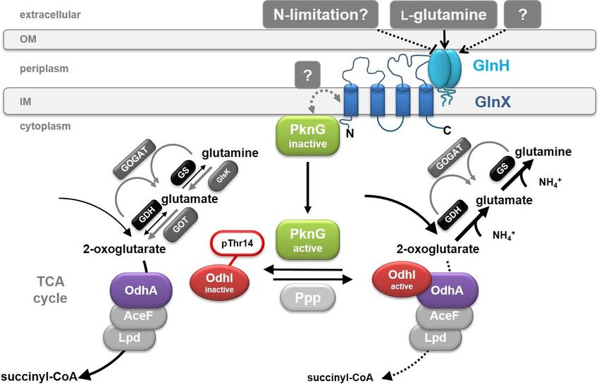
Die vorliegende Arbeit wurde am Institut für Bio- und Geowissenschaften 1: Biotechnologie
(IBG-1), Forschungszentrum Jülich, von Januar 2014 bis April 2017 unter der Betreuung von
Prof. Dr. Michael Bott und Dr. Meike Baumgart durchgeführt.

Gedruckt mit der Genehmigung der
Mathematisch-Naturwissenschaftlichen Fakultät der
Heinrich-Heine-Universität Düsseldorf

1.           Prof. Dr. Michael Bott
             Institut für Bio- and Geowissenschaften 1: Biotechnologie
             Forschungszentrum Jülich

2.           Prof. Dr. Martina Pohl
             Institut für Bio- and Geowissenschaften 1: Biotechnologie
             Forschungszentrum Jülich

Tag der mündlichen Prüfung:
07.12.2020
In dieser Dissertation beschriebene Ergebnisse wurden in der folgenden Originalpublikation
veröffentlicht:

Kim Julia Kraxner, Tino Polen, Meike Baumgart, Michael Bott (2019) The conserved
actinobacterial transcriptional regulator FtsR controls expression of ftsZ and further target
genes and influences growth and cell division in Corynebacterium glutamicum. BMC
Microbiology 19:179
Table of contents I
Table of Contents
Table of Contents ...................................................................................................... I
Abbreviations ........................................................................................................... III
1. Summary ............................................................................................................... 1
1.1. English Summary .................................................................................................. 1
1.2. Deutsche Zusammenfassung ............................................................................... 2
2. Introduction .......................................................................................................... 3
2.1. Corynebacterium glutamicum .............................................................................. 3
2.2. 2-Oxoglutarate dehydrogenase and its regulation in C. glutamicum ................ 4
2.3. Cell division in C. glutamicum .............................................................................. 6
2.4. Aim of this work ....................................................................................................10
3. Materials and Methods ....................................................................................... 11
3.1. Bacterial strains, plasmids, and growth conditions ...........................................11
3.2. Recombinant DNA work and construction of insertion and deletion mutants .14
3.3. Fluorescence microscopy....................................................................................18
3.4. Purification of FtsR...............................................................................................18
3.5. Electrophoretic mobility shift assays (EMSAs) ..................................................19
3.6. Promoter studies with PftsZ fused to mVenus .....................................................20
3.7. Chromatin affinity purification with subsequent Sequencing (ChAP-Seq) ......20
3.8. DNA microarrays ..................................................................................................21
3.9. DNA affinity purification and MALDI-ToF-MS analysis ......................................21
3.10. Genome re-sequencing ........................................................................................22
3.11. Quantitative PCR ..................................................................................................22
3.12. Coulter counter measurements ...........................................................................24
4. Results ................................................................................................................ 25
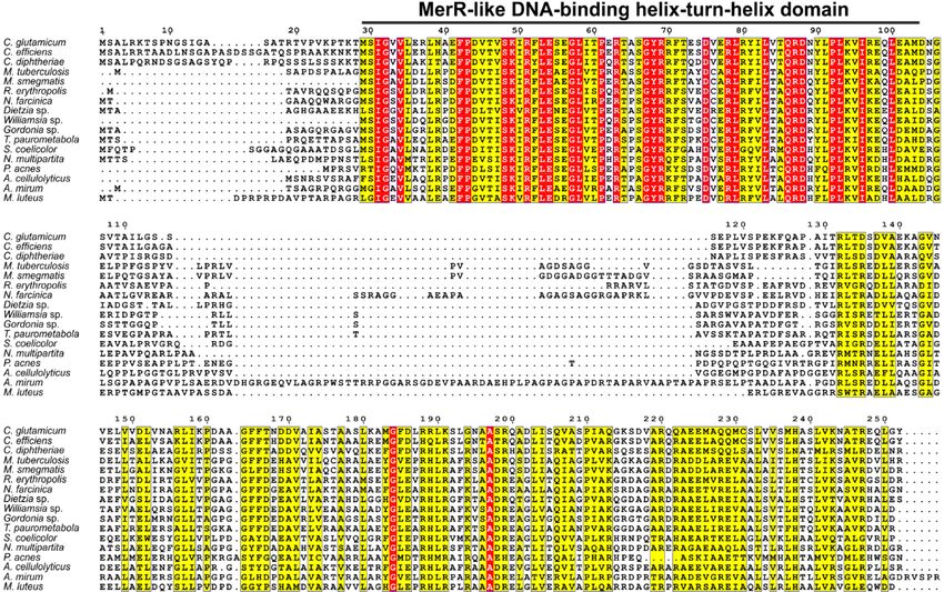
4.1. Phylogenetic conservation of odhI (cg1630) and adjacent genes.....................25
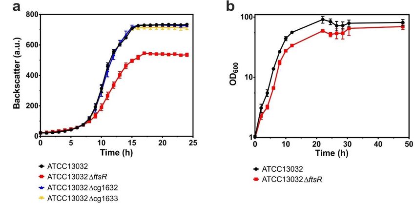
4.2. Deletion of ftsR affects growth behavior ............................................................29
4.3. Complementation experiments with C. glutamicum ATCC13032ftsR.............29
4.4. The morphological phenotype of C. glutamicum caused by ftsR deletion and
overexpression .....................................................................................................31
4.5. Transcriptome comparison of the ftsR mutant with its parent wild type .......33
4.6. Genome re-sequencing of the ftsR deletion mutant and amplification of the
trehalose cluster ...................................................................................................36
4.7. DNA affinity purification for unraveling transcriptional regulation of odhI ......38
Table of contents II
4.8. Focusing on investigation of the uncharacterized regulator FtsR ....................39
4.9. Deletion and overexpression of the ftsR gene in the MB001 background .......40
4.10. Complementation of the MB001ftsR phenotype with native FtsR and
homologs of C. diphtheriae and M. tuberculosis ...............................................41
4.11. Quantitative PCR to further investigate trehalose cluter amplification ............42
4.12. Effect of ftsR deletion on ftsZ promoter activity and FtsZ distribution ............43
4.13. Genome-wide profiling of in vivo FtsR binding sites .........................................45
4.14. in vitro binding of purified FtsR to the proposed binding motif in the ftsZ
promoter region ....................................................................................................51
4.15. DNA affinity chromatography with the ftsZ promoter ........................................53
4.16. Analysis of the transcriptome of a ftsR mutant in the MB001 background .......55
4.17. FtsR-independent expression of FtsZ .................................................................56
4.18. Influence of FtsR on ftsZ promoter activity in strains with FtsR-independent
ftsZ-expression .....................................................................................................60
5. Discussion .......................................................................................................... 62
5.1. Regulation of OdhI in C. glutamicum ..................................................................62
5.2. Do secondary effects contribute to the ftsR mutant phenotype? ...................63
5.3. The switch to MB001 as background strain ........................................................65
5.4. FtsR, the first transcriptional regulator of FtsZ identified for the
Corynebacteriales order.......................................................................................65
5.5. The importance of fine-tuning .............................................................................66
5.6. FtsR’s mode of action and binding site ..............................................................66
5.7. FtsR must have additional targets besides FtsZ ................................................68
5.8. The physiological function of FtsR .....................................................................70
6. References .......................................................................................................... 71
Abbreviations                                                                III

Abbreviations
®
                registered trademark
°C              degree Celsius
µ               growth rate
µg              microgram
µL              microliter
A               nucleobase adenine
ag              attogram
ATCC            American Type Culture Collection
BHI(S)          Brain Heart Infusion (+ Sorbitol)
bp              base pairs
C-              carboxyl-terminal end
C               nucleobase cytosine
CA              California
CDC             Centers for Disease Control and Prevention
CGP3            Corynebacterium glutamicum prophage 3
CGXII           minimal medium for Corynebacterium glutamicum
ChAP-Seq        chromatin affinity purification with subsequent sequencing
ChIP            chromatin immunoprecipitation
CmR             chloramphenicol resistance
CO2             carbon dioxide
CoA             coenzyme A
D               dexter, D-configuration (e.g. of an amino acid)
Da              Dalton
dcw             division cell wall
DNA             deoxyribonucleic acid
DNase           desoxyribonuclease
DSMZ            Deutsche Sammlung von Mikroorganismen und Zellkulturen
DTT             dithiothreitol
e.g.            exempli gratia, for example
EDTA            ethylenediaminetetraacetic acid
EMSA            electrophoretic mobility shift assay
et al.          et alii
etc.            et cetera
eYFP            enhanced yellow fluorescent protein
FHA             forkhead-associated
g               standard gravity, 9.80665 m/s2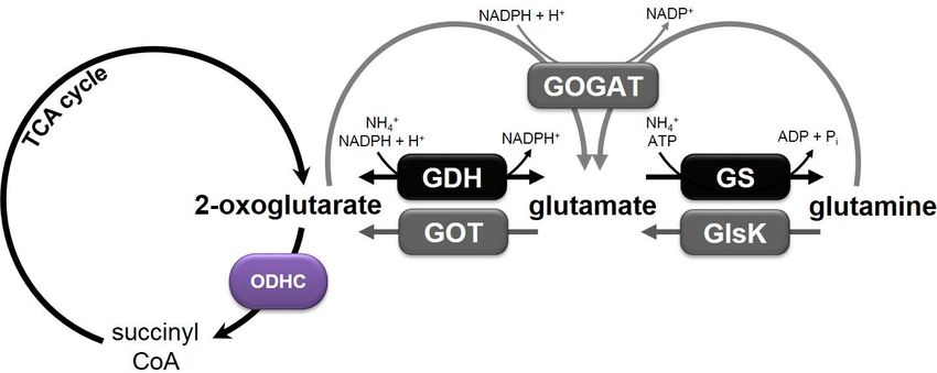
Abbreviations IV
G nucleobase guanine
GDH glutamate dehydrogenase
GmbH Gesellschaft mit beschränkter Haftung
GOGAT glutamate-2-oxoglutarate aminotransferase
GS glutamate synthase
HPLC high performance liquid chromatography
HTH helix-turn-helix
IL Illinois
Inc. Incorporation
IPTG isopropyl -D-1-thiogalactopyranoside
k kilo
Kan R
kanamycin resistance
L laevus, L-configuration (e.g. of an amino acid)
L liter
LB lysogeny broth
Ltd. private company limited by shares
MALDI matrix-assisted laser desorption/ionization
MEME Multiple Em for Motif Elicitation
mg milligram
mL milliliter
mM millimolar
mRNA messenger RNA
MS mass spectrometry
MSG monosodium glutamate
N- amino-terminal end
NAD+/NADH nicotinamide adenine dinucleotide, oxidized/reduced
NADP /NADPH+
nicotinamide adenine dinucleotide phosphate, oxidized/reduced
NCBI National Ceter for Biotechnology Information
ng nanogram
nm nanometer
OD600 optical density at 600 nm
ODHC 2-oxoglutarate dehydrogenase complex
ori origin of replication
PAGE polyacrylamide gel electrophoresis
PBS phosphate-buffered saline
PCR polymerase chain reaction
pg picogram
Abbreviations V
pH potential of hydrogen
PhD philosophiae doctor
pI isoelectric point
pmol picomol
psi pound-force per square inch
qPCR quantitative PCR
RBS ribosome binding site
RNA ribonucleic acid
rpm revolutions per minute
SDS sodium dodecyl sulfate
SNP single nucleotide polymorphism
T nucleobase thymine
t ton
TCA trichloroacetic acid
TCA cycle tricarboxylic acid cycle
TE transposable element
TEV tobacco etch virus
TM
Trademark
ToF time of flight
TSS transcriptional start site
UK United Kingdom
USA United States of America
V Volt
v/v volume per volume
w/v weight per volume
WHO World Health Organization
WT wild type
delta/deletion
T temperature difference
Abbreviations not included in this section are according to international standards, as for
example listed in the author guidelines of the American Society for Biochemistry and Molecular
Biology.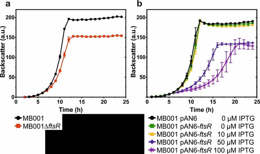
Summary 1 1. Summary 1.1. English Summary In the first part of this doctoral thesis the transcriptional regulation of the odhI gene (cg1630) of Corynebacterium glutamicum was analyzed. OdhI in its unphoshorylated state functions as inhibitor of the 2-oxoglutarate dehydrogenase complex (ODHC) by binding to the OdhA subunit. Phosphorylation of OdhI by serine/threonine protein kinases abolishes this effect. Inhibition of ODHC activity by OdhI was shown to be crucial for overproduction and secretion of L-glutamate, which is used as a flavour enhancer. Since downstream of odhI two genes presumably encoding transcriptional regulators (cg1631 and cg1633) are located, it was speculated that these could be involved in transcriptional regulation of odhI. However, transcriptome analysis of deletion mutants lacking cg1631 or cg1633 and DNA affinity chromatography with the odhI promoter did not support this hypothesis. Furthermore, no other potential transcriptional regulators of odhI could be identified. Thus, there is currently no evidence for transcriptional regulation of odhI. The second part of this thesis addresses the regulation of cytokinesis in C. glutamicum. In contrast to e.g. Escherichia coli and Bacillus subtilis, knowledge about regulators of cytokinesis in Actinobacteria is very limited. In this study, the so far uncharacterized Cg1631 protein was discovered to be a transcriptional regulator of the ftsZ gene in C. glutamicum encoding the key player of bacterial cell division. Therefore, Cg1631 was named FtsR, standing for FtsZ regulator. Both deletion and overexpression of ftsR caused growth defects and an altered cell morphology, emphasizing an important function of FtsR in cell division or cell wall synthesis. The wild-type phenotype could be restored by plasmid-based complementation. Chromatin affinity purification with subsequent next generation sequencing (ChAP-Seq) identified a region in the ftsZ promoter as a major FtsR binding site, but revealed also additional potential target genes. With the ChAP-Seq results a putative DNA-binding motif could be identified for FtsR. Transcriptional activation of ftsZ expression by FtsR was underlined by DNA microarray experiments, electrophoretic mobility shift assays (EMSAs), and reporter gene studies. Analysis of strains expressing ftsZ under control of the gluconate-inducible gntK promoter revealed that the phenotype of the ftsR mutant is not solely caused by reduced ftsZ expression but involves additional factors. In summary, FtsR was identified as the first transcriptional regulator of ftsZ in C. glutamicum. Furthermore, since FtsR and its DNA-binding site in the promoter region of ftsZ are highly conserved in Actinobacteria, it can be assumed that this regulatory mechanism is also relevant for the control of cell division in related Actinobacteria. This makes FtsR a promising target for the development of new antimicrobial drugs against pathogenic relatives of C. glutamicum.
Summary 2 1.2. Deutsche Zusammenfassung Im ersten Teil dieser Doktorarbeit wurde die Transkriptionsregulation des odhI-Gens (cg1630) von Corynebacterium glutamicum analysiert. OdhI wirkt in seinem nichtphosphorylierten Zustand als Inhibitor des 2-Oxoglutarat-Dehydrogenase-Komplexes (ODHC) durch Bindung an die OdhA-Untereinheit. Die Phosphorylierung von OdhI durch Serin/Threonin- Proteinkinasen hebt diesen Effekt auf. Die Hemmung der ODHC-Aktivität durch OdhI ist entscheidend für die Überproduktion und Sekretion von L-Glutamat, das als Geschmacksverstärker verwendet wird. Da sich stromabwärts von odhI zwei Gene befinden, die vermutlich für Transkriptionsregulatoren (cg1631 und cg1633) kodieren, wurde spekuliert, dass diese an der Transkriptionsregulation von odhI beteiligt sein könnten. Die Transkriptomanalyse der Deletionsmutanten cg1631 und cg1633 sowie DNA-Affinitäts- chromatographie mit dem odhI-Promotor stützten diese Hypothese jedoch nicht. Darüber hinaus konnten keine anderen potenziellen Transkriptionsregulatoren von odhI identifiziert werden. Daher gibt es derzeit keine Hinweise auf eine Transkriptionsregulation von odhI. Der zweite Teil dieser Arbeit befasst sich mit der Regulation der Zytokinese bei C. glutamicum. Im Gegensatz zu z.B. Escherichia coli und Bacillus subtilis ist das Wissen über Regulatoren der Zytokinese bei Actinobakterien sehr begrenzt. In dieser Arbeit wurde entdeckt, dass das bisher nicht charakterisierte Cg1631-Protein ein Transkriptionsregulator des ftsZ-Gens in C. glutamicum ist, das den Schlüsselakteur der bakteriellen Zellteilung codiert. Daher wurde Cg1631 als FtsR bezeichnet und steht für FtsZ-Regulator. Sowohl Deletion als auch Überexpression von ftsR verursachten Wachstumsdefekte und eine veränderte Zell- morphologie, was auf eine Funktion von FtsR bei Zellteilung oder Zellwandsynthese hindeutet. Der Wildtyp-Phänotyp konnte durch plasmidbasierte Komplementation der ftsR-Mutante wiederhergestellt werden. Chromatin-Affinitätsreinigung mit anschließender Sequenzierung (ChAP-Seq) bestätigte die Bindung von FtsR an den ftsZ-Promotor und identifizierte weitere potenzielle FtsR-Zielgene. Mit den ChAP-Seq-Daten konnte ein mutmaßliches DNA- Bindungsmotiv für FtsR identifiziert werden. Die Aktivierung der ftsZ-Expression durch FtsR wurde durch DNA-Microarray-Experimente, elektrophoretische Retardationstests und Reportergenstudien bestätigt. Die Analyse von Stämmen, die ftsZ unter Kontrolle des Gluconat-induzierbaren gntK-Promotors exprimierten, ergab, dass der Phänotyp der ftsR- Mutante nicht nur durch eine verringerte ftsZ-Expression verursacht wird, sondern zusätzliche Faktoren beinhaltet. Zusammenfassend wurde FtsR als erster Transkriptionsregulator von ftsZ in C. glutamicum identifiziert. Da FtsR und seine DNA-Bindungsstelle im ftsZ-Promotor in Actinobakterien hoch konserviert sind, wird angenommen, dass dieser Regulationsmechanismus auch für die Kontrolle der Zellteilung in verwandten Actinobakterien relevant ist. Dies macht FtsR zu einem vielversprechenden Ziel für die Entwicklung neuer antimikrobieller Wirkstoffe gegen pathogene Verwandte von C. glutamicum.
Introduction 3 2. Introduction 2.1. Corynebacterium glutamicum Corynebacterium glutamicum is a non-pathogenic, aerobic, Gram-positive soil bacterium which is used for the large-scale production of several L-amino acids and other industrially relevant compounds (Wendisch et al., 2016, Eggeling & Bott, 2015, Eggeling & Bott, 2005). Moreover, it is a useful model organism for the Corynebacteriales, including pathogenic species such as Corynebacterium diphtheriae and Mycobacterium tuberculosis, which cause the fatal infectious diseases diphtheria and tuberculosis in humans, respectively (WHO, 2015, CDC, 2014, Lawn & Zumla, 2011, Hadfield et al., 2000). Due to its industrial relevance and close relationship to pathogenic species, great efforts have been made in the last decades to unravel the regulatory network of C. glutamicum. About 140 transcriptional regulators were identified (Schröder & Tauch, 2010, Brinkrolf et al., 2007) and approximately half of them have been characterized to date. This leads to a better understanding of the biomolecular circuits in this organism, which is of major importance in order to further improve biotechnological production strains or drug discovery processes. In the past, C. glutamicum, which has been isolated in Japan by Kinoshita et al. (1957), became famous due to its native ability to produce the flavor enhancer monosodium glutamate (MSG). Originally, MSG was discovered in 1908 by Professor Kikunae Ikeda, as he isolated it from the seaweed Laminaria japonica because he wanted to unravel what constitutes the unique taste of Japanese seaweed broth (Lindemann et al., 2002). Generally, Ikeda had the vision that the findings of his research could result in a commercial application, for example in a seasoning that would help improve human nutrition and therefore lead to a longer life expectancy of the Japanese population (Sano, 2009). And indeed, it has been shown that MSG and its umami taste have various positive effects in relation to improving nutrition and health (Mouritsen, 2012). Back then, MSG has been produced by hydrolysis of plant proteins using hydrochloric acid or later on by direct chemical synthesis. The discovery and characterization of C. glutamicum opened up the possibility of bacterial MSG synthesis, which had the advantages of enantiopure production of the natural L-form of glutamic acid and to satisfy the increasing demand while reducing production costs and relieving the environment (Sano, 2009). Today, C. glutamicum is an important industrial amino acid producer which is used to produce amongst others several million tons of the above-mentioned flavor enhancer L-glutamate and the feed additive L-lysine (Becker & Wittmann, 2020). By now, there are several conditions known which trigger glutamate overproduction and secretion: Biotin limitation (Shiio et al., 1962), addition of detergents like Tween-40 or Tween-60 (Duperray et al., 1992, Takinami et al., 1966), addition of -lactam antibiotics like penicillin G (Nunheimer et al., 1970), or inhibitors of cell wall synthesis like ethambutol
Introduction                                                                                    4

(Radmacher et al., 2005), glycerol, or fatty acid auxotrophy (Kimura et al., 1997, Nakao et al.,
1972, Kanzaki et al., 1967, Okazaki et al., 1967), and high temperature cultivation of
temperature-sensitive strains (Delaunay et al., 1999, Momose & Takagi, 1978), which are all
thought to influence cell wall composition or membrane permeability, but the actual molecular
mechanism is not entirely unraveled to date (Kimura, 2002, Eggeling et al., 2001). Indeed, a
transporter involved in glutamate secretion, named YggB (Cg1434), has been identified
(Nakamura et al., 2007) and metabolic flux analyses confirmed a strongly reduced activity of
the 2-oxoglutarate dehydrogenase complex (ODHC) during cultivation of C. glutamicum under
producing versus non-producing conditions, indicating that regulation of the ODHC plays an
important role in glutamate production (Kataoka et al., 2006, Shirai et al., 2005, Shimizu et al.,
2003, Kawahara et al., 1997).

2.2. 2-Oxoglutarate dehydrogenase and its regulation in C. glutamicum

ODHC is a multimeric enzyme composed of three subunits that catalyzes the conversion of
2-oxoglutarate, NAD+, and coenzyme A to succinyl-CoA, NADH + H+, and CO2 in the
tricarboxylic acid (TCA) cycle (Usuda et al., 1996). The first subunit E1 is the OdhA protein
encoded by the gene cg1280, harboring the actual 2-oxoglutarate dehydrogenase activity. The
E2 and E3 subunits of the ODHC are called AceF (Cg2421) and LpdA (Cg0790) and encode
a dihydrolipoamide acetyltransferase and a dihydrolipoamide dehydrogenase, respectively
(Ikeda & Nakagawa, 2003, Kalinowski et al., 2003, Schwinde et al., 2001). The substrate
2-oxoglutarate marks an important branch point in the metabolism of C. glutamicum. Besides
the conversion to succinyl-CoA by the ODHC in the TCA cycle it can also be reductively
aminated to glutamate by the glutamate dehydrogenase Gdh (Cg2280), which is mainly active
under nitrogen excess (Börmann et al., 1992). Glutamate is converted to glutamine by the
glutamine synthetase GlnA (Cg2429), which serves together with the glutamate-2-oxoglutarate
aminotransferase GOGAT (encoded by gltBD, cg0229, and cg0230) for the assimilation of
ammonium under nitrogen-limiting conditions (Jakoby et al., 1997). The 2-oxoglutarate
dehydrogenase reaction and the nitrogen metabolism in C. glutamicum are schematically
illustrated in Figure 1.
Introduction                                                                                  5

Figure 1: Schematic illustration of the 2-oxoglutarate dehydrogenase reaction and of the
nitrogen metabolism in C. glutamicum. Ammonium assimilation is mediated by the reactions carried
out by the glutamate dehydrogenase (GDH) and glutamine synthetase (GS)/glutamate-2-oxoglutarate
aminotransferase (GOGAT), whereas glutamine catabolism involves the glutaminase K (GlsK) and the
glutamate-oxaloacetate transaminase reaction.

Because the GDH shows a 30 to 70 times lower affinity to the substrate 2-oxoglutarate
compared to the ODHC (Shiio & Ozaki, 1970), inhibition of ODHC activity is necessary for
efficient glutamate production. How this occurs has been unraveled by the discovery of a novel
kind of regulation of the ODHC, which results in an increased flux of 2-oxoglutarate to
glutamate (Bott, 2007). It was found out that the 15 kDa small inhibitor protein OdhI (Cg1630)
is involved in the regulation of the ODHC, which in turn is regulated by a complex signalling
cascade. The findings obtained in previous studies so far led to the assumption of the following
mechanism, which is schematically illustrated in Figure 2: in the presence of L-glutamine as
sole carbon and nitrogen source, it is bound by the membrane-associated lipoprotein GlnH
(Cg3045). The signal is then transmitted from GlnH via protein-protein interaction to the
integral membrane protein GlnX (Cg3044), consisting of four transmembrane helices with the
N- and C-terminus located in the cytoplasm. GlnX activates hereupon the soluble
serine/threonine protein kinase PknG (Cg3046). Hence, PknG dissociates from the
cytoplasmic membrane and phosphorylates the OdhA inhibitor protein OdhI at its threonine-
14 residue. In its phosphorylated state, OdhI is inactive and not able to inhibit the ODHC, so
that the complex is present in its active state. Under these conditions, a normal flux of
glutamine through the TCA cycle towards succinyl-CoA occurs. The loss of interaction of
phosphorylated OdhI with the ODHC is very likely caused by a conformational change which
is triggered by the phosphorylation through PknG. OdhI consists of a non-folded N-terminal
domain with two phosphorylation sites (Thr14 and Thr15) and a C-terminal forkhead-
associated (FHA) domain. Phosphorylation through PknG at the threonine-14 residue causes
a conformational change of the non-folded N-terminus which in turn can be bound by the FHA
domain. This auto-inhibition leads to an inactive OdhI protein.
Introduction                                                                                            6

Figure 2: Model of the GlnX-GlnH-PknG-OdhI signal transduction cascade in C. glutamicum.
Illustrated is the model of the GlnX-GlnH-PknG-OdhI signal transduction cascade in C. glutamicum as
proposed based on the previous findings. A detailed description of the mechanism can be found in the text.

In the absence of the proteins of the cascade, the signal can neither be detected nor
transmitted and consequently, OdhI can no longer be phosphorylated by the kinase PknG.
In its unphosphorylated state, the inhibitor protein binds to the OdhA subunit of the ODHC,
inhibiting its activity, which leads to a reduced conversion of 2-oxoglutarate to succinyl-CoA in
the TCA cycle and an increased flux towards glutamate. Due to dephosphorylation of OdhI by
the phospho-serine/threonine protein phosphatase Ppp (Cg0062), this process is reversible.
The proposed model is based on the findings of Bosco (2011), Krawczyk et al. (2010), Schultz
et al. (2009), Schultz et al. (2007), Gebel (2006), and Niebisch et al. (2006).

2.3. Cell division in C. glutamicum

Bacterial reproduction is usually characterized by cellular growth followed by binary fission of
a mother cell into two daughter cells. During this process, the bacteria need to coordinate
several distinct processes such as DNA replication, biogenesis of the new cell wall, and the
division process itself. Although the overall process is similar, several different concepts have
evolved in distinct bacterial groups. Escherichia coli and Bacillus subtilis represent two of the
best studied species in this respect (Du & Lutkenhaus, 2017, Hajduk et al., 2016).

Corynebacteria are rod-shaped with frequently engrossed cell poles during exponential
growth, leading to the typical “club”-shaped morphology by which the name of this genus is
Introduction                                                                                                   7

inspired (Cure & Keddie, 1973). Like for almost all other bacteria, the highly conserved FtsZ
protein is also the key player of cell division in C. glutamicum (Donovan & Bramkamp, 2014,
Adams & Errington, 2009). It is essential and the first protein that moves to the future division
site, recruiting other proteins involved in this process (Margolin, 2005). Several FtsZ proteins
together form the so-called Z-ring, which in rod-shaped bacteria is located in the middle of the
cell, and constriction of this ring leads to a formation of two daughter cells (Adams & Errington,
2009, Margolin, 2005) (see Figure 3).

Figure 3: Assembly and disassembly of FtsZ and the Z-ring. On the left, a newborn cell with FtsZ
proteins (blue) arranged in a spiral pattern is depicted, as known for E. coli. The cell in the middle already
shows a formed Z-ring and the recruitment of other proteins of the divisome (violet, green, and orange).
On the right site, a dividing cell with a contracting Z-ring is illustrated. Picture inspired by Margolin (2005).

Interestingly, C. glutamicum – as well as other Actinobacteria – is lacking homologs for several
important cell division-related genes described for E. coli or B. subtilis (Table 1). Cell division
in C. glutamicum is for example independent of the actin homolog MreB, which is essential for
elongation of the lateral cell wall in E. coli, Caulobacter crescentus, and B. subtilis (Kruse et
al., 2005, Figge et al., 2004, Jones et al., 2001). In contrast to these species, C. glutamicum
inserts new cell wall material at the poles, representing an apical elongation mechanism
(Daniel & Errington, 2003). Besides mreB, also other typically essential cell division genes are
missing, such as ftsL and ftsN (Letek et al., 2008). Most of the genes required for cell division
and peptidoglycan synthesis are located in the so-called division cell wall (dcw) cluster, which
is highly conserved throughout the bacterial kingdom (Mingorance et al., 2004). Several
studies of this cluster in Actinobacteria revealed that its arrangement in Corynebacteria and
Mycobacteria clearly differs from many other bacteria (Tamames et al., 2001). However, the
most striking difference might be the complete absence of homologs of other known spatial
and temporal, positive and negative regulators of cell division, as for example ftsA, ezrA, slmA,
sulA, zipA, zapA, min, and noc are missing (Donovan & Bramkamp, 2014, Letek et al., 2007).
FtsA is a positive regulator of FtsZ assembly and supports membrane association of the Z-ring
(Pichoff & Lutkenhaus, 2005, Beall & Lutkenhaus, 1992). The integral membrane protein EzrA
negatively influences Z-ring assembly and is involved in mid-cell localisation of the Z-ring
(Haeusser et al., 2004, Levin et al., 1999). SlmA and NocA are nucleoid occlusion effector
proteins, known to inhibit assembly of the Z-ring over the nucleoid (Bernhardt & de Boer, 2003,
Wu & Errington, 2004). SulA also inhibits Z-ring formation and is induced upon DNA damage
Introduction                                                                                            8

(Bi & Lutkenhaus, 1993, Huisman et al., 1984). ZipA and ZapA are positive regulators of
Z-ring assembly (Slayden et al., 2006, Gueiros-Filho & Losick, 2002, Hale & de Boer, 1997),
and the Min system consists of several proteins preventing division occurring at the cell poles
(Bramkamp et al., 2008, Raskin & de Boer, 1999, Levin et al., 1992, de Boer et al., 1989).
Taking all these differences into account, it is obvious that in Actinobacteria cell division and
its regulation must differ profoundly from the processes described for other well-studied rod-
shaped bacteria like E. coli, and B. subtilis (Letek et al., 2007, Flardh, 2003, Ramos et al., 2003).

Table 1: Putative cell division genes of E. coli, B. subtilis, C. glutamicum, and M. tuberculosis.
Known positive regulators (green), negative regulators (red), and essential cell division genes (blue)
which are missing in C. glutamicum are written in bold letters. The table is based on Letek et al. (2008)
and Donovan and Bramkamp (2014). For an overview of the predicted protein functions, see Donovan
and Bramkamp (2014).

 gene name          E. coli             B. subtilis             C. glutamicum         M. tuberculosis
 amiC               b2817               BSU09420 (lytE)         cg3424                rv3915
                                        BSU09370 (lytF)
 clpX               b0438               BSU28220                cg2620                rv2457c

 crgA               ?                   ?                       cg0055                rv0011c

 cwsA               ?                   ?                       ?                     rv008c

 divIVA             ?                   BSU15420                cg2361                rv2154c (wag31)

 divS               b0958 (sulA)        BSU17860 (yneA)         cg2133                rv2719 (chiZ)

 ezrA               ?                   BSU29610                ?                     ?

 ftsA               b0094               BSU15280                ?                     ?

 ftsB (divIC)       b2748               ?                       cg1112                rv1024

 ftsE               b3463               BSU35260                cg0914                rv3102c

 ftsI               b0084               BSU15170                cg2375                rv2163c

 ftsK               b0890               BSU29800 (ytpT)         cg2158                rv2748c
                                        BSU16800 (spoIIIE)
 ftsL               b0083               BSU15150                ?                     ?

 ftsN               b3933               ?                       ?                     ?

 ftsQ (divB)        b0093               BSU15240                cg2367                rv2151c

 ftsW               b0089               BSU14850                cg2370                rv2154c
                                        BSU15210 (spoVE)
 ftsX               b3462               BSU35250                cg0915                rv3101c

 ftsZ               b0095               BSU15290                cg2366                rv2150c

                                                                                            (Continued)
Introduction 9
Table 1. Continued.
gene name E. coli B. subtilis C. glutamicum M. tuberculosis
ftsX b3462 BSU35250 cg0915 rv3101c
ftsZ b0095 BSU15290 cg2366 rv2150c
minC b1176 BSU28000 ? ?
minD b1175 BSU27990 ? ?
minJ ? BSU35220 ? ?
minE b1174 ? ? ?
mreB b3251 BSU14470 (mreBH) ? ?
BSU28030 (mreB)
BSU36410 (mbl)
mreC b3250 BSU28020 ? ?
mreD b3249 BSU28010 ? ?
noc ? BSU40990 ? ?
pldP ? ? cg1610 rv1708
rodA b0634 BSU38120 cg0061 rv0017c
sepF ? BSU15390 cg2363 rv2147c
slmA b3641 ? ? ?
sulA b0958 ? ? ?
ugtP ? BSU21920 ? ?
zapA b2910 ? ? ?
zapB b3928 ? ? ?
zapC b0946 ? ? ?
zapD b0102 ? ? ?
zipA b2412 ? ? ?
To date, C. glutamicum is an established model organism for industrial biotechnology and for
pathogenic relatives, but we have a relatively narrow understanding about the exact regulation
of its cytokinesis (Donovan & Bramkamp, 2014). However, it is known that accurate expression
of FtsZ is critical for normal growth of C. glutamicum and M. tuberculosis and small variations
lead to severe morphological changes and affect cell viability, suggesting that the intracellular
levels of FtsZ are critical for proper cell division in these organisms (Letek et al., 2007, Ramos
et al., 2005, Dziadek et al., 2003). Thus, FtsZ levels must be subject to a tight regulation, which
is supported by different studies of the ftsZ promoter region, showing a very complexIntroduction 10 transcription of ftsZ in various bacteria (Letek et al., 2007, Roy & Ajitkumar, 2005, Flardh et al., 2000, Flardh et al., 1997, Gonzy-Treboul et al., 1992). A complex of the DNA-binding proteins WhiA (Cg1792) and WhcD (Cg0850) was reported to bind to the ftsZ promoter region (Lee et al., 2018), however, the effect of this interaction on ftsZ expression remains to be elucidated. Previously, it was shown that the FtsZ protein can be phosphorylated in vitro by the serine/threonine protein kinases PknA, PknB, and PknL and is an in vivo substrate of the phospho-serine/threonine protein phosphatase Ppp (Schultz et al., 2009), suggesting that the properties and activities of FtsZ can also be influenced by posttranslational regulation (Donovan & Bramkamp, 2014). However, a regulatory protein involved in direct control of ftsZ transcription has only been described for Caulobacter crescentus (Kelly et al., 1998). For the Corynebacteriales order, the regulation of cell division processes remains largely undiscovered. 2.4. Aim of this work Originally, the focus of this thesis lay on studying the role of GlnX, GlnH, PknG, and OdhI in C. glutamicum in order to attain a deeper understanding of the signal transduction cascade which consists of these proteins. More specifically, one major objective was to investigate whether the inhibitory protein of the ODHC is potentially regulated on the transcriptional level. This seemed sensible because odhI (cg1630) is located upstream of the two genes (cg1631 and cg1633), which are annotated to encode transcriptional regulators. However, the experiments performed in this direction (DNA affinity chromatography with the odhI promoter, DNA microarrays) did not provide any evidence to support transcriptional regulation of odhI. Nevertheless, since studying the GlnH-GlnX-PknG-OdhI-ODHC signal transduction cascade was the initial objective of this work, a part of this introduction and the first chapters of the results section are related to this topic. In these initial studies, deletion mutants of the genes cg1631, cg1632, and cg1633 were constructed and analyzed. Thereby it was observed that deletion of cg1631 led to a severe growth defect and drastic morphological changes. Because there is hitherto no transcriptional regulator of cell division known for C. glutamicum, this phenotype was highly interesting and let us redefine the aims of this thesis towards the functional analysis of the previously uncharacterized protein Cg1631. In this context, we searched for the target genes of Cg1631 by a variety of complementary approaches and identified ftsZ to be activated by Cg1631. Therefore, Cg1631 was renamed FtsR, standing for ftsZ regulator. We show that FtsR is critical for normal growth and cell morphology of C. glutamicum, suggesting an important role of this protein in the regulation of cell division.
Materials and Methods                                                                       11

3. Materials and Methods
3.1. Bacterial strains, plasmids, and growth conditions

Bacterial strains and plasmids used in this study are listed in Table 2. The C. glutamicum type
strain ATCC13032 or its prophage-free variant C. glutamicum MB001 were used as wild type
or reference strain, as indicated. The parental strain ATCC13032 (DSM No. 20300) was
obtained from the Deutsche Sammlung von Mikroorganismen und Zellkulturen GmbH (DSMZ,
Braunschweig, Germany). The prophage-free strain MB001 was derived from an in-house
stock but can also be obtained from the DSMZ (DSM No. 102070). First experiments were
performed with ATCC13032, but to be able to discriminate between effects primarily caused
by ftsR deletion or secondarily emerged due to phage-related effects, we later switched to the
C. glutamicum MB001 strain. Deletion of the prophages did not lead to any negative effects on
the physiology of C. glutamicum MB001 and due to its reduced complexity it is a useful platform
organism for basic research (Baumgart et al., 2013b).

For growth experiments, C. glutamicum was pre-cultivated for six to eight hours at 30 °C and
170 rpm in 5 mL BHI medium (BD BactoTM Brain Heart Infusion, Becton Dickinson and
Company, Heidelberg, Germany). The cells were harvested by centrifugation, washed with
phosphate-buffered saline (PBS, 137 mM NaCl, 2.7 mM KCl, 4.3 mM Na2HPO4, 1.4 mM KH2PO4,
pH 7.3) and used as inoculum for a second pre-culture in 20 mL CGXII minimal medium
(Keilhauer et al., 1993) supplemented with 3,4-dihydroxybenzoate (30 mg/L) as iron chelator
and, if not stated otherwise, 2% (w/v) glucose as carbon source. This second pre-culture was
incubated overnight at 30 °C and 120 rpm. After harvesting by centrifugation and washing with
PBS, the cells were used for inoculation of the main culture, using CGXII medium with 3,4-
dihydroxybenzoate (30 mg/L) and 2% (w/v) glucose as carbon source, if not stated otherwise.

Growth experiments were either performed in 100 mL shake flasks with 20 mL medium (initial
optical density at 600 nm (OD600) of 1.0) that were shaken at 30 °C and 120 rpm or in 48-well
FlowerPlates® (m2p-labs GmbH, Baesweiler, Germany) containing a final culture volume of
800 µL (initial OD600 of 0.5) that were shaken in a BioLector® system (m2p-labs GmbH,
Baesweiler, Germany) at 30 °C and 1200 rpm. Growth was monitored as cell density by
determining either OD600 (shake flasks experiments, OD600 measured with an Ultrospec 500
Pro UV/Vis spectrophotometer, Amersham Biosciences, Little Chalfont, United Kingdom) or as
scattered light at 620 nm in the BioLector® (Kensy et al., 2009), which is termed “backscatter”
throughout this study.

For growth experiments with promoter exchange strains in which expression of the ftsZ gene
was under control of the gntK promoter, the first pre-culture was supplemented with 0.1% (w/v)
and the second pre-culture with 0.01% (w/v) gluconate and 1.99% (w/v) glucose as carbon
source. The gluconate concentration of the main culture is indicated for each experiment.
Materials and Methods 12
For cloning purposes, Escherichia coli DH5 was used and routinely cultivated at 37 °C in
lysogeny broth (LB, (Sambrook & Russell, 2001)). When required, the media were
supplemented with 25 µg/mL kanamycin or 10 µg/mL chloramphenicol for C. glutamicum or
with 50 µg/mL kanamycin or 34 µg/mL chloramphenicol for E. coli.
Table 2: Bacterial strains and plasmids used in this study.
Strain or plasmid Characteristics Source or
Reference
Bacterial strains
E. coli
DH5α F- Φ80dlac∆(lacZ)M15 ∆(lacZYA-argF) U169 Hanahan (1983)
endA1 recA1 hsdR17 (rK-, mK+) deoR thi-1 phoA
supE44 λ- gyrA96 relA1; strain used for cloning
procedures
C. glutamicum
ATCC13032 biotin-auxotrophic wild type. Kinoshita et al.
(1957)
ATCC13032ftsR ATCC13032 with in-frame deletion of ftsR this work
(cg1631)
ATCC13032cg1632 ATCC13032 with in-frame deletion of cg1632 this work
ATCC13032cg1633 ATCC13032 with in-frame deletion of cg1633 this work
ATCC13032ramB ATCC13032 with in-frame deletion of ramB Gerstmeir et al.
(cg0444) (2004)
ATCC13032::ftsZ-venus ATCC13032 with a chromosomal insertion of this work
pK18mob-ftsZ-venus at the ftsZ locus; this strain
has two chromosomal copies of ftsZ under
control of its native promoter, one with and one
without the fusion to venus
ATCC13032ftsR::ftsZ- ATCC13032ftsR with a chromosomal insertion this work
venus of pK18mob-ftsZ-venus at the ftsZ locus; this
strain has two chromosomal copies of ftsZ under
control of its native promoter, one with and one
without the fusion to venus
MB001 ATCC13032 with in-frame deletions of the Baumgart et al.
prophages CGP1 (cg1507-cg1524), CGP2 (2013b)
(cg1746-cg1752), and CGP3 (cg1890-cg2071)
MB001ftsR MB001 with in-frame deletion of ftsR (cg1631) this work
MB001::PgntK-ftsZ MB001 with a chromosomal promoter exchange this work
of the native ftsZ promoter against the
gluconate-inducible promoter of gntK (cg2732)
MB001ftsR::PgntK-ftsZ MB001::PgntK-ftsZ with in-frame deletion of ftsR this work
C. diphtheriae genomic DNA of this strain was used as PCR DSM 44123
ATCC27010 template for amplification of the ftsR homolog
CDC7B_1201
M. tuberculosis H37Rv genomic DNA of this strain was used as PCR ATCC 25618
template for amplification of the ftsR homolog
rv1828
(Continued)Materials and Methods 13
Table 2. Continued.
Strain or plasmid Characteristics Source or
reference
Plasmids
pK18mob KanR; plasmid for insertion of a DNA fragment Schäfer et al.
into the chromosome of C. glutamicum (1994)
(pK18 oriVE.c., lacZα)
pK18mob-ftsZ-venus KanR; pK18mob derivative for chromosomal this work
insertion of the coding sequence for a fusion
protein of FtsZ and the fluorescent protein
Venus under control of the native ftsZ promoter
pK19mobsacB KanR; plasmid for allelic exchange in Schäfer et al.
C. glutamicum (pK18 oriVE.c., sacB, lacZα). (1994)
pK19mobsacB-ftsR KanR; pK19mobsacB derivative for in-frame this work
deletion of ftsR; contains PCR product covering
the fused up- and downstream regions of ftsR
pK19mobsacB-PgntK-ftsZ KanR; pK19mobsacB derivative for construction this work
of MB001::PgntK-ftsZ; contains a PCR fragment
encompassing 691 bp of the upstream region of
cg2366 (ftsZ) covering its promoter, followed by
a terminator sequence, the gntK (cg2732)
promoter and 523 bp of the ftsZ (cg2366) coding
region
pK19-P2732-lcpA KanR; pK19mobsacB derivative used as Baumgart et al.
template for the promoter exchange plasmid (2016)
pK19mobsacB-PgntK-ftsZ
pAN6 KanR; C. glutamicum/E. coli shuttle vector for Frunzke et al.
regulated gene expression using Ptac (Ptac lacIq (2008)
pBL1 oriVCg pUC18 oriVEc)
pAN6-ftsR KanR; pAN6 derivative for expression of ftsR this work
(252 amino acids) under control of Ptac
pAN6-ftsR-short KanR; pAN6 derivative encoding an N-terminally this work
shortened FtsR protein (224 amino acids) under
control of Ptac
pAN6-ftsR-Strep KanR; pAN6 derivative for expression of ftsR this work
under control of Ptac with a C-terminal Strep-tag®
pAN6-CDC7B_1201 KanR; pAN6 derivative for expression of this work
C. diphtheriae CDC7B_1201 (ftsR homolog)
under control of Ptac.
pAN6-rv1828 KanR; pAN6 derivative for expression of this work
M. tuberculosis rv1828 (ftsR homolog) under
control of Ptac.
pEC-XC99E CmR; C. glutamicum/E. coli shuttle vector for Kirchner and
regulated gene expression using Ptrc, (Ptrc, catI, Tauch (2003)
laclq, rrnB, oriVE.c, per and repA (pGA1)C.g.).
pEC- ftsR CmR; pEC-XC99E derivative for expression of this work
ftsR under control of Ptrc.
pJC1-venus-term KanR; pJC1 derivative carrying the Venus coding Baumgart et al.
sequence and additional terminators (pCG1 (2013a)
oriCg, pACYC177 oriEc).
pJC1-PftsZ-venus KanR, pJC1-venus-term derivative carrying the this work
ftsZ promoter controlling expression of venus for
promoter activity studies.Materials and Methods 14
3.2. Recombinant DNA work and construction of insertion and deletion mutants
Routine methods such as PCR, DNA restriction and ligation, Gibson assembly, and
transformation were performed using standard protocols (Sambrook & Russell, 2001,
Hanahan, 1983, van der Rest et al., 1999, Gibson, 2011). Phusion Green High Fidelity DNA
Polymerase (Thermo Fisher Scientific Inc., Rockford, IL, USA) was used for cloning purposes.
For all other PCRs, either the KAPA2G Fast ReadyMix PCR Kit (Kapa Biosystems, Wilmington,
USA) or DreamTaq DNA Polymerase (Thermo Fisher Scientific Inc., Rockford, IL, USA) were
used. The oligonucleotides used in this study (see Table 3) were purchased from Eurofins
Genomics GmbH (Ebersberg, Germany) and DNA sequencing was also performed by this
company. The ftsR mutant of C. glutamicum and the promoter exchange strains carrying a
DNA fragment with a transcription terminator sequence and the gntK promoter inserted into
the chromosome between the native ftsZ promoter and the ftsZ coding region were constructed
via a two-step homologous recombination protocol as described previously (Baumgart et al.,
2016, Niebisch & Bott, 2001), using plasmids pK19mobsacB-ftsR and pK19mobsacB-PgntK-
ftsZ. The strains ATCC13032::ftsZ-venus and ATCC13032ftsR::ftsZ-venus were constructed
by chromosomal insertion of pK18mob-ftsZ-venus by single homologous recombination. In all
tested clones of both strains, the plasmid did not insert into the intergenic region between
cg1121 and cg1122, but into the ftsZ region. As the insertion plasmid was constructed in such
a way that these mutants also carry one native copy of ftsZ and one fused to venus, these
strains were used for microscopy, anyway.
Table 3: Oligonucleotides used in this study for cloning, EMSAs, and DNA affinity purification.
Restriction sites are underlined. Bold letters represent the overlapping sequences needed for Gibson
Assembly. If not stated otherwise, C. glutamicum genomic DNA was used as template.
Oligonucleotide Sequence (5’ → 3’) and properties Commentary
DNA affinity purification with PodhI and PftsZ
AP_PodhI_fw TGATCAGTCCGTGAGGGAAC
AP_PodhI_rv_bio GAGGAGTCGTCGATGTGGAGACCCAGC overlap (in italics) homologous to the
GCGGAATACTGAGGTG biotin-oligo used as reverse primer in a
second PCR to create a biotinylated
fragment
AP_PftsZ_fw CATTAGCTCACCCTCAATGG
AP_PftsZ_rv_bio GAGGAGTCGTCGATGTGGAGACCGAGG overlap (in italics) homologous to the
CCTTCTTCAATCATGC biotin-oligo used as reverse primer in a
second PCR to create a biotinylated
fragment
biotin_oligo 5'BIO- biotin-labelled sequence homologous to
GAGGAGTCGTCGATGTGGAGACC the overlap of the rv_bio primers for
attachment of a biotin-tag to the
resulting fragments; biotinylated
molecules bind with high affinity to the
Streptavidin-coated magnetic beads
used for DNA affinity purification.
(Continued)Materials and Methods 15
Table 3. Continued.
Oligonucleotide Sequence (5’ → 3’) and properties Commentary
Construction of pK18mob- and pK19mobsacB derivatives
M13-fw CGCCAGGGTTTTCCCAGTCAC for sequencing of pK18mob-ftsZ-venus
M13-rv AGCGGATAACAATTTCACACAGGA and pK19mobsacb-ftsR
Plasmid pK18mob-ftsZ-venus for chromosomal insertion of ftsZ-venus
pK18_IRG-fw-V2 CCTGCAGGTCGACTCTAGAGGTTTACG for amplification of a fragment covering
CAGCACAAGACCCC about 450 bp of the intergenic region
IGR-rv TCCTCCATAATTAGAGAGCGTAAGGCC between cg1121 and cg1122
C
PftsZ-fw CGCTCTCTAATTATGGAGGATGATGGT for amplification of the promoter and the
GACCATGTCATTGACACCG coding region of ftsZ
FtsZ-rv TCCTCGCCCTTGCTCACCATCTGGAGG
AAGCTGGGTACATCCAG
venus-fw ATGGTGAGCAAGGGCGAGGAG for amplification of the venus gene
pK18_venus-rv-V2 CAGCTATGACCATGATTACGTTACTTG encoding the fluorescent protein Venus
TACAGCTCGTCCATGCC
FtsZ-Seq1 CATTAGCTCACCCTCAATGGTG for sequencing of pK18mob-ftsZ-venus
FtsZ-Seq2 GAACCTGTCCATCATGGAAGC
int-reg-fw AGCACCTTCGGCAAGAAGTA test of integration strains for integration
into the intergenic region of cg1121-
int-reg-rv CATCGAAGGTGTCGCAAAC
cg1122
ftsQ-rv AGCAATAACCGCAGGAAGCAC
M13-fw CGCCAGGGTTTTCCCAGTCAC
ftsQ-fw ACAAGGCAGGACTAGCGTGAAC test of integration strains for integration
into the chromosomal ftsZ-region
ftsZ-downstream-rv TCGTGAAGACCTTGCGGAC
pK18-IGR-fw CTTGGTTCGAATATGCAGTTCGG
eYFP-int_rv CGACCAGGATGGGCACCAC
Deletion plasmid pK19mobsacb-ftsR
D_ftsR_1_fw GATCGGATCCTCCGCACTCAACATCTA PCR product contains BamHI site for
GAC cloning in BamHI-cut pK19mobsacb
D_ftsR_2_rv TGTTTAAGTTTAGTGGATGGGGATGTT
GCTGCTACCTGCTGTGAATTAAA
D_ftsR_3_fw CCCATCCACTAAACTTAAACATAGAAA PCR product contains HindIII site for
AAATGAGTTTTGTTGAACTT cloning in HindIII-cut pK19mobsacb
D_ftsR_4_rv GATCAAGCTTCGTCGCCTGAAGCAGAT
TCC
map_DftsR_fw TCCGCACTCAACATCTAGAC for verification of chromosomal ftsR
deletion
map_DftsR_rv CAGGTAAAGCCATCTGGTTC
Deletion plasmid pK19mobsacb-cg1632
D_cg1632_1_fw GATCGGATCCTCAGCCCGGAGAAGTTT PCR product contains BamHI site for
CAG cloning in BamHI-cut pK19mobsacb
D_ cg1632_2_rv TGTTTAAGTTTAGTGGATGGGTTTTTC
TATCAGTATCCAAGCTGCTCGCGAGTT
GC
D_ cg1632_3_fw CCCATCCACTAAACTTAAACAAATGTG PCR product contains HindIII site for
ACTAATCACACCCTCAGATTTCAAC cloning in HindIII-cut pK19mobsacb
D_ cg1632_4_rv GATCAAGCTTAGGTCGTTGGTGCCCAT
GTC
(Continued)Materials and Methods 16
Table 3. Continued.
Oligonucleotide Sequence (5’ → 3’) and properties Commentary
Deletion plasmid pK19mobsacb-cg1633
D_cg1633_1_fw GATCGGATCCTACATGGCTACCATCAC PCR product contains BamHI site for
TAC cloning in BamHI-cut pK19mobsacb
D_ cg1633_2_rv TGTTTAAGTTTAGTGGATGGGTAGTTT
CTCCGATTATTCAGCACCCATGTTTTA
TTC
D_ cg1633_3_fw CCCATCCACTAAACTTAAACAAAACTC PCR product contains HindIII site for
AGCGCAGTAAATCTTCAAGCC cloning in HindIII-cut pK19mobsacb
D_ cg1633_4_rv GATCAAGCTTCAAGCCCTCGACCGACT
TGG
Promoter exchange plasmid pK19mobsacb-PgntK-ftsZ
ftsZ_upstream_fw CAGGTCGACTCTAGAGGATCGAAGTGC for amplification of a fragment upstream
TTCCTGCGGTTAT of the ftsZ coding region with
ftsZ_upstream_rv CACTACCATCGGCGCTACTGTCGATGT homologies to the pK19mobsacb-
CTCGCCTTTCG backbone and to the terminator
sequence
term_fw GTAGCGCCGATGGTAGTG for amplification of a fragment harboring
the terminator and the PgntK coding
Pcg2732-rv GTCTTATCCTTTCTTTGGTGGCG
sequence; template: pK19-P2732-
cg0847 (Baumgart et al., 2016)
PgntK_ftsZ_fw CACCAAAGAAAGGATAAGACATGACCT for amplification of a fragment of the first
CACCGAACAACTA 523 bp of the ftsZ (cg2366) coding
ftsZ_start_rv AAAACGACGGCCAGTGAATTGTCGTTT region (including start codon) with
GGAATAACGATGA homologies to the PgntK sequence and
the pK19mobsacb-backbone
map_term- TGAGTGCGGAACCAGCTTCG for verification of insertion of the
PgntK_insertion_fw terminator-PgntK-fragment between PftsZ
and the ftsZ coding region on the
map_term- ACCATCACCGTGGAGCTGAC
chromosome
PgntK_insertion_rv
Construction of pAN6 derivatives
pAN6_check_fw CATCGGAAGCTGTGGTATGG for sequencing of pAN6 derivatives
pAN6_check_rv CCTGGCAGTTCCCTACTCTC
pAN6-ftsR, pAN6-ftsR-short & pAN6-ftsR-Strep
pAN6_ftsR_fw CTGCAGAAGGAGATATACATATGAGTG PCR product with overlaps for cloning of
CACTCCGTAAAAC ftsR in the NdeI/EcoRI-cut pAN6
pAN6_ftsR_short_fw CTGCAGAAGGAGATATACATATGTCAA plasmid; with fw-primer, the ftsR start
TTGGTGTGGTACT codon GTG is changed to ATG
pAN6_ftsR_rv AAAACGACGGCCAGTGAATTTCAGTAT
CCAAGCTGCTCGC
pAN6_ftsR_Strep_rv CTGTGGGTGGGACCAGCTAGTGTATCC with pAN6_ftsR_fw amplification of a
AAGCTGCTCGCGA PCR product without stop codon with
overlaps for in-frame cloning of ftsR
upstream of a Strep-tag® coding region
for expression of FtsR-Strep in the
NdeI/NheI-cut pAN6 vector
pAN6-CDC7B_1201
CDC7B_1201_pAN6_fw GCCTGCAGAAGGAGATATACATATGAG PCR product with overlaps for cloning of
TGCACTTCCGCAACG the CDC7B_1201 gene (ftsR homolog)
CDC7B_1201_pAN6_rv AAAACGACGGCCAGTGAATTTTAACGG in the NdeI/EcoRI-cut pAN6 vector;
TTCAGCTCATCGCGC template: C. diphtheriae ATCC27010
genomic DNA
(Continued)Materials and Methods 17
Table 3. Continued.
Oligonucleotide Sequence (5’ → 3’) and properties Commentary
pAN6-rv1828
rv1828_pAN6_fw GCCTGCAGAAGGAGATATACATGTGAG PCR product with overlaps for cloning of
CGCACCCGATAGCC the rv1828 gene (ftsR homolog) in the
rv1828_pAN6_rv AAAACGACGGCCAGTGAATTTCAGCGG NdeI/EcoRI-cut pAN6 vector; template:
TGAAGAACGTCGCGAAC M. tuberculosis H37rv genomic DNA
Construction of pEC-ftsR
ftsR_BamHI_fw GATCGGATCCGCCTGCAGAAGGAGATA PCR product contains BamHI and XbaI
TAC sites for cloning in pEC-XC99E vector
cut with these enzymes; template:
ftsR_XbaI_rv GATCTCTAGATCAGTATCCAAGCTGCT pAN6-ftsR, amplified fragment harbors
CGC RBS from pAN6
pEC-check_fw TAATCATCGGCTCGTATAATGTGTG for sequencing of pEC derivatives
pEC-check_rv GCTTCTGCGTTCTGATTTAATCTG
Construction of pJC1-PftsZ-venus
pJC1_PftsZ_fw AGCGACGCCGCAGGGGGATCTTCCTGC amplification of the ftsZ-promoter region
GGTTATTGCTGTA equipped with overlaps homologous to
PftsZ_venus_rv CTCCTCGCCCTTGCTCACCATTGTCGA the pJC1-venus-term vector backbone
TGTCTCGCCTTTCG cut with SpeI and to the venus coding
region
venus_fw ATGGTGAGCAAGGGCGAGGAG PCR product with overlap for cloning
venus_pJC1_rv AAAACGACGGCCAGTACTAGTTACTTG into BamHI-cut pJC1-venus-term;
TACAGCTCGTCCATGC template: pJC1-venus-term
pJC1_check_fw TGAAGACCGTCAACCAAAGG for sequencing of pJC1-venus-term
derivatives
pJC1_check_rv TACGGCGTTTCACTTCTGAG
30-bp oligonucleotides for Electrophoretic Mobility Shift Assays
ftsZ_oligo CGCTACCCTCAACCTTTACTTTAGGGT
TGT
ftsZ_oligo_compl ACAACCCTAAAGTAAAGGTTGAGGGTA
GCG
cg1081_oligo GAAGCCACATGACATATGTCATGAAAA
TTA
cg1081_oligo_compl TAATTTTCATGACATATGTCATGTGGC
TTC
Competition-EMSA
Cy3-ftsZ_prom_rv Cy3*TCTTGGCGAGGTAGTTGTTC 5’-Cy3-label
ftsZ_prom_rv TCTTGGCGAGGTAGTTGTTC
ftsZ_prom250_fw AGTTCCGGTTCACCCGTTTC
ftsZ_prom500_fw AGTGCTTCCTGCGGTTATTG
ftsZ_prom250up_rv CTACCCAGCCACTTTAGCGG
qPCR for determination of the copy number of cg0834 and cg0840
qPCR_cg0834_fw TTCGAATTTGCCTGCGGTTG
qPCR_cg0834_rv AAACCAACCTCGCATTCACC
qPCR_cg0840_fw TCTGACCAACCAGTTGCAAG
qPCR_cg0840_rv CCACAGTGCGTTTTTATGGC
qPCR_recF_fw GGCTATCTTGCGCATTTGTC
qPCR_recF_rv TCTCGGCCTTGATTAACAGCMaterials and Methods 18 3.3. Fluorescence microscopy The C. glutamicum cells were centrifuged and suspended in PBS containing 100 ng/mL Hoechst 33342 dye to stain DNA and and 300 ng/mL Nile red (Sigma-Aldrich Chemie GmbH, Taufkirchen, Germany) to stain membranes. After 10 minutes of incubation in the dark at room temperature, samples were spotted onto a glass slide covered with a thin agarose layer (1% (w/v) in TAE buffer (40 mM Tris, 1 mM Na2EDTA, 20 mM glacial acetic acid, pH 8) and analyzed using a Zeiss Axio Imager M2 microscope equipped with a Zeiss AxioCam MRm camera and a Plan-Apochromat 100x/1.40-numerical aperture phase-contrast oil immersion objective and AxioVision 4.8 software (Carl Zeiss Microscopy GmbH, Jena, Germany). Hoechst 33342 fluorescence was visualized with filter set 49 and Nile red with filter set 63 HE (both Carl Zeiss AG). C. glutamicum strains producing the FtsZ-Venus fusion protein were directly used for microscopy without further staining. 3.4. Purification of FtsR FtsR was purified using C. glutamicum ATCC13032ftsR carrying the plasmid pAN6-FtsR- Strep. The preculture was prepared using a single colony from a fresh BHI agar plate to inoculate 20 mL BHI with 2% (w/v) glucose and 25 µg/mL kanamycin and incubated at 30 °C and 120 rpm for 6-8 hours. The main culture (500 mL of the same medium in a 2 L baffled flask) was inoculated with 1 mL of the preculture. Due to the growth defect of the strain, the culture was incubated overnight at 30 °C and 90 rpm. On the following morning, expression of ftsR was induced by addition of 50 µM isopropyl -D-1-thiogalactopyranoside (IPTG) and cultivation was continued for four hours at 30 °C and 120 rpm. Subsequently, the cells were harvested by centrifugation (4 °C, 30 min, 3399 g) and resuspended in buffer A (100 mM Tris- HCl, 100 mM NaCl, pH 7.5) supplemented with cOmplete Mini EDTA-free protease inhibitor cocktail tablets (Roche Diagnostics GmbH, Mannheim, Germany). Cell disruption was performed by five passages through a French® pressure cell using an HTU-Digi-F-Press (G. Heinemann Ultraschall- und Labortechnik, Schwaebisch Gmuend, Germany) at a pressure of 103.4 MPa (15,000 psi). Cell debris was removed by centrifugation for 20 minutes at 5300 g and 4 °C, followed by ultracentrifugation for one hour at 84,000 g and 4 °C. The supernatant was directly used for affinity chromatography using a Strep-Tactin®-Sepharose® column (IBA, Göttingen, Germany) with 1 mL bed volume, equilibrated with buffer W (50 mM Tris-HCl, 250 mM NaCl, pH 7.5). After the protein extract had passed, the column was washed three times with 15 mL buffer W and FtsR-Strep was eluted ten times with 1 mL buffer E (buffer W with 15 mM D-desthiobiotin (Sigma-Aldrich Chemie GmbH, Steinheim, Germany)). Aliquots of the elution fractions were analysed by SDS-PAGE (Laemmli, 1970) and Coomassie staining and the fractions containing FtsR-Strep were pooled and concentrated using Amicon® Ultra-4
You can also read