Improved Flagging Rates on the Sysmex XE-5000 Compared With the XE-2100 Reduce the Number of Manual Film Reviews and Increase Laboratory Productivity
←
→
Page content transcription
If your browser does not render page correctly, please read the page content below
Hematopathology / Improved Flagging Rates on the Sysmex XE-5000

Improved Flagging Rates on the Sysmex XE-5000
Compared With the XE-2100 Reduce the Number
of Manual Film Reviews and Increase Laboratory
Productivity
Carol J. Briggs, FIBMS,1 Joachim Linssen, PhD,2 Ian Longair,1 and Samuel J. Machin, FRCP1

Key Words: Sysmex XE-5000; Flags; Efficiency; Microscopic review

DOI: 10.1309/AJCPDLR4KGKAFW4W

Abstract                                                                During recent years, there has been increasing demand
     Hematology analyzers generate suspect flags in                for hematologic tests with clearly defined turnaround times
the presence of abnormal cells. False-positive rates               and also within the context of cuts to laboratory budgets.
for flags are high on all analyzers. Sysmex, Kobe,                 Staff members represent the major expenditure, and, in many
Japan, has developed new software for its XE-5000                  laboratories in which staff numbers have even been reduced,
with improved algorithms for flagging blast cells,                 there is now more work for fewer laboratory scientists.
abnormal lymphocytes or lymphoblasts, and atypical                 Automated blood cell counters offer leukocyte, RBC, and
lymphocytes.                                                       platelet counts and a 5-part (some 6-part) leukocyte differential
     This study evaluated the efficiency of these flags in         count. In addition, some instruments provide a nucleated RBC
1,002 samples. The XE-5000 was compared with the                   (NRBC) count. Hematology instrument differentials provide
XE-2100 (Sysmex) and microscopic examination of cell               only limited information about cell morphologic features
morphologic features.                                              using various algorithms to generate abnormal cell flags and
     On the XE-2100, the blast flag demonstrated 90                are often unable to reliably classify abnormal and immature
false-positives, 13 true-positives, and 3 false-negatives.         cells. The usefulness of instrument-generated flags depends
The values on the XE-5000 were 27 false-positives, 14              on their sensitivity and specificity.
true-positives, and 2 false-negatives. The abnormal                     The Sysmex XE-2100 (Sysmex, Kobe, Japan) was
lymphocyte/lymphoblast flag was assessed with                      introduced in 1999. It is a widely used, fully automated
the atypical lymphocyte flag. The XE-2100 showed                   hematology analyzer using direct current sheathed flow for
114 false-positives, 23 true-positives, and 20 false-              the RBC count, hematocrit measurement, and impedance
negatives, and on the XE-5000, there were 45 false-                platelet count and fluorescence flow cytometry to provide
positives, 22 true-positives, and 21 false-negatives.              a leukocyte differential, NRBC count, reticulocyte count,
     This more specific flagging reduces the number                and optical fluorescent platelet count.1 The leukocyte
of films that require manual review.                               differential is measured using optical information (forward-
                                                                   scattered light, side-scattered light, and side fluorescence)
                                                                   to produce the differential (DIFF) scattergram. Information
                                                                   on immature cells is derived from the immature myeloid
                                                                   information (IMI) channel.
                                                                        Software upgrades have been introduced, the first of which
                                                                   allows reliable automated counting of immature granulocytes
                                                                   (promyelocytes, myelocytes, and metamyelocytes) in the
                                                                   DIFF channel.2 Another upgrade is the new automated
                                                                   method to quantitate reticulated platelets, expressed as the

© American Society for Clinical Pathology                                                        Am J Clin Pathol 2011;136:309-316   309
                                                                                          309    DOI: 10.1309/AJCPDLR4KGKAFW4W       309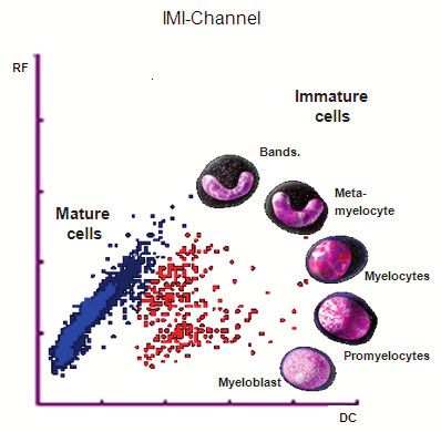
Briggs et al / Improved Flagging Rates on the Sysmex XE-5000

immature platelet fraction. The immature platelet fraction is        be an increase in specificity, fewer false-positives, and no loss
identified by flow cytometry with the use of a nucleic acid–         of sensitivity or decrease in true-positives.
specific dye in the reticulocyte/optical platelet channel. The
clinical usefulness of this parameter has been established in
the laboratory diagnosis of thrombocytopenia due to increased
                                                                     Materials and Methods
peripheral platelet destruction, particularly autoimmune
thrombocytopenic purpura and thrombotic thrombocytopenic
purpura,3,4 and as a predictor of platelet recovery following        Sample Selection and Study Design
hematopoietic progenitor cell transplantation.5 The latest                For the study, 1,002 adult patient residual K2EDTA
software to be introduced was for the measurement of                 samples were randomly selected from University College
the reticulocyte hemoglobin concentration. The reticulocyte          London Hospital Haematology laboratory (London, England)
hemoglobin is a measure of the forward scatter of stained            after all routine testing had been completed. Samples were
reticulocytes and provides an indirect measure of the functional     selected at different times of the day and on different days
iron available for new RBC production during the previous 3          of the week during a 3-month period to try to mimic 1 day’s
or 4 days. It also provides an early measure of the response to      workload. University College London Hospital receives a high
iron therapy, increasing within 2 to 4 days of the initiation of     proportion of abnormal samples as a tertiary reference center.
intravenous iron therapy.6                                                Samples were analyzed on the XE-2100 and the
     The most recently introduced analyzer from Sysmex is            XE-5000 in CBC and DIFF mode. Most XE-5000 instruments
the XE-5000, launched in 2007. This instrument performs              automatically report the NRBC count for all samples; however,
a CBC and differential in the same way as the XE-2100,               some models have NRBC counts as a selectable test to save
but new parameters have been introduced, and it also has             on NRBC reagent, and the count is performed in response to
the ability to measure body fluids. The instrument measures          the NRBC flag. The XE-5000 used in this evaluation did not
the hemoglobin content of individual RBCs, calculates the            routinely report NRBCs in every CBC and differential; when
percentages of hypochromic and hyperchromic RBCs, and                an NRBC flag was triggered, samples were rerun in NRBC
quantifies the proportion of marginally sized erythrocytes. The      mode to enumerate the NRBCs and correct the WBC count
availability of extended RBC parameters should allow earlier         and differential. If the reticulocyte (RETIC) action message
diagnosis of abnormal iron metabolism and the response to            appeared, samples were rerun in the RETIC mode, not only
iron or folate supplementation.7                                     to count reticulocytes, but also in case an optical fluorescent
     The sensitivity and specificity of flags for blasts, abnormal   platelet count, instead of impedance, was needed. The number
lymphocytes/lymphoblasts, and atypical lymphocytes on the            of reruns on each instrument was recorded.
XE-2100 have been reported previously,8,9 and, generally, the             Two blood films were made and stained on all samples
results are similar to those for other instruments available.10      using the Sysmex SP100 slide maker. Blood films were
These flags show good sensitivity but poorer specificity,            examined for leukocyte morphologic features by 2 experienced
which, in the routine laboratory, leads to the unnecessary           observers (C.J.B. and I.L.), a third if there was disagreement
examination of blood films.                                          between the first 2 results. Observers were not aware at
     A new set of algorithms for the detection of the presence       that time of the instruments’ results. Lymphocytes were
of these cells, efficient multichannel messaging (eMM)               classified as normal or abnormal. Abnormal lymphocytes
software, was developed for the XE-5000 in October 2009.             included atypical (likely associated with viral infection),
The software integrates the flagging areas from the DIFF and         plasmacytoid, or malignant (lymphocytes with coarse nuclear
the IMI channels.                                                    chromatin, nuclear clefts, prolymphocytes, plasma cells, or
     On the XE-2100 version for flagging in the IMI channel,         smear cells). Abnormal lymphocytes and atypical lymphoid
it was possible to have an overlap between the blast and             cells are heterogeneous populations; even very experienced
immature granulocyte flagging areas; however, because                observers cannot always be sure to which category they
lymphocytes are not measured in this channel, there cannot           belong. Although there are 2 flags for abnormal and atypical
be any overlap between blasts and atypical lymphocytes. In           lymphocytes on the Sysmex instruments, these 2 flags were
the DIFF channel, there is no overlap between blasts and             assessed together and counted as true-positive if either
immature granulocytes, but there is possible overlap between         flag was triggered in the presence of atypical or abnormal
blasts and atypical lymphocytes. The XE-5000 eMM software            lymphocytes.
was designed to improve the specificity of these flags.                   Despite the fact that only the blast cell, abnormal
     This aim of this study was to evaluate the efficiency of        lymphocyte/lymphoblast, and atypical lymphocyte flags have
these flags by examining the results from the XE-2100 and            been changed for the XE-5000, for this study, any sample with
XE-5000 on a routine daily workload. The ideal result would          an abnormal cell flag or incomplete differential results was

310    Am J Clin Pathol 2011;136:309-316                                                             © American Society for Clinical Pathology
310    DOI: 10.1309/AJCPDLR4KGKAFW4W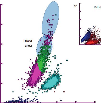
Hematopathology / Original Article
considered to need a manual blood film review. Quantitative The basophil count is derived in the WBC/BASO channel in
flags alone or numeric results outside the reference range which basophils are resistant to the reagent system and retain
were not considered to need a manual review. This approach their size and shape, forming a cluster of larger cells distinct
allowed the assessment of the changes in flagging on the total from other nonbasophil nucleated cells whose membranes are
number of blood films made in the laboratory on a typical perforated and cytoplasm is lost.
working day. In the IMI channel, a combination of radio frequency
and impedance, direct current resistance methods are used in
Principle of Leukocyte Differential on the XE-2100 conjunction with special reagents. Immature WBCs contain
and XE-5000 less lipid than do mature cells. In the presence of special
The optical system on both XE instruments uses a lysing reagents, mature cells are disrupted, their granules
stable red diode laser producing a light beam of 633 nm are eluted, and only their nuclei remain. Immature cells of
wavelength and a polymethine-based fluorescent dye. When the myeloid series behave in a different way: before the
the laser beam collides with a stained cell, 3 signals are intracellular components are eluted, the lysing reagent enters
produced: forward-scattered light, providing information the cells and binds to the membrane and granules, thus fixing
on cell size; side-scattered light, providing information on the cells and differentiating them from mature cells. The
internal cell structure; and side fluorescence, providing effect of the lysing reagent differs with each type of immature
information on DNA and RNA content. The light scatter cell, allowing quantitative differentiation ❚Image 2❚.
signals are detected by photo diodes and the fluorescence
signal by a photomultiplier via a dichroic mirror. In the DIFF Abnormal Cell Flags
scattergram ❚Image 1❚, side fluorescence is on the y-axis Abnormal cells have characteristics different from those
and side-scattered light on the x-axis. In a healthy person, of normal cells, such as cell size, nuclear size, and granule
cluster analysis reveals cell ghosts, lymphocytes, monocytes, content. In the presence of abnormal cells, most instruments
eosinophils, combined neutrophil and basophil populations, will generate an “abnormal” cell or “suspect” flag. The
and, if present, immature granulocytes, which are the sum of instrument detects the “cloud” (as seen in Image 1) for a
metamyelocytes, myelocytes, and promyelocytes (Image 1). particular cell population as having an abnormal size or
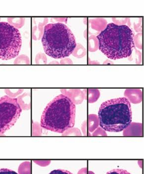
Promyelocytes
Fluorescence Intensity
Myelocytes
Metamyelocytes
Side Scatter
❚Image 1❚ Sysmex differential (DIFF) channel scatterplot. Different leukocyte types have different locations in the scattergram.
Fluorescence is depicted on the y-axis and side-scattered light on the x-axis. The blue dots close to the x-axis are cell ghosts;
the turquoise clusters, neutrophils and basophils; pink dots, lymphocytes; green dots, monocytes; and orange dots, eosinophils.
Immature granulocytes show higher fluorescence than neutrophils and are represented by blue dots appearing above the
neutrophils with the earliest cells, promyelocytes, demonstrating the highest fluorescence; more mature cells, metamyelocytes,
are in the area closest to the neutrophil cluster.
© American Society for Clinical Pathology Am J Clin Pathol 2011;136:309-316 311
311 DOI: 10.1309/AJCPDLR4KGKAFW4W 311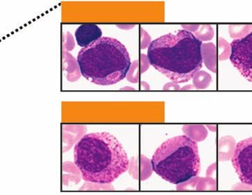
Briggs et al / Improved Flagging Rates on the Sysmex XE-5000

                                                                                                          shape by cluster analysis. The instrument will also detect
                                                                                                          events outside the normal cell population areas. The flags are
                                                                                 Immature cells           generated by combining pattern abnormalities from the DIFF
                                                                                                          and IMI channels.
                                                                               Bands                           The areas of abnormal WBCs in the DIFF and IMI
                                                                                                          scatterplots are shown in ❚Image 3❚.
Radio Frequency

                                                                                         Metamyelocytes
                           Mature cells                                                                   The New Blast Cell Flag Algorithm
                                                                                                              The blast cell flag is given when number of cells in the
                                                                                            Myelocytes    IMI channel blast area exceeds a preset trigger limit and when
                                                                                                          more cells in the IMI channel immature granulocyte area are
                                                                                                          detected than in their signature position in the DIFF channel.
                                                                                         Promyelocytes
                                                                                                          This information is then combined with the number of cells
                                                                                                          in the DIFF channel blast area ❚Image 4A❚, which must
                                                                    Myeloblasts
                                                                                                          exceed the trigger limit and be more than in the DIFF channel
                                                               Direct Current
                                                                                                          atypical lymphocyte area.

❚Image 2❚ Sysmex immature myeloid information channel                                                     The New Abnormal Lymphocyte/Lymphoblast Flag
with direct current resistance on the x-axis and radio                                                    Algorithm
frequency on the y-axis. Mature WBCs are completely                                                            For this flag to be triggered, the blast flag is not present
lysed and shrunken by the reagent and are located in the                                                  (no cells in IMI blast area) and the number of cells on the
blue cluster. The effect of the lysing reagent is different                                               lymphocytes and monocytes border ❚Image 4B❚ is more than
on immature myeloid cells; they are not completely lysed,                                                 normal (Image 1) and exceeds the trigger limit. The trigger
allowing for their quantitative differentiation from mature                                               limit varies depending on the total WBCs or if the number
cells. The red dots represent immature myeloid cells. In a                                                of cells in the DIFF channel blast areas (Images 4A and 4B)
normal scattergram there are very few or no red events.                                                   exceeds the trigger limit and is more than in the DIFF channel
                                                                                                          atypical lymphocyte area combined with absence of cells in
                                                                                                          the IMI channel blast area.

     A                                                                                                    B
                                                       Atypical
                                                     lymphocyte
                                                         area
                  Fluorescence Intensity

                                                                                                              Radio Frequency

                                                Blast or                           IG
                                               abnormal                           area
                                              lymphocyte
                                                 area

                                                                                                                                  IG area

                                                                                                                                 Blast area

                                                                      Side Scatter                                                  Direct Current

❚Image 3❚ A, Schematic representation of the Sysmex differential channel scatterplot showing abnormal cell locations. B,
Schematic representation of the Sysmex immature myeloid information channel scatterplot showing immature myeloid cell
locations. IG, immature granulocyte.

312                                        Am J Clin Pathol 2011;136:309-316                                                                © American Society for Clinical Pathology
312                                        DOI: 10.1309/AJCPDLR4KGKAFW4W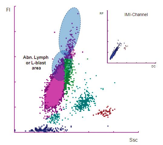
Hematopathology / Original Article

The New Atypical Lymphocyte Flag Algorithm                                               Statistical Methods
     This flag means that the high-fluorescent lymphocyte                                    The rates of efficiency for the blast cell and abnormal/
count is more than 1% of the total lymphocyte count ❚Image                               atypical lymphocyte flags were compared statistically by
4C❚ and the number of cells in the DIFF channel atypical                                 using the McNemar test on S-PLUS, version 6.1 (Insightful,
lymphocyte area exceeds the trigger limit and the number of                              Palo Alto, CA).
cells in the DIFF channel atypical lymphocyte area exceeds
those in the DIFF channel blast area (Image 4A). Under
these conditions, high-fluorescent lymphocytes have been                                 Results
proven to be activated B lymphocytes or plasma cells,11                                       ❚Table 1❚ shows the total number of abnormal cell
resulting in high specificity and sensitivity for screening for                          flags seen on each instrument. The XE-5000 had 185 fewer
plasma cells with the atypical lymphocyte flag.12                                        abnormal cell flags than the XE-2100, which would lead to

 A                                                                                       B
                                                     Radio Frequency

                                                                                                                                                   Radio Frequency
 Fluorescence Intensity

                                                                                          Fluorescence Intensity

                                                                                                                      Abnormal
                              Blast                                                                                lymphocyte or
                              area                                                                                  lymphoblast
                                                                       Direct Current                                   area                                         Direct Current

                                      Side Scatter                                                                                 Side Scatter

                                                                                        ❚Image 4❚ A, The new blast cell flag algorithm on the XE-5000.
C
                                                                                        Information from the differential (DIFF) scattergram and the
                             Atypical
                                                                                        immature myeloid information (IMI) channel is used. The lower
                                                     Radio Frequency

                           lymphocyte                                                   blue shaded area in the DIFF channel is the area where blasts
                               area                                                     are found, but the flag will be triggered only if there are also
                                                                                        events in the blast area (red dots) in the IMI channel. B, The
  Fluorescence Intensity

                                                                                        new abnormal lymphocyte/lymphoblast flag algorithm on the
                                                                                        XE-5000. Information from the DIFF scattergram and the IMI
                                                                                        channel is used. The lower blue shaded area in the DIFF channel
                                                                       Direct Current   is the area where blasts, abnormal lymphocytes, or lymphoblasts
                                                                                        may reside. With no events in the blast area in the IMI channel,
                                                                                        these cannot be myeloblasts, so only the abnormal lymphocyte/
                                                                                        lymphoblast flag will be triggered. C, The new atypical
                                                                                        lymphocyte flag algorithm on the XE-5000. The top blue shaded
                                                                                        area in the DIFF channel is where atypical (high-fluorescent)
                                                                                        lymphocytes reside. There are no events in the abnormal
                                                                                        lymphocyte/lymphoblast area or in the blast area in the IMI
                                      Side Scatter                                      channel, so only the atypical lymphocyte flag will be triggered.

© American Society for Clinical Pathology                                                                                                Am J Clin Pathol 2011;136:309-316        313
                                                                                                                                   313   DOI: 10.1309/AJCPDLR4KGKAFW4W            313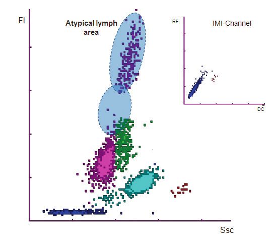
Briggs et al / Improved Flagging Rates on the Sysmex XE-5000
❚Table 1❚ ❚Table 2❚
Total Number of Flags Generated by Both Instruments, Clinical Usefulness and Efficiency of Abnormal Cells Flags
Number of Blood Films That Would Have Been Made in the on the XE-2100 and XE-5000 for 1,002 Samples
Laboratory, Number of Samples That Needed to Be Rerun,
and Number of Differential Vote-Outs for Each Instrument Flag XE-2100 XE-5000
XE-2100 XE-5000 Blasts
TP 13 14
Total No. of samples analyzed 1,002 1,002 TN 896 959
Total No. of flags 525 340 FP 90 27
No. (%) of blood films 243 (24.3) 138 (13.8) FN 3 2
No. of rerun samples* 75 86 Sensitivity (%) 81.2 87.5
No. (%) of reruns that gave additional 49 (65) 55 (64) Specificity (%) 90.8 97.2
clinical information† Efficiency (%) 90.7 97.1†
No. of vote-outs‡ Abnormal lymphocytes
Neutrophils 27 24 or lymphoblasts/atypical lymphocytes
Lymphocytes 25 23 TP 23 22
Monocytes 20 17 TN 845 914
Eosinophils 9 9 FP 114 45
Basophils 10 11 FN 20 21
Immature granulocytes 27 25 Sensitivity (%) 53.5 51.2
Specificity (%) 88.1 95.3
* Rerun because of an nucleated RBC (NRBC) flag or the reticulocyte (RETIC) Efficiency (%) 86.6 93.4†
action message. Left shift
† Reruns that gave additional clinical in formation are those with NRBCs present or TP 32 28
an optical platelet count reported as measured in the RETIC channel. TN 910 929
‡ Instrument did not report the leukocyte differential cell count.
FP 24 15
FN 36 30
Sensitivity (%) 47.1 35.7
Specificity (%) 97.4 98.4
a reduction in blood film reviews from 24.3% to 13.8% of Efficiency (%) 94.0 95.5
Nucleated RBCs
samples analyzed in the laboratory. TP 20 22
The number of differential vote-outs, where the TN 945 936
FP 27 37
instrument does not report a differential count in the presence FN 10 7
of abnormal cells, was similar for both instruments (9.1% on Sensitivity (%) 66.7 75.8
the XE-5000 and 8.4% on the XE-2100); however, on both Specificity (%) 97.2 96.2
Efficiency (%) 96.3 95.6
instruments, all voted-out differentials can be seen in the Platelet clumps
WBC research screen. TP 1 1
TN 960 976
The XE-5000 required the rerunning of slightly more FP 31 16
samples because of more NRBC flags on first analysis on FN 10 9
Sensitivity (%) 9.1 10.0
the XE-5000 than on the XE-2100. The number of justified Specificity (%) 96.9 98.4
reruns, ie, NRBCs were present in the sample or an optical Efficiency (%) 95.9 97.5
platelet count from the RETIC channel was reported, was FN, false-negative; FP, false-positive; TN, true-negative; TP, true-positive.
almost the same for both instruments, 65% (49/75) for the * Values are given as number of samples unless otherwise indicated.
† P < .001.
XE-2100 and 64% (55/86) for the XE-5000.
❚Table 2❚ shows the flagging performance for blasts,
abnormal lymphocytes/lymphoblasts with atypical Although the immature granulocyte count is derived
lymphocytes, left shift, NRBCs, and platelet clumps. The from the DIFF channel, if there is an abnormal cell pattern,
eMM software, version 00-06, only has new algorithms a suspect immature granulocyte flag is generated but the
for the blast cell, abnormal lymphocyte/lymphoblast, and count is still reported. Each laboratory should have a defined
atypical lymphocyte flags, but the other flags were assessed protocol on how to deal with samples generating this flag,
to determine how many manual blood film reviews would but for this evaluation, a stained peripheral blood film was
be performed on a daily basis in the routine laboratory using examined. On the XE-2100, this flag was seen 66 times (6.6%
either instrument. of samples) and on the XE-5000, 50 times (5.0% of samples).
It is interesting that the XE-5000 tended to give more All samples that were positive for immature granulocytes on
false-positives for NRBCs, 37 compared with 27 on the the blood film were also positive by both instruments, and all
XE-2100. This finding means that extra samples are rerun in samples with an automated immature granulocyte count of
the NRBC mode on the XE-5000, but does not necessarily 3% or more were positive on the blood film, unless the WBC
mean that these samples would need manual review. There count was less than 500/μL (0.5 × 10⁹/L).
were fewer false-positive platelet clump flags on the XE-5000, The red cell fragment flag was not sensitive on either
but this flag did not perform efficiently on either analyzer. instrument: 33 blood films demonstrated fragments on the
314 Am J Clin Pathol 2011;136:309-316 © American Society for Clinical Pathology
314 DOI: 10.1309/AJCPDLR4KGKAFW4W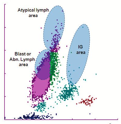
Hematopathology / Original Article

blood film, but the XE-2100 flagged only 7 of these samples,             The objective for the laboratory is to reduce the number
and the XE-5000 flagged 3. Neither instrument showed any            of samples needing further action as far as possible without
false-positives. There is a fragmented red cell count available     endangering patients by reporting false or misleading results,
on both instruments on the RBC research screen,13 but this          especially false-negative results. Any analyzer that decreases
was not assessed in this evaluation.                                the number of false-positive flags without losing sensitivity
     One sample with red cell autoagglutination was included        will increase laboratory efficiency.
in the study, and both instruments correctly flagged the                 The flagging efficiency of the XE-2100 has been
abnormality. There were no false-positives.                         published,8,9 and, generally, the results are similar to those for
     The efficiency of the flagging for blasts and abnormal         other instruments available. The XE-2100 blast flag has been
or atypical lymphocytes was significantly better (P < .001)         reported as the most sensitive when compared with the Abbott
on the XE-5000 compared with the XE-2100. One sample                CELL-DYN Sapphire (Abbott Diagnostics, Santa Clara, CA),
was false-negative for the blast flag on both instruments.          the Siemens Advia 120 (Siemens Diagnostics, Tarrytown,
This sample was from a patient with acute myeloid leukemia          NY), and the Beckman Coulter LH 750 (Beckman Coulter,
undergoing chemotherapy, and the WBC count was only 690/            Miami, FL).10
μL (0.69 × 10⁹/L). There were 2 more false-negatives on the              The efficiency evaluation of the XE-2100 and XE-5000
XE-2100 and 1 on the XE-5000. Overall, the number of false-         was obtained by assessment of the number of blood films
positives was greatly reduced on the XE-5000, 27 compared           made in the laboratory owing to the presence of abnormal cell
with 90 on the XE-2100, without any loss of sensitivity. This       flags. This approach allows a simple means of determining
result was also true for the abnormal/atypical lymphocyte           how efficient the instrument is in detecting true abnormalities
flag: The number of false-positives was reduced to 45 on the        and how inefficient it is in generating false flags.
XE-5000 from 114 on the XE-2100, and both instruments                    Our findings indicate that the use of the XE-5000 in the
showed similar numbers of false-negatives and true-positives.       routine laboratory would reduce the number of manual film
Of the false-positives, 17 were the same samples on both            reviews triggered by abnormal cell flags from about 24% on
instruments and were from patients with various diagnoses           the XE-2100 to about 14% on the XE-5000. With a daily
and WBC and lymphocyte counts.                                      workload of about 1,000 samples, this difference represents
                                                                    a reduction of 105 blood films. Nearly all of this reduction
                                                                    is from fewer false-positive blast cell and abnormal/atypical
Discussion                                                          lymphocyte flags. High sensitivity of blast cell detection in
     Examination of blood films is not only labor-intensive,        routine hematology analyzers is important for the diagnosis
but it is also requires highly trained staff. The impact of         and follow-up of hematologic malignancies. There were
a wrong diagnosis necessitates that experienced staff be            3 false-negatives on the XE-5000 and 2 on the XE-2100.
present in the laboratory 24 hours a day. However, manual           These samples were from patients with leukopenia, and it
cell classification is subjective, with significant interobserver   has been reported that flagging sensitivities are reduced with
and intraobserver variation,14 and any count is also subject to     low WBC counts.8
significant statistical variance.15                                      Many instruments available only have 1 flag for the
     With continuing pressure on laboratory resources and           presence of abnormal or atypical lymphocytes, but the Sysmex
the need for faster turnaround times, it is essential to reduce     instruments have 2, the abnormal lymphocyte/lymphoblast
the number of manual blood film reviews and manual                  and atypical lymphocyte flags. In this study, as in a previous
differential counts. A film is reviewed to provide information      evaluation,9 these flags were assessed together. Morphologic
additional to or missing from the analyzer report or to             definitions of atypical or abnormal lymphocytes are highly
confirm results provided by the analyzer. The challenge is          variable and would make the assessment of the individual
to reduce the number of blood films examined without                flags problematic. If abnormal or atypical lymphocytes
missing important diagnostic information. In 20% to 25% of          were seen in the blood film, the presence of either one of
CBCs with differentials, leukocyte-related abnormal cell flags      the flags constituted a true-positive. There was a reduction
are generated, which means a manual microscopic review              in the number of false-positive flags on the XE-5000, 4.5%
on a stained blood smear may be required.16,17 However,             compared with 11.4% on the XE-2100. There was no loss of
depending on the clinical population and local guidelines for       sensitivity, with both instruments showing similar numbers
making blood films, film review rates range from 10% to             of false-negatives, which were mostly the same samples on
50% in different laboratories.17,18 More than 80% of manual         both instruments from patients with various diagnoses and
film reviews are triggered by hematology analyzer flags16;          lymphocyte counts.
however, more than 62% of reviews undertaken in the routine              Despite the fact that the eMM software only has new
laboratory are due to false-positive flags.17                       algorithms for blast cell, abnormal lymphocyte/lymphoblast,

© American Society for Clinical Pathology                                                          Am J Clin Pathol 2011;136:309-316   315
                                                                                            315    DOI: 10.1309/AJCPDLR4KGKAFW4W       315
Briggs et al / Improved Flagging Rates on the Sysmex XE-5000

and atypical lymphocyte flags, some differences were noted             6. Brugnara C. Use of reticulocyte cellular indices in the
for NRBC flag, with the XE-5000 giving slightly more false-               diagnosis and treatment of haematological disorders. Int J
                                                                          Clin Lab Res. 1998;28:1-11.
positives, which would mean more reruns for the NRBC
                                                                       7. Urrechaga E, Borque L, Escanero JF. Potential utility of the
count but not necessarily more manual film reviews.                       new Sysmex XE 5000 red blood cell extended parameters
     The platelet clump flag did not perform well on either               in the study of disorders of iron metabolism. Clin Chem Lab
analyzer.                                                                 Med. 2009;47:411-416.
     Laboratory productivity is inversely related to the number        8. Ruzicka K, Veitl M, Thalhammer-Scherrer R, et al. The
                                                                          new hematology analyzer Sysmex XE-2100: performance
of manual film reviews,16 the higher the manual review rates,             evaluation of a novel white blood cell differential technology.
the lower the productivity. Using the XE-5000 with eMM                    Arch Pathol Lab Med. 2001;125:391-396.
software in the routine hematology laboratory reduces the              9. Stamminger G, Auch D, Diem H, et al. Performance of
number of manual blood film reviews needed and so increases               the XE-2100 leucocyte differential. Clin Lab Haematol.
                                                                          2002;24:271-280.
workflow efficiency and improves turnaround times by
                                                                      10. Kang S, Kim HK, Ham CK, et al. Comparison of four
speeding up sample processing. In the future, it is hoped that            hematology analyzers, CELL-DYN Sapphire, ADVIA 120,
more abnormal cells, especially blast cells, will be accurately           Coulter LH 750 and Sysmex XE-2100, in terms of clinical
quantitated by hematology analyzers rather than their possible            usefulness. Int J Lab Hematol. 2008;30:480-486.
presence being indicated by a flag.                                   11. Linssen J, Jennissen V, Hildmann J, et al. Identification and
                                                                          quantification of high fluorescence-stained lymphocytes as
                                                                          antibody synthesizing/secreting cells using the automated
From the 1Department of Haematology, University College                   routine hematology analyzer XE-2100. Cytometry B Clin
London Hospital, London, England; and 2Sysmex Europe,                     Cytom. 2007;72B:157-166.
Norderstedt, Germany.                                                 12. Linssen J, Jennissen V. Identification of high fluorescence-
    Address reprint requests to Ms Briggs: Dept of Haematology,           stained lymphocytes (HFL) count on the XE-5000 with
                                                                          efficient multichannel messaging (eMM) as antibody
University College London Hospital, 60 Whitfield St, London W1T
                                                                          synthesizing, c.q. plasma cells. Sysmex J Int. 2009;19:1-8.
4EU, England.
    University College London Hospital has received an                13. Jiang M, Saigo K, Kumagai S, et al. Quantification of red
                                                                          blood cell fragmentation by automated haematology analyser
unrestricted educational grant from Sysmex Europe.
                                                                          XE-2100. Clin Lab Haematol. 2001;23:167-172.
    Acknowledgment: We thank Arthur Childs for help in the
examination of blood films.                                           14. Koepke JA, Dotson MA, Shifmann MA. A critical
                                                                          evaluation of the manual/visual differential leukocyte
                                                                          counting method. Blood Cells. 1985;11:173-186.
                                                                      15. Rumke CL. Statistical reflections on finding atypical cells.
References                                                                Blood Cells. 1985;11:141-144.
 1. Briggs C, Harrison P, Grant D, et al. New quantitative            16. Novis DA, Walsh M, Wilkinson D, et al. Laboratory
    parameters on a recently introduced automated blood cell              productivity and the rate of manual peripheral blood smear
    counter: the XE-2100. Clin Lab Haematol. 2000;22:345-350.             review: a College of American Pathologists Q-Probes study
 2. Briggs C, Kunka S, Fujimoto H, et al. Evaluation of immature          of 95,141 complete blood count determinations performed in
    granulocyte counts by the XE-IG master: upgraded software             263 institutions. Arch Pathol Lab Med. 2006;130:596-601.
    for the XE-2100 automated hematology analyzer. Lab                17. Barnes PW, McFadden SL, Machin SJ, et al. The
    Hematol. 2003;9:117-124.                                              International Consensus Group for Hematology Review:
 3. Briggs C, Hart D, Kunka S, et al. Assessment of an immature           suggested criteria for action following automated CBC and
    platelet fraction (IPF) in peripheral thrombocytopenia. Br J          WBC differential analysis. Lab Hematol. 2005;11:83-90.
    Haematol. 2004;126:93-99.                                         18. Briggs C, Longair I, Slavik M, et al. Can automated blood
 4. Pons I, Monteagudo M, Lucchetti G, et al. Correlation                 film analysis replace the manual differential? an evaluation of
    between immature platelet fraction and reticulated platelets:         the CellaVision DM96 automated image analysis system. Int J
    usefulness in the etiology diagnosis of thrombocytopenia. Eur         Lab Hematol. 2009;31:48-60.
    J Haematol. 2010;85:158-163.
 5. Zucker ML, Murphy CA, Rachel JM, et al. Immature
    platelet fraction as a predictor of platelet recovery following
    hematopoietic progenitor cell transplantation. Lab Hematol.
    2006;12:125-130.

316   Am J Clin Pathol 2011;136:309-316                                                                © American Society for Clinical Pathology
316   DOI: 10.1309/AJCPDLR4KGKAFW4W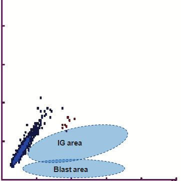
You can also read