A TISSUE CULTURE MODEL OF ESTROGEN-PRODUCING PRIMARY BOVINE GRANULOSA CELLS - JOVE
←
→
Page content transcription
If your browser does not render page correctly, please read the page content below
Journal of Visualized Experiments                                                                                            www.jove.com

Video Article
A Tissue Culture Model of Estrogen-producing Primary Bovine Granulosa
Cells
              1                  1
Anja Baufeld , Jens Vanselow
1
Institute of Reproductive Biology, Leibniz Institute for Farm Animal Biology (FBN)

Correspondence to: Anja Baufeld at baufeld@fb-dummerstorf.de

URL: https://www.jove.com/video/58208
DOI: doi:10.3791/58208
Keywords: Biology, Issue 139, Serum-free culture, ovary, reproduction, cell culture model, molecular biology, follicle, cryopreservation
Date Published: 9/6/2018
Citation: Baufeld, A., Vanselow, J. A Tissue Culture Model of Estrogen-producing Primary Bovine Granulosa Cells. J. Vis. Exp. (139), e58208,
doi:10.3791/58208 (2018).

Abstract
    Ovarian granulosa cells (GC) are the major source of estradiol synthesis. Induced by the preovulatory luteinizing hormone (LH) surge, cells
    of the theca and, in particular, of the granulosa cell layer profoundly change their morphological, physiological, and molecular characteristics
    and form the progesterone-producing corpus luteum that is responsible for maintaining pregnancy. Cell culture models are essential tools to
    study the underlying regulatory mechanisms involved in the folliculo-luteal transformation. The presented protocol focuses on the isolation
    procedure and cryopreservation of bovine GC from small- to medium-sized follicles (< 6 mm). With this technique, a nearly pure population
    of GC can be obtained. The cryopreservation procedure greatly facilitates time management of the cell culture work independent of a direct
    primary tissue (ovaries) supply. This protocol describes a serum-free cell culture model that mimics the estradiol-active status of bovine GC.
    Important conditions that are essential for a successful steroid-active cell culture are discussed throughout the protocol. It is demonstrated
    that increasing the plating density of the cells induces a specific response as indicated by an altered gene expression profile and hormone
    production. Furthermore, this model provides a basis for further studies on GC differentiation and other applications.

Video Link
    The video component of this article can be found at https://www.jove.com/video/58208/

Introduction
    Successful ovulation and luteinization depend on finely tuned and well-orchestrated molecular alterations in different somatic follicular cell types.
    Since details of these developmental processes are not yet fully understood, further clarification is required. In vivo approaches are elaborate
    and costly and, in particular, fail to address specific molecular mechanisms that occur during folliculogenesis. Therefore, reductionistic in vitro
    models are needed in addition, to provide insight into cellular and molecular details. Different studies describe the culture of whole follicles in
                                                    1,2,3
    the context of in vitro fertilization techniques     . Because researchers are interested in mechanisms of differentiation, many studies focus on
    follicular GC. These cells, directly associated with the oocyte, are the major sources of estrogen production and, thus, play an essential role
                                                    4
    throughout folliculogenesis and luteinization .

    Immortalized cell lines of GC have been developed from different species. Most of them, however, do not show a sufficient steroid hormone
               5                                                               6                                                                      7
    production . So far, only one cell line of bovine GC has been established , but this line lost its steroidogenic activity after several passages .
    Therefore, since steroidogenesis and, in particular, estradiol production is an essential feature of GC functionality, it is advisable to study these
    aspects in primary cell culture models. In previous studies, it was demonstrated that a considerable estradiol production can only be observed
                                          8,9
    under serum-free culture conditions . Further on, the supplementation of a precursor of estradiol synthesis is another prerequisite, as GC fail
                                                                                            10
    to express the necessary enzyme that can convert progesterone to androstenedione . Additionally, the synergistic effect of FSH and IGF-1
                                                                                                                    11
    supplementation in vitro revealed an optimized activity of aromatase, the key enzyme of estradiol synthesis . In the present protocol, other
    important factors that have a substantial impact on the GC culture model are also described. In particular, the cell plating density has tremendous
                                                12
    effects on the outcome of the experiment . Furthermore, a cryopreservation technique of bovine GC that does not significantly interfere with GC
    physiology in culture could be established. This technique helps to improve the organization of cell culture work and to optimize the preferred
    plating density.

Protocol
    NOTE: Bovine ovaries were obtained from a commercial slaughterhouse. The collection of abattoir byproducts does not require an ethical
    approval according to the German law.

Copyright © 2018 Creative Commons Attribution-NonCommercial-NoDerivs 3.0 Unported                         September 2018 | 139 | e58208 | Page 1 of 7
License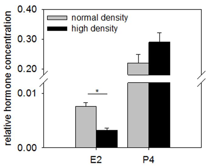
Journal of Visualized Experiments                                                                                            www.jove.com

1. Working Conditions and Preparations
  1. To guarantee sterility, perform all media and tissue preparation as well as all culture work in a specialized cell culture lab using a laminar flow
     bench.
  2. Prepare 1x phosphate buffered saline (PBS, pH 7.4) supplemented with 100 IU penicillin, 0.1 mg/mL streptomycin and 0.5 µg/µL
     amphotericin for the transport of ovaries.
     NOTE: The maximal duration between receiving the ovaries and isolating the GC is 2 h in this set-up.

2. Isolation of Bovine Granulosa Cells
  1. Wash the ovaries several times in 1x PBS (with 100 IU penicillin, 0.1 mg/mL streptomycin, and 0.5 µg/µL amphotericin) to remove
     any blood from the surface before starting the isolation procedure. Use a beaker, place the ovaries inside, fill it with 1x PBS, and
     discard the PBS again.
       1. Repeat this washing step 3–4x, until the ovaries are cleaned from any remaining blood.

  2. Wipe one ovary using a lab wipe soaked with 70% alcohol, to minimize possible contaminations.
  3. With a 3 mL syringe and an 18 G needle, aspirate the GC by puncturing small- to medium-sized follicles (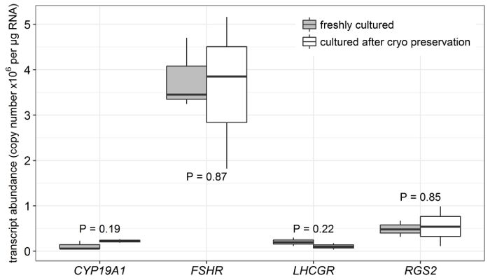
Journal of Visualized Experiments www.jove.com
2. Prepare media and supplements for culture work under a laminar flow hood. For a successful recovery of the GC, they have to be processed
immediately after thawing.
3. Prepare the following media.
1. Supplement α-Minimal Essential Medium (α-MEM) with 2 mM L-glutamine, 0.084% sodium bicarbonate, 0.1% bovine serum albumin
(BSA), 20 mM HEPES, 4 ng/mL sodium selenite, 5 µg/mL transferrin, 10 ng/mL insulin and 1 mM non-essential amino acids.
2. Additionally, add 100 IU penicillin and 0.1 mg/mL streptomycin to the media.
NOTE: The antibiotics, once warmed, are stable for approximately 48 h. Therefore, aliquot the desired amount of media for the culture
set-up and planned media exchange. Pre-warm only the media aliquots to 37 °C in a water bath. The prepared media can be stored at
4 °C for no longer than 3 months.
3. To induce steroid activity in cultured bovine GC, supplement the α-MEM additionally with 20 ng/mL follicle-stimulating hormone (FSH),
3
50 ng/mL R IGF-1, and 2 µM androstenedione.
NOTE: Always supplement these components shortly before the onset of a culture or media exchange. Avoid repeated freeze-and-
thaw cycles for FSH and IGF-1 stock solutions, as this might compromise the steroid activity.
5. Cell Culture Work
1. Thaw the cells quickly in a water bath at 37 °C for 3–4 min and transfer the cell suspension into 37 °C pre-warmed α-MEM (without hormone
supplement). For centrifugation, a final volume of 10 mL is recommended.
2. Centrifuge the GC for 3 min at room temperature and at 500 x g. Discard the supernatant. Add the pre-warmed and supplemented α-MEM
and resuspend the cells carefully.
5 6
3. Seed the GC at a density of 1 x 10 or 1 x 10 cells per well in a final volume of 500 µL. Include technical replicates of at least three culture
wells per condition.
4. Incubate the cell culture in an incubator at 37 °C with 5% CO2 for 8 d.
5. Perform a media exchange every other day. Replace only two-thirds of the culture media to reduce stress and to ease the adaptation of the
cells to the fresh media.
NOTE: The GC form a typical fibroblast-like phenotype after a few days in culture and tend to cluster together. In the high cell-density culture,
larger clusters are found more frequently as compared to the normal density GC culture (Figure 1, left vs. right panel).
6. Subsequent Analysis of Cultured Granulosa Cells
1. To perform steroid hormone analysis, collect the media and freeze it at -20 °C. Use appropriate methods to analyze the estradiol and
14
progesterone concentration in the spent media [e.g., radioimmunoassay (RIA)] .
2. For the subsequent analysis of different target molecules (RNA, DNA, proteins, etc.), lyse the cells directly in the culture dish with an
appropriate lysing agent as recommended by the supplier.
NOTE: For most isolation procedures, it is possible to stop the protocol here, as the lysed cells can be stored at -20 °C for up to 1 week. For
longer storage, the cells should be kept at -80 °C.
3. To determine the transcript abundance, synthesize cDNA and measure specific transcripts by quantitative real-time polymerase chain
15
reaction (qPCR) techniques . A statistical evaluation should include at least three replications of different cell cultures, originated from
different cell preparations (biological replicates).
Representative Results
5
GC plated at 1 x 10 cells/well display a typical fibroblast-like appearance as they form a cell extension comparable to fibroblasts and tend to
6
build clusters (Figure 1a and 1c). Increasing the plating density by 10-fold to 1 x 10 cells/well did not change the morphology but more cell
clusters can be observed (Figure 1b and 1d, arrows). As already shown in a previous publication, the collagen coating of the culture dishes
13
largely improved the attachment of the cells .
Initial cryopreservation did not considerably change the physiological characteristics of cells in culture as compared to those directly cultured
from freshly isolated samples. This is shown in Figure 2 as the transcript abundance (measured by qPCR) of several marker genes did not differ
when comparing cultured cells derived from either frozen or freshly isolated pools.
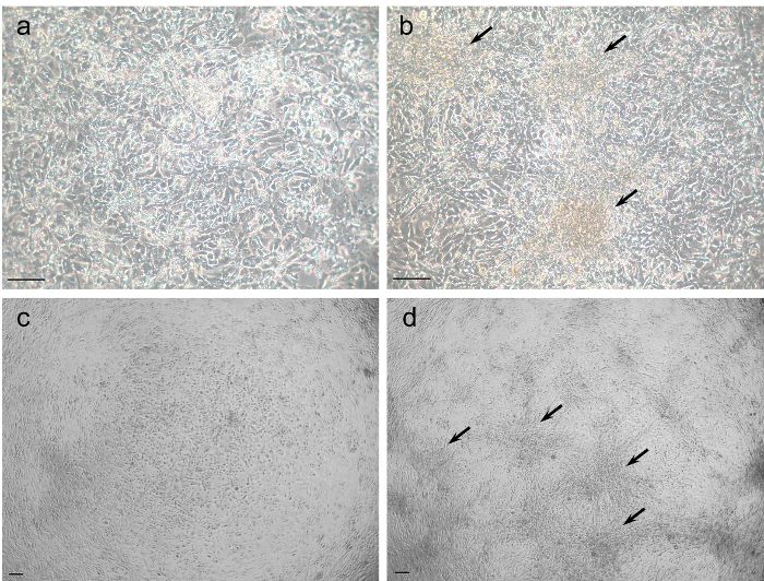
Figure 3 shows representative results of the steroid hormone analysis (measured by RIA) in bovine GC cultured at normal vs. high density. The
estradiol concentration is significantly decreased in the high-density culture compared to the normal-density culture, whereas the progesterone
production tended to be higher.
A comparative analysis of several genes (measured by qPCR) in high- vs. normal-density cultures revealed a significant effect (Figure 4).
CYP19A1, encoding the key enzyme of estradiol biosynthesis aromatase, as well as the gonadotropin receptor FSHR, were significantly down-
regulated. In contrast, the genes RGS2 and VNN2 showed a significant up-regulation. These results clearly suggest that specific processes of
cellular differentiation are induced by increasing the cell plating density.
Copyright © 2018 Creative Commons Attribution-NonCommercial-NoDerivs 3.0 Unported September 2018 | 139 | e58208 | Page 3 of 7
License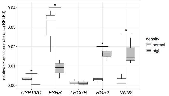
Journal of Visualized Experiments                                                                                           www.jove.com

  Figure 1: Cultured bovine granulosa cells at normal (left panels) and high cell density (right panels). (a and c) Cells cultured at a density
           5                                                                                                                 6
  of 1 x 10 cells/well displayed a typical fibroblast-like phenotype. (b and d) GC cultured at a high plating density (1 x 10 cells/well) tended to
  form large cell clusters (arrows) more frequently as compared to cells under a normal density (left panel). Scale bars = 100 µm. Please click here
  to view a larger version of this figure.

  Figure 2: Comparison of cells cultured directly after isolation and after cryopreservation. The cells were cultured at a density of 1 x
     5
  10 cells per well. The gene expression of several marker transcripts was evaluated and revealed no difference between cultured cells with or
  without an initial cryopreservation. The boxplot shows the median of n = 3. Two-tailed Student's t-test, the p-values are included. This figure is
                                           16
  reproduced from Baufeld and Vanselow . Please click here to view a larger version of this figure.

Copyright © 2018 Creative Commons Attribution-NonCommercial-NoDerivs 3.0 Unported                      September 2018 | 139 | e58208 | Page 4 of 7
License
Journal of Visualized Experiments www.jove.com
Figure 3: Steroid hormone concentration in granulosa cells cultured at different plating densities. The estradiol (E2) concentration
decreased significantly when GC were cultured at a high cell density, whereas the progesterone (P4) concentration only tended to be higher. The
hormone concentration was corrected for the DNA content to normalize for different cell numbers. The mean and SEM of n = 3 are shown. p >
15
0.05, two-tailed Student's t-test. This figure is reproduced from Baufeld et al. . Please click here to view a larger version of this figure.
Figure 4: Abundance of functional key transcripts in granulosa cells cultured at a normal vs. a high plating density. Bovine GC cultured
under serum-free conditions for 8 d revealed a specific regulation of several selected marker genes. The boxplot displays the median of n = 3
individual replicates. p < 0.05, two-tailed Student's t-test. Please click here to view a larger version of this figure.
Discussion
The presented cell culture model provides a tool to analyze granulosa cell differentiation in vitro. Several studies showed that a serum-free
8,9
cultivation is a prerequisite to maintaining steroid activity in cultured bovine GC or GC of other species . Additionally, coating the culture dish
13
with components of the extracellular matrix (e.g., collagen R) , improved the attachment of the cells significantly. Another important feature is
the prolonged culture period. Recently, it has been demonstrated that a long-term culture is necessary to obtain sufficient steroidogenic activity
17
and a balanced expression of granulosa cell identity markers . It appears that GC require the time to recover from the physical stress during the
isolation procedure.
The media supplements FSH, IGF-1, and androstenedione are known to induce aromatase activity in cultured GC. Especially, the
supplementation with androstenedione is absolutely necessary, as the GC need a precursor for estradiol synthesis. This has been published
Copyright © 2018 Creative Commons Attribution-NonCommercial-NoDerivs 3.0 Unported September 2018 | 139 | e58208 | Page 5 of 7
LicenseJournal of Visualized Experiments www.jove.com
11,18
previously and, therefore, was not further investigated during the present study. However, an adaptation of FSH, IGF-1, and androstenedione
concentrations might be necessary for other experimental set-ups.
The cryopreservation technique described here can help to improve the organization of tissue culture experiments by making them more
independent from the varying supply with ovaries. According to previous testing, cryopreservation does not affect the GC phenotype or steroid
production in culture. Also, the abundance of marker transcripts in cultured cells did not reveal significant differences comparing samples
16
prepared from freshly isolated cells with those previously subjected to cryopreservation .
A crucial parameter for the present GC culture model is the cell plating density. As shown by the Representative Results, increasing the
plating density induced remarkable changes of physiological and molecular characteristics. Several genes are regulated in a specific manner,
4,19
resembling the changes that are induced by LH stimulation in vivo . The fact that an increasing cell density can drive differentiation-like
processes in cultured bovine GC has to be meticulously considered in this GC in vitro model to avoid conflicting results between replicates.
Therefore, contradictory results with other studies might be ascribed to different cell densities and should be examined more closely.
The culture model described here revealed to be non-responsive to LH, as the transcripts of the receptor LHCGR are close to the detection limit.
13
Hence, a simulation of the LH surge alike the in vivo situation failed to induce differentiation . Nonetheless, this model provides a helpful tool to
study estradiol-active GC in primary culture, in particular as no functional bovine GC lines exist at present.
Different treatment protocols can be tested in the present GC culture model that help to unravel regulatory mechanisms of steroid production or
GC differentiation. Further, single factors that are involved in developmental processes can be separately analyzed. Therefore, this culture model
provides a basis for many different applications.
Disclosures
The authors have nothing to disclose.
Acknowledgements
We thank Veronica Schreiter for her excellent technical assistance and helpful modifications of the established cryopreservation technique and
cell culture model. Additionally, we like to thank Maren Anders and Swanhild Rodewald for their excellent technical assistance in the subsequent
analysis.
References
1. Gutierrez, C. G., Ralph, J. H., Telfer, E. E., Wilmut, I., Webb, R. Growth and antrum formation of bovine preantral follicles in long-term culture
in vitro. Biology of Reproduction. 62 (5), 1322-1328 (2000).
2. Cortvrindt, R., Hu, Y., Smitz, J. Recombinant luteinizing hormone as a survival and differentiation factor increases oocyte maturation in
recombinant follicle stimulating hormone-supplemented mouse preantral follicle culture. Human Reproduction. 13 (5), 1292-1302 (1998).
3. Paes, V. M. et al. Effect of heat stress on the survival and development of in vitro cultured bovine preantral follicles and on in vitro maturation
of cumulus-oocyte complex. Theriogenology. 86 (4), 994-1003 (2016).
4. Christenson, L. K. et al. Research resource: preovulatory LH surge effects on follicular theca and granulosa transcriptomes. Molecular
Endocrinology. 27 (7), 1153-1171 (2013).
5. Havelock, J. C., Rainey, W. E., Carr, B. R. Ovarian granulosa cell lines. Molecular and Cellular Endocrinology. 228 (1-2), 67-78 (2004).
6. Bernath, V. A. et al. Cyclic AMP inhibits fibronectin gene expression in a newly developed granulosa cell line by a mechanism that
suppresses cAMP-responsive element-dependent transcriptional activation. The Journal of Biological Chemistry. 265 (30), 18219-18226
(1990).
7. Lerner, A. A., Salamone, D. F., Chiappe, M. E., Baranao, J. L. Comparative studies between freshly isolated and spontaneously immortalized
bovine granulosa cells: protein secretion, steroid metabolism, and responsiveness to growth factors. Journal of Cellular Physiology. 164 (2),
395-403 (1995).
8. Gutierrez, C. G., Campbell, B. K., Webb, R. Development of a long-term bovine granulosa cell culture system: induction and maintenance of
estradiol production, response to follicle- stimulating hormone, and morphological characteristics. Biology of Reproduction. 56 (3), 608-616
(1997).
9. Campbell, B. K., Scaramuzzi, R. J., Webb, R. Induction and maintenance of oestradiol and immunoreactive inhibin production with FSH by
ovine granulosa cells cultured in serum-free media. Journal of Reproduction and Fertility. 106 (1), 7-16 (1996).
10. Hillier, S. G., Whitelaw, P. F., Smyth, C. D. Follicular Oestrogen Synthesis - The Two-Cell, Two- Gonadotrophin Model Revisited. Molecular
and Cellular Endocrinology. 100, 51-54 (1994).
11. Silva, J. M., Price, C. A. Effect of follicle-stimulating hormone on steroid secretion and messenger ribonucleic acids encoding cytochromes
P450 aromatase and cholesterol side-chain cleavage in bovine granulosa cells in vitro. Biology of Reproduction. 62 (1), 186-191 (2000).
12. Portela, V. M., Zamberlam, G., Price, C. A. Cell plating density alters the ratio of estrogenic to progestagenic enzyme gene expression in
cultured granulosa cells. Fertility and Sterility. 93 (6), 2050-2055 (2010).
13. Baufeld, A., Vanselow, J. Increasing cell plating density mimics an early post-LH stage in cultured bovine granulosa cells. Cell and Tissue
Research. 354, 869-880 (2013).
14. Schneider, F., Brüssow, K. P. Effects of a preovulatory administered depot gonadotrophin-releasing hormone agonist on reproductive
hormone levels and pregnancy outcome in gilts. Reproduction, Fertility and Development. 18 (8), 857-866 (2006).
15. Baufeld, A., Koczan, D., Vanselow, J. Induction of altered gene expression profiles in cultured bovine granulosa cells at high cell density.
Reproductive Biology and Endocrinology. 15 (1), 3 (2017).
16. Baufeld, A., Vanselow, J. Lactate promotes specific differentiation in bovine granulosa cells depending on lactate uptake thus mimicking an
early post-LH stage. Reproductive Biology and Endocrinology. 16 (1), 15 (2018).
Copyright © 2018 Creative Commons Attribution-NonCommercial-NoDerivs 3.0 Unported September 2018 | 139 | e58208 | Page 6 of 7
LicenseJournal of Visualized Experiments www.jove.com
17. Yenuganti, V. R., Vanselow, J. Cultured bovine granulosa cells rapidly lose important features of their identity and functionality but partially
recover under long-term culture conditions. Cell and Tissue Research. 368 (2), 397-403 (2017).
18. Hamel, M., Vanselow, J., Nicola, E. S., Price, C. A. Androstenedione Increases Cytochrome P450 Aromatase Messenger Ribonucleic Acid
Transcripts in Non-Luteinizing Bovine Granulosa Cells. Molecular Reproduction and Development. 70, 175-183 (2005).
19. Gilbert, I., Robert, C., Dieleman, S., Blondin, P., Sirard, M. A. Transcriptional effect of the LH surge in bovine granulosa cells during the peri-
ovulation period. Reproduction. 141 (2), 193-205 (2011).
Copyright © 2018 Creative Commons Attribution-NonCommercial-NoDerivs 3.0 Unported September 2018 | 139 | e58208 | Page 7 of 7
LicenseYou can also read