YAP1 and the Hippo Signaling Pathway Regulate Progenitor Proliferation
←
→
Page content transcription
If your browser does not render page correctly, please read the page content below
YAP1 and the Hippo Signaling Pathway Regulate
           Progenitor Proliferation

                      Inaugural-Dissertation
                  to obtain the academic degree
              Doctor rerum naturalium (Dr. rer. nat.)

 submitted to the Department of Biology, Chemistry and Pharmacy,
                      Freie Universität Berlin

                          submitted by

               Dipl.-Biochem. Karin Schlegelmilch

                     born in Berlin, Germany

                              2013
I conducted my doctoral studies from July 2009 until February 2013 at the Boston Children’s
Hospital/ Department for Stem Cell and Regenerative Biology of Harvard University in
Boston, USA under the supervision of Assistant Prof. Dr. Fernando D. Camargo.

1st Reviewer:      Assistant Prof. Dr. Fernando D. Camargo, Harvard University,
                   Boston, USA

2nd Reviewer:      Prof. Dr. Petra Knaus, Freie Universität Berlin,
                   Berlin, Germany

Date of defense:    09. September 2013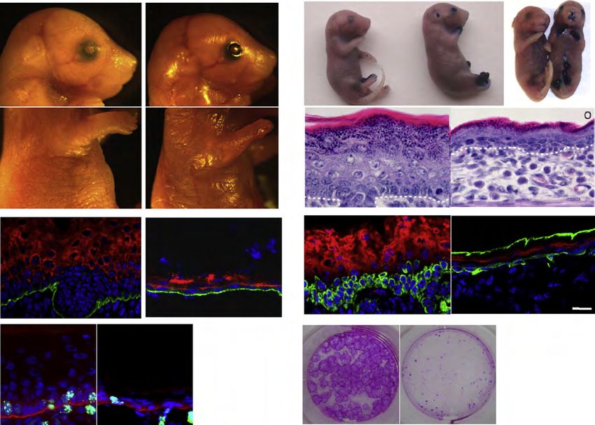
Acknowledgements

Most importantly, I would like to thank Assistant Professor Fernando Camargo for giving me
the great opportunity to work in his laboratory, for always being there for stimulating
discussions and providing guidance and support.

I would also like to thank Professor Petra Knaus. Without her support and commitment, my
PhD thesis abroad would not have been possible.

I thank my whole family and in particular my parents, Sigrid and Walter Schlegelmilch, who
have supported me in my decisions all my life. I am moreover truly grateful to my brother
Ingo Schlegelmilch for all his encouragement.

I am immensely grateful to Jan Riethmayer for supporting me in every possible way
throughout my PhD, it means so much to me and made all the difference.

I am especially thankful to have met my amiga Ingrid Carvacho during the time at Children’s
Hospital. Thanks so much for always being there for me and finding the right words. You
definitely brought sunshine into my days. I will miss our lunches.

I am also truly grateful to the two other members of my “lunch-group”, Alexander von Gise
and Mathias Pawlak for their friendship during my entire time in Boston, which means a lot to
me, not to mention all the opportunities to laugh and great scientific support.

I would also like to thank Grit Thurlow for her invaluable friendship and all the motivational
support during the time of my PhD and especially during the time of writing.

Great thanks goes also to all my Italian friends, for great culinary experiences and for making
my time in Boston more fun and special.

Thank you to all my friends all over the world, especially Cindy Bloch, Viola Rasch, Jasmina
Murad, Sandra Stehling-Sun, Joanna Himmel and Stefanie Städtler, who make this life so
much better.

I would like to thank the members of the Camargo laboratory for fruitful discussions, in
particular Jianlong Sun. And I am especially thankful for Azucena Ramos, Rosemarie de la
Rosa and Basanta Gurung for bringing so much fun to the lab and always being immensely
helpful.

I would like to thank Thijn Brummelkamp, Jan Pruszak and Oktay Kirak for all the helpful
discussions and a great collaboration.

Furthermore, I would like to thank Alexander von Gise, Azucena Ramos, Rosemarie de la
Rosa and Lindsay Herman for critical reading of this manuscript and valuable suggestions.

Sincere gratitude goes to my grandmother, Agate Schlegelmilch, a truly outstanding person,
for your boundless support throughout my life and sharing your wisdom with me. This is
dedicated to you.
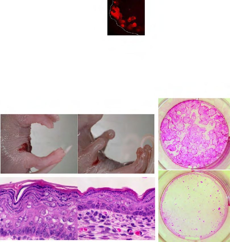
Table of Contents
1. Introduction..................................................................................................................... 4
1.1 The Hippo Signaling Pathway ...............................................................................................4
1.1.1 The Hippo Signaling Pathway Core Kinase Cascade ........................................................5
1.1.2 YAP....................................................................................................................................8
1.1.3 Downstream Regulators.....................................................................................................9
1.1.4 Upstream Regulators........................................................................................................10
1.1.5 The Physiological Role of Mammalian Hippo Signaling ................................................11
1.2 The Skin .................................................................................................................................12
1.2.1 Structure of the Skin ........................................................................................................12
1.2.2 Epidermal Stem Cells ......................................................................................................15
1.2.2.1 Heterogeneity of Epidermal Stem Cells .....................................................................16
1.2.2.2 Independent Stem Cell Populations in Hair Follicle and Interfollicular Epidermis...18
1.2.3 Cell Adhesion in the Skin ................................................................................................19
1.2.3.1 Adherens Junctions.....................................................................................................19
1.2.3.2 Key Cytoplasmic Regulators at the Adherens Junctions............................................21
1.2.3.3 α-Catenin’s Function in Cell Proliferation and Carcinogenesis .................................22
1.2.4 Hippo Signaling in the Skin .............................................................................................24
2. Aims................................................................................................................................ 25
Aim 1: Investigate the Effect of Yap1 Activation in the Epidermis ..............................................25
Aim 2: Investigate the Effect of Yap1 Deletion in the Epidermis .................................................26
Aim 3: Investigate the Function of the TEAD Transcription Factors in Epidermal Biology........26
Aim 4: Investigate the Function of the MST1/2 Kinases in Epidermal Biology ..........................27
3. MANUSCRIPT I........................................................................................................... 28
3.1 SCC-like Tumor Formation upon Yap1 activation is reversible .......................................51
3.2 Yap1 cKO skin grafts can be sustained ................................................................................52
4. Discussion........................................................................................................................ 53
4.1 YAP as a Critical Regulator of Epidermal Stem Cell Proliferation .................................53
4.2 YAP and Skin Cancers ..........................................................................................................55
4.3 A Connection of α-Catenin and the Hippo Pathway ..........................................................56
4.4 Regulation of YAP Activity by 14-3-3 and PP2A................................................................57
4.5 Crowd Control and the Hippo Pathway ..............................................................................57
4.6 YAP Regulates Cardiomyocyte Proliferation .....................................................................58
4.7 Tissue Specificity of the Hippo Pathway..............................................................................59
4.8 Future Directions ...................................................................................................................60
5. Summary......................................................................................................................... 64
6. Zusammenfassung.......................................................................................................... 66
7. Abbreviation List ........................................................................................................... 68
8. Eidesstattliche Erklärung.............................................................................................. 69
9. Bibliography ................................................................................................................... 70
Appendix .................................................................................................................................... i
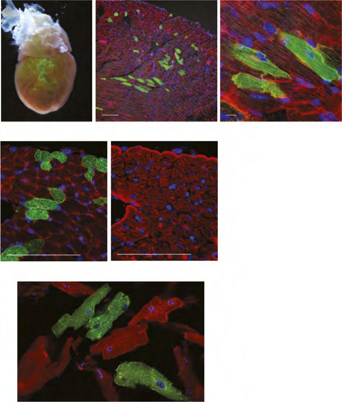
The Heart .............................................................................................................................................i
MANUSCRIPT II....................................................................................................................iii
Introduction

1.       Introduction

1.1      The Hippo Signaling Pathway

Looking at the different species of the animal kingdom, it is striking to see such an immense
variation in organism size. An elephant, for example, grows to be a million times larger than a
mouse. Additionally, the internal organs of an organism receive information telling them to
grow proportionally to the overall body size.          The mechanisms directing these growth
regulatory processes of organisms and organs fascinate biologists all over the globe, however,
they are still poorly understood. A better understanding of growth regulatory mechanisms
might ultimately lead to the development of better medical treatments for human diseases
associated with abnormal proliferation, such as cancer. An interesting organ in terms of
growth potential is the liver, which is able to regenerate its original mass, even when two-
thirds of it had been removed by hepatectomy. It is unclear, however, how it senses when to
stop growing (Michalopoulos and DeFrances, 1997).

Several studies were conducted to obtain more information about these signals and the results
suggest that the information regulating organ growth can be intrinsic to the organ as well as
extrinsic. When juvenile organs such as kidney, thymus, or the growth plate were transplanted
into adult recipients, these organs retained their growth potential until they had reached their
adult sizes, arguing for a growth-sensor intrinsic to the organ (Metcalf, 1963; Stevens et al.,
1999; Pape et al., 2006). In contrast, a long-standing theory suggests, that soluble factors
secreted by cells, so-called chalones, are responsible for organ growth (Bullough and
Laurence, 1964). Recent transplantation studies support the theory that information regulating
the growth process is obtained extrinsically through circulating factors. In an experiment
where livers were transplanted from small dogs to large dogs, the transplanted livers grew
until they had reached the size expected for the recipient (Kam et al., 1987). It had been
previously demonstrated that a hepatectomy performed in one rat joined to another rat by
parabiosis led to stimulation of hepatocyte proliferation in both animals (Moolten and Bucher,
1967). More recent parabiosis experiments connecting the vascular system of young and old
mice showed that factors of the young organism were able to increase the proliferation
capacity of muscle satellite cells and hepatocytes in the older mice (Conboy et al., 2005).


                                             4
Introduction

Stem cells are a prerequisite for organ growth and have been shown to affect the final size of
certain organs when their amount or activity is manipulated (Depaepe et al., 2005; Stanger et
al., 2007). Though numerous signaling molecules are known to regulate cell proliferation, it is
unclear how the information about organ size is integrated and translated to result in stem cell
proliferation and apoptosis. Genetic screens in Drosophila melanogaster have led to the
discovery of a novel signaling network, the Hippo signaling pathway, that is able to restrict
tissue growth in the fly by limiting cell proliferation and promoting apoptosis, thereby
determining organ size. Interestingly, it was demonstrated later that this pathway is highly
conserved in mammals, which led to novel discoveries regarding organ growth regulation in
mammals.

1.1.1    The Hippo Signaling Pathway Core Kinase Cascade

In genetic screens aimed at identifying tumor suppressor genes in Drosophila melanogaster,
the kinase Warts (wts) was identified in 1995 as the first component of the Hippo signaling
pathway (Justice et al., 1995; Xu et al., 1995). In subsequent studies three additional tumor
suppressor genes were identified: the kinase Hippo (Hpo), which gave the pathway its name,
and the scaffolding proteins Salvador (Sav) and Mob as tumor suppressor (Mats) (Kango-
Singh, 2002; Tapon et al., 2002; Harvey et al., 2003; Udan et al., 2003; Wu et al., 2003; Lai et
al., 2005). These four components form the core protein cascade of the Hippo signaling
pathway with the Hpo-Sav kinase complex phosphorylating and thus activating the Wts-Mats
kinase complex (Figure 1; Wu et al., 2003; Huang et al., 2005; Wei et al., 2007).

The downstream effector of this pathway, the transcriptional coactivator Yorkie (Yki) was
discovered in a yeast two-hybrid screen for Wts-interacting proteins (Huang et al., 2005). It
was demonstrated that Wts directly phosphorylates Yki on serine residue S168 leading to 14-
3-3 protein binding, which renders Yki inactive in the cytoplasm (Dong et al., 2007; Zhao et
al., 2007; Oh and Irvine, 2008). In its unphosphorylated state, the transcriptional coregulator
Yki translocates to the nucleus, where it interacts with the transcription factor Scalloped (Sd)
(Wu et al., 2008; Zhang et al., 2008b). Loss-of-function mutations for Hpo, Sav, Wts and
Mats cause strong tissue overgrowth phenotypes in the fly, that are characterized by enhanced
proliferation and reduced apoptosis (Kango-Singh, 2002; Tapon et al., 2002; Harvey et al.,


                                             5
Introduction

2003; Pantalacci et al., 2003; Udan et al., 2003; Wu et al., 2003; Lai et al., 2005). Similar
phenotypes are observed in gain-of-function mutations for Yki, in line with Yki being the
downstream target of the Hippo cascade (Huang et al., 2005).

In mammals the core set of Hippo signaling components is highly conserved. The mammalian
orthologs MST1/2 (Hpo), SAV1 (Sav), LATS1/2 (Wts), MOB1 (Mats) and YAP/TAZ (Yki)
form a kinase cascade analogous to their fly counterparts (Figure 1; Chan et al., 2005; Zhao et
al., 2007). In mammalian cells nuclear YAP induces growth, as observed for Yki in the fly
(Zhao et al., 2007). The evolutionary conservation is further underscored by the finding that
human MST2, MOB, LATS1 and YAP are able to rescue the loss-of-functions mutant
phenotypes of their corresponding Drosophila counterparts in vivo (Tao et al., 1999; Wu et
al., 2003; Huang et al., 2005; Lai et al., 2005).

Figure 1 presents an overview of the Hippo signaling pathway in Drosophila melanogaster
and mammals. The Sterile 20-like kinases MST1 and MST2 and their regulatory protein
SAV1 (also known as WW45) interact to form an activated complex. In mammalian cells the
Ras association domain family (RASSF) proteins can activate MST1/2 (Khokhlatchev et al.,
2002; Oh et al., 2006; Guo et al., 2007; 2011). Interestingly, in Drosophila, dRASSF instead
inactivates Hpo (Polesello et al., 2006). Activated MST1/2 can directly phosphorylate and
thus activate the large tumor suppressor homolog kinases LATS1 and LATS2 and their
regulatory proteins MOB (MOBKL1A/B) (Chan et al., 2005; Dong et al., 2007; Hirabayashi
et al., 2008; Chow et al., 2010). Activated LATS1/2 phosphorylate the transcriptional
coactivators YAP at S127 and TAZ at S89 in humans, or S112 and S87 in mouse YAP and
TAZ respectively. Phosphorylation at these residues leads to the cytoplasmic retention and
thus inactivation of YAP and TAZ through 14-3-3 binding (Dong et al., 2007; Zhao et al.,
2007; Hao et al., 2008; Lei et al., 2008; Oh and Irvine, 2008; Oka et al., 2008; Zhang et al.,
2008a). When unphosphorylated, YAP and TAZ translocate to the nucleus where they interact
with different transcription factors, many of which have been implicated in stem cell biology
(Mauviel et al., 2011). However, the group of transcription factors that has been described
best to mediate YAP and TAZ activities belongs to the TEA-domain family (TEAD, also
known as transcription enhancer factors (TEFs), which consists of four family members
TEAD1-4 (Zhao et al., 2008; Zhang et al., 2009a).


                                                  6
Introduction

         50
                                                          !"#$#%&'()                                                                  '%BB%C0
                                                                        K)+                                                                         K)+
       56.     $%&               $H                                                                  O

                                                                                                                                                            D%C09
                     FG&

                                                                                12&

                                                                                                                                                               DJL3
                                                     '()                                                             $I'5           P$Q

                                                                                  DJL3
                                            >E                                                                                               !%-ML%N                  L3
                                                                                                                     @@@O
                                                                                                                                                    J'VL
                                                 *#+)%                                                                      *#+)%

                                                                 2IJ11$
         5%670
                                                                                                                                             IJ11$

                                             ,-.                                                                          '0&9MQ
                                      1%8                                                                          1%89

                                                         '%&0                                                                       '.+
        234+                                 /&0                      9:                         JH4+%                    F%&09MQ               9:
                                                                         ;
Introduction

1.1.2    YAP

The Yes-associated protein (YAP) was originally cloned as an interaction partner of the non-
receptor tyrosine kinase c-Yes, which YAP binds at the SH3 domain. Today, YAP is
primarily recognized as the major downstream target of the Hippo signaling pathway (Sudol,
1994; Sudol et al., 1995). The gene for Yap1 is located on the human chromosome 11q22
locus. The Yap1 gene generates two major protein isoforms, YAP1 (454aa) and the longer
isoform YAP2 (488aa), which differ only in the number of WW domains. YAP1 contains one
WW domain whereas YAP2 contains two. In this work YAP will be used to describe
YAP1/YAP2, and the specific YAP isoforms will be indicated if applicable. Figure 2 shows
the human YAP2 protein and its structural domains. The WW domains are important for the
interaction between YAP and LATS1/2. The serine residue S127 gets phosphorylated by
LATS1/2, and serves as a binding site for 14-3-3 proteins (Basu et al., 2003; Zhao et al.,
2007). LATS1/2 also phosphorylate the serine residues S61, S109, S164, and S381 of YAP2
(Zhao et al., 2010).

Of these serine residues, two are known to be important for YAP inactivation. One is S127,
whose phosphorylation results in the cytoplasmic localization of YAP, rendering it inactive
through spatial regulation. The other is S381 (YAP2), where LATS1/2 phosphorylation
causes casein kinase 1 (CK1δ/ε) to phosphorylate two additional serine residues (S384 and
S387) located within a phosphodegron sequence. Activation of the phosphodegron results in
the ubiquitination of YAP by the SCFβ-TRCP E3 ubiquitin ligase, similar to that observed for β-
catenin. Ubiquitination targets YAP for degradation, thus providing a means for temporal
regulation (Liu et al., 2002; Fuchs et al., 2004; Zhao et al., 2010). It is not only serine residues
that play a role in the regulation of YAP. One study showed that the phosphorylation of YAP
on Tyrosine Y357 (YAP1) or Y391 (YAP2) by c-Abl in response to DNA damage leads to
YAP stabilization and promotes p73-mediated apoptosis. This indicates a possible mechanism
limiting the oncogenic potential of YAP under certain circumstances (Levy et al., 2008).


                                               8
Introduction

                     5641,-$./$.0                          5#7.87%9$:79$;.

                                             )*+,-$./$.0

                     ! ! !         ! !                               ! ! !          !

       34! !"#$%&                            '' ''         ((                              BCC,77
                    )? )=@A )=
Introduction Several studies using gene expression profiling have revealed a set of target genes that are induced by YAP (Dong et al., 2007; Zhao et al., 2007; Hao et al., 2008; Ota and Sasaki, 2008; Lu et al., 2010). Some of these genes, such as the IAP family member BIRC5 and the EGF family member amphiregulin (AREG), have been confirmed in vitro to mediate YAP- activated growth (Dong et al., 2007; Zhang et al., 2009b). However, the best-characterized direct target gene of YAP and TAZ is the connective tissue growth factor (CTGF). CTGF contains several TEAD-binding elements in its promoter region and was shown to mediate YAP-TAZ/TEAD-dependent proliferative and oncogenic functions. Microarray analysis identified an additional member of the CCN family, cystein-rich protein (Cyr61), as a target of YAP/TEAD (Zhao et al., 2007; 2008; Zhang et al., 2009a; Lai et al., 2011). Interestingly, CTGF and Cyr61 were found to be highly expressed in human cancers, where normal growth- restricting mechanisms are lost (Xie et al., 2001). 1.1.4 Upstream Regulators Intense research over the past years established a relatively well-defined kinase cascade at the core of the Hippo pathway. However, the upstream signals, regulating this core kinase cascade, are not well defined and the subject of ongoing research. Most of what is currently known about the upstream signals was discovered in Drosophila. The first upstream regulators to be identified were the FERM domain containing apical cytoskeleton-binding proteins Merlin (Mer) and Expanded (Ex) (Hamaratoglu et al., 2006; Pellock et al., 2007). The deletion of both Mer and Ex led to tissue overgrowth phenotypes, similar to those observed by the deletion of core kinase components Hpo, Sav and Wts. Consistent with this observation, coexpression of Mer and Ex led to an activation of the pathway through an increase in Wts phosphorylation (Hamaratoglu et al., 2006). The protein Kibra was later shown to also interact with Mer and Ex, resulting in activation of the pathway (Baumgartner et al., 2010; Genevet et al., 2010; Yu et al., 2010). A milestone was the identification of the atypical cadherin Fat as the first transmembrane receptor protein signaling to the Hippo pathway (Bennett and Harvey, 2006; Cho et al., 2006; Hariharan, 2006; Silva et al., 2006; Willecke et al., 2006; Tyler and Baker, 2007). Fat functions by interacting with another atypical cadherin, Dachsous (Ds), on the adjacent cell. Other proteins shown to regulate Fat activity are the casein kinase Disc overgrown (Dco), the Golgi-resident kinase Four-jointed 10
Introduction

(Fj) and the Fat/Ds-interacting protein Lowfat (Lft) (Matakatsu and Blair, 2006; Feng and
Irvine, 2007; Ishikawa et al., 2008; Rogulja et al., 2008; Willecke et al., 2008; Mao et al.,
2009; Sopko et al., 2009; Simon et al., 2010). Fat regulates the Hippo pathway by influencing
expression and localization of Wts and Ex through mechanisms that involve the
unconventional myosin Dachs (Bennett and Harvey, 2006; Cho et al., 2006; Silva et al., 2006;
Willecke et al., 2006; Feng and Irvine, 2007; Tyler and Baker, 2007). In the search for
additional cell surface proteins regulating the Hippo pathway, the apical transmembrane
protein Crumbs (Crb) was recently discovered. This protein is important for the regulation of
apical-basal cell polarity (Chen et al., 2010; Grzeschik et al., 2010; Robinson et al., 2010).

Even though mammalian homologs exist for most of the upstream components described for
Drosophila, the significance and relevance of these proteins in regulating the activity of YAP
in vivo in mammals remains poorly understood. An exception is the Neurofibromatosis 2 gene
product (NF2), the Mer homolog. The deletion of NF2 in vivo in the murine liver leads to
tumor formation, while additional deletion of one Yap1 allele largely rescues the overgrowth
phenotype. This suggests that both genes reside within the same pathway (reviewed in
McClatchey and Giovannini, 2005; Zhang et al., 2010a).

1.1.5    The Physiological Role of Mammalian Hippo Signaling

Progress has been made in the quest to understand the physiological role of the Hippo
signaling pathway components in mammals. Identifying the essential function of YAP in cell
proliferation came from a study showing increased YAP expression levels in embryonic stem
cells (Ramalho-Santos et al., 2002). It was later shown that deletion of Yap1 in mice led to
developmental defects that resulted in embryonic lethality at E8.5 (Morin-Kensicki et al.,
2006). The phenotype for Taz (also called Wwtr1)-null mice is less drastic with embryos
being viable at birth (Hossain et al., 2007; Tian et al., 2007; Makita et al., 2008). However,
when Yap1 and Taz are both deleted, anomalies occur during pre-implantation development
and embryos die before the morula stage (Nishioka et al., 2009).

Great efforts have been made to elucidate the role of mammalian Hippo signaling in organ
size regulation of the liver, the body’s organ with the highest regeneration potential. In mice,


                                             11
Introduction

over a four-fold increase in liver size (hepatomegaly) was observed when the constitutively
active YAP1-S127A was inducibly expressed. Remarkably, after ending the ectopic
expression, the liver reversed to its original size (Camargo et al., 2007; Dong et al., 2007).
Hepatomegaly also occurred when Mst1/2 or Sav1 were lost (Zhou et al., 2009; Lu et al.,
2010; Song et al., 2010). However, it remains uncertain to what extent the discovered
functional relationships of the Hippo signaling transduction in the liver are conserved and
active in other tissues (Zhou et al., 2009). Stem cells are essential for organs to reach their
final size. Interestingly, YAP expression in the intestine and in the epidermis was found to be
restricted to the compartment that comprises the tissue resident stem cells (Camargo et al.,
2007).

1.2      The Skin

The skin is the largest organ in mammals, covering the entire body surface (in humans an
average of 1.5-2 m2) and forming a barrier between the organism and its surrounding
environment. One of its various functions is to protect the organism against environmental
stresses, such as pathogens, UV radiation, chemical and mechanical stress. Other functions
are the regulation of hydration and temperature of the body (Chuong et al., 2002; Blanpain
and Fuchs, 2006; Segre, 2006). The skin serves an important role beyond the barrier-function.
It provides a tactile sense, which enables the animal to obtain information about the
environment. Additionally, it is important for social interactions between animals, as a form
of communication via pigment pattern, hair and fur (Blanpain and Fuchs, 2006). Moreover, it
plays an important role for immunologic, endocrine and metabolic processes (Chuong et al.,
2002). Many appendages are formed in the mammalian skin, which include hair follicles,
nails, sebaceous (oil) and sweat glands, all of which are crucial for its functions.

1.2.1    Structure of the Skin

In mammals the skin comprises the epidermis, the dermis and the subcutis (or hypodermis).
Figure 3 shows the structure of the skin. The innermost layer of the skin, the subcutis, consists


                                              12
Introduction

of adipocytes and lies below the dermis (Montagna and Parakkal, 1974; Alberts et al., 2008).
Right above the subcutis lies the dermis, a mesenchymal connective tissue. The dermis is
composed of fibroblasts and extracellular matrix (ECM) components (e.g. collagen and elastic
fibers). The subcutis and dermis contain many blood vessels and nerves, and hence, provide
not only stability but also nutrients for the outermost layer of the skin, the epidermis. The
cross talk between dermis and epidermis is critical for proliferation and morphogenesis of the
epidermal compartment (Sengel, 1986; Chuong, 1998). The epidermis is a multilayered
(stratified) and highly specialized epithelium that is separated from the underlying dermis by
the basement membrane (BM). It comprises the interfollicular epidermis (IFE), which
describes the epidermis in between the hair follicles. The IFE contains as associated
appendages hair follicles (HFs), sebaceous glands (SGs) as well as sweat glands (Figure 3).

Figure 3: Structure of the Skin.
The skin consists of the epidermis, dermis and subcutis. Appendages of the interfollicular epidermis (IFE) are
hair follicles, sebaceous glands and sweat glands. Modified after Aasen et al. 2010.

The basal layer of the IFE contains the interfollicular proliferative cells, which eventually
withdraw from the cell cycle and commit to terminal differentiation (Koster and Roop, 2007).
In the process of building the multilayered epidermis (Figure 4), the basal cells detach from
the BM and move upwards, through different stages of differentiation (Fuchs, 2008). The first


                                                     13
Introduction

suprabasal differentiated layer is the spinous layer, containing transcriptionally active cells
that produce keratin filaments connected to desmosomes, laying the groundwork for the
mechanical infrastructure of the suprabasal epidermis. The process of the basal-spinous
transition is regulated by various signals, for example the switch from keratin K14/K5
expression to keratin K1/K10 expression (Fuchs and Green, 1980; Stoler et al., 1988; Yuspa
et al., 1989; Bickenbach et al., 1995). Right above the spinous layer follows the granular
layer. The cells of the granular layer produce lipid-rich lamellar granules and contribute to
establishing the cornified barrier function of the epidermis. Finally, the outermost protective
layer of the epidermis, the stratum corneum, consists of enucleated, dead cells that are ready
to be shed (Koster and Roop, 2007; Fuchs, 2008).

Figure 4: Structure of the Epidermis.
Schematic showing the different layers of the epidermis. The epidermis is separated from the dermis by the
basement membrane (BM). The basal layer comprises the progenitor cells of the epidermis and expresses keratin
5 (K5) and keratin 14 (K14). The basal cells eventually withdraw from the cell cycle and enter a differentiation
program, detaching from the BM and moving upwards. The BM expresses integrin-β4 (β-int4), a component of
hemidesmosomes. The first differentiated layer above the basal layer is the spinous layer, followed by the
granular layer. The spinous layer expresses keratin 1 (K1) and keratin 10 (K10), while the granular layer
expresses loricrin. Modified from Alonso and Fuchs 2003.


                                                     14
Introduction

One of the epidermal appendages is the hair follicle (HF), which is composed of the outer root
sheath (ORS), companion layer, inner root sheath (IRS), hair shaft and the rapidly
proliferating matrix cells in the hair bulb, which give rise to the different hair lineages
(Niemann and Watt, 2002; Blanpain and Fuchs, 2006; Shimomura and Christiano, 2010). The
best-characterized multipotent hair follicle stem cells have been shown to reside within the
permanent part of the HF, the so-called bulge (Cotsarelis et al., 1990; Nowak et al., 2008).
Whereas under homeostatic conditions regeneration of the IFE is a continuous process, the
hair follicle undergoes cycles of growth (anagen), apoptosis-mediated regression (catagen),
and quiescence (telogen) (Alonso and Fuchs, 2006).

Another epidermal appendage is the sebaceous gland (SG), which is attached to the HF. The
SG contains an undifferentiated layer of keratinocytes at the periphery of the gland and
terminally differentiated sebocytes in the center. Mature sebocytes are lipid-filled and
eventually burst, releasing their contents onto the surface of the skin (Niemann, 2009;
Schneider et al., 2009).

1.2.2    Epidermal Stem Cells

Similar to the gastrointestinal and hematopoietic systems, the epidermis is a rapidly renewing
tissue, which makes it ideal for studying stem cell biology. Characteristics by which stem
cells (SCs) are being identified are their capability of unlimited or prolonged self-renewal,
their proliferation potential, their long-term repopulation capacity as well as their potential to
generate at least one type of highly differentiated cell type (Lajtha, 1979; Hall and Watt,
1989; Morrison et al., 1997; Potten, 1997; He et al., 2009).

Epidermal SCs have been shown to be multipotent, and are needed for building the epidermis
during development (Watt, 1998). One single adult skin SC has the proliferative capacity to
produce epidermis covering the complete body surface (Rochat et al., 1994). But SCs are also
important in adult life, for the process of continual and balanced cell replacement, called
tissue homeostasis, which is critical for maintaining an intact adult epidermis. Human
epidermis is thicker compared to murine epidermis and has a frequent cell turn-over, self-
renewing within four weeks (Blanpain et al., 2007; Pincelli and Marconi, 2010). In mice, the


                                             15
Introduction

       proliferation rate slows down in the postnatal epidermis as the hair coat develops and takes
       over most of the protective functions (Fuchs, 2008). In the epidermis stem cells have been
       found to reside in the bulge area of the HF, the IFE and the SG (Figure 5).

                                                                ()*

                                              ,7

               0123+45%%26%'
                                      #$%&'

                                                ()*+,-'.+/'%%
                                                #$%&'+,-'.+/'%%
                                                ,7+,-'.+/'%%

                        !"     #$%8
                                                #191%+/'%%

       Figure 5: Stem Cells in the Epidermis.
       Stem cells in the epidermis are located in the interfollicular epidermis (IFE), the sebaceous gland (SG) and the
       bulge region of the hair follicle (HF). The bulb of the hair follicle is in contact with the dermal papilla (DP).

       1.2.2.1 Heterogeneity of Epidermal Stem Cells

       In the quest for the identification of epidermal stem cells, lineage-tracing and label-retaining
       studies utilizing the characteristic of these cells to be slow-cycling brought some important
       answers. By injecting nucleotide analogs (BrdU or 3H-thymidine) at a time when the
       epidermis is actively proliferating and thus incorporating the label, it was now possible to
       trace the cells that were retaining the label over a long time period, which were considered to
       be the stem cells (label-retaining cells, LRCs) (Potten, 1974; Bickenbach, 1981; Morris et al.,
       1985; Potten and Morris, 1988; Braun and Watt, 2004; Fuchs, 2009). It became clear in these
       label-retaining studies that the epidermis contains at least two distinct LRC populations with a
       different cell cycle duration and moreover a non-label-retaining, post-mitotic population.


                                                             16
Introduction

Accordingly, the most widely accepted model for the explanation of cell replacement in the
epidermis is that of the epidermal proliferative unit (EPU), which implies proliferative
heterogeneity (Mackenzie, 1969; 1970; Allen and Potten, 1974; Potten, 1974). Each EPU
contains in its center of approximately 10 to 11 basal cells one slow-cycling and thus long-
term label-retaining cell (SC), which gives rise to SC daughters, as well as rapidly
proliferating daughter cells which are termed transit amplifying (TA) cells (Bickenbach,
1981; Potten, 1981; Cotsarelis et al., 1989; Pincelli and Marconi, 2010). Furthermore the EPU
contains three flattened suprabasal cells and five to seven cornified cells, making a total of 18
to 21 cells per EPU (Potten, 1974; Mackenzie, 1975; Ghazizadeh and Taichman, 2001).

The finding, that infrequently dividing epidermal SCs produce frequently proliferating TA
daughters that eventually start to differentiate after a limited number of cell divisions, has
been supported by studies analyzing the cell kinetics in the epidermis (Potten, 1974; Jones et
al., 1995; Fuchs and Horsley, 2008). It has been shown that in the epidermis cell division is
restricted to the cells in the basal layer, but there are observations that not all of these cells
capable of dividing are in fact stem cells (Morris et al., 1985). About 10% of the basal cells
are assumed to be stem cells, which has been concluded from a study in which murine
epidermis had been severely damaged by radiation and the number of basal cells with a
clonogenic potential to renew the epidermis was quantified (Withers, 1967; Potten and
Hendry, 1973). This leaves an additional 50% of dividing basal cells, which might represent
the TA population, whereas post-mitotic cells make up the remaining 40% of basal cells
(Iversen et al., 1968; Potten and Morris, 1988). A similar percentage of stem cells in murine
epidermis was also confirmed with in vitro studies looking at the colony-forming efficiency
and showed that the frequency of clonogenic keratinocytes in culture lies between 2% and 8%
(Morris et al., 1988; Bickenbach and Chism, 1998).

The hierarchy of epidermal stem cells has also been supported by clonogenic assays with
human epidermal cells, where in vitro replating-experiments have identified three types of
keratinocyte clones: holoclones (28%), paraclones (49%) and meroclones (23%) (Barrandon
and Green, 1987). Holoclones have the highest proliferation potential in long-term culture,
give rise to the largest colonies and are thought to arise from stem cells. Paraclones have a
more limited growth potential (approximately 15 cell generations), form smaller colonies that


                                             17
Introduction

ultimately differentiate and are thus assumed to represent the TA cells. Meroclones are an
intermediate between holoclones and paraclones in terms of their appearance and their
reproductive behavior (Barrandon and Green, 1987).

Recently the EPU model with its stem cell heterogeneity has been questioned by lineage-
tracing experiments showing that adult epidermis is instead maintained by a single population
of progenitors (Clayton et al., 2007). It has to be taken into account, however, that this study
was contemplated in murine tail epidermis, whereas most previous studies have been done
either in murine back epidermis or cultivated primary human keratinocytes. Regional as well
as age-related differences might be reflected in the varying results. The tail epidermis, for
example, not only contains fewer HFs but is also organized in squames or scales, and thus it
cannot be ruled out that there might be a different underlying system of stem cell proliferation
in this particular region.

1.2.2.2 Independent Stem Cell Populations in Hair Follicle and Interfollicular
Epidermis

Already in the 1960s, it could be demonstrated that the HF completely regenerated after
surgical removal of the bulb region of the HF, indicating that the SCs are not located in this
region (Oliver, 1966a; 1966b). However, the hair bulb does contain the so-called matrix cells,
which are highly proliferative cells (TA cells) that give rise to the hair and IRS. Label-
retaining studies in the murine HF made it clear that the SCs of the HF, which are responsible
for the cyclic regeneration of the HF, are located in the bulge region (Figure 5 ; Cotsarelis et
al., 1990; Rochat et al., 1994; Oshima et al., 2001). It was calculated from subsequent LRC
studies that approximately 95% of the slow-cycling epidermal LRCs reside in the bulge
(Braun et al., 2003; Blanpain et al., 2004; Tumbar, 2004). Moreover, cells isolated from the
bulge of rat as well as human HFs by microdissection showed the highest colony-forming
efficiency (Rochat et al., 1994; Oshima et al., 2001).

It was assumed for a while that the SCs in the HF bulge constitute the true SCs of the
epidermis, giving rise to all the different lineages of the epidermis. And while indeed it is
confirmed that HF SCs can regenerate the complete epidermis with all its lineages upon


                                             18
Introduction

injury, it has also been demonstrated that under homeostatic conditions the HF SCs do not
contribute to the maintenance of the IFE (Braun et al., 2003; Tumbar, 2004; Levy et al., 2007;
Langton et al., 2008). Additionally, evidence for a long-term populating interfollicular SC
came from in vivo lineage-tracing studies (Ghazizadeh and Taichman, 2001; Niemann and
Watt, 2002; Schneider et al., 2003; Ghazizadeh and Taichman, 2005; Ito et al., 2005; Langton
et al., 2008). An interesting study demonstrates that upon loss of HFs, the survival of the
adjacent IFE was not affected (Ito et al., 2005). These studies provide strong evidence for an
independent SC population in the IFE, albeit this population is not well characterized to date.

1.2.3     Cell Adhesion in the Skin

The formation of a multi-cellular organism of higher complexity, with various tissues and
organs, requires the interaction of different cell types. A close interaction between adjacent
cells through intercellular junctions is a pre-requisite for achieving this goal. Intercellular
junctions play a crucial role in epithelial tissues such as the skin, since they provide the
necessary stability that is needed for tissue morphogenesis and also for body movement. They
are essential for the proper physiology and barrier function of the skin (Niessen, 2007). The
epidermis is a dynamic tissue with its cells moving upwards in their differentiation process
during homeostatic self-renewal. And also during development, organ growth, and wound
repair, adhesive contacts need to get remodeled. Therefore, intercellular junctions face a
special challenge. They need to ensure the adhesive properties necessary for epithelial sheet
and barrier formation, but at the same time they need to be adaptive and ready to rearrange to
allow for the dynamic changes in cell adhesion (van Roy and Berx, 2008).

1.2.3.1 Adherens Junctions

Three of the junctional complexes that are important for proper epithelial sheet formation are
tight junctions (TJs), adherens junctions (AJs) and desmosomes (Matter and Balda, 2003;
Perez-Moreno et al., 2003; Yin and Green, 2004).
  Together they form the apical junctional
complex (Figure 6), which regulates cell polarity, permeability and adhesion (Farquhar and
Palade, 1963).


                                            19
Introduction

Most apically located within the vertebrate cell are the TJs (zona occludens), which serve two
main functions. They seal neighboring cells closely together and function as a selective
permeability barrier (limiting free diffusion of molecules). In addition they also ensure the
separation of apical and basolateral membrane components, which is their second so-called
“fence function” (D'Atri and Citi, 2002; Tsukita and Furuse, 2002). Desmosomes play,
similar to AJs, an important role for intercellular adhesion, but they are located basolateral
within the cell and provide anchorage sites for intermediate filaments, which belong to the
keratin family in most epithelial cells (Garrod et al., 2002; Huber, 2003; Garrod and Chidgey,
2008).

Figure 6: Intercellular Junctions.
Tight junctions (TJs), adherens junctions (AJs) and desmosomes form the apical junctional complex. Figure
from Kobielak and Fuchs 2004.

AJs are positioned directly below the TJs and are as their name already suggests crucial for
cell-cell adhesion (Tepass, 2002; Perez-Moreno et al., 2003). In epithelial tissues, such as the
skin, AJs provide a connection to the cytoskeleton of the cell, forming a continuous adhesion
belt. Lately functions for AJs have been identified that go beyond their mere role in securing
tissue integrity. A view of AJs as a hub for signaling molecules and polarity cues is emerging,
which make them a likely candidate for sensing and relaying information during tissue growth
(Nelson and Nusse, 2004; Perez-Moreno and Fuchs, 2006). Indeed, there is evidence
emerging that AJs play a role in restricting basal cell proliferation and in sensing epidermal
cell density (Lien et al., 2006a).


                                                 20
Introduction

The core components of the AJ complex are the cadherin adhesion molecules (Takeichi,
1990). In epithelial tissues E(epithelial)-cadherin is prevalent, which like all cadherins,
contains two or more extracellular cadherin domains that form a homotypical Ca2+-dependent
interaction with the E-cadherins of the neighboring cell (Leckband and Prakasam, 2006;
Pokutta and Weis, 2007). While their extracellular domains ensure the adhesion to the
adjacent cell, their conserved cytoplasmic tail interacts with a set of cytoplasmic proteins, the
catenins, β-catenin, p120-catenin, and α-catenin, that build the bridge to the actin cytoskeleton
(Perez-Moreno and Fuchs, 2006; Pokutta and Weis, 2007; Nishimura and Takeichi, 2009).

1.2.3.2 Key Cytoplasmic Regulators at the Adherens Junctions

The catenins, which form the connection to the cytoplasmic E-cadherin domains, are very
important for the function of the AJs since they are ensuring a stable intercellular adhesion by
providing the link to the actin cytoskeleton. While this structural role of the catenins is well
established, recent studies have shown that they also serve an additional function as signaling
molecules (Perez-Moreno and Fuchs, 2006). The best-studied example is β-catenin, with its
dual role in cell adhesion and as a transcriptional coactivator in the Wnt signaling pathway
(Nelson and Nusse, 2004; Bienz, 2005). But also for p120 catenin and α-catenin studies are
revealing additional roles in cellular processes that go beyond cell-adhesion (Vasioukhin et
al., 2001; Reynolds and Roczniak-Ferguson, 2004). Another component of the AJs is
plakoglobin (also called γ-catenin).

While p120-catenin, β-catenin and plakoglobin belong to the armadillo family of proteins and
share sequence similarity, α -catenin differs notably in both sequence and structural
organization. p120-catenin, β-catenin and plakoglobin bind directly to the cytoplasmic
domain of E-cadherin, whereas α -catenin joins the complex via binding to β -catenin or
plakoglobin and providing the link to the actin cytoskeleton (Figure 7). The binding of α -
catenin to β-catenin and actin has been shown to be mutually exclusive (Drees et al., 2005;
Yamada et al., 2005).


                                             21
Introduction

Figure 7: Structure of the Adherens Junctions.
p120-catenin and β-catenin/plakoglobin directly bind to the cytoplasmic domain of E-cadherin. α-catenin binds
to β -catenin or plakoglobin and provides the link to the actin cytoskeleton through different actin binding
proteins.

1.2.3.3 α-Catenin’s Function in Cell Proliferation and Carcinogenesis

The α -catenin protein family contains three different forms. The founding member α -E-
catenin (hereafter α-catenin) is expressed in epithelial tissues such as the skin (Nagafuchi and
Takeichi, 1989; Ozawa et al., 1989). The other forms are α-N-catenin, which is restricted to
neural tissues and α-T-catenin, which is predominantly found in the testis and the heart.

Genetic studies in mice have demonstrated the requirement for α -catenin during
embryogenesis at the blastocyst stage. Loss of α -catenin results in an embryonic lethal
phenotype, similar to that observed upon E-cadherin loss (Larue et al., 1994; Kofron et al.,
1997; Torres et al., 1997). Moreover, it was shown that α-catenin is essential for the proper
function of AJs in epithelial cells in vitro and in vivo and in fact serves an important function
in epidermal biology (Vasioukhin et al., 2000; 2001). Whereas β -catenin has long been
acknowledged to play dual roles in cell adhesion and as a signaling molecule, the same has
only recently become recognized for α -catenin as well (Nelson and Nusse, 2004). The
conditional deletion of α-catenin in the developing epidermis on embryonic day 14.5 led to an
impairment in hair follicle development and epidermal morphogenesis (Vasioukhin et al.,


                                                  22
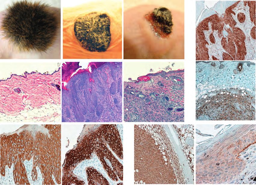
Introduction

2001). Intercellular adhesion through AJs was compromised, even though the AJ components
E-cadherin and β -catenin were still localizing correctly to the cell membrane. Surprisingly,
the conditional loss of α -catenin in the epidermis also led to hyperproliferation in the
epidermis. When plated in vitro, α -catenin-null keratinocytes were poorly contact inhibited
and showed a high proliferation rate (Vasioukhin et al., 2001). These results suggest that α-
catenin regulates stem cell proliferation, and demonstrate an inverse correlation of adhesion
and proliferation.

The implication for AJs in proliferative processes is further supported by observations
showing that mutations in AJ-proteins not only result in tissue degeneration, but they also
lead to tumor formation and metastasis, processes where proliferation has gone awry (Yagi
and Takeichi, 2000; Tepass, 2002; Perez-Moreno et al., 2003). It is known by now that a
reduction of α-catenin and also E-cadherin levels in a variety of tissues contributes to tumor
formation in humans (Kadowaki et al., 1994; Ochiai et al., 1994; Shiozaki et al., 1994; Ewing
et al., 1995; Rimm et al., 1995; Kozyraki et al., 1996). Null mutations of α-catenin are also
frequently found in epithelial cancers, where they correlate more highly with the degree of
tumor invasiveness than those of E-cadherin (Shimoyama et al., 1992; Morton et al., 1993;
Kadowaki et al., 1994; Matsui et al., 1994; Ewing et al., 1995; Bullions et al., 1997). The
function of α -catenin as a tumor suppressor was moreover demonstrated in the skin by a
transplantation experiment, showing that the grafting of α-catenin-null skin onto Nude mice
led to the formation of squamous cell carcinoma (SCC) (Kobielak and Fuchs, 2006). The
results obtained upon α -catenin ablation are making it necessary to reconsider the existing
view that defects in intercellular adhesion are occurring later in the process of carcinogenesis,
and instead suggest that they are earlier steps than previously assumed. The mechanisms how
exactly α-catenin mediates these processes are unclear.


                                             23
Introduction

1.2.4   Hippo Signaling in the Skin

The epidermis is a rapidly renewing tissue. Epidermal stem cells are essential for both skin
homeostasis and regeneration during wound repair. Aberrant proliferation in the epidermis
leads to skin diseases such as psoriasis and epidermal cancer formation. Therefore, it is
important to understand the mechanisms that control proliferation in the epidermis. The
Hippo signaling pathway has been shown to be involved in the regulation of epidermal
proliferation by one previous study. In this study, loss of Sav1 (WW45), a core component of
the Hippo cascade, in the epidermis induced an expansion of the basal progenitor cell
population (Lee et al., 2008). Moreover, the phosphorylation of YAP, the regulatory
downstream event rendering YAP inactive in the cytosol, could no longer be observed in vitro
in cultured Sav1-null keratinocytes, implying an effect on YAP activity (Lee et al., 2008).
Interestingly, YAP has been shown to be expressed in the basal layer of the epidermis, the
compartment that comprises the tissue resident stem and progenitor cells (Camargo et al.,
2007). Future research will have to elucidate how the Hippo signaling pathway controls tissue
homeostasis and tissue growth in the epidermis.


                                           24
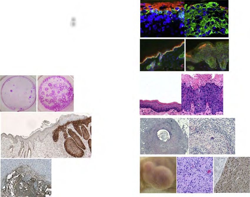
Aims

2.       Aims
Research in recent years has begun to discover a role for the Hippo signaling pathway in the
regulation of organ size not only in Drosophila but also in mammals. In the murine liver,
constitutive activation of the pathway’s downstream effector, YAP, caused a more than four-
fold increase in liver size (Camargo et al., 2007; Dong et al., 2007). However, little is known
about the endogenous mechanisms that provide information about organ size and relay this
information to the organ’s resident stem cells.

Interestingly, YAP has been found to be expressed in stem cell compartments of the intestine
and also of the epidermis (Camargo et al., 2007). Moreover, the deletion of one of the core
cascade components, Sav1, resulted in an expansion of progenitor cells in the epidermis (Lee
et al., 2008). Based on these results, the aim of this thesis was to further investigate the
function of Hippo signaling in the epidermis, with a focus on YAP. Understanding the
function of this novel pathway in the epidermis could prove therapeutically relevant for
developing treatments for skin diseases associated with aberrant proliferation (e.g. psoriasis)
as well as skin cancers. Moreover, it could ultimately lead to an advancement of current skin
transplantation therapies.

Aim 1:
Investigate the Effect of Yap1 Activation in the Epidermis

The deletion of core proteins of the Hippo pathway in Drosophila produces phenotypes
similar to that obtained upon Yki (YAP homolog)-overexpression. In the liver activation of
Yap1 led to an increase in liver size (Camargo et al., 2007; Dong et al., 2007). Based on this
finding, combined with the published results obtained for Sav1 loss in the epidermis, we
hypothesize that expression of mutated, constitutively active form of YAP (YAP1-S127A),
which is predominantly nuclear, leads to an expansion of epidermal progenitors. To test this,
we will utilize transgenic mice carrying a doxycycline (Dox)-inducible allele of the mutated
Yap1-S127A . We plan to use a genetic strategy to limit YAP1-S127A expression to the basal
layer of the epidermis, utilizing the Cre-mediated activation of a reverse tetracycline-


                                            25
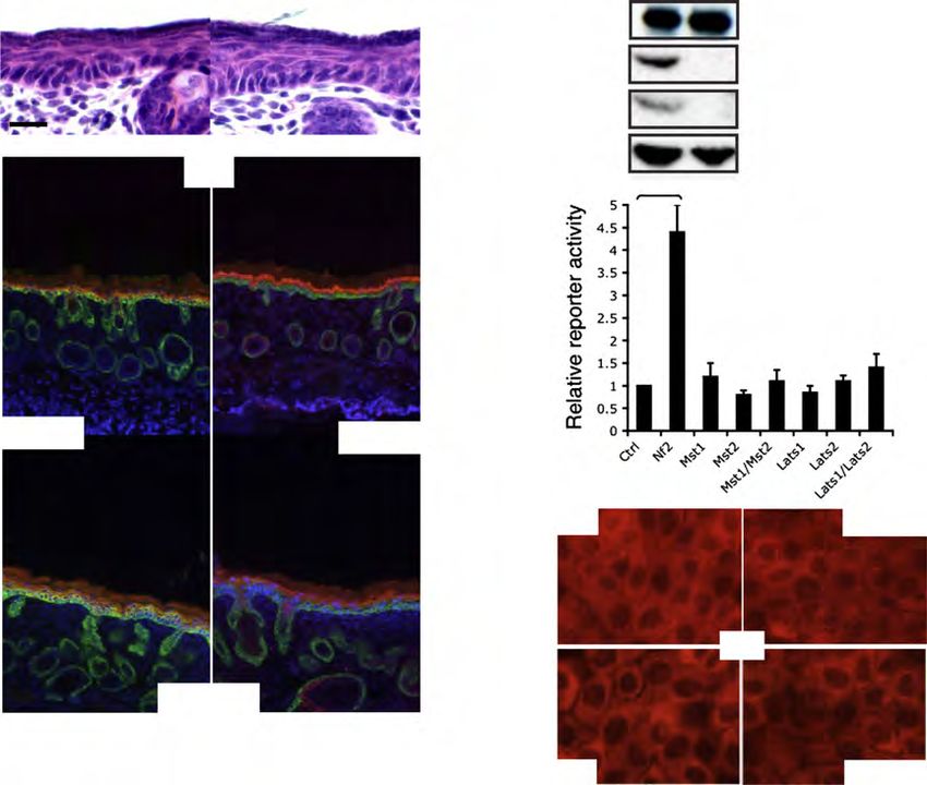
Aims

dependent transactivator (rtTA) selectively in the progeny of K14-expressing cells of
stratified epithelia (Dassule et al., 2000; Belteki et al., 2005). Mice carrying these transgenes
will be referred to as Tg in this work.

Aim 2:
Investigate the Effect of Yap1 Deletion in the Epidermis

The systemic deletion of Yap1 in the mouse is lethal at embryonic day E8.5, therefore
preventing the examination of Yap1 loss in the epidermis. To circumvent this problem, we
generated a mouse with a conditional Yap1 allele, where exon 1 and 2 were flanked by loxP
sites, allowing tissue-specific excision upon Cre-expression. We plan to delete Yap1 by
expressing Cre under the K14-promoter, and to study the potential effect on epidermal
biology. K14 expression is restricted to the basal layer in the epidermis, known to comprise
the epidermal progenitor cells.

Aim 3:
Investigate the Function of the TEAD Transcription Factors in
Epidermal Biology

It is known that the serine residue S94 in human YAP is crucial for the interaction with the
transcription factor TEAD, which is thought to mediate most of the Hippo pathway dependent
signals. In one study, the pro-proliferate functions of YAP were greatly diminished when S94
was mutated to alanine (S94A), thus impairing the interaction of YAP and TEAD (Zhao et al.,
2008). However, YAP has been demonstrated to bind to other transcription factors as well,
and a tissue-dependent effect cannot be ruled out (Yagi et al., 1999; Strano et al., 2001;
Vassilev et al., 2001; Ferrigno et al., 2002; Zaidi et al., 2004). We thus wanted to evaluate the
function of the TEAD transcription factors in Hippo signaling, specifically in epidermal
biology. To address this question in vivo, we generated a Yap1 mutant allele containing the
S79A mutation, which is the murine equivalent to S94A in human YAP. We plan to study the
effect of this allele in the epidermis by combining it with the Yap1 conditional loss-of-
function allele (Aim 2), which will be deleted by Cre expressed under the K14-promoter,
leaving YAP1-S79A as the remaining functional protein.


                                             26
Aims

Aim 4:
Investigate the Function of the MST1/2 Kinases in Epidermal Biology

Deletion of both Mst1 and Mst2 in the liver results in YAP activation and hepatomegaly,
suggesting a function for these kinases as negative regulators in vivo (Zhou et al., 2009; Song
et al., 2010). We wanted to assess whether deletion of both Mst1 and Mst2 in the skin would
lead to YAP activation and produce a similar phenotype of tissue overgrowth. We plan to
study mice generated in the laboratory of Joseph Avruch (MGH), carrying the Mst1-/- Mst2-/fl
allele by crossing them with mice expressing K14-Cre to ablate MST1/2 expression in the
basal cells of the epidermis. We plan to complement the in vivo studies with in vitro
experiments in the keratinocyte cell line HaCaT, using siRNAs to knock down the transcript
of Mst1 and Mst2. These results will provide valuable information about the conservation of
the Hippo core kinase cascade across different tissues of the body.


                                            27
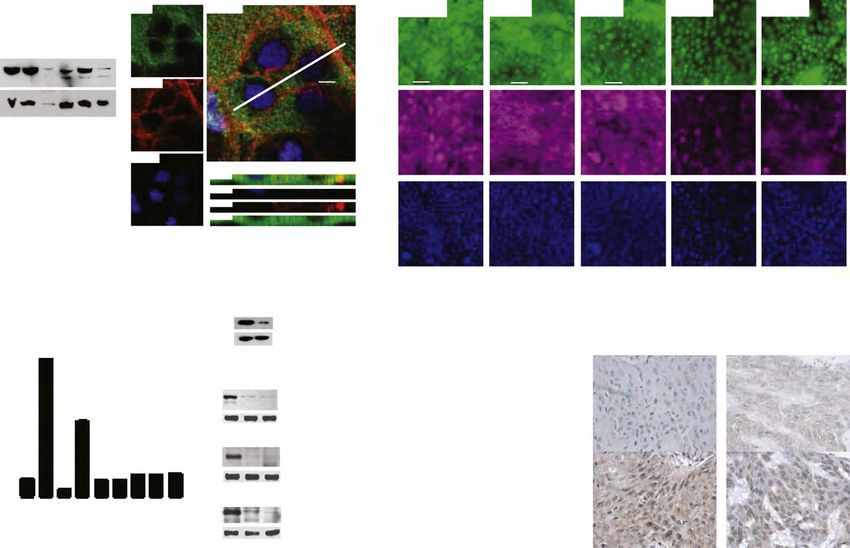
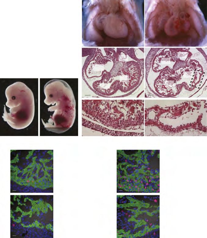
MANUSCRIPT I

3.       MANUSCRIPT I

Yap1 Acts Downstream of α -Catenin to Control Epidermal
Proliferation

Karin Schlegelmilch*, Morvarid Mohseni*, Oktay Kirak, Jan Pruszak, J. Renato Rodriguez,
Dawang Zhou, Bridget T. Kreger, Valera Vasioukhin, Joseph Avruch, Thijn R.
Brummelkamp, and Fernando D. Camargo

* These authors contributed equally to this work

Cell. 2011 Mar 4; 144(5):782-95.

Experimental Contribution

The part of my work presented in the publication “Yap1 Acts Downstream of α -Catenin to
Control Epidermal Proliferation” was the in vivo characterization of the role of the Hippo
signaling pathway in the epidermis, utilizing transgenic mouse models for gain- and loss-of-
function of Yap1, the mutated Yap1-S79A and Mst1/2 loss-of-function. The Yap1 cKO and
Yap1-S79A mice were generated during my diploma thesis work. The initial ideas and the
conceptual design of my work were developed together with my advisor, Assistant Professor
Fernando Camargo. Experiments were planned, performed and evaluated by myself. I wrote
the manuscript together with Fernando Camargo and Morvarid Mohseni.

The following figures present my work:
Figure 1 A, C-G; Figure 2 A-E; Figure 3 A-G; Figure 4 D-E; Figure 7 C
Figure S1 A-H; Figure S2 A-C; Figure S3 A-C; Figure S4 A-C, E

The original article including the supplemental information is included on the following pages
and online available at:
http://www.cell.com/abstract/S0092-8674(11)00183-8


                                           28
You can also read


