Reversible Membrane Pearling in Live Cells upon Destruction of the Actin Cortex - Core
←
→
Page content transcription
If your browser does not render page correctly, please read the page content below
Biophysical Journal Volume 106 March 2014 1079–1091                                                                                           1079

Reversible Membrane Pearling in Live Cells upon Destruction of the
Actin Cortex

Doris Heinrich,†‡ Mary Ecke,§ Marion Jasnin,§ Ulrike Engel,{ and Günther Gerisch§*
†
    Leiden Institute of Physics, LION, Leiden University, The Netherlands; ‡Fraunhofer-Institut für Silicatforschung ISC, Würzburg, Germany;
§
    Max Planck Institute of Biochemistry, Martinsried, Germany; and {Nikon Imaging Center, Heidelberg, Germany

ABSTRACT Membrane pearling in live cells is observed when the plasma membrane is depleted of its support, the cortical
actin network. Upon efficient depolymerization of actin, pearls of variable size are formed, which are connected by nanotubes
of ~40 nm diameter. We show that formation of the membrane tubes and their transition into chains of pearls do not require
external tension, and that they neither depend on microtubule-based molecular motors nor pressure generated by myosin-II.
Pearling thus differs from blebbing. The pearling state is stable as long as actin is prevented from polymerizing. When polymer-
ization is restored, the pearls are retracted into the cell, indicating continuity of the membrane. Our data suggest that the alter-
nation of pearls and strings is an energetically favored state of the unsupported plasma membrane, and that one of the functions
of the actin cortex is to prevent the membrane from spontaneously assuming this configuration.

INTRODUCTION
The shape of the plasma membrane in a motile eukaryotic                        Shape variations in model membranes have been studied
cell is normally dominated by the organization of the actin                 in giant unilamellar vesicles (GUVs) (5). In GUVs com-
network in the cell cortex, which is linked by noncovalent                  posed of defined multicomponent lipid mixtures and also
bonds to the lipid bilayer on the cell surface. In cooperation              in plasma membrane vesicles lacking an underlying cyto-
with associated proteins, the actin network forces the                      skeleton, lipids and proteins can segregate into two fluid
membrane to bend, forming protrusions of different shapes                   phases, a liquid phase with short-range order (L0) and a
like lamellipodia and filopodia, or invaginations like phago-               liquid-disordered (Ld) phase (6,7). This separation of phases
somes and other endocytic vesicles. The question addressed                  occurs at temperatures below a mixing point, and is typical
here is how the complex membrane of a living cell self-                     of systems that are destabilized by competing interactions
organizes when support by the submembraneous actin layer                    (8). The segregated domains differ in lipid composition
is impaired.                                                                and may also differ in membrane curvature. The coupling
   The scenario we study is as follows. The highly motile                   between segregation and curvature has two aspects: 1),
cells of Dictyostelium discoideum rapidly change shape                      segregation can be induced by fluctuations in local mem-
and efficiently internalize plasma membrane by endocy-                      brane curvature (9); and 2), segregation results in stabilizing
tosis. Osmotic pressure in the cells is regulated through                   the membrane in its curved state (10).
the outward pumping of water by contractile vacuoles that                      Even in uniform GUVs that consist only of one type of
periodically form a channel to the plasma membrane (1),                     lipid, local shape differences can be induced by applying
thus keeping cell volume within narrow limits. Both the                     external force. Nanotubes can be extracted from a GUV
irregular and variable shape of migrating cells and endocytic               by axial strain (11). Close to natural conditions, nanotubes
activity depend on the availability of membrane area. We                    are extracted by the activity of microtubule-dependent
challenged the regulation of surface area by a blocker of                   motor proteins (12–14). The use of nonprocessive motors
actin polymerization, latrunculin A (latA), at concentrations               showed that these nanotubes persist only under the contin-
that still allow the contractile vacuoles to work as osmoregu-              uous application of force, because they retract when the
lators. Because phagocytosis, macropinocytosis, as well as                  motors dissociate (15).
endocytosis of clathrin-coated vesicles (2) are arrested at                    Local pulling force applied through a laser tweezer may
the latA concentrations used, the internalization of excess                 cause a membrane tube to transform into pearls on a string.
membrane is prevented. Under these conditions, the cells                    Dynamic pearling states can be set off in tubes composed
round up without expanding their volume. The excess mem-                    of a single lipid (16). This long-range, slowly relaxing
brane has been observed to fold under these conditions into                 Rayleigh-like instability is due to the competition of
tubes, which are converted into chains of interconnected                    curvature energy and surface tension. Pearling can also be
pearls in Dictyostelium (3) as in mammalian cells (4).                      induced in the absence of a pulling force. Thread-like
                                                                            oleate vesicles are induced to pearl by the photooxidation
                                                                            of thiols, probably causing a change in surface tension
Submitted July 29, 2013, and accepted for publication December 11, 2013.    (17), and pearls on a chain can be generated by the absor-
*Correspondence: gerisch@biochem.mpg.de                                     bance of cationic nanoparticles to the inner leaflet of
Editor: Cecile Sykes.
Ó 2014 by the Biophysical Society
0006-3495/14/03/1079/13 $2.00                                                                            http://dx.doi.org/10.1016/j.bpj.2013.12.054
1080                                                                                                                                      Heinrich et al.

DOPC (dioleoyl-glycero-phosphocholine) vesicles (18) or                       Nikon Eclipse TE 300 microscope equipped with a 100/1.4 NA oil
by increasing the osmotic pressure within a tube (19).                        PlanApo objective (Nikon, Tokyo, Japan), in combination with an Ultra
                                                                              View confocal scanner (Perkin Elmer, Waltham, MA) and an Orca
   The study of membrane pearling in live cells has been                      ERC4742-95-12 ERG camera (Hamamatsu Photonics, Hamamatsu, Japan).
pioneered by Bar-Ziv et al. (4). These authors discovered                     Images of Fig. 5 were obtained using an Ultra View system (Perkin Elmer)
pearling in membrane tubes that were under tension, which                     on a Nikon TE-2000 microscope equipped with a VC 100/1.4 NA
was supplied by local attachment of the tubes to a solid sub-                 PlanApo objective (Nikon, Tokyo, Japan) and an EM-CCD camera (Andor
strate. If the rigidity of the submembraneous actin shell is                  Technology, Belfast, UK). For the 3D reconstructions (see Fig. 5, A–C),
                                                                              z-stacks generated with a spacing of 0.3 mm were deconvolved using
reduced by partial depolymerization, the membrane tubes                       Huygens Essential 3.0.0 (Scientific Volume Imaging, 1213 VB Hilversum,
are converted into pearls on a chain.                                         The Netherlands) and surface rendered using Volocity 3.7.0 (Perkin Elmer).
   Here, we explore the transformation of the plasma mem-                     The equipment for total internal reflection fluorescence microscopy has
brane of live cells into tubes and pearls, and the reversal of                been described in (2).
this transformation upon reconstitution of the actin cortex.                     Fluorescence recovery after photobleaching (FRAP) experiments were
                                                                              performed using an Olympus IX 81 microscope with a 60/1.35 NA oil
A distinguishing feature of the pearling studied by us is its                 UPlanSApo objective (Olympus, Tokyo, Japan), a Yokogawa spinning-
independence of tension exerted by stretching the mem-                        disc scan head CSU_X1 (Andor Technology, Belfast, UK), an IQ version
brane tubes between adhesion points, as evidenced by the                      1.9 FRAPPA configuration (Andor Technology), and an IXON DU897_
pearl formation on free, unattached portions of the cell sur-                 BV_3880-iXon plus camera (Andor Technology). For bleaching, an ALC
face. We show that membrane pearling in Dictyostelium                         laser combined with a 488 nm/50 mW Coherent Sapphire laser (Andor
                                                                              Technology) was used.
cells does not require force generation by microtubule-based
motors. Moreover, the pearling is independent of internal
pressure generated by myosin-II, and is thus distinguished                    Cryo-ET
from blebbing.
                                                                              Cells were plated on glow-discharged, holey-carbon-film-coated gold EM
   By cryo-electron tomography (cryo-ET) we show the
                                                                              grids (C-flat 2/1-2Au, Protochips, Raleigh, NC) and kept for 2 h, before
persistence of short bundles of actin filaments in pearls                     the cells were treated with 5 or 30 mM latA for 15 min. For cryo-preparation
formed in the presence of 5 mM latA and the absence of                        we added BSA-coated 15 nm colloidal gold (Aurion, Wageningen,
filaments in tubes or pearls after the depolymerization                       The Netherlands) on top of the grid, removed excess liquid by blotting
of actin at 30 mM latA. We discuss pearling as a process                      from the reverse side, and rapidly plunge-froze the grids in liquid pro-
                                                                              pane-ethane.
of membrane self-organization, which is prevented by an
                                                                                 Tomograms were obtained under low-dose conditions using a Tecnai
intact actin network in the cell cortex.                                      G2 Polara transmission electron microscope (FEI, Eindhoven, The
                                                                              Netherlands) equipped with a 300-kV field emission gun, a Gatan GIF
                                                                              2002 post-column energy filter, and a 2048  2048 slow-scan CCD camera
MATERIALS AND METHODS                                                         (Gatan, Pleasanton, CA). The electron microscope was operated at an
                                                                              accelerating voltage of 300 kV; the pixel size at the specimen level was
Strains, treatment of cells, and imaging                                      0.713 nm. Tilt series were recorded using the SerialEM software (25), typi-
conditions                                                                    cally covering an angular range from 60 to þ60 with a tilt increment
                                                                              of 2 , defocus values of 6 and 12 mm, and a total electron dose of
Cells of D. discoideum strain AX2-214 that expressed various fluorescent      200 electrons /Å2. The projection images were aligned using the gold beads
proteins were cultivated at 21–23 C in nutrient medium supplemented          as fiducial markers. Tomograms with a once binned pixel size of 1.42 nm
with appropriate selection markers. The cells expressed LimED-GFP             were calculated using the software IMOD (26).
(20), GFP-a-tubulin (21), the PH-domain of human phospholipase Cd1               For automated filament segmentation, tomograms were subjected to
(22), GFP-coronin (22), or free GFP. In the AX2-derived myosin II-HC          nonlocal means filtering. Actin filaments were traced using an automated
deficient mutant strain HS2205 (23), mRFPM-LimED (24) was expressed.          segmentation algorithm implemented in the Amira software (27), using
For confocal fluorescence imaging, the cells were washed in 17 mM Na/K        a generic filament as a template (28). Diameter and length of the generic
phosphate buffer, pH 6.0, and transferred into an open chamber on a glass     filament were 8 and 42 nm, respectively; short filamentous structures
coverslip. LatA (Life Technologies, Grand Island, NY) was routinely used      of
Membrane Pearling with No Actin Support                                                                                           1081

                                                                                    FIGURE 1 Membrane pearling and destruction
                                                                                    of the actin cortex in response to actin depolymer-
                                                                                    ization. (A and B) Confocal images of an untreated
                                                                                    cell of D. discoideum (A) and of cells incubated
                                                                                    with 5 mM latA (B). The cells expressed LimED-
                                                                                    GFP as a label for filamentous actin (green)
                                                                                    and were stained with FM4-64, which integrates
                                                                                    into the plasma membrane and from there into
                                                                                    the membranes of the contractile vacuole system
                                                                                    (red). In the motile cell of (A), actin is assembled
                                                                                    along the filopodia and in the cortical region
                                                                                    beneath the plasma membrane. In the two cells
                                                                                    of (B) showing extensive membrane pearling, the
                                                                                    LimED-GFP is dispersed in the cytoplasm and
                                                                                    the lumen of the pearls. A time series of pearling
                                                                                    in the cell displayed in (A) is shown in Movie
                                                                                    S1. (C) Coronin, a protein preferentially associated
                                                                                    with disassembling actin filaments (22), is en-
                                                                                    riched in the pearls. The cell expressing GFP-coro-
                                                                                    nin has been imaged by total internal reflection
                                                                                    fluorescence microscopy. (D and E) A pearl sub-
                                                                                    jected to cryo-ET, showing bundles of actin
                                                                                    filaments in its interior. The cell was treated for
                                                                                    15 min with 5 mM latA. In (D) a slice through
                                                                                    the tomogram is displayed. Segmentation of the
                                                                                    filaments and the membrane is shown in (E).
                                                                                    Bar for (A–C) 10 mm; for (D and E) 200 nm. The
                                                                                    tomogram is shown in Movie S2.

5 mM latA. Under these conditions, the microtubule-              plasma membrane. GFP-PLCd1 was expressed in the cells,
dependent motility of intracellular vesicles is not disturbed    which recognizes phosphatidylinositol- (4,5) bisphosphate
and may even be enhanced (29). The contractile vacuoles          (PIP2) (31), and cells were labeled with FITC-concanavalin
continue to pulse, indicating that their osmoregulatory          A, which preferentially binds to glycosyl residues that are
activity is preserved.                                           N-linked to membrane proteins of Dictyostelium cells
   After the addition of latA, the leading edges became          (32). These labels did not distinguish the membrane of the
indistinguishable, and actin-based cell-surface protrusions      pearls from the plasma membrane on the cell body indi-
such as filopodia disappeared. The loss of protrusions           cating that, together with lipids, membrane proteins enter
created an excess plasma membrane area, which was trans-         the pearling membrane protrusions (Fig. 2, A and B, and
formed into chains of pearls sticking out of the area of the     Movie S3).
plasma membrane that enveloped the rounded cell body
(Fig. 1 B).
                                                                 Pearling by membrane protrusion or retraction
   At the beginning of latA action, actin remained tran-
                                                                 of the cell body
siently clustered at the plasma membrane, primarily in con-
tact with clathrin-coated pits (2). After 15 min the actin was   Two modes of transformation of the plasma membrane into
depolymerized to an extent that in most cells no structures      pearls were observed in cells adhering to a glass surface:
of microscopic size remained recognizable with LimED-            1), protrusion of chains of pearls, some of them in contact
GFP (Movie S1 in the Supporting Material). However,              with the substrate surface, other ones freely extending into
with GFP-coronin, which associates with depolymerizing           the fluid medium as shown by their Brownian movement
actin structures (22,30) the pearls were intensely labeled       (Fig. 2 C and Movie S4); 2), retraction of the cell from
(Fig. 1 C), suggesting that slowly dissociating complexes        the substrate, leaving substrate-attached pearls behind
of actin filaments were still present. To visualize these com-   (Fig. 2 D and Movie S5). The different modes of pearl
plexes, we subjected latA-treated cells to cryo-ET. In a sub-    formation were reflected in highly variable pearl sizes,
set of pearls, bundles of straight or slightly bent actin        ranging from diameters of 0.22 mm, as revealed by cryo-
filaments were detectable, which were too short for deform-      ET, to 3 mm. We obtained also extensive pearling by
ing the pearls (Fig. 1, D and E, and Movie S2).                  combining 10 mM lat A with 10 mM cytochalasin A, a
   Because pearling is a phenomenon observed in pure             blocker of the plus ends of actin filaments, indicating
lipid membranes, we explored the possibility that the pearl-     that even efficient depolymerization of actin does not
ing of membrane tubes in Dictyostelium cells is caused by        prevent the membrane from pearling. Combination of the
unmixing of the lipid phase from the protein-containing          two drugs resulted again in pearls-on-a-string and variable

                                                                                            Biophysical Journal 106(5) 1079–1091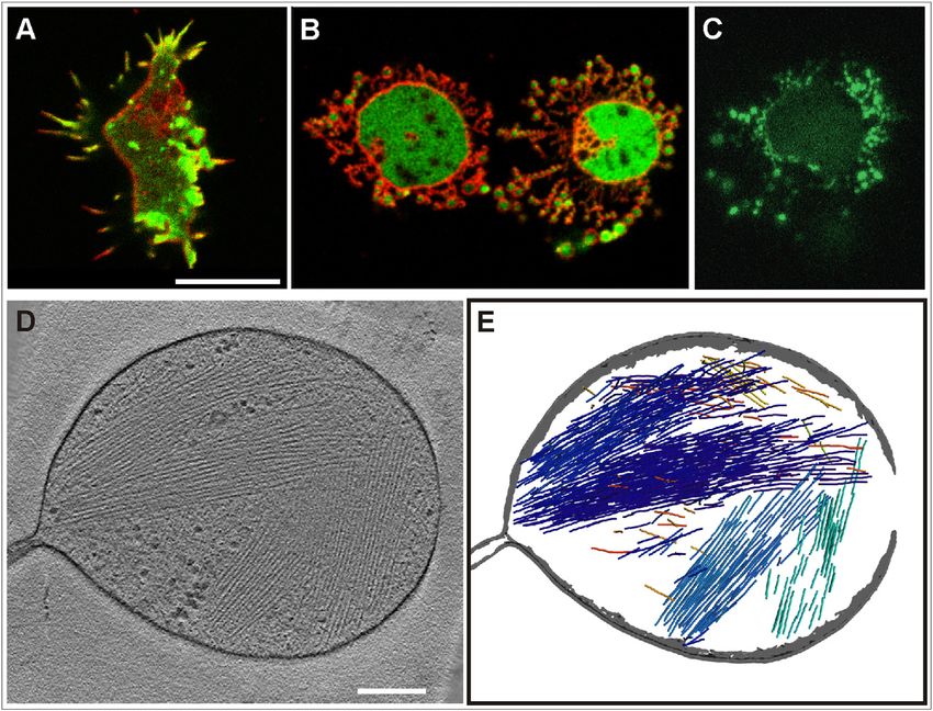
1082                                                                                                                                              Heinrich et al.

FIGURE 2 Pearling membranes labeled with concanavalin A or PLCd1. (A) Cells labeled with FITC–concanavalin A (green), a lectin binding preferen-
tially to a-mannosyl residues on N-glycosylated proteins, and counterstained with Alexa Fluor 568 succinimidyl ester, a cell surface marker (red). The three
panels display merged images (left), the concanavalin A label (middle), and the cell-surface label (right). (B) Cell expressing mRFP- LimED (red) and GFP-
PLCd1, which recognizes PI(4,5)P2 (green). PI(4,5)P2 is localized to the membrane of the pearls as well as to the plasma membrane enveloping the cell body.
(C) Pearl formation by protrusion of the cell surface. (D) Pearl formation by retraction of the cell. In both (C) and (D) pearl formation begins at a late stage of
actin depolymerization. (E) Withdrawal of pearls during the repolymerization of actin. Cells in (C–E) were labeled similar to the cell in (B). Patches of PLCd1
indicate local enrichment of PIP2 under conditions of intense clathrin-dependent endocytosis (2), in accord with a role of PIP2 in membrane invagination
(69). Numbers indicate seconds after the addition (C and D) or the removal of 5 mM latA (E). Bars, 10 mm. The time series corresponding to (B–E) are
displayed in Movies S3–S5, S7.

pearl sizes (Movie S6), suggesting that incomplete or de-                           into the plasma membrane. Only a few protrusions, in
layed actin depolymerization is not responsible for the                             particular those strongly adhering to the substrate, became
variability.                                                                        disconnected.
   To inquire whether pearling membranes are still in a
native state, we explored the reversibility of pearling
                                                                                    Reversible compartmentation of the cell
after latA removal during the recovery of normal cell
motility. Fig. 2 E and Movie S7 show that pearled protru-                           Reintegration of the membrane of pearls into the plasma
sions of the cells can be pulled back and reintegrated                              membrane suggests continuity of the pearl membrane with

Biophysical Journal 106(5) 1079–1091
Membrane Pearling with No Actin Support 1083
FIGURE 3 Tomograms of pearls and tubes.
Pearling was induced by incubating cells with
either 5 mM (A–F) or 30 mM (G and H) latA. (A)
Pearls connected to each other or separated by
short tubes. (B and C) Pearls extending into long
tubes, showing constrictions at the pearl-tube inter-
face. The inset in (B) shows a clathrin-coated pit
visible in another slice of the tomogram. In (C)
bundled actin filaments are retained, similar to
Fig. 1 D. (D–H) Gallery of long nanotubes, two
with actin filaments (D and E) and three with no
detectable filaments (F–H). The arrowheads in
(D) and (E) point to long filaments; in E the fila-
ment extends from the pearl into the upper part
of the tube. The inset in (E) shows a cross section
through the tube at the position indicated by bars,
displaying a bundle of two filaments inside. A
bent tube is shown in (F), and a tube with periodic
variations of diameter in (G). All images show sli-
ces through tomograms. A 3D reconstruction of
(C) is displayed in Movie S8 together with the
3D segmentation of the filaments and the mem-
brane. Bars, 200 nm; the insets in (B) and (E) are
twofold magnified relative to the main figure.
that enveloping the cell body. The question of whether the displayed in Fig. 3 G, suggest an incomplete stage of
contents of the pearls is connected with the cytoplasm was tube-pearl differentiation.
addressed using FRAP in cells that expressed free GFP, The tubes and pearls were unilamellar; the pearls
which is purely cytosolic and has a diffusion constant D ¼ commonly containing vesicles of variable sizes (Fig. 3, A
42 mm2 s1 in Dictyostelium cells in the absence of fila- and B), and also clathrin-coated pits (Fig. 3 B). The cyto-
mentous actin (33). FRAP of GFP in the cell body was plasm entrapped in the pearls was rich in ribosomes and
accordingly fast (Fig. S1 A). When pearls were bleached, also populated with larger particles of ~50 nm diameter
recovery was slow, incomplete, or absent. No recovery (Fig. 3 C). The presence of such particles in tubes (Fig. 3
meant that GFP molecules with a size of 3–4 nm were pre- G) might be responsible for narrowing the space for diffu-
vented from diffusion through the tubes that separated a sion of GFP molecules, as shown in Fig. S1.
pearl from other pearls or from the cell body. Incomplete In cells treated with 5 mM latA, a fraction of the pearls
recovery indicated no connection to the GFP reservoir of and of the tubes connecting them still contained microfila-
the cell body (Fig. S1 B). ments (Fig. 1, D and E, and Fig. 3, C–E).
Other pearls and tubes formed at 5 mM latA appeared to
be free of microfilaments (Fig. 3 F). To substantiate the
Pearl and tube structures revealed by cryo-ET
independence of pearl formation from an actin support,
To determine the diameter of the tubes connecting the we applied 30 mM latA, which rapidly and efficiently
pearls and to examine whether these tubes can be formed eliminated any actin clusters but did not prevent pearling.
in the absence of microfilaments extending along their At this very high concentration, chains of pearls were
axis, we have applied cryo-ET on cells treated with either formed during the disappearance of actin structures, either
5 or 30 mM latA. Cells were incubated with the drug on a on the substrate or protruding freely into the extracellular
carbon film and vitrified after ~15 min. In one experiment space (Movie S9). In the tomograms, we no longer detected
the cells were resuspended by pipetting, to disconnect the microfilaments in the tubes or pearls (Fig. 3, G and H). This
tubes from any support that could exert tension, before the means, the tubes were free of filaments that could support
cells were allowed to settle on the film. myosins in applying tension for pulling out membrane
The pearls were directly connected to each other (Fig. 3 tubes.
A) or separated by tubes (Fig. 3, B–H). Sometimes these The diameters of the microfilament-depleted tubes varied
tubes were branched, or bent as shown in Fig. 3 F; this not only from one tube to the other but also along a single
argues against forces acting in stretching the tubes between tube, most evidently in tubes with an oscillating diameter
two points along their surface that might be fixed on the as shown in Fig. 3 G. The diameters of uniform tubes that
carbon film. Periodic variations of the tube diameter, as were depleted of actin ranged from 38 to 46 nm, measured
Biophysical Journal 106(5) 1079–10911084 Heinrich et al.
as the distance between the middle of the electron-dense They changed their shape only slowly by rounding up
membranes. Often a constriction reducing the tube diameter (Movie S12).
to ~29 nm was observed close to the pearl-tube interface To distinguish the mechanism of pearling from that
(Fig. 3, B, C, and H). This constriction is in accord with of blebbing, we treated myo-II HC-null cells with latA.
theoretical predictions (34) and observations on tubes pulled These mutant cells showed intense pearling (Fig. 4, E
out of giant lipid vesicles (35). and F, and Movie S13). The formation of pearls in the
mutant cells resembled that of WT cells (compare in
Fig. 2 D and Fig. 4 E the pearling by retraction of the
Pearl formation is microtubule independent
cell body). During recovery of the mutant cells, the pearls
The possibility that chains of pearls are protruded or re- are efficiently withdrawn; in the example shown in Fig. 4
tracted by the polymerization/depolymerization of micro- G, the pearling stage is followed by the propagation of
tubules or by forces applied by microtubule-dependent an actin wave (Movie S14), similar to wave formation in
motors, was examined by the labeling of microtubules using WT cells (2).
GFP-a-tubulin (21) and by depolymerizing the microtu-
bules using nocodazole. In the absence of the drug, we never
saw a microtubule form along the axis of an emerging chain Spreading of pearling cells on a glass surface
of pearls (Fig. S2 A), nor have we seen microtubules enter The spreading of cells on an adhesive surface involves
these chains during retraction of the pearls into the cell shape changes, phase transitions, as well as regulation of
body (Fig. S2 D). the surface area and membrane tension. These regulations
In cells incubated with nocodazole, the microtubule are mediated by the actin system in Dictyostelium (38) as
length was strongly reduced such that their ends could no in mammalian cells (39–41). Nevertheless, Dictyostelium
longer touch the plasma membrane from where pearls are cells can spread even in the pearling state. LatA-treated
emanating. Nevertheless, pearls were formed in the pres- Dictyostelium cells produce pearling tubes also in suspen-
ence of latA (Fig. S2, B and C, and Movie S10) and were sion. Because these cells will adhere to a glass surface not
retracted after its removal (Movie S11). An exceptional only with the compact surface of their body but also with
mode of recovery is shown in Fig. S2 E, where cytoplasm the pearling protrusions, we have monitored the spreading
filled the lumen of a chain of pearls, in that way opening behavior of cells in the presence of latA. As shown in
the constrictions that separate the pearls. Fig. 5 and Movie S15, a latA-treated cell can turn from
initial substrate contact, made by the tips of pearling mem-
Formation and retraction of pearls does not brane tubes, to expansion of the cell body on the substrate.
require myosin-II and is thus distinguished Subsequently, contact with the substrate induced new tubes,
from blebbing followed by pearling as displayed in Fig. 6 and Movie S16.
These time series show that cells devoid of an actin cortex
Myosin-II is the motor protein that in cooperation with fila- can spread on an adhesive surface, and they do so by the
mentous actin is responsible for tail contraction in migrating formation of pearling tubes.
Dictyostelium cells. Myosin-II generates also the rigor
resulting in the contraction of cells that are depleted of
ATP (36). The increase of membrane tension caused by DISCUSSION
the depletion of ATP in cells of D. discoideum results
Reversible pearling of plasma membranes
in local disruption of the actin cortex and blebbing of the
lacking an actin support
cell surface. Although a thin layer of filamentous actin
covers the membrane of the blebs, the majority of actin The membrane pearling studied here is an autonomous
filaments are clustered in the interior of the cell together process that occurs in live cells when support by the cortical
with myosin-II and certain actin-binding proteins (37). actin network is lacking. Sizes of the pearls ranged in our
Wild-type (WT) and mutant cells that lack myosin-II experiments from 0.2 to 3 mm diameter, as seen in Fig. 6
heavy chains (myo-II HC-null cells) were exposed to 2,4- B and Movie S16, and thin tubes connecting the pearls
dinitrophenol. This uncoupler of oxidative phosphorylation had a diameter of ~40 nm. Microtubules are not involved
is known to decrease cellular ATP to ~10% of its normal in protrusion or retraction of the chains of pearls formed
level. Under these conditions, which reduce the resistance upon the depolymerization of actin, and the pressure-
of the actin cortex, WT cells are induced to bleb within generating activity of myosin-II is dispensable. Pearls can
3 min (37). The cells stay alive and return to normal shape emanate from the free cell surface, which means that tension
and motility as soon as the uncoupler is removed. Fig. 4, produced by adhesion to the substrate is not required,
A–D, shows that myosin-II is required for cell-surface although the pearls may adhere to a glass surface if they
blebbing under the conditions of ATP depletion: in con- come into contact with it. To find out whether the contents
trast to WT cells, the myo-II HC-null cells did not bleb. of the pearls is continuous with, or separated from, the
Biophysical Journal 106(5) 1079–1091Membrane Pearling with No Actin Support 1085
FIGURE 4 Pearling distinguished from blebbing by its independence of myosin-II. (A–D) Blebbing induced in WT cells by the decline of cellular ATP.
WT cells expressing GFP- LimED (green) mixed with myo-II HC-null cells expressing mRFP-LimED (red) were exposed to 50 mM or, in (C), to 100 mM
2,4-dinitrophenol. The bottom panels show differential interference contrast bright-field images, the top panels confocal fluorescence scans. In the overview
of (A) a cell forming a chain of blebs is indicated by an arrowhead. In (B) the large mutant cell on top displays filopodia, whereas the WT cell below forms
multiple blebs. In WT cells of (C) a thin actin layer persists at the membrane of the blebs, whereas the majority of actin is separately clustered. In the optical
sections of (D) septa of actin are recognizable through which blebs are broken (arrowheads). A time series corresponding to (A–D) is shown in Movie S12.
(E–G) Pearling in myo-II HC-null cells. These mutant cells expressed LimED- GFP to label filamentous actin (green); they were stained with FM4-64 (red)
and incubated in 5 mM latA. The arrowhead at the large cell of (E) points to an area where transformation of the membrane into pearls begins. A mutant cell
forming large pearls is shown in (F), and the withdrawal of pearls in a mutant cell in (G). Here, a propagating actin wave was formed at the final stage of
reintegration of the pearled membrane into the cell surface. Actin waves are typical of an intermediate stage of recovery from actin depolymerization (2).
Numbers in (E) and (F) indicate seconds before or after the addition of latA, in (G) after its removal. Bars, 10 mm. Time series corresponding to (E) and (G)
are shown in Movie S13 and Movie S14.
cytoplasm of the cell, we have applied FRAP to cells ex- of the pearls can be reintegrated into the envelope of a
pressing free GFP. The tubes, which separate each pearl cell during recovery of actin polymerization.
from its neighbors in a chain, together with the particles The fact that at a concentration of 5 mM, latA did not
within these tubes, largely reduce or completely abolish completely depolymerize the actin, enabled us to recon-
the exchange of proteins between the pearls and between struct tomograms of the most resistant actin assemblies.
the pearls and the cell body. Nevertheless, the membranes These consisted of microfilament bundles of variable
Biophysical Journal 106(5) 1079–10911086 Heinrich et al.
FIGURE 5 Spreading of cells preincubated with
5 mM latA before subjecting them onto a coverslip.
Membranes were stained using FM4-64. (A–C)
Shape of two cells preincubated with latA and
attached for 2 min (A) or 50 min (B and C) to a
glass surface. In the 3D reconstruction of the cell
shown in (A), membrane tubules are primarily
pointing into the fluid space. The 3D reconstruc-
tion in (B) shows many pearled tubes spread along
the substrate surface, the cross section in (C) the
cell body to rise from the substrate at an angle
of ~73 . (D) Stages of spreading on a strongly
adhesive fluoralkane-coated surface. The framed
upper cell proceeds to spread, whereas the bottom
cell is already in a stable spreading state. The
arrowhead points to a long chain of pearls.
Numbers indicate seconds after the first contact
of the upper cell with the glass surface. The full
time series corresponding to this figure is presented
in Movie S15. Bar, 10 mm.
orientation, which were of limited length and did no longer that myosin-II null mutant cells of D. discoideum (Fig. 4,
deform the pearls (Fig. 1, D and E, Fig. 3 C, Movie S2, and A–D), or M2 macrophages treated with the myosin-II
Movie S8). ATPase inhibitor blebbistatin (43,44) do not bleb. In
contrast, pearling tubes are still formed in myosin-II null
mutants (Fig. 4, E–G).
Membrane pearling is distinct from myosin-II-
We note that myosin-II null cells have been reported to
based blebbing and from microtubule-supported
bleb if they are stimulated with a high concentration of
tubulation
cyclic AMP (45). From blebbing under our conditions
The pearling phenomenon is distinct from membrane bleb- this chemoattractant-induced blebbing is distinguished
bing caused by an increase in rigor, which leads to local by the formation of rounded protrusions that are filled
rupture of the actin cortex followed by the repolymerization with filamentous actin, as indicated by phalloidin labeling.
of actin beneath the membrane of a bleb (42). An increase in Obviously, this type of blebbing does not require an increase
rigor is observed in Dictyostelium cells when the synthesis in tension that is generated by myosin-II.
of ATP is inhibited by azide (36). In a reversible manner, Microtubules can support the tubulation of membranes in
the blebbing is induced by 2,4-dinitrophenol, a membrane two ways. First, phospholipid vesicles are deformed from
permeable uncoupler of oxidative phosphorylation (37). within by the polymerization of microtubules (46). Second,
The distinguishing feature of blebbing is that the force is lipid membranes can be pulled into nanotubes by the
generated by the contractile activity of myosin-II, such walking of microtubule-based motors along their outer
Biophysical Journal 106(5) 1079–1091Membrane Pearling with No Actin Support 1087
FIGURE 6 Transformation of tubes into pearls
in spreading cells. The cells expressing LimED-
GFP (green) were incubated for 30 min with latA
in gently shaken suspension before they were
transferred onto a glass surface and FM4-64 (red)
was added. At that time no filamentous actin struc-
tures were detectable by the LimED-GFP label.
(A) A spreading cell protruding multiple tubes,
the left one is subdivided into five pearls. (B) A
tube protruded by a spreading cell, which is from
top to bottom progressively organized into pearls.
Numbers indicate seconds after the first frame in
each panel. Bars, 10 mm. The pearling sequence
shown in (B) is displayed in Movie S16.
surface. In that way, membranes of the endoplasmic reticu- portions, the envelope of the cell body and the excess mem-
lum are drawn in vivo into tubes (47), and in vitro the lipid brane area. The cell body is rounded; when attached to glass
membranes of GUVs are pulled along microtubules by its surface arises from the substrate at a large angle of ~73
kinesins (12). The pearling membrane tubes studied by us (Fig. 5 C). This shape suggests that the smooth membrane
in live cells were not populated with microtubules, and these closely surrounding the cell body is under tension, and the
membrane tubes can be formed and retracted after micro- size of its area is kept at a minimum, whereas the excess
tubule depolymerization (Fig. S2). We conclude that forma- plasma membrane not supported by an actin layer has a
tion and pearling of the membrane tubes in Dictyostelium high propensity of organizing into tubes that undergo a tran-
cells are not based on microtubule-dependent force gene- sition into chains of pearls.
ration. In tubes formed at a latA concentration as high A phenomenon that may be compared with tube forma-
as 30 mM, no actin filaments were detected by cryo-ET, tion in actin-depleted cells has been discovered in giant lipid
excluding the possibility that any myosin motors generate vesicles that encapsulate two aqueous phases, dextran and
the force for tubulation. polyethylene glycol (PEG) (48). Upon deflation of these
vesicles, excess membrane is generated and transformed
into nanotubes that extend from the vesicle surface into
Pearling tubes are a favored state of unsupported
the interior of the PEG phase. This shape transformation
plasma membranes
is reversible, as the increase of membrane tension results
In Dictyostelium cells depleted of a submembraneous actin in retraction of the nanotubes back into the surface mem-
layer, the plasma membrane is divided into two distinct brane of the vesicle. Theoretical analysis revealed that the
Biophysical Journal 106(5) 1079–10911088 Heinrich et al.
conversion of excess membrane into nanotubes is driven by The study of membrane pearling in live cells has been
the spontaneous curvature that the membrane assumes in pioneered by Bar-Ziv et al. (4). These authors discovered
contact with the internal PEG phase. By the same argument, that in membrane tubes of SVT2 cells, pearling resulted
the pearled membrane tubes that are reversibly protruded from the competition of surface tension with curvature
from the surface of Dictyostelium cells into the extracellular energy. The tension was supplied by local attachment of
space might represent an energetically favored state of the the tubes to a solid substrate. As the pearling in lipid vesi-
unsupported plasma membrane of a live cell. In this case, cles, that of the plasma membrane was interpreted as a
one may assume that the membrane is transformed into Rayleigh-like instability. A distinguishing feature of the
pearled tubes until this transformation is counteracted by pearling studied by us is its independence of an external
the increasing membrane tension on the surface of the cell source of tension, as evidenced by the formation of pearls
body. This notion is underscored by the fact that pearling on the free portion of the cell surface. Whereas the pearling
is reversible. Reversibility indicates that the pearled portion in (4) required actin to be only partially depolymerized, that
of the membrane is continuous with the fraction of the in Dictyostelium cells occurred even after efficient depoly-
plasma membrane that envelops the rounded cell body. merization (Fig. 3, G and H, Movie S6, and Movie S9).
In contrast to the pearls in stretched lipid vesicles, those
in Dictyostelium cells assume a fixed position along the
Pearl dynamics in spreading cells
tubes even if they are not attached to a substrate (as seen
When cells attached to a glass surface are incubated with in Movies S3, S4, and S9).
latA, tube formation and pearling will end up in a station- An alternative mechanism underlying shape changes in
ary state of pearls that are either free or attached to the sur- vesicles is the coupling of membrane curvature and local
face, as shown in Fig. 1 B and Movie S1. When cells are composition in two- or multicomponent systems (49,50),
incubated in suspension, they also form tubes and pearls. which can lead to the formation of a bud separated from
The nonrequirement of cell-substrate adhesion for pearl the vesicle by a neck at a site where two domains meet
formation is disclosed by imaging cells on a nonadhesive (51). The local curvature of membranes can be determined
polyethylene glycol-coated surface that are incubated in by their protein or lipid composition. The shape of vesicles
suspension with 5 mM latA (data not shown). However, composed of a ternary mixture of two phospholipids and
the stationary state is disturbed if the suspended cells are cholesterol is determined by several parameters. 1), Line
brought into contact with an adhesive substrate. Spreading tension drives the boundary between a L0 and a Ld domain
of these cells implies the protrusion of new tubes and their to a minimum radius, resulting in a constriction along the
transformation into chains of pearls (Fig. 5 D and Fig. 6). boundary (10). 2), In the Ld phase, lipids have a higher diffu-
This reorganization indicates that, even after long incuba- sion coefficient (52) and bending is facilitated (10). Because
tion with latA, the states of the membrane are still convert- the L0 phase is more rigid, it is excluded from membrane
ible and adhesion energy is capable of shifting the state tubes that are pulled out of GUVs (53). Accordingly, the
toward pearling. more flexible Ld phase will sort into regions of higher cur-
vature than the L0 phase with short-range order (54). 3), If
the separated domains differ in the geometry of lipid mole-
Mechanisms of membrane pearling
cules, they tend to assume concave or convex curvature (55).
The behavior of the complex plasma membrane of living Cone-shaped lipids such as double-chain phospholipids will
cells may be compared with that of model lipid membranes. prefer concave membrane areas as opposed to inverted-
If local forces are acting on a giant unilamellar phospholipid cone-shaped lipids such as saturated lysophospholipids,
vesicle (GUV), tubes with a radius in the tens of nanometer which prefer convex areas. This coupling of curvature to
range can be pulled out of the vesicle (9). In membrane lipid segregation needs to be specified because experimental
tubes that are stretched using a laser trap, a Rayleigh-like data revealed that individual conical or inverted-cone-
instability can be elicited, resulting in the generation of shaped phospholipid molecules are too small to show
pearls on a string that can travel along the thin tube connect- more than a weak curvature preference (56). Larger lipids,
ing them (16). When the tension is released by turning off such as gangliosides, may have stronger curvature prefer-
the tweezers that stretch the tube, the pearling state relaxes ences. However, even small phospholipids may be strongly
into a straight cylinder. coupled to curvature if they are clustered.
Conversion of a tube into a chain of pearls may also be Examples for proteins that force membranes to bend are
caused by an increase in its volume due to high osmotic dynamins and BAR or I-BAR domain-containing proteins.
pressure (19). In the case of pearling tubes that exchange Dynamins bind to PIP2-supplemented nanotubes with a
GFP molecules with the cell body (Fig. S1 B) this is unlikely preferred radius of 11 nm, and stabilize these tubes by poly-
the mechanism to work because the pressure would be merizing on their outer surface (57). F-BAR or I-BAR
released from the lumen of the tubes into the cell body proteins accumulate at the cytoplasmic face of convex or
where the contractile vacuoles serve as osmoregulators. concave membrane regions and force the membranes to
Biophysical Journal 106(5) 1079–1091Membrane Pearling with No Actin Support 1089
bend (58). The F-BAR proteins polymerize and form lattices tion of more lipids of the same class and in turn enhance
on the outer surface of membrane tubes (59). the bending. According to this hypothesis, the feedback
Dictyostelium cells contain two dynamins, A and B, circuit would be cut off and consequently unmixing and
which participate in endocytic trafficking (60,61). One I- pearling prevented if the membrane bending is suppressed
BAR protein has been identified in D. discoideum, which ac- by an actin support.
cumulates at clathrin-coated structures immediately before In summary, our data are consistent with a function of
endocytosis (62) and at the neck region of particles that the membrane-associated actin network in cutting off the
are being phagocytized (22). Two proteins containing an positive feedback of lipid (and probably protein) sorting
F-BAR domain are involved in transforming the membrane and membrane curvature. This feedback may, in the absence
of the contractile vacuole into tubules (63). of an actin network, enhance the sorting of lipids (and
A protein that shapes membrane tubes by associating proteins) that create a certain membrane curvature and favor
with their inner, i.e., cytoplasmic face, would be needed to this curvature for accumulating. Experiments that combine
sculpt the tubes in pearling Dictyostelium cells. We cannot model lipid membranes with in vitro generated actin net-
strictly rule out the presence of such a protein. However, works of defined mechanical properties would be needed
our tomograms provide no evidence for a regular lattice of to analyze the mechanisms by which the cortical actin layer
proteins on the tubular membrane. Therefore, we consider prevents membrane pearling.
the sorting out of cone-shaped and inverse-cone-shaped
lipids as a reasonable mechanism for the pearl-tube differen-
tiation of the plasma membrane in the absence of an actin SUPPORTING MATERIAL
network. In the complex membranes of eukaryotic cells Two figures and sixteen movies are available at http://www.biophysj.org/
that contain sterols (primarily D22-stigmasten-3b-ol in the biophysj/supplemental/S0006-3495(14)00134-9.
case of Dictyostelium cells (64,65)), such coupling is likely
We thank Wolfgang Baumeister, MPI for Biochemistry, for his support and
to be effective and responsible for the periodicity of convex Harald Engelhardt for discussions on Cryo-ET, Britta Schroth-Diez, MPI-
and concave areas in the pearling state. CBG, for cooperation in FRAP experiments and TIRF imaging.
The separation of Ld and Lo phases in membranes re- D.H. is grateful to Joachim Spatz, MPI for Intelligent Systems, for support-
quires that temperatures are below a demixing point (66). ing her and also acknowledges funding by the Fraunhofer Society within the
The fact that the optimal temperature for the growth ‘‘Attract’’ scheme. We acknowledge equipment and technical support of the
and development of Dictyostelium cells is ~23 C rather Nikon Imaging Center at the University of Heidelberg, and thank the Max
than 37 C as for mammalian cells, is supposed to favor Planck Society for support to G.G.
phase segregation in the membranes under natural living
conditions. REFERENCES
1. Heuser, J., Q. Zhu, and M. Clarke. 1993. Proton pumps populate the
How does the actin network prevent pearling? contractile vacuoles of Dictyostelium amoebae. J. Cell Biol. 121:
1311–1327.
Because the plasma membrane of a live cell has a complex
2. Schroth-Diez, B., S. Gerwig, ., G. Gerisch. 2009. Propagating waves
composition of lipids and integral or associated proteins, separate two states of actin organization in living cells. HFSP J. 3:
it is reasonable to assume that lipids and proteins known 412–427.
to enforce membrane bending play a role in pearling. Actin 3. Gerisch, G., T. Bretschneider, ., K. Anderson. 2004. Mobile actin
networks may hinder membrane components from sorting clusters and traveling waves in cells recovering from actin depolymer-
ization. Biophys. J. 87:3493–3503.
by two mechanisms that are not mutually exclusive. 1),
4. Bar-Ziv, R., T. Tlusty, ., A. Bershadsky. 1999. Pearling in cells: a clue
Cross-linked actin filaments and bundles form a fence that to understanding cell shape. Proc. Natl. Acad. Sci. USA. 96:10140–
restricts fast diffusion in the membrane to small meshes, 10145.
with a low probability of hopping over the barriers (67). 5. Sackmann, E. 1994. The seventh Datta Lecture. Membrane bending
Experimental analysis of a minimal membrane-cortex energy concept of vesicle- and cell-shapes and shape-transitions.
FEBS Lett. 346:3–16.
model and Monte Carlo simulations revealed partial con-
6. Baumgart, T., A. T. Hammond, ., W. W. Webb. 2007. Large-scale
finement of lipid and protein within actin meshes, with a fluid/fluid phase separation of proteins and lipids in giant plasma mem-
stronger effect on the larger protein (68). 2), The submem- brane vesicles. Proc. Natl. Acad. Sci. USA. 104:3165–3170.
braneous actin layer increases the bending stiffness of a 7. Veatch, S. L., P. Cicuta, ., B. Baird. 2008. Critical fluctuations in
composite membrane, in that way cutting off the positive plasma membrane vesicles. ACS Chem. Biol. 3:287–293.
feedback of curvature generation and protein or lipid sort- 8. Seul, M., and D. Andelman. 1995. Domain shapes and patterns: the
ing. In the absence of a supporting actin network, the unmix- phenomenology of modulated phases. Science. 267:476–483.
ing of cone-shaped and inverted-cone-shaped lipids will be 9. Sens, P., and M. S. Turner. 2011. Microphase separation in nonequilib-
rium biomembranes. Phys. Rev. Lett. 106:238101.
enhanced by the positive feedback: stochastic clustering of
10. Baumgart, T., S. T. Hess, and W. W. Webb. 2003. Imaging coexisting
one or the other type of lipid should cause slight local fluid domains in biomembrane models coupling curvature and line
bending of the membrane, which will favor the accumula- tension. Nature. 425:821–824.
Biophysical Journal 106(5) 1079–10911090 Heinrich et al.
s. 1999. Vesicle deformation by an
11. Heinrich, V., B. Bozic, ., B. Zek 33. Potma, E. O., W. P. de Boeij, ., D. A. Wiersma. 2001. Reduced protein
axial load: from elongated shapes to tethered vesicles. Biophys. J. diffusion rate by cytoskeleton in vegetative and polarized dictyostelium
76:2056–2071. cells. Biophys. J. 81:2010–2019.
12. Roux, A., G. Cappello, ., P. Bassereau. 2002. A minimal system al- 34. Derényi, I., F. Jülicher, and J. Prost. 2002. Formation and interaction of
lowing tubulation with molecular motors pulling on giant liposomes. membrane tubes. Phys. Rev. Lett. 88:238101.
Proc. Natl. Acad. Sci. USA. 99:5394–5399. 35. Heinrich, M., A. Tian, ., T. Baumgart. 2010. Dynamic sorting of
13. Koster, G., M. VanDuijn, ., M. Dogterom. 2003. Membrane tube for- lipids and proteins in membrane tubes with a moving phase boundary.
mation from giant vesicles by dynamic association of motor proteins. Proc. Natl. Acad. Sci. USA. 107:7208–7213.
Proc. Natl. Acad. Sci. USA. 100:15583–15588. 36. Pasternak, C., J. A. Spudich, and E. L. Elson. 1989. Capping of surface
14. Leduc, C., O. Campas, ., P. Bassereau. 2010. Mechanism of mem- receptors and concomitant cortical tension are generated by conven-
brane nanotube formation by molecular motors. Biochim. Biophys. tional myosin. Nature. 341:549–551.
Acta. 1798:1418–1426. 37. Jungbluth, A., V. von Arnim, ., G. Gerisch. 1994. Strong increase in
15. Shaklee, P. M., T. Idema, ., M. Dogterom. 2008. Bidirectional mem- the tyrosine phosphorylation of actin upon inhibition of oxidative phos-
brane tube dynamics driven by nonprocessive motors. Proc. Natl. Acad. phorylation: correlation with reversible rearrangements in the actin
Sci. USA. 105:7993–7997. skeleton of Dictyostelium cells. J. Cell Sci. 107:117–125.
16. Bar-Ziv, R., and E. Moses. 1994. Instability and ‘‘pearling’’ states pro- 38. Heinrich, D., S. Youssef, ., G. Gerisch. 2008. Actin-cytoskeleton dy-
duced in tubular membranes by competition of curvature and tension. namics in non-monotonic cell spreading. Cell Adhes. Migr. 2:58–68.
Phys. Rev. Lett. 73:1392–1395. 39. Döbereiner, H.-G., B. Dubin-Thaler, ., M. P. Sheetz. 2004. Dynamic
17. Zhu, T. F., K. Adamala, ., J. W. Szostak. 2012. Photochemically phase transitions in cell spreading. Phys. Rev. Lett. 93:108105–108109.
driven redox chemistry induces protocell membrane pearling and divi- 40. Cuvelier, D., M. Théry, ., L. Mahadevan. 2007. The universal dy-
sion. Proc. Natl. Acad. Sci. USA. 109:9828–9832. namics of cell spreading. Curr. Biol. 17:694–699.
18. Yu, Y., and S. Granick. 2009. Pearling of lipid vesicles induced by
41. Pietuch, A., and A. Janshoff. 2013. Mechanics of spreading cells
nanoparticles. J. Am. Chem. Soc. 131:14158–14159.
probed by atomic force microscopy. Open Biol. 3:130084–130094.
19. Zeitz, M., and P. Sens. 2012. Reversibility of red blood cell deforma-
42. Bergert, M., S. D. Chandradoss, ., E. Paluch. 2012. Cell mechanics
tion. Phys. Rev. E Stat. Nonlin. Soft Matter Phys. 85:051904.
control rapid transitions between blebs and lamellipodia during migra-
20. Bretschneider, T., S. Diez, ., G. Gerisch. 2004. Dynamic actin pat- tion. Proc. Natl. Acad. Sci. USA. 109:14434–14439.
terns and Arp2/3 assembly at the substrate-attached surface of motile
43. Straight, A. F., A. Cheung, ., T. J. Mitchison. 2003. Dissecting
cells. Curr. Biol. 14:1–10.
temporal and spatial control of cytokinesis with a myosin II Inhibitor.
21. Neujahr, R., R. Albrecht, ., G. Gerisch. 1998. Microtubule-mediated Science. 299:1743–1747.
centrosome motility and the positioning of cleavage furrows in multi-
44. Shu, S., X. Liu, and E. D. Korn. 2005. Blebbistatin and blebbistatin-
nucleate myosin II-null cells. J. Cell Sci. 111:1227–1240.
inactivated myosin II inhibit myosin II-independent processes in
22. Clarke, M., U. Engel, ., G. Gerisch. 2010. Curvature recognition and Dictyostelium. Proc. Natl. Acad. Sci. USA. 102:1472–1477.
force generation in phagocytosis. BMC Biol. 8:154–176.
45. Fukui, Y., A. De Lozanne, and J. A. Spudich. 1990. Structure and func-
23. Manstein, D. J., M. A. Titus, ., J. A. Spudich. 1989. Gene replacement tion of the cytoskeleton of a Dictyostelium myosin-defective mutant.
in Dictyostelium: generation of myosin null mutants. EMBO J. 8: J. Cell Biol. 110:367–378.
923–932.
46. Fygenson, D. K., J. F. Marko, and A. Libchaber. 1997. Mechanics
24. Fischer, M., I. Haase, ., A. Müller-Taubenberger. 2004. A brilliant of microtubule-based membrane extension. Phys. Rev. Lett. 79:4497–
monomeric red fluorescent protein to visualize cytoskeleton dynamics 4500.
in Dictyostelium. FEBS Lett. 577:227–232.
47. Waterman-Storer, C. M., and E. D. Salmon. 1998. Endoplasmic retic-
25. Mastronarde, D. N. 2005. Automated electron microscope tomography ulum membrane tubules are distributed by microtubules in living cells
using robust prediction of specimen movements. J. Struct. Biol. using three distinct mechanisms. Curr. Biol. 8:798–806.
152:36–51.
48. Li, Y., R. Lipowsky, and R. Dimova. 2011. Membrane nanotubes
26. Kremer, J. R., D. N. Mastronarde, and J. R. McIntosh. 1996. Computer induced by aqueous phase separation and stabilized by spontaneous
visualization of three-dimensional image data using IMOD. J. Struct. curvature. Proc. Natl. Acad. Sci. USA. 108:4731–4736.
Biol. 116:71–76.
49. Andelman, D., T. Kawakatsu, and K. Kawasaki. 1992. Equilibrium
27. Stalling, D., M. Westerhoff, and H.-C. Hege. 2005. Amira: a highly shape of two-component unilamellar membranes and vesicles. Euro-
interactive system for visual data analysis. In Visualization Handbook. phys. Lett. 19:57–62.
D. H. Charles and R. J. Chris, editors. Butterworth-Heinemann,
Burlington, MA, p. 749. 50. Seifert, U. 1993. Curvature-induced lateral phase segregation in two-
component vesicles. Phys. Rev. Lett. 70:1335–1338.
28. Rigort, A., D. Günther, ., H.-C. Hege. 2012. Automated segmentation
of electron tomograms for a quantitative description of actin filament 51. Jülicher, F., and R. Lipowsky. 1993. Domain-induced budding of
networks. J. Struct. Biol. 177:135–144. vesicles. Phys. Rev. Lett. 70:2964–2967.
29. Clarke, M., J. Köhler, ., G. Gerisch. 2002. Endosome fusion and 52. Korlach, J., P. Schwille, ., G. W. Feigenson. 1999. Characterization of
microtubule-based dynamics in the early endocytic pathway of dictyos- lipid bilayer phases by confocal microscopy and fluorescence correla-
telium. Traffic. 3:791–800. tion spectroscopy. Proc. Natl. Acad. Sci. USA. 96:8461–8466.
30. Ishikawa-Ankerhold, H. C., G. Gerisch, and A. Müller-Taubenberger. 53. Roux, A., D. Cuvelier, ., B. Goud. 2005. Role of curvature and phase
2010. Genetic evidence for concerted control of actin dynamics in transition in lipid sorting and fission of membrane tubules. EMBO J.
cytokinesis, endocytic traffic, and cell motility by coronin and Aip1. 24:1537–1545.
Cytoskeleton (Hoboken). 67:442–455. 54. Parthasarathy, R., C.-H. Yu, and J. T. Groves. 2006. Curvature-
31. Stauffer, T. P., S. Ahn, and T. Meyer. 1998. Receptor-induced transient modulated phase separation in lipid bilayer membranes. Langmuir. 22:
reduction in plasma membrane PtdIns(4,5)P2 concentration monitored 5095–5099.
in living cells. Curr. Biol. 8:343–346. 55. McMahon, H. T., and J. L. Gallop. 2005. Membrane curvature and
32. Hohmann, H. P., S. Bozzaro, ., G. Gerisch. 1987. Two-step glycosyl- mechanisms of dynamic cell membrane remodelling. Nature.
ation of the contact site A protein of Dictyostelium discoideum and 438:590–596.
transport of an incompletely glycosylated form to the cell surface. 56. Kamal, M. M., D. Mills, ., J. Howard. 2009. Measurement of the
J. Biol. Chem. 262:16618–16624. membrane curvature preference of phospholipids reveals only weak
Biophysical Journal 106(5) 1079–1091Membrane Pearling with No Actin Support 1091
coupling between lipid shape and leaflet curvature. Proc. Natl. Acad. 63. Heath, R. J. W., and R. H. Insall. 2008. Dictyostelium MEGAPs:
Sci. USA. 106:22245–22250. F-BAR domain proteins that regulate motility and membrane tubula-
tion in contractile vacuoles. J. Cell Sci. 121:1054–1064.
57. Roux, A., G. Koster, ., P. Bassereau. 2010. Membrane curvature con-
trols dynamin polymerization. Proc. Natl. Acad. Sci. USA. 107:4141– 64. Heftmann, E., B. E. Wright, and G. U. Liddel. 1960. The isolation of
4146. D22-stigmasten-3b-ol from Dictyostelium discoideum. Arch. Biochem.
Biophys. 91:266–270.
58. Mim, C., and V. M. Unger. 2012. Membrane curvature and its genera- 65. Gilkes, N. R., and G. Weeks. 1977. The purification and characteriza-
tion by BAR proteins. Trends Biochem. Sci. 37:526–533. tion of Dictyostelium discoideum plasma membranes. Biochim. Bio-
59. Yu, H., and K. Schulten. 2013. Membrane sculpting by F-BAR do- phys. Acta. 464:142–156.
mains studied by molecular dynamics simulations. PLOS Comput. 66. Sorre, B., A. Callan-Jones, ., P. Bassereau. 2009. Curvature-driven
Biol. 9:e1002892. lipid sorting needs proximity to a demixing point and is aided by pro-
teins. Proc. Natl. Acad. Sci. USA. 106:5622–5626.
60. Wienke, D. C., M. L. W. Knetsch, ., D. J. Manstein. 1999. Disruption
of a dynamin homologue affects endocytosis, organelle morphology, 67. Kusumi, A., T. K. Fujiwara, ., K. G. N. Suzuki. 2012. Dynamic orga-
and cytokinesis in Dictyostelium discoideum. Mol. Biol. Cell. 10: nizing principles of the plasma membrane that regulate signal transduc-
225–243. tion: commemorating the fortieth anniversary of Singer and Nicolson’s
fluid-mosaic model. Annu. Rev. Cell Dev. Biol. 28:215–250.
61. Rai, A., H. Nöthe, ., D. J. Manstein. 2011. Dictyostelium dynamin B 68. Heinemann, F., S. K. Vogel, and P. Schwille. 2013. Lateral membrane
modulates cytoskeletal structures and membranous organelles. Cell. diffusion modulated by a minimal actin cortex. Biophys. J. 104:1465–
Mol. Life Sci. 68:2751–2767. 1475.
62. Veltman, D. M., G. Auciello, ., R. H. Insall. 2011. Functional analysis 69. Sun, Y., and D. G. Drubin. 2012. The functions of anionic phospho-
of Dictyostelium IBARa reveals a conserved role of the I-BAR domain lipids during clathrin-mediated endocytosis site initiation and vesicle
in endocytosis. Biochem. J. 436:45–52. formation. J. Cell Sci. 125:6157–6165.
Biophysical Journal 106(5) 1079–1091You can also read