Super-enhancer mediated regulation of adult -globin gene expression: the role of eRNA and Integrator
←
→
Page content transcription
If your browser does not render page correctly, please read the page content below

Published online 21 January 2021 Nucleic Acids Research, 2021, Vol. 49, No. 3 1383–1396
doi: 10.1093/nar/gkab002
Super-enhancer mediated regulation of adult -globin
gene expression: the role of eRNA and Integrator
Aishwarya Gurumurthy1 , David T. Yu1 , Jared R. Stees1 , Pamela Chamales1 ,
Ekaterina Gavrilova1 , Paul Wassel1 , Lu Li1 , Daniel Stribling 1,2 , Jinyang Chen3 ,
Marissa Brackett1 , Alexander M. Ishov4 , Mingyi Xie1 and Jörg Bungert 1,*
1
Department of Biochemistry and Molecular Biology, Center for Epigenetics, Genetics Institute, UF Health Cancer
Center, Powell-Gene Therapy Center, Gainesville, FL 32610, USA, 2 Department of Molecular Genetics and
Microbiology, Gainesville, FL 32610, USA, 3 Department of Statistics, University of Georgia, Athens, GA 30602, USA
Downloaded from https://academic.oup.com/nar/article/49/3/1383/6104440 by guest on 28 November 2021
and 4 Department of Anatomy and Cell Biology, UF Health Cancer Center, University of Florida, Gainesville, FL,
32610, USA
Received May 19, 2020; Revised December 14, 2020; Editorial Decision January 02, 2021; Accepted January 04, 2021
ABSTRACT INTRODUCTION
Super-enhancers (SEs) mediate high transcription Enhancers are cis-regulatory DNA elements usually 200–
levels of target genes. Previous studies have shown 400 bp long, that recruit specific combinations of transcrip-
that SEs recruit transcription complexes and gener- tion factors and activate transcription initiation or elonga-
ate enhancer RNAs (eRNAs). We characterized tran- tion at target gene promoters (1,2). Many enhancers also
scription at the human and murine -globin locus initiate transcription of eRNAs, and genome-wide studies
have shown that recruitment of RNA polymerase II (Pol
control region (LCR) SE. We found that the human
II) occurs at a large number of enhancers (3,4). Impor-
LCR is capable of recruiting transcription complexes tantly, association of Pol II at enhancers and transcription
independently from linked globin genes in transgenic of eRNA often precedes activation of target genes during
mice. Furthermore, LCR hypersensitive site 2 (HS2) cell differentiation (5). Super-enhancers (SEs) are extended
initiates the formation of bidirectional transcripts in cis-regulatory elements that associate with extremely high
transgenic mice and in the endogenous -globin levels of Mediator and Pol II (6,7). Locus control regions
gene locus in murine erythroleukemia (MEL) cells. (LCRs) are SEs that have been demonstrated to provide
HS2 3 eRNA is relatively unstable and remains in position-independent expression of linked genes in trans-
close proximity to the globin gene locus. Reducing genic mice through dominant chromatin opening activities
the abundance of HS2 3 eRNA leads to a reduction in (8,9). Like other SEs, LCRs associated with the human and
murine -globin gene loci consist of multiple DNase I hy-
-globin gene transcription and compromises RNA
persensitive sites (HSs) that function together to mediate ex-
polymerase II (Pol II) recruitment at the promoter.
tremely high-level expression of the cis-linked globin genes
The Integrator complex has been shown to terminate (10–12).
eRNA transcription. We demonstrate that Integrator The Tuan and Proudfoot laboratories were the first
interacts downstream of LCR HS2. Inducible abla- to document non-coding transcription originating within
tion of Integrator function in MEL or differentiating HS sites of the human LCR (13,14). Subsequent reports
primary human CD34+ cells causes a decrease in demonstrated that LCR HS sites harbor promoter-like ac-
expression of the adult -globin gene and accumula- tivity (15,16). While Tuan et al. proposed a facilitated track-
tion of Pol II and eRNA at the LCR. The data suggest ing mechanism by which the enhancer is delivered to the
that transcription complexes are assembled at the globin gene promoters by a transcription mediated pro-
LCR and transferred to the globin genes by mecha- cess (13), others hypothesized that LCR recruited transcrip-
tion complexes are transferred to the globin gene promoters
nisms that involve Integrator mediated release of Pol
by looping mechanisms (17–19). Evidence for both mecha-
II and eRNA from the LCR.
nisms exist. For example, in support of a tracking mech-
anism investigators have shown that placing an insulator
between the LCR and the globin genes caused accumula-
* To whom correspondence should be addressed. Tel: +1 352 273 8098; Fax: +1 352 392 2953; Email: jbungert@ufl.edu
C The Author(s) 2021. Published by Oxford University Press on behalf of Nucleic Acids Research.
This is an Open Access article distributed under the terms of the Creative Commons Attribution-NonCommercial License
(http://creativecommons.org/licenses/by-nc/4.0/), which permits non-commercial re-use, distribution, and reproduction in any medium, provided the original work
is properly cited. For commercial re-use, please contact journals.permissions@oup.com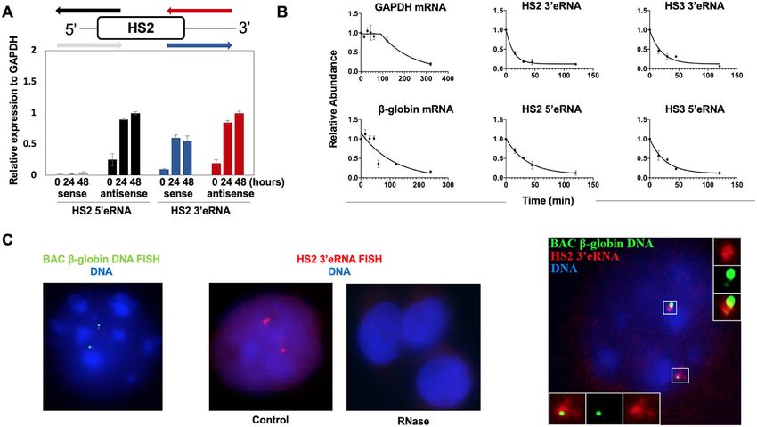
1384 Nucleic Acids Research, 2021, Vol. 49, No. 3
tion of Pol II at the respective locations and reduced expres- MATERIALS AND METHODS
sion of globin genes (20,21). On the other hand, the LCR
Generation and characterization of human LCR transgenic
is positioned in relatively close proximity to the adult -
mice
globin gene promoter in differentiated cells expressing the
gene (22). In erythroid cells lacking the transcription fac- Enzymes ClaI and NotI were used to cut out the human -
tor NF-E2, adult -globin expression was reduced and Pol globin locus control region from a plasmid (pRS316/LCR)
II accumulated at the LCR (17). Moreover, in vitro stud- previously described (16). The products were run on a 1%
ies demonstrated that Pol II is transferred from an immobi- agarose gel and the 15kbp band containing the LCR was
lized LCR to a -globin gene template in a process stimu- extracted from the gel and purified using the QIAquick Gel
lated by NF-E2 (18). Deletion of the murine LCR reduced Extraction Kit (Qiagen, 28706). The DNA was diluted to
recruitment of Pol II at the adult globin gene promoter concentrations of 1 and 0.5 ng/l in injection buffer and
by about 50%; however, the remaining Pol II recruited to injections were performed on fertilized FVB oocytes as de-
the globin gene was not transcriptionally competent (23). scribed previously (33). Eggs were implanted into the uterus
Downloaded from https://academic.oup.com/nar/article/49/3/1383/6104440 by guest on 28 November 2021
These data suggest that the LCR regulates both recruit- of pseudo-pregnant mothers and after birth, DNA was iso-
ment of Pol II and transcription elongation at the -globin lated and subjected to PCR for genotyping as described pre-
gene. viously (33).
The functional role of transcription initiation at en- The integration sites of the transgenes were determined
hancers or that of eRNAs is not completely understood by inverse PCR technique. For this, 3 g transgenic mouse
(24,25). Some studies have shown that eRNAs participate genomic DNA was digested using SacI and then run on
in the transcription activation function of enhancers (26). a 1% agarose gel to verify digestion. 1 g of this di-
Other studies demonstrated that the process of transcrip- gested DNA was used in a 400 ul intramolecular ligation
tion itself contributes to enhancer function (27). Mount- reaction and then the DNA was purified using phenol-
ing evidence suggests that eRNAs play a role in orchestrat- chloroform/isoamyl alcohol extraction and ethanol precip-
ing gene expression in cis or trans (24,25). Knockdown of itation. PCR reactions using inverse PCR primers comple-
eRNAs by RNA interference mechanisms or by drug me- mentary to sequences near the 3 and the 5 ends of the
diated transcription termination decreased target gene ex- inserted LCR were performed and the products were run
pression (28,29). eRNAs are relatively short in length and on 1% agarose gels. Resulting bands were purified using
it was shown that early termination of enhancer mediated the QIAquick Gel Extraction kit and were ligated with the
transcription requires the Integrator complex, which also pGEM-T vector (Promega, A3600). These plasmids were
terminates transcription of small nuclear RNAs (snRNAs) then transformed into Stbl2 competent cells (Invitrogen,
(30). Inhibition of Integrator complex function led to pro- 10268-019) and selected using Ampicillin. Colonies were
longed transcription at enhancers and a reduction in en- picked and selected by colony PCR (using the same inverse
hancer function (30). PCR primers) for positive clones. Minipreps (QIAprep Spin
The mammalian -type globin genes are expressed exclu- Miniprep Kit, 27106) were performed and the samples were
sively and at extremely high levels in erythroid cells in a de- sent for Sanger Sequencing. The primers used in the PCR
velopmental stage-specific manner (9). Those globin genes are listed in the Supplementary Table.
close to the LCR are expressed during the embryonic and Real-time quantitative PCR for copy number determi-
fetal stages, whereas the more distant globin genes are ex- nation was carried out as previously described with minor
pressed during the adult stages of hematopoiesis. The stage- modifications (34). Three to four mice from each transgenic
specific expression is mediated primarily by gene proximal line were analyzed on at least two separate occasions with
regulatory elements; however, the order of the genes with reactions prepared in triplicate. The 2−Ct method was
respect to the LCR also contributes to developmental reg- used for analysis and results were confirmed using the stan-
ulation (31,32). dard curve method (35). Mouse HS2 primers were used to
Here, we show that Pol II is recruited to the human amplify a reference sequence and DNA from a -YAC line
-globin LCR in transgenic mice and initiates generation was used as a one-copy calibrator.
of eRNAs independently from the linked human -globin
genes. RNA fluorescence in situ hybridization (RNA-FISH)
Cell culture and stable transfection
combined with DNA-FISH revealed that eRNA generated
from HS2 remains associated with the -globin gene locus. MEL cells were grown in DMEM medium (Cellgro,
Depletion of the eRNA 3 to HS2 (HS2 3 eRNA) in MEL Manassas, VA) supplemented with 10% (vol/vol) fe-
cells caused a reduction in adult -globin gene transcrip- tal bovine serum (FBS; Sigma) and 1% (vol/vol)
tion and an accumulation of Pol II at LCR element HS2. penicillin–streptomycin (P–S; Cellgro) in the presence
Ablation of Integrator subunit 11 (INTs11) in MEL or in of 5% CO2 at 37◦ C. The cell density was maintained at
differentiating human CD34+ cells reduced globin expres- 106 cells/ml. MEL cells were transfected with the TripZ
sion and increased the levels of LCR HS2 and HS3 associ- vector containing two different shRNAs (shRNA-1 and
ated eRNA. Furthermore, depletion of Integrator complex shRNA-2) against INTs11 and two shRNAs against
function led to accumulation of Pol II at LCR elements HS2 3 HS2eRNA [3 HS2(A) and 3 HS2(B)] or scrambled
and HS3 and a reduction of Pol II recruitment to the - control shRNAs. Single cell clones were generated using
globin gene. These data support a model of Pol II transfer an established protocol developed by John A Ryan at
from the LCR to the globin genes and implicate eRNA and Corning (https://www.corning.com/catalog/cls/documents/
Integrator in this process. protocols/Single cell cloning protocol.pdf). The plate was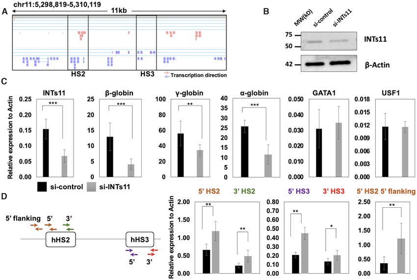
Nucleic Acids Research, 2021, Vol. 49, No. 3 1385
kept at 37◦ C for 7–9 days to allow single cells to grow into (Sigma Aldrich Catalog No. H4522), 2 U/ml human re-
colonies. The colonies were picked and seeded into 96-well combinant erythropoietin (EPO; STEMCELL Technolo-
plates. The 96-well plates were cultured for about 4 days gies Catalog No. 78007) for inducing differentiation. The
until the cells reached confluency. Clones were then scaled cells were subjected to siRNA treatment 24 h after induc-
up to 24-well plates and finally adapted for culture in flasks. ing with EPO. Accell non-targeting pool control siRNA
The sequences of shRNAs are listed in the Supplementary (control siRNA) (Accell Catalog No. D-001910-01) and Ac-
Table. cell Human INTs11 (54973) siRNA (INTs11 kd siRNA)
The pTRIPZ vector (for the INTs11kd cell line) or SMARTpool (Accell Catalog No. E-013798) were obtained
pGIPZ vector (for the 3 HS2 eRNA KD cell lines) con- from GE Dharmacon. A total of 50 000 human UCB
taining shRNA sequences were transfected into MEL cells CD34+ cells were suspended in Accell Delivery Medium
via the Trans-Lentiviral packaging kit (Thermo Scientific). (Dharmacon Catalog B-005000) with 2 units/ml human re-
Lenti-X 293T cells (Clontech) were passed at ∼70% con- combinant EPO, 1% penicillin–streptomycin, and seeded in
fluency for 2 days to ensure rapid growth. In six-well cell a 96-well plate. The siRNAs were maintained at a final con-
Downloaded from https://academic.oup.com/nar/article/49/3/1383/6104440 by guest on 28 November 2021
culture plates (Costar), 1.2 × 10⁶ Lenti-X 293T cells in centration of 1 mol/l according to the manufacturer’s pro-
two mLs of standard DMEM cell culture media [Dul- tocol. The cells were transferred to basic growth medium 72
becco’s modified Eagle’s medium (Cellgro), 10% (vol/vol) h after transfection. Aliquots of cells were collected at 72 h
fetal bovine serum (FBS) and 1% (vol/vol) penicillin– and at 6 days for downstream experiments.
streptomycin (Cellgro)] were incubated overnight at 37◦ C
in the presence of 5% CO2 . The transfection mix was cre-
Western blotting
ated by adding 4.3 l of Trans-Lentiviral packaging mix
with 6 g of pTRIPZ/pGIPZ-shRNA vector and bringing Western blotting procedures were performed using a pre-
the solution to 135 l with sterile water. To each transfec- viously published protocol (36). Briefly, cells were collected
tion mix 15 l of CaCl2 reagent was added (catalog number and lysed in radioimmunoprecipitation assay (RIPA) buffer
TLP5911), and the solution was gently mixed while adding (10 mmol/liter Tris–HCl, pH 8.0, 1 mmol/liter EDTA,
150 l 2× HEPES-buffered saline solution (catalog num- 0.5 mmol/l EGTA, 1% Triton X-100, 0.1% sodium de-
ber TLP5910). The completed transfection mix was incu- oxycholate, 0.1% SDS, 140 mmol/l NaCl, 1× protease
bated at room temperature for 3 min before being added in a inhibitor). The supernatant containing the proteins was
drop wise manner to the Lenti-X 293T cells. Cells were incu- loaded onto 4–15% (wt/vol) SDS-PAGE gels (Bio-Rad,
bated for 12 h at 37◦ C in the presence of 5% CO2 . The media Hercules, CA). After electrophoresis the proteins were
with transfection mix was exchanged for Dulbecco’s mod- transferred to polyvinylidene difluoride (PVDF) mem-
ified Eagle’s medium with 5% (vol/vol) fetal bovine serum branes (Bio-Rad). The PVDF membranes were incubated
(FBS) and 1% (vol/vol) penicillin–streptomycin. The Lenti- with anti-INTs11 (A301-274A; Bethyl Laboratories Inc.),
X 293T cells were then incubated with the reduced serum anti-GAPDH (catalog number CST5174; Cell Signaling,
media at 37◦ C in the presence of 5% CO2 for 48 h. The vi- Beverly, MA), or anti--actin (catalog number sc-47778,
ral particles were released into the media over a period of Santa Cruz, Biotechnology, Santa Cruz, CA) antibodies.
48 h. The media with viral particles was cleared of detached The membranes were washed and incubated with secondary
cells and debris by centrifugation at 3600 rpm for 5 min. goat anti-rabbit IgG-HRP (catalog number sc-2004) or goat
A mixture of 1 ml virus containing media, 1 ml of stan- anti-mouse IgG-HRP (catalog number sc-2005; Santa Cruz
dard DMEM cell culture media, and 8 g/ml polybrene Biotechnology, Santa Cruz, CA). Antibodies were detected
(Sigma) was used to resuspend 4 × 10⁵ MEL cells. MEL using an enhanced chemiluminescence (ECL) reagent (Mil-
cells were incubated with virus for 48 h before the media was lipore, Danvers, MA) and visualized with X-ray films (Ko-
refreshed. The transfected MEL cells were then subjected dak, Rochester, NY).
to selection with 1 g/ml puromycin for 7 days. The trans-
fected MEL cells were maintained with standard DMEM
RNA extraction and gene expression analysis
cell culture media containing 0.25 g/ml puromycin. Sin-
gle cell clones for each shRNA stably transfected cell line Total RNA was extracted using the RNeasy kit (Qiagen,
were generated as described above. For induction of differ- Hilden, Germany) following the manufacturer’s protocol.
entiation, MEL cells were grown in 2% (vol/vol) dimethyl RNA was reverse transcribed into cDNA using the IScript
sulfoxide (DMSO) for up to 72 h. For Integrator shRNA cDNA synthesis kit (Bio-Rad). The cDNAs were subjected
expression, transfected MEL cells were treated with both 1 to qPCR in a 10-l reaction mix with SYBR green and
g/ml Doxycycline (Sigma, D9891) and 2% final volume 1.5 mol/l of each forward and reverse primer. Gene ex-
DMSO (Sigma, D2650) for 24 and 48 h. pression was analyzed by the normalization of expression
The Umbilical Cord Blood (UCB) CD34+ cells were ob- to that of GAPDH/ß-Actin using the CT method. The
tained from STEMCELL Technologies. A total of 2 × 10⁵ primer sequences used for RT-qPCR are listed in the Sup-
cells were expanded in Stem Span Serum Free Expansion plementary Table.
Medium II (STEMCELL Technologies, Catalog 09605)
supplemented with StemSpan™ Erythroid Expansion Sup-
Chromatin immunoprecipitation
plement (100×) (Catalog No. 02691) and 1% penicillin–
streptomycin. The cells were allowed to expand for 9 days The chromatin immunoprecipitation (ChIP) protocol used
with frequent media changes. On the ninth day, the me- in this study was described previously (36). Briefly, 5 ×
dia was changed and supplemented with 3% human serum 106 cells were collected and cross-linked with 1% (vol/vol)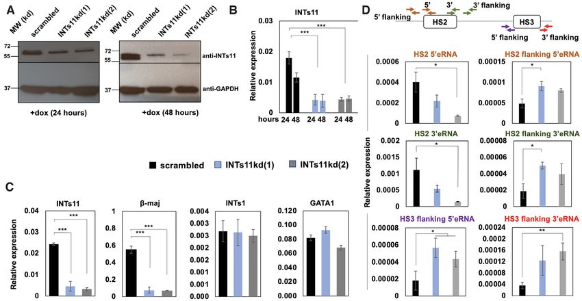
1386 Nucleic Acids Research, 2021, Vol. 49, No. 3
formaldehyde at room temperature for 10 min. The reac- mg of salmon sperm DNA. DNA was lyophilized in a heat-
tion was quenched with 125 mmol/l glycine, and the cells ing block, resuspended in 7 l of deionized formamide and
were incubated in cell lysis buffer (5 mmol/l PIPES [1,4- mixed well (at 15–25◦ C for at least 30 min). Probe was de-
piperazinediethanesulfonic acid], pH 8.0, 85 mmol/l KCl, natured and pre-labelled with Cot-1 DNA (85◦ C for 5 min,
0.5% NP-40 [nonyl octylphenoxypolyethoxylethanol], 1× 37◦ C for 20 min, 12◦ C hold). Fixed chromosome speci-
protease inhibitor) and nucleus lysis buffer (50 mmol/l mens were denatured by immersing the slides in denatu-
Tris, pH 8.0, 10 mmol/l EDTA, 0.32% SDS, 1× protease ration buffer (70% formamide, 2× SSC [pH 7.0]) at 82◦ C
inhibitor). Chromatin was fragmented to average 300-bp for 2 min. 10 l of the denatured probe was applied onto
fragments on ice using a Bioruptor disruptor (Diagen- an 18 × 18-mm glass coverslip (prewarmed to 37◦ C) and
ode, Denville, NJ). For each immunoprecipitation reaction, the slide was lowered onto the probe mix to avoid trap-
chromatin from 2 × 10⁵ cells was incubated with 2.5 g anti- ping air bubbles. Rubber cement was applied to seal around
body [anti-INTS11 (A301-274A; Bethyl Laboratories Inc.,), the coverslip. The slides with samples were hybridized in
anti-GAPDH (CST5174; Cell Signaling), anti-RNA poly- a humidified chamber in a hot-air oven at 37◦ C for 4 h.
Downloaded from https://academic.oup.com/nar/article/49/3/1383/6104440 by guest on 28 November 2021
merase II, clone CTD4H8 (Millipore, 05-623), anti-RNA The slides were washed with 2× SSC followed by wash-
polymerase II CTD repeat YSPTSPS (phospho S5) anti- ing in wash buffer (70% formamide, 2× SSC (pH 7), pre-
body (4H8 ab5408; Abcam), or rabbit polyclonal anti-RNA heated to 42◦ C). 200 l of detecting solution (4% dried milk
polymerase II CTD repeat YSPTSPS (phospho S2) ab5095; powder, 4× SSC) was added to the hybridized sample and
Abcam] plus protein A/G magnetic beads (Pierce) rotat- incubated at 25◦ C for 20 min. 50l of detecting solution
ing at 4◦ C overnight. The chromatin-antibody-bead com- containing Anti Digoxigenin Alexa 488 antibody (1:1000)
plexes were subjected to washing, eluting, and reverse cross- was added to a coverslip and incubated for 2 h at RT. The
linking at 65◦ C overnight. The DNA was purified using a slides were then washed in wash buffer (containing 2× SSC,
PCR purification kit (Qiagen) and subjected to qPCR as 0.1% Tween 20 and DAPI to stain chromatin), three times at
described earlier. The sequences of the primers used in the 42◦ C. The slides were mounted, and images were analyzed
ChIP assays are listed in the Supplementary Table. and documented using a Leica TCS SP5 confocal micro-
scope. The sequences of the probes used in DNA FISH are
listed in the Supplementary Table. For immunofluorescence
Chromatin-associated RNA isolation
microcopy MEL cells were incubated with the Pol II-S2P
The chromatin associated RNA was isolated along with the primary and FITC-conjugated secondary antibody (Santa
cytoplasmic and nucleoplasmic fraction following a pub- Cruz, sc2777).
lished protocol (37). Briefly, 105 –106 cells were homoge- The RNA FISH probes were prepared and labelled by
nized by lysis with cell lysis buffer (10 mM Tris pH 7.4, nick translation using a nick translation kit. (Catalog No.
150 mM NaCl, 0.15% Igepal CA-630). The lysate was cen- 11745824910, Roche). The hybridization solution was pre-
trifuged through a 24% sucrose cushion (sucrose buffer, 10 pared by mixing 50% formamide, 2× SSC, dextran sulfate,
mM Tris pH 7.4, 150 mM NaCl, 24% sucrose), which yields yeast t-RNA and nick-translated probe and was incubated
a pellet with purified nuclei and the supernatant represent- at 3◦ C for 10 min. The probe was denatured by heating at
ing the cytoplasmic fraction. The sedimented nuclei were 90◦ C for 5–10 min. The probe was chilled on ice immedi-
gently resuspended in glycerol buffer (20 mM Tris pH 7.4, ately. Fixed MEL cells were hybridized with the denatured
75 mM NaCl, 0.5 mM EDTA, 50% glycerol) followed by 3 HS2 LCR probe overnight. The probe was detected us-
rapid lysis in nuclear lysis buffer (10 mM Tris pH 7.4, 1 M ing a 5-layer Ab system (biotin-StAv) for 12 h (2× signal
Urea, 0.3 M NaCl, 7.5 mM MgCl2 , 0.2 mM EDTA, 1% amplification) (1st antibody StAv Alexa 488 (1:1000) 2 h at
Igepal CA-630). After a brief centrifugation at 13 000g for RT, wash with 3× PBS for 5 min, second Ab Biotinylated
2 min, the supernatant represented the nucleoplasm and the Anti Streptavidin (1:200) 2 h at RT, wash with 3× PBS for
pellet contained the chromatin associated RNA. RNA from 5 min, first antibody StAv Alexa 488 (1:1000) 2 h at RT,
all fractions was isolated by Trizol extraction (15596026, In- wash with 3× PBS for 5 min, second Ab Biotinylated Anti
vitrogen). The RNA was reverse transcribed into cDNA us- Streptavidin (1:200) 2 h at RT, wash with 3× PBS for 5 min,
ing the IScript cDNA synthesis kit (Bio-Rad). The cDNAs first antibody StAv Alexa 488 (1:1000) 4◦ C overnight). Post-
were subjected to qPCR in a 10-l reaction mix with SYBR hybridization wash was performed using 2× SSC for 5 min,
green and 1.5 mol/l of each forward and reverse primer. followed by 1× SSC for 5 min and another 1× SSC wash
Gene expression was analyzed by the normalization of ex- for 5 min at 37◦ C. DNA was counterstained with DAPI.
pression to that of the chromatin fraction of each sample In control experiments cells were treated with RNase. The
using the CT method. The primer sequences used for RT- slides were mounted, and images were analyzed and docu-
qPCR are listed in the Supplementary Table. mented using a Leica TCS SP5 confocal microscope. The
sequence of the probe used in RNA FISH is listed in the
Supplementary Table.
DNA and RNA fluorescence in situ hybridization
Mouse Fetal Liver cells/MEL cells were fixed on glass slides
Statistics
and incubated in methanol at –20◦ C for 10 min, dried and
then incubated in 300 l of 10% formaldehyde and 2× SSC The experiments were repeated at least three times unless
for 8 min. A probe cocktail was prepared by mixing 20 ng stated otherwise. In experiments involving qPCR, each sam-
of probe DNA (ds Beta major probe labelled with Digox- ple from the three repeats was analyzed by PCR three times.
igenin using DIG Nick Translation Mix by Sigma) and 3 Error bars represent the standard error of the mean (SEM).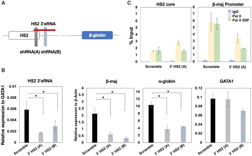
Nucleic Acids Research, 2021, Vol. 49, No. 3 1387
Statistical significance (P value) was determined using Stu- at the 5 end, to distinguish the signal from the potential
dent’s t-test. sense transcription of the GRPELI gene. We did indeed de-
tect eRNA at the transgenic HS2 5 region. This transcript
RESULTS was absent in the non-transgenic control. Together, the data
demonstrate that the transgenic human LCR is capable of
Recruitment of Pol II to human LCR HS2 in transgenic
independently recruiting Pol II transcription complexes in
mouse fetal liver
the absence of linked globin genes.
Previous data have shown that LCR HSs associate with
transcription complexes (16,17). To determine if the LCR
Stability, location and function of HS2 3 eRNA in MEL cells
is able to recruit transcription complexes in the absence of
linked globin genes we generated transgenic mice with the To further analyze the importance of eRNAs with re-
human LCR (Figure 1A). The fragment encompassed all spect to globin gene regulation we examined their expres-
of the human -globin LCR HSs but none of the human sion, stability and localization in differentiating murine ery-
Downloaded from https://academic.oup.com/nar/article/49/3/1383/6104440 by guest on 28 November 2021
globin genes. We generated two transgenic lines harboring throleukemia (MEL) cells (Figure 2). We focused on char-
one or up to six copies of the human LCR (Figure 1A). acterizing transcripts around LCR HS2, which has been
The copy number was determined by quantitative PCR us- shown to display the strongest enhancer activity. Examina-
ing a human -globin yeast artificial chromosome (YAC) tion of sense and antisense transcription 5 and 3 of LCR
transgenic mouse as a one copy calibrator (32). We used in- HS2 revealed that both sense eRNA 3 of HS2 and antisense
verse PCR to map the integration sites of the transgenes. eRNA transcription 5 and 3 of HS2 increased 24 and 48
In the two transgenic lines, the human LCR integrated into h after induction of differentiation (Figure 2A). We did not
an intron of protein-coding genes. In line 622, four to six detect upregulation of a sense transcript 5 of HS2. These
copies of the transgenic LCR integrated into the last intron data are largely consistent with previous reports which also
of the GrpE-like I gene (GRPELI), which encodes a pro- demonstrated that formation of eRNAs at the LCR pre-
tein of the mitochondrial PAM (presequence translocase- cedes the transcription of the -globin gene (41).
associated motor) complex (38). In line 680, one copy of Previous studies have shown that eRNAs are unstable
the transgenic LCR integrated into an intron of the OS- (24). We measured the stability of HS2 and HS3 5 and
GEP gene, which encodes a putative O-sialoglycoprotein 3 eRNAs as well as GAPDH and maj-globin RNA by
endopeptidase (39). RT-qPCR of MEL cells treated with the transcription in-
We next examined the interaction of Pol II with the trans- hibitor actinomycin D (Figure 2B). Actinomycin D inter-
genic human LCR and with the endogenous mouse LCR calates into DNA and prevents unwinding of DNA thus
using chromatin immunoprecipitation (ChIP) (Figure 1A). inhibiting RNA polymerase activity (42). The transcripts
We used an antibody that does not discriminate between the of the -maj-globin and GAPDH genes revealed stabilities
different phosphorylated forms of Pol II (S5P or S2P). The with a half-life (t1/2 ) of 98 and 78 min, respectively. In con-
data show that Pol II interacted with human HS2 in the two trast, HS2 3 eRNA revealed a significant shorter half-life of
transgenic lines examined. We also examined association about 9 min. The half-life of the other LCR eRNAs was ∼14
with a region flanking the human LCR HSs 2 and 3 (Human (HS3 3 eRNA), ∼18 (HS3 5 eRNA) and 25 (HS2 5 eRNA)
HS2–3 Linker). Pol II did not interact with the transgenic min.
HS flanking region, ruling out the possibility that interac- We next performed combined RNA and DNA FISH to
tion of Pol II is detected because the transgene integrated examine the location of HS2 3 eRNA with respect to the
into an active gene locus. The binding of Pol II at HS2 in -globin gene locus (Figure 2C). We used a bacterial artifi-
line 680 was as efficient as the binding of Pol II at the en- cial chromosome (BAC) probe for the murine -globin gene
dogenous LCR HS elements, and as expected, less efficient locus and probes specific for HS2 3 eRNA. The data show
compared to binding of Pol II at the endogenous mouse - that most cells revealed two signals for the -globin gene
maj-globin promoter. This is relevant as line 680 harbored locus (green) in induced MEL cells (representative nucleus,
one copy of the human LCR. Figure 2C, left). Cells induced to differentiate for 48 hours
We next examined association of the transgenic LCR revealed two RNA foci (red) as well (representative nucleus,
with transcription foci in mouse fetal liver cells. We isolated Figure 2C, middle-left), suggesting that a large fraction of
fetal liver cells from transgenic line 622 and subjected the the eRNA is concentrated at a specific nuclear location.
cells to immunofluorescence microscopy using antibodies The HS2 3 eRNA foci were not visible in cells treated with
specific for the serine 2 phosphorylated form of Pol II (Pol RNase, confirming the specificity of the signals (three rep-
II-S2P) and a fluorescently labeled DNA probe correspond- resentative nuclei, Figure 2C, middle-right). Combination
ing to the human LCR (Figure 1B). Pol II-S2P represents of RNA and DNA FISH revealed that the HS2 3 eRNA is
the transcription elongating form of Pol II (40). The data present in cloud-like foci and located in close proximity to
show that the transgenic LCR associates with Pol II-S2P and partially overlapping with the -globin gene locus (rep-
foci, suggesting that the LCR is in close proximity to tran- resentative nucleus, Figure 2C, right).
scription domains. It is possible that transcription of the eRNA molecules per se have been shown to contribute
GRPELI gene contributes to associations with transcrip- to the activation of gene expression by enhancers, whereas
tion foci. To examine if transgenic LCR HSs initiate for- other studies demonstrated that it is the process of eRNA
mation of eRNA, we subjected fetal liver RNA from trans- transcription that is crucial for gene regulation (24,27). We
genic line 622 to reverse transcriptase-PCR analysis (Figure reduced the abundance of the HS2 3 eRNA by shRNA me-
1C). In this experiment, we analyzed HS2 antisense eRNA diated knockdown in MEL cells (Figure 3). We generated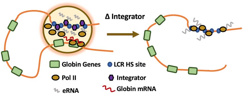
1388 Nucleic Acids Research, 2021, Vol. 49, No. 3
Downloaded from https://academic.oup.com/nar/article/49/3/1383/6104440 by guest on 28 November 2021
Figure 1. Recruitment of Pol II to human LCR HS2 in transgenic mice. (A) Two transgenic mouse lines were generated in which the human LCR integrated
with four to six copies or one copy into introns of the GRPEL1 (line 622) or OSGEP (line 680) genes, respectively. Fetal liver (12.5 dpc) was isolated from
the transgenic mice and subjected to ChIP followed by qPCR using antibodies specific for Pol II and control IgG, and primers specific for mouse or
human HS2, for a region in between human HS2 and HS3 (HS2-3 Linker), or for the mouse -maj-globin promoter. Error bars reflect SEM from three
independent experiments. (B) Association of the transgenic human LCR with Pol II foci in mouse fetal liver cells (12.5 dpc). Fetal liver cells were isolated
from transgenic line 622 and subjected to immunofluorescence using antibodies specific for Pol II-S2P (green) and a fluorescently labeled probe specific for
the transgenic human LCR (red; DNA in blue). The magnified insert shows association between the transgenic LCR and Pol II-S2P (five out of six nuclei
showed association of the transgenic LCR with transcription foci). (C) Analysis of HS2 5 eRNA. RNA was isolated from fetal liver of transgenic line 622
(12.5 dpc) and subjected to PCR analysis with or without reverse transcriptase (noRT). Results from analyzing DNA from wild-type, non-transgenic mice,
and from a negative H2 O control are also shown. The diagram at the bottom shows the location of primers used for the RT-PCR analysis.
two single cell clones expressing different shRNAs against albeit not to the extent observed for the -globin gene. We
the HS2 3 eRNA (3 HS2 A and B; Figure 3A). Stably trans- next assessed the binding of Pol II using ChIP at HS2 and
fected cells expressing the 3 HS2 shRNAs revealed a signif- the adult -globin promoter in cells expressing the scram-
icant reduction in the HS2 3 eRNA compared to clones ex- bled shRNA and in cells expressing the shRNA directed
pressing scrambled shRNA (Figure 3B). The reduction in against HS2 3 eRNA (Figure 3C). We used two different an-
HS2 eRNA resulted in a decrease in adult -globin gene tibodies; antibodies specific for the serine 5 phosphorylated
expression. Expression of the GATA1 gene, which encodes form (Pol II S5P) and antibodies that do not distinguish be-
for an essential erythropoietic transcription factor, was not tween the differentially phosphorylated forms of Pol II (Pol
affected in the 3 HS2 (A) KD cells, and was only mildly re- II). Figure 3C shows that reducing the abundance of the
duced in the 3 HS2 (B) KD cells (43). However, we also de- HS2 3 eRNA caused a reduction in the association of Pol II
tected a decrease in expression of the adult ␣-globin gene, and Pol II-S5P with the -maj-globin gene promoter, con-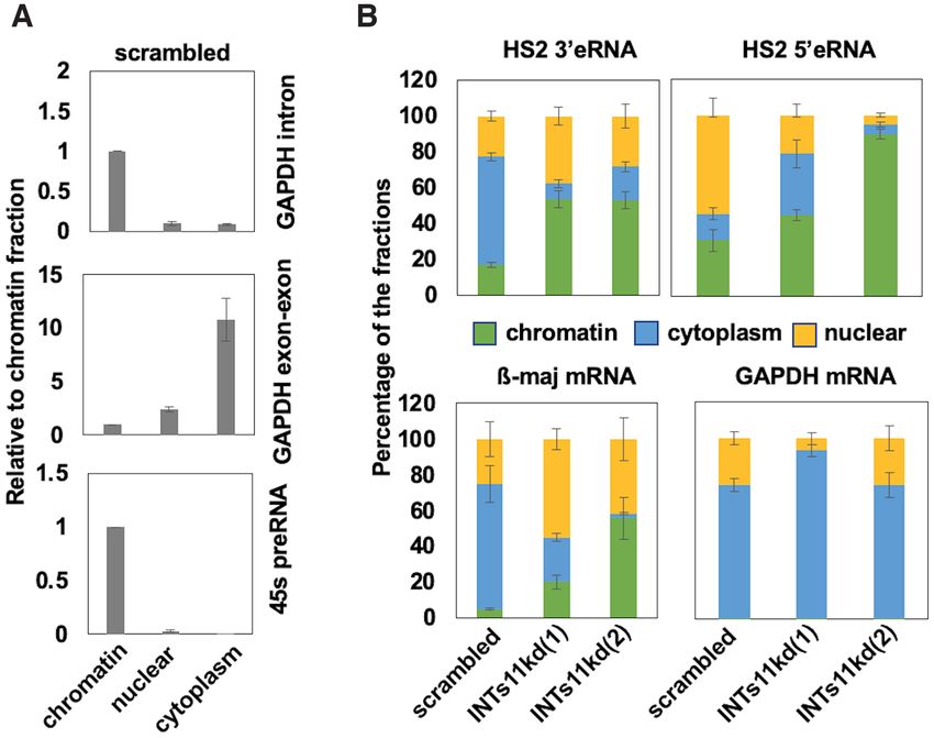
Nucleic Acids Research, 2021, Vol. 49, No. 3 1389
Downloaded from https://academic.oup.com/nar/article/49/3/1383/6104440 by guest on 28 November 2021
Figure 2. Formation, stability and localization of LCR associated eRNA. (A) Transcription of 5 and 3 HS2 eRNAs during differentiation of MEL cells.
Diagram on top showing primers used to assess HS2 5 and 3 antisense and sense transcription. MEL cells were subjected to differentiation for 24 and 48 h
with 2% (final v/v) DMSO. RNA was isolated, subjected to reverse transcription using strand-specific primers, and analyzed by qPCR. (B) Measurements
of GAPDH, -maj-globin RNA as well as HS2 and HS5 5 and 3 eRNA half-life. RNA was isolated from differentiated MEL cells incubated with
actinomycin D for up to 8 h and subjected to RT-qPCR using primers specific for GAPDH, -maj-globin, and HS2 or HS3 5 and 3 eRNAs. Error
bars reflect SEM from three independent experiments. (C) Differentiated MEL cells were subjected to -globin gene locus DNA-FISH (left, representative
nucleus) or HS2 3 eRNA-FISH (middle-left, representative nucleus; middle-right: RNAse treatment to show specificity of RNA signal, three representative
nuclei) using fluorescently labeled probes specific for the murine -globin gene locus (green) or HS2 3 eRNA (red; DNA: blue). Right: combination of HS2
3 eRNA and -globin gene locus DNA FISH. HS2 3 eRNA (red) is associated with the -globin gene locus (green; DNA: blue; four out of four nuclei
showed association between the globin gene locus on HS2 3 eRNA).
sistent with reduced expression of the gene. In contrast, the INTs11 protein (Figure 4A) and RNA (Figure 4B) com-
levels of Pol II, but not Pol II-S5P, were increased at LCR pared to MEL cells expressing scrambled shRNA. Gene
HS2 in cells expressing HS2 3 shRNA. These data show that expression analysis demonstrated that INTs11 and -maj-
the HS2 3 eRNA plays an active role in the regulation of globin expression was reduced in differentiated MEL cells
adult -globin gene expression. with inducible INTs11 KD compared to cells expressing
scrambled control shRNA (Figure 4C). In contrast, we did
not observe a change in expression of INTs1, the largest
Role of Integrator in LCR mediated control of globin gene subunit of integrator, or GATA1 genes. We also observed
expression a reduction in expression of HS2 5 and 3 eRNA that are
Recently, it was shown that many enhancers recruit the Inte- expressed immediately downstream of the HS2 core region
grator complex which mediates termination of eRNA tran- (Figure 4D). Using primers located further away from the
scription (30). Integrator is a component of the transcrip- core, we observed an increase in the levels of eRNAs both
tion termination machinery implicated in the processing of further upstream and downstream of HS2 (HS2 flanking 5
splicing associated small nuclear RNAs (snRNAs) (44). The and 3 eRNA). The same was true for outer eRNA associ-
involvement of Integrator in the early transcription termi- ated with HS3, which increased both at the 5 and 3 end
nation of eRNA suggests that the release of Pol II from en- (HS3 flanking 5 and 3 eRNA).
hancers could aide in the transfer of Pol II to promoters. We We next analyzed the occupancy of Pol II in the globin
used doxycycline (dox) inducible shRNA mediated knock- gene locus as well as at the GAPDH gene in the INTs11
down of Integrator subunit 11 (INTs11) in differentiating KD and control differentiated MEL cells using ChIP (Fig-
MEL cells to examine the function of Integrator in LCR ure 5). We used antibodies specific for the serine 5 (Pol II
mediated recruitment of Pol II to the adult -globin gene S5P) and serine 2 (Pol II S2P) phosphorylated forms of Pol
(Figure 4). INTs11 is the catalytic subunit of the Integrator II. Pol II-S2P represents the elongating form of Pol II (40).
complex exhibiting endonuclease activity (44). Single MEL We also used antibodies specific for INTs11. The binding
cell clones expressing two different shRNAs against INTs11 of Pol II-S5P or Pol II-S2P was not altered at the GAPDH
[INTs11kd(1) and INTs11kd(2)] showed reduced levels of promoter in INTs11 KD versus control cells, while the inter-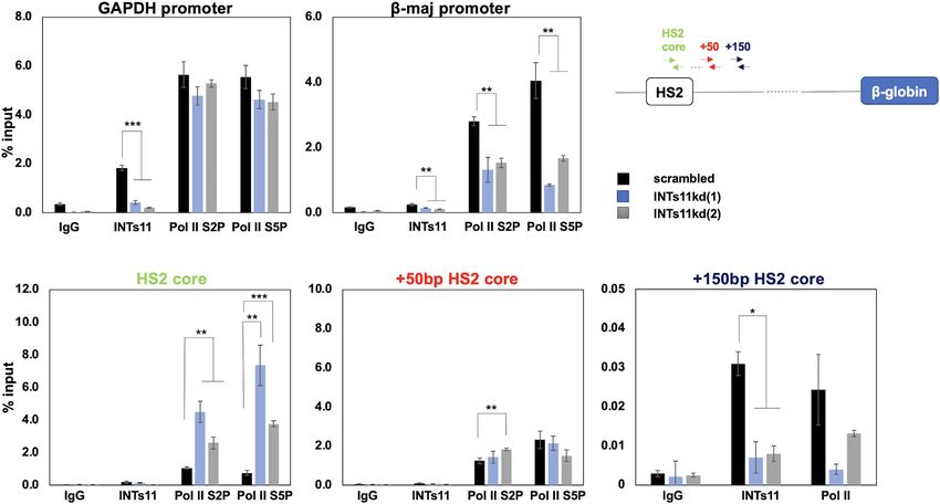
1390 Nucleic Acids Research, 2021, Vol. 49, No. 3
Downloaded from https://academic.oup.com/nar/article/49/3/1383/6104440 by guest on 28 November 2021
Figure 3. Changes of transcription and Pol II association in MEL cells expressing shRNA against HS2 3 eRNA. (A) Diagram showing targeting of HS2
3 eRNA by two shRNAs. (B) Depletion of HS2 3 eRNA reduces -maj-globin gene expression. MEL cells stably expressing shRNA against HS2 3 eRNA
(clones A and B) or scrambled control shRNA, were subjected to RT-qPCR using primers specific for HS2 3 eRNA or the -maj-globin, ␣-globin, and
GATA1 genes. HS2 3 eRNA expression was normalized to GATA1 RNA because of the high expression of -actin. (C) Changes in Pol II accumulation in
the -globin gene locus in cells expressing HS2 3 eRNA shRNA. MEL cells stably expressing shRNA against HS2 3 eRNA (clone A) or scrambled shRNA
were subjected to ChIP-qPCR using antibodies specific for Pol II, Pol II-S5P, and control IgG, and primers specific for the HS2 core and the -maj-globin
gene. Error bars reflect SEM from three independent experiments. (*P < 0.05)
action of INTs11 was reduced in INTs11 KD cells. We ob- presence of 45S precursor ribosomal RNA (pre-rRNA) in
served little binding of Integrator subunit 11 at the adult - the chromatin fraction was consistent with previous find-
maj-globin gene promoter. INTs11 KD reduced association ings (Figure 6A) (37). We observed an increase in the chro-
of both forms of Pol II with the -globin gene consistent matin associated fraction of HS2 5 and 3 eRNA in INTs11
with reduced expression levels. Interestingly, we observed KD cells while at the same time there was a decrease of -
an increase in the association of Pol II-S2P and Pol II-S5P maj-globin mRNA in the cytoplasm. Consistent with the
with the HS2 core region in INTs11 KD cells. There was observation that INTs11 KD did not affect GAPDH gene
little binding of INTs11 at the HS2 core. However, INTs11 expression, the relative abundance of GAPDH RNA in
associated with a region further downstream of HS2 (+150 the three fractions did not change after INTs11 KD (Fig-
bp). The association of INTs11 with the HS2 downstream ure 6B). The data were normalized to the chromatin frac-
region (+150 bp HS2 core) was reduced in INTs11 KD cells, tions and the presence of RNA species in each fraction has
as was the association of Pol II (Pol II-S2Pand Pol II-S5P). been calculated as a percentage as described by Conrad and
We did not observe a reduction in the association of Pol II Orum (37).
in the region at +50 bp downstream of the HS2 core. To validate our findings with respect to Integrator func-
Lai et al., showed that in the absence of Integrator sub- tion in the -globin gene locus, we reduced Integrator func-
unit 11, eRNAs remained bound to RNA Pol II and their tion in differentiating primary human erythroid cells. Fig-
primary transcripts accumulated at chromatin (30). To ex- ure 7A documents the presence of HS2 and HS3 5 and
amine if Integrator KD causes alterations in the associa- 3 eRNA in differentiated CD34+ cells, according to data
tion of eRNA with chromatin, we isolated cytoplasmic, nu- from the ENCODE project (45). The CD34+ cells were de-
clear, and chromatin associated RNA from INTs11 KD and rived from umbilical cord and transcribe both the adult
scrambled control MEL cells differentiated for 48 hours - and fetal ␥ -globin genes upon erythroid differentiation
(Figure 6). The control experiments confirmed the success- (Figure 7C). We subjected the differentiated cells to siRNA
ful separation of the RNAs showing that intronic GAPDH mediated knock-down of INTs11 (si-INTs11) or to a con-
RNA is chromatin associated, whereas intron-less GAPDH trol siRNA (si-control). The data show that RNA and pro-
RNA is located mostly in the cytoplasm. Furthermore, the tein expression of INTs11 was reduced in siINTs11 treatedNucleic Acids Research, 2021, Vol. 49, No. 3 1391
Downloaded from https://academic.oup.com/nar/article/49/3/1383/6104440 by guest on 28 November 2021
Figure 4. Transcription changes in MEL cells stably expressing shRNA against INTs11. (A) MEL cells stably expressing shRNA against INTs11 (clones
1 and 2) or scrambled control were subjected to western blotting experiments using antibodies specific for INTs11 or GAPDH. (B) MEL cells stably
expressing shRNA against INTs11 (clones 1 and 2) or scrambled control were subjected to RT-qPCR using primers specific for INTs11. Expression was
normalized to that of GAPDH. Error bars reflect SEM from three independent experiments (***, P < 0.001). (C, D) MEL cells expressing shRNA against
INTs11 [INTs11kd(1) and INTs11kd(2)] or scrambled control shRNA (scrambled) were subjected to RT-qPCR using primers specific for the INTs11,
-maj-globin, INTs1, GATA1 genes (C) as well as primers specific for HS2 and HS3 eRNAs (D). The diagram on top of panel D illustrates the location
of primers used in the analysis of LCR HS2 and HS3 associated eRNA. Error bars reflect SEM from three independent experiments (***P < 0.001; **P
< 0.01; *P < 0.05).
Figure 5. Increased association of Pol II with the LCR HS2 core region in INTs11 depleted MEL cells. MEL cells stably expressing shRNA against INTs11
(clones 1 and 2) or scrambled control were subjected ChIP-qPCR using antibodies specific for Pol II-S2P, Pol II-S5P and INTs11, and primers specific for
GAPDH, -maj-globin, and different regions downstream of LCR HS2 as indicated. Error bars reflect SEM from three independent experiments (***P
< 0.001; **P < 0.01; *P < 0.05).1392 Nucleic Acids Research, 2021, Vol. 49, No. 3
Downloaded from https://academic.oup.com/nar/article/49/3/1383/6104440 by guest on 28 November 2021
Figure 6. Increased association of HS2 3 eRNA with chromatin in INTs11 depleted cells. (A) Cytoplasmic, nuclear, and chromatin associated RNA was
isolated from differentiated MEL cells and subjected to RT-PCR using primers specific for a GAPDH intron, GAPDH exon-exon junctions, and 45Spre-
rRNA. (B) Cytoplasmic, nuclear, and chromatin associated RNA was isolated from differentiated MEL cells stably expressing shRNA against INTs11
(clones 1 and 2) or scrambled control and subjected to RT-qPCR using primers specific for HS2 3 eRNA, HS2 5 eRNA, -maj-globin, and GAPDH. The
data were normalized to the chromatin fraction and calculated as a percentage as described by Conrad and Orum (37). Error bars reflect SEM from three
independent experiments (**P < 0.01).
cells compared to the si-control treated cells (Figure 7 B and formation of bidirectional eRNAs (6,7). We studied the role
C). We observed a decrease in expression of the ␣- and - of eRNA and Pol II recruitment at the human and mouse
globin genes, and to a lesser extent expression of ␥ -globin. -globin LCR. Consistent with previous studies, we found
Consistent with our observations in MEL cells, we detected that the transgenic human LCR recruits transcription com-
an increase in HS2 and HS3 associated 5 and 3 eRNA in plexes and associates with transcription foci in fetal liver
si-INTs11 treated cells compared to si-control treated cells cells independently from the presence of the -type globin
(Figure 7D). This increase in eRNA was also observed in a genes (Figure 1) (48). This observation underscores the no-
region further upstream (5) of HS2 (HS2 5 flanking). tion that the association of Pol II at the LCR is not a re-
flection of interactions with linked transcriptionally active
globin genes. Previous studies have shown that during the
DISCUSSION
process of hematopoiesis, many enhancers recruit Pol II be-
Based on the current and previous data, we propose that fore Pol II is detectable at target genes, suggesting that Pol
the -globin LCR provides a highly accessible recruitment II recruitment to enhancers is involved in priming these reg-
site for coactivators and Pol II (19,46). A fraction of Pol ulatory DNA-elements (5). This is consistent with observa-
II is assembled into transcription complexes and generates tions that elevated eRNA transcription at the LCR precedes
eRNAs (Figure 8). Integrator terminates transcription at activation of the adult -globin gene after induction of ery-
enhancers and releases Pol II and eRNA. The eRNAs to- throid differentiation (41).
gether with Pol II, as well as co-activators such as Mediator The ablation of HS2 3 eRNA transcripts by shRNA
and Brd4 generate a transcription domain that transiently caused a decrease in the expression of the adult -globin
interacts with the globin promoters to load Pol II onto the gene up to 4-fold but did not affect expression of the
genes. Disruption of Integrator function prevents formation GATA1 gene (Figure 4). Surprisingly, reduction of HS2
of the LCR transcription domain due to the lack of release 3 eRNA using two different shRNAs also decreased expres-
of Pol II and eRNA. Recently, it was found that Integra- sion of the adult ␣-globin genes by about 2-fold. We can-
tor not only terminates transcription but also dephospho- not exclude the possibility that the two shRNAs have off-
rylates the C-terminal domain of Pol II (47). This seems to target effects. However, the two shRNAs contain different
be an important finding with respect to the Integrator me- sequences and we consider it unlikely that they both would
diated Pol II transfer model, as it is the unphosphorylated reduce ␣-globin as a consequence of off-target effects. The
form of Pol II that is recruited to Pol II promoters. ␣- and -globin gene loci have been shown to associate with
SEs recruit a large number of transcription factors, co- the same nuclear speckles suggesting some form of coor-
activators, as well as Pol II and as a consequence initiate the dinated regulation (49). However, removal of the -globinNucleic Acids Research, 2021, Vol. 49, No. 3 1393
Downloaded from https://academic.oup.com/nar/article/49/3/1383/6104440 by guest on 28 November 2021
Figure 7. Reduced globin gene expression and increased LCR HS2 and HS3 eRNA levels in differentiating CD34+ cells deficient for INTs11. (A) RNA
sequence reads in the human LCR HS2 and HS3 genomic region. Data were obtained from CD34+ blood progenitor cells from the ENCODE database
(ENCSR000CUA,(45). (B) Western blot analysis of INTs11 and -actin in differentiating CD34+ cells treated with INTs11 siRNA (si-INTs11) or non-
targeting control siRNA (si-control) for 72 h. (C) RT-qPCR expression analysis of INTs11, -globin, ␥ -globin, ␣-globin, GATA1 and USF1 genes in
differentiating CD34+ cells treated with si-INTs11 or si-control for 72 h. (D) RT-qPCR expression analysis of LCR HS2 and HS3 5 and 3 eRNAs in
differentiating CD34+ cells treated with si-INTs11 or si-control for 72 hours. The diagram on the left illustrates position of primers used to amply different
regions flanking LCR HS2 and HS3. Error bars reflect SEM from three independent experiments (***P < 0.001; **P < 0.01; *P < 0.05). Expression in
(C) and (D) was normalized to that of -actin (Actin).
LCR in mice had no effect on expression of the ␣-globin NAs are constantly being produced and could thus function
genes (50). It is possible that compensatory mechanisms in the process of gene regulation by contributing to the for-
in mice maintain ␣-globin expression in the absence of the mation of a transcription domain that concentrates Pol II or
LCR. by mediating proximity between enhancers and promoters.
The exact mechanisms by which eRNAs contribute to en- The turn-over of the eRNAs would allow rapid changes in
hancer function is unclear. It was recently shown that super- SE configuration. The observation that KD of HS2 3 eRNA
enhancers establish domains of a high concentration of Me- reduced expression of the adult -globin gene clearly sug-
diator and unphosphorylated Pol II (46,51). RNA could gests a functional role of at least a subset of LCR associated
play a role in the formation of SE associated transcription eRNAs.
domains. SE associated eRNAs have also been shown to The Integrator terminates transcription of snRNAs as
be involved in mediating proximity between SEs and tar- well as eRNAs and attenuates transcription at protein cod-
get genes (52).To further examine a potential role of LCR ing genes possibly by removing unproductive transcription
eRNA in -globin gene regulation, we analyzed the loca- complexes (30,44,53). Ablation of Integrator function led
tion of HS2 3 eRNA in relation to the -globin gene locus to reduced transcription and enhancer-promoter looping at
(Figure 2). We found that HS2 3 eRNA remains largely as- epidermal growth factor (EGF) induced genes (30). That
sociated with the -globin gene locus. The association of study not only implicated the Integrator complex in en-
the HS2 3 eRNA with the globin gene locus does not nec- hancer function but also suggested that eRNAs released
essarily reflect a specific function, it could rather be a con- from enhancers contribute to the communication between
sequence of the relatively short half-life, similar to intron enhancers and promoters. We conditionally reduced expres-
containing pre-mRNA. However, we found that the eRNAs sion of INTs11, the catalytic subunit of Integrator, in dif-
associated with LCR HSs exhibit a half-life of 9 min and ferentiating MEL cells and primary human CD34+ cells
longer. Furthermore, despite a relative short half-life, eR- and examined the consequence on eRNA formation, Pol1394 Nucleic Acids Research, 2021, Vol. 49, No. 3
Downloaded from https://academic.oup.com/nar/article/49/3/1383/6104440 by guest on 28 November 2021
Figure 8. Model of LCR mediated regulation of adult -globin gene expression focusing on the role of Integrator. The LCR associates with erythroid-
specific and basal transcription factors, which recruit co-activators, including Mediator, and Pol II. A fraction of Pol II initiates transcription of eRNA.
Integrator terminates transcription at the LCR and releases Pol II and eRNA which contributes to the formation of a transcription domain with high
concentration of Pol II (yellow circle).The -globin gene transiently associates with the transcription domain and Pol II is transferred to strong basal
promoter elements at the -globin gene. Disruption of Integrator function (shown on the right) prevents release of Pol II and eRNA from the LCR and,
as a consequence, prevents the loading of Pol II to the globin gene promoter.
II recruitment, and -globin gene transcription. The data the core in INTs11 KD MEL cells. In contrast, an increase
demonstrate that Integrator is required for the release of in eRNA was mostly observed downstream of the HS2 core
eRNA and Pol II from the -globin LCR (Figures 4–7). region in these cells. It is possible that the lack of termina-
The accumulation of Pol II at the LCR and the decreased tion of transcription downstream of HS2 leads to an accu-
recruitment to the -globin gene in the INTs11 KD cells mulation of Pol II at the site of initiation. Moreover, it has
suggests that Integrator is involved in the transfer of Pol II been reported that INTs13 regulates early steps in the for-
from the LCR to the -globin gene promoter. In addition, mation of enhancers (57). Thus, Integrator could play a role
Integrator mediated release of eRNA could contribute to in the initiation of transcription at SEs as well.
the formation of transcription domains that concentrate Pol In summary, the data demonstrate that Integrator re-
II (54). leases Pol II and eRNAs from the LCR and thereby con-
Using RNA proximity ligation, Cai et al. (52) demon- tributes to the loading of Pol II to the globin gene promot-
strated that eRNA frequently associates with promoter up- ers.
stream transcripts (PROMPTS) and RNA binding proteins
like hnRNPK. Ablation of a MYC SE associated noncod-
ing RNA reduced MYC transcription. The authors sug- SUPPLEMENTARY DATA
gest that hnRNPK oligomerization, orchestrated by eRNA, Supplementary Data are available at NAR Online.
contributes to the delivery of Pol II from the SE to the
target gene promoter. Recently, it was shown that noncod-
ing chromatin associated RNAs, including eRNAs, are en- ACKNOWLEDGEMENTS
riched for 5 splice sites and interact with U1snRNP, likely
via transcribing Pol II (55). It is possible that HS2 3 eRNA We thank our colleagues in the Bungert and Xie labora-
remains at the globin locus first via retention by U1snRNPs tories for discussions and encouragement. We thank Wara
and subsequently, after release by Integrator, participates Rattanaphong for technical assistance. We thank Drs Mike
in transcription domain formation and/or communication Kilberg, Rolf Renne, and Jixiu Shan (UF) for helpful dis-
between the LCR and the genes. This will be the subject of cussions and comments on the manuscript.
future investigations.
We and others provided evidence for a Pol II transfer FUNDING
mechanism in the -globin gene locus (19). For example,
Johnson et al. showed accumulation of Pol II at the LCR American Society of Hematology (to J.B.); National
and reduced -globin expression in MEL cells lacking tran- Institutes of Health [R56 DK111439, R01 DK052356
scription factor NF-E2 (17). Moreover, we demonstrated to J.B., R00-CA190886, R35-GM128753 to M.X.,
that Pol II recruited to an immobilized LCR template is R01DE026707, R21CA198820 to A.M.I.]. Funding
transferred to the -globin gene in a process depending on for open access charge: American Society of Hematology.
the -globin TATA-box and stimulated by NF-E2 (18). Fi- Conflict of interest statement. None declared.
nally, targeting a synthetic DNA-binding protein to the -
globin downstream promoter inhibited transcription elon-
gation and accumulated Pol II at the LCR (56). These data REFERENCES
together with the data presented here support a Pol II trans- 1. Andersson,R. and Sandelin,A. (2020) Determinants of enhancer and
fer model. promoter activities of regulatory elements. Nat. Rev. Genet., 21,
71–87.
There are some observations in our study that appear to 2. Gasperini,M., Tome,J.M. and Shendure,J. (2020) Towards a
be incongruent. For example, we found that Pol II levels in- comprehensive catalogue of validated and target-linked human
creased in the HS2 core but not in a region downstream of enhancers. Nat. Rev. Genet., 21, 292–310.Nucleic Acids Research, 2021, Vol. 49, No. 3 1395
3. Kim,T.K., Hemberg,M., Gray,J.M., Costa,A.M., Bear,D.M., Wu,J., 24. Arnold,P.R., Wells,A.D. and Li,X.C. (2019) Diversity and emerging
Harmin,D.A., Laptewicz,M., Barbara-Haley,K., Kuersten,S. et al. roles of enhancer RNA in regulation of gene expression and cell fate.
(2010) Widespread transcription at neuronal activity-regulated Front. Cell Dev. Biol., 7, 377.
enhancers. Nature, 465, 182–187. 25. Tippens,N.D., Vihervaara,A. and Lis,J.T. (2018) Enhancer
4. Denisenko,E., Guler,R., Mhlanga,M.M., Suzuki,H., Brombacher,F. transcription: what, where, when, and why? Genes Dev., 32, 1–3.
and Schmeier,S. (2017) Genome-wide profiling of transcribed 26. Lam,M.T., Cho,H., Lesch,H.P., Gosselin,D., Heinz,S.,
enhancers during macrophage activation. Epigenet. Chromatin, 10, 50. Tanaka-Oishi,Y., Benner,C., Kaikkonen,M.U., Kim,A.S., Kosaka,M.
5. Arner,E., Daub,C.O., Vitting-Seerup,K., Andersson,R., Lilje,B., et al. (2013) Rev-Erbs repress macrophage gene expression by
Drabløs,F., Lennartsson,A., Rönnerblad,M., Hrydziuszko,O., inhibiting enhancer-directed transcription. Nature, 498, 511–515.
Vitezic,M. et al. (2015) Transcribed enhancers lead waves of 27. Fitz,J., Neumann,T., Steininger,M., Wiedemann,E.M., Garcia,A.C.,
coordinated transcription in transitioning mammalian cells. Science, Athanasiadis,A., Schoeberl,U.E. and Pavri,R. (2020) Spt5-mediated
347, 1010–1014. enhancer transcription directly couples enhancer activation with
6. Hnisz,D., Abraham,B.J., Lee,T.I., Lau,A., Saint-André,V., physical promoter interaction. Nat. Genet., 52, 505–515.
Sigova,A.A., Hoke,H.A. and Young,R.A. (2013) Super-enhancers in 28. Hsieh,C.L., Fei,T., Chen,Y., Li,T., Gao,Y., Wang,X., Sun,T.,
the control of cell identity and disease. Cell, 155, 934–947. Sweeney,C.J., Lee,G.S., Chen,S. et al. (2014) Enhancer RNAs
7. Whyte,W.A., Orlando,D.A., Hnisz,D., Abraham,B.J., Lin,C.Y., participate in androgen receptor-driven looping that selectively
Downloaded from https://academic.oup.com/nar/article/49/3/1383/6104440 by guest on 28 November 2021
Kagey,M.H., Rahl,P.B., Lee,T.I. and Young,R.A. (2013) Master enhances gene activation. Proc. Natl. Acad. Sci. U.S.A., 111,
transcription factors and mediator establish super-enhancers at key 7319–7324.
cell identity genes. Cell, 153, 307–319. 29. Li,W., Notani,D., Ma,Q., Tanasa,B., Nunez,E., Chen,A.Y.,
8. Grosveld,F., van Assendelft,G.B., Greaves,D.R. and Kollias,G. (1987) Merkurjev,D., Zhang,J., Ohgi,K., Song,X. et al. (2013) Functional
Position-independent, high-level expression of the human beta-globin roles of enhancer RNAs for oestrogen-dependent transcriptional
gene in transgenic mice. Cell, 51, 975–985. activation. Nature, 498, 516–520.
9. Stamatoyannopoulos,G. (2005) Control of globin gene expression 30. Lai,F., Gardini,A., Zhang,A. and Shiekhattar,R. (2015) Integrator
during development and erythroid differentiation. Exp. Hematol., 33, mediates the biogenesis of enhancer RNAs. Nature, 525, 399–403.
259–271. 31. Peterson,K.R. and Stamatoyannopoulos,G. (1993) Role of gene order
10. Tuan,D., Solomon,W., Li,Q. and London,I.M. (1985) The in developmental control of human gamma- and beta-globin gene
“beta-like-globin” gene domain in human erythroid cells. Proc. Natl. expression. Mol. Cell. Biol., 13, 4836–4843.
Acad. Sci. U.S.A., 82, 6384–6388. 32. Tanimoto,K., Liu,Q., Bungert,J. and Engel,J.D. (1999) Effects of
11. Forrester,W.C., Takegawa,S., Papayannopoulou,T., altered gene order or orientation of the locus control region on
Stamatoyannopoulos,G. and Groudine,M. (1987) Evidence for a human beta-globin gene expression in mice. Nature, 398, 344–348.
locus activation region: the formation of developmentally stable 33. Bungert,J., Davé,U., Lim,K.C., Lieuw,K.H., Shavit,J.A., Liu,Q. and
hypersensitive sites in globin-expressing hybrids. Nucleic Acids Res., Engel,J.D. (1995) Synergistic regulation of human beta-globin gene
15, 10159–10177. switching by locus control region elements HS3 and HS4. Genes Dev.,
12. Li,Q., Peterson,K.R., Fang,X. and Stamatoyannopoulos,G. (2002) 9, 3083–3096.
Locus control regions. Blood, 100, 3077–3086. 34. Ballester,M., Castelló,A., Ibáñez,E., Sánchez,A. and Folch,J.M.
13. Tuan,D., Kong,S. and Hu,K. (1992) Transcription of the (2004) Real-time quantitative PCR-based system for determining
hypersensitive site HS2 enhancer in erythroid cells. Proc. Natl. Acad. transgene copy number in transgenic animals. BioTechniques, 37,
Sci. U.S.A., 89, 11219–11223. 610–613.
14. Ashe,H.L., Monks,J., Wijgerde,M., Fraser,P. and Proudfoot,N.J. 35. Livak,K.J. and Schmittgen,T.D. (2001) Analysis of relative gene
(1997) Intergenic transcription and transinduction of the human expression data using real-time quantitative PCR and the 2(-Delta
beta-globin locus. Genes Dev., 11, 2494–2509. Delta C(T)) Method. Methods, 25, 402–408.
15. Routledge,S.J. and Proudfoot,N.J. (2002) Definition of transcriptional 36. Hossain,M.A., Shen,Y., Knudson,I., Thakur,S., Stees,J.R., Qiu,Y.,
promoters in the human beta globin locus control region. J. Mol. Pace,B.S., Peterson,K.R. and Bungert,J. (2016) Activation of fetal
Biol., 323, 601–611. ␥ -globin gene expression via direct protein delivery of synthetic
16. Leach,K.M., Nightingale,K., Igarashi,K., Levings,P.P., Engel,J.D., zinc-finger DNA-binding domains. Mol. Ther. Nucleic Acids, 5, e378.
Becker,P.B. and Bungert,J. (2001) Reconstitution of human 37. Conrad,T. and Ørom,U.A. (2017) Cellular fractionation and isolation
beta-globin locus control region hypersensitive sites in the absence of of chromatin-associated RNA. Methods Mol. Biol., 1468, 1–9.
chromatin assembly. Mol. Cell. Biol., 21, 2629–2640. 38. Srivastava,S., Savanur,M.A., Sinha,D., Birje,A., R,V., Saha,P.P. and
17. Johnson,K.D., Christensen,H.M., Zhao,B. and Bresnick,E.H. (2001) D’Silva,P. (2017) Regulation of mitochondrial protein import by the
Distinct mechanisms control RNA polymerase II recruitment to a nucleotide exchange factors GrpEL1 and GrpEL2 in human cells. J.
tissue-specific locus control region and a downstream promoter. Mol. Biol. Chem., 292, 18075–18090.
Cell, 8, 465–471. 39. Seki,Y., Ikeda,S., Kiyohara,H., Ayabe,H., Seki,T. and Matsui,H.
18. Vieira,K.F., Levings,P.P., Hill,M.A., Crusselle,V.J., Kang,S.H., (2002) Sequencing analysis of a putative human O-sialoglycoprotein
Engel,J.D. and Bungert,J. (2004) Recruitment of transcription endopeptidase gene (OSGEP) and analysis of a bidirectional
complexes to the beta-globin gene locus in vivo and in vitro. J. Biol. promoter between the OSGEP and APEX genes. Gene, 285, 101–108.
Chem., 279, 50350–50357. 40. Zaborowska,J., Egloff,S. and Murphy,S. (2016) The pol II CTD: new
19. Levings,P.P. and Bungert,J. (2002) The human beta-globin locus twists in the tail. Nat. Struct. Mol. Biol., 23, 771–777.
control region. Eur. J. Biochem., 269, 1589–1599. 41. Kim,Y.W., Lee,S., Yun,J. and Kim,A. (2015) Chromatin looping and
20. Zhu,X., Ling,J., Zhang,L., Pi,W., Wu,M. and Tuan,D. (2007) A eRNA transcription precede the transcriptional activation of gene in
facilitated tracking and transcription mechanism of long-range the -globin locus. Biosci. Rep., 35, e00179.
enhancer function. Nucleic Acids Res., 35, 5532–5544. 42. Ayupe,A.C. and Reis,E.M. (2017) Evaluating the stability of mRNAs
21. Zhao,H. and Dean,A. (2004) An insulator blocks spreading of and noncoding RNAs. Methods Mol. Biol., 1468, 139–153.
histone acetylation and interferes with RNA polymerase II transfer 43. Katsumura,K.R., Bresnick,E.H. and Group,G.F.M. (2017) The
between an enhancer and gene. Nucleic Acids Res., 32, 4903–4919. GATA factor revolution in hematology. Blood, 129, 2092–2102.
22. Allahyar,A., Vermeulen,C., Bouwman,B.A.M., Krijger,P.H.L., 44. Rienzo,M. and Casamassimi,A. (2016) Integrator complex and
Verstegen,M.J.A.M., Geeven,G., van Kranenburg,M., Pieterse,M., transcription regulation: Recent findings and pathophysiology.
Straver,R., Haarhuis,J.H.I. et al. (2018) Enhancer hubs and loop Biochim. Biophys. Acta, 1859, 1269–1280.
collisions identified from single-allele topologies. Nat. Genet., 50, 45. Consortium,E.P. (2012) An integrated encyclopedia of DNA
1151–1160. elements in the human genome. Nature, 489, 57–74.
23. Sawado,T., Halow,J., Bender,M.A. and Groudine,M. (2003) The 46. Sabari,B.R., Dall’Agnese,A., Boija,A., Klein,I.A., Coffey,E.L.,
beta-globin locus control region (LCR) functions primarily by Shrinivas,K., Abraham,B.J., Hannett,N.M., Zamudio,A.V.,
enhancing the transition from transcription initiation to elongation. Manteiga,J.C. et al. (2018) Coactivator condensation at
Genes Dev., 17, 1009–1018. super-enhancers links phase separation and gene control. Science,
361, eaar3958.You can also read