Organoids in image-based phenotypic chemical screens
←
→
Page content transcription
If your browser does not render page correctly, please read the page content below
www.nature.com/emm

REVIEW ARTICLE                          OPEN

Organoids in image-based phenotypic chemical screens
                   1,3                                                      ✉
Ilya Lukonin          , Marietta Zinner1,2,3 and Prisca Liberali1,2

© The Author(s) 2021

    Image-based phenotypic screening relies on the extraction of multivariate information from cells cultured under a large variety of
    conditions. Technical advances in high-throughput microscopy enable screening in increasingly complex and biologically relevant
    model systems. To this end, organoids hold great potential for high-content screening because they recapitulate many aspects of
    parent tissues and can be derived from patient material. However, screening is substantially more difficult in organoids than in
    classical cell lines from both technical and analytical standpoints. In this review, we present an overview of studies employing
    organoids for screening applications. We discuss the promises and challenges of small-molecule treatments in organoids and give
    practical advice on designing, running, and analyzing high-content organoid-based phenotypic screens.

    Experimental & Molecular Medicine (2021) 53:1495–1502; https://doi.org/10.1038/s12276-021-00641-8

INTRODUCTION                                                                              massive assays, HTS trades readout complexity for high coverage:
Since the first high-throughput imaging devices became commer-                             single-endpoint measurements such as cell viability6, proliferation7,
cially available ~20 years ago1,2, technological advances in                              or single gene reporter assays8 are typically used for hit selection.
automated microscopy have enabled researchers to perform                                  High-content screening (HCS), on the other hand, is a phenotype-
image-based high-content screens with a large number of                                   based approach that relies on the extraction of phenotypic data. It
conditions in a relatively short amount of time. Such screens                             captures information that is not available with classical high-
either use genetic approaches to perturb gene expression or use                           throughput methods (such as target-unrelated readouts, cytotoxicity,
small molecules to modulate protein function3. Chemical com-                              protein localization, and others9,10) and generally aims at obtaining a
pounds have advantages over genetic manipulations, especially in                          comprehensive picture of the system.
mammalian systems, due to their versatility: they can be used in                             High-throughput high-content imaging assays are at the
systems that are not readily accessible by genetic techniques, they                       interface of both approaches, enabling fully automated primary
allow the inhibition and activation of protein function and the                           screening of large-compound11 and siRNA12–14 collections with
precise timing of treatment starting time and duration. Small-                            high resolution and multiparametric readouts, revealing more
molecule-based screens have thus become the tool of choice for                            detail about the target of interest and effects on general cell
biological and drug discovery in academia and industry4. At the                           morphology. The pioneering technologies in this field were
same time, the image-based readout of microscopy-based screens                            termed cell-based phenotypic assays11,15,16 and aimed at the
offers high information content, enabling phenotypic profiling. To                         characterization of the screened compounds in cellular contexts.
fully utilize the potential of this experimental approach requires                        Furthermore, whereas classical HTS readouts are well based, high-
both a biologically relevant model system and a data-driven,                              content analysis often relies on features extracted from single cells
unsupervised analysis strategy. In the following sections, we give                        to unmask pleiotropic phenotypes and cell-to-cell variability in
practical advice with literature examples on the application of                           drug response17,18. HCS assays have thus earned their place in
complex cellular systems, namely, organoids, in image-based                               both the scientific discovery process and the modern drug
high-content chemical screening, covering the main pillars of                             development pipeline, illustrating the importance of complex, cell-
assay design, data acquisition, and analysis.                                             based model systems coupled with multiparametric, phenotypic
                                                                                          readouts2,19.
Target-based and phenotype-driven screening pipelines                                        In the last decade, organoids and three-dimensional (3D)
There are two main approaches for designing chemical screening                            organotypic systems20 have become widely available to research-
assays: either based on perturbing the activity of a defined molecular                     ers. These complex structures recapitulate the architecture and
target (target-based screening) or based on a known target                                function of in vivo organs and tissues and develop from stem cells
phenotype (phenotype-driven screening). The target-based approach                         or organ-specific progenitors in a self-organizing process21–24.
aims to identify compounds that affect the target in the desired way,                     Currently, a wide selection of tissues has been recapitulated
so-called “hits” or “leads,” from a large library of candidates5.                         in vitro with the help of organoid systems, and the list is
Advances in assay automation, particularly liquid handling and                            continually growing. Naturally, scientists now aim to use
automated microscopy, have enabled high-throughput screens (HTS)                          organoids in chemical screening to leverage the power of both
for libraries ranging up to millions of chemicals. To leverage such                       systems for biological discoveries.

1
 Friedrich Miescher Institute for Biomedical Research (FMI), Maulbeerstrasse 66, 4058 Basel, Switzerland. 2University of Basel, Petersplatz 1, 4001 Basel, Switzerland. 3These
authors contributed equally: Ilya Lukonin, Marietta Zinner. ✉email: prisca.liberali@fmi.ch

Received: 16 November 2020 Revised: 8 April 2021 Accepted: 4 May 2021
Published online: 18 October 2021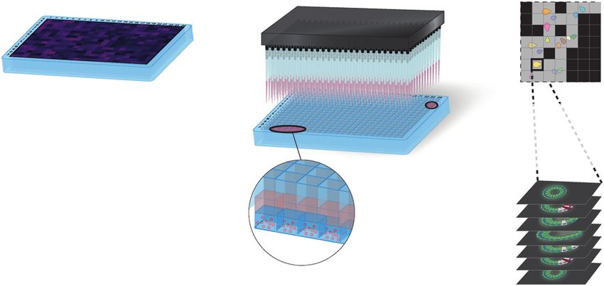
I. Lukonin et al.
                  1496
                         Adult and pluripotent stem-cell (PSC)-derived organoids                  cell-type composition and morphology but also enables assay
                         Organoids can be divided into two categories depending on the            miniaturization and upscaling. A study employing this approach
                         cells used to generate them: PSCs, including embryonic and               resulted in the identification of therapeutics preventing ZIKA virus
                         induced PSCs, and adult stem cells (ASCs). The generation of             infection by using stem-cell-derived brain organoids42. Assessing
                         organoids from PSCs recapitulates the sequence of events of              the drug response of a specific genotype is, on the other hand,
                         embryonic development by exposing them to specific combina-               best approached by screening patient-derived organoids with the
                         tions and concentrations of morphogens, allowing the patterning          goal of mapping patient-specific responses to available standard-
                         of germ layers and, subsequently, tissues and organs. PSC-derived        of-care drugs. Of note, a thorough understanding of a process
                         organoids include optic cup25, intestinal26, cerebral27, kidney28,       may result in a nontrivial choice of model system, which
                         thyroid29, lung30, and retinal31 organoids and others. Although          nevertheless is best suited for the question at hand. In a study
                         they hold great potential, are used to generate a wide array of          pivotal for the organoid field, intestinal organoids were used to
                         tissues, and can do so in a patient-specific context, the resulting       establish a diagnostic assay to predict the patient-specific
                         organoids resemble embryonic rather than adult tissues23,25,32.          response to standard-of-care drugs for treating pulmonary cystic
                            In contrast, ASC-derived organoids do not recapitulate devel-         fibrosis43. Phenotypes can be analyzed at the organoid level to
                         opmental steps; rather, the regenerative capacity of parent tissues      answer broader biological questions: multivariate phenotypes of
                         is utilized by the dissociation of biopsies and the subsequent           400,000 intestinal organoids were recently used to systematically
                         culture of derived cells in artificial extracellular matrices. These      map functional interactions during organoid development and
                         include small intestinal33, colon34, lung35, mammary36 and salivary      identify key players in intestinal regeneration41.
                         gland37, and pancreatic38 organoids and others. Consistently, the           As discussed above, higher biological relevance can ultimately
                         growth conditions for ASC-derived organoids typically include            deliver novel biological insight and translate to better quality
                         factors that control tissue repair or homeostasis, and the resulting     leads for the drug discovery pipeline, yet the assay has to be
                         structures are more mature than PSC-derived organoids22.                 feasible in terms of cost and technology. In the next sections, we
                         However, the composition of culture media with respect to added          will focus on the underlying concepts and enabling technologies
                         growth factors and compounds often favors proliferation and              employed in organoid system screening.
                         culture expansion over cellular differentiation, which often limits
1234567890();,:

                         the phenotypic heterogeneity that can be achieved. ASC-derived           Assay development
                         organoid systems usually mimic tissues that either have a high cell      After defining the biological question and identifying a suitable
                         turnover rate or are capable of regeneration, such as the small          organoid system, one needs to design an experimental assay,
                         intestine, stomach, and lung.                                            which often involves multiple rounds of protocol optimization.
                                                                                                  Furthermore, this process demands fundamental knowledge of
                         Organoid model systems in chemical screening                             the biology and culture conditions of the chosen model system to
                         Whereas target-based HTS assays can use simple readouts39 and            decide on the screening setup. In the following section, we outline
                         minimalistic model systems40 because of their focus on perturbing        considerations in planning the experimental setup (Fig. 1) and
                         target activity, phenotype-driven approaches require complex             give recommendations for individual steps of assay development,
                         model systems with high disease or tissue relevance. Increasing          imaging, and data analysis (summarized in Fig. 2).
                         the depth of compound profiling with phenotypic resolution thus
                         goes hand-in-hand with increasing the physiological relevance of         Marker selection. Selecting informative markers is a crucial step
                         the screened system. Organoids are ideal candidates because they         of assay design and strongly influences how a screen can be
                         provide self-contained organotypic structures that mimic the cell-       interpreted. Traditionally, dyes that stain the DNA content,
                         type composition and function of parent tissues with the major           including DAPI and Hoechst, are used to identify cell nuclei.
                         advantage that they are amenable to use in large screening               Staining of a cell’s protein content (e.g., CellTrace), cytoskeleton
                         assays41. In particular, organoids are accessible for imaging and        (e.g., phalloidin), or membrane (e.g., CellMask) can be included to
                         standard liquid handling automation, meaning they can be used            aid cell segmentation. Additional markers largely depend on the
                         for standardized and well-established automated chemical screen-         scientific question and may include markers of cell type,
                         ing pipelines. Thus, progressing from using isolated cells cultured      subcellular structures, or cellular processes, and identification of
                         in a monolayer to organoid structures in screening experiments is        these markers is achieved either by immunofluorescent staining or
                         the next logical step. Combining organoids with small-molecule           by fluorescent tagging of the protein of interest. Importantly, one
                         treatment offers several major advantages. First, organoids              needs to verify both the correct labeling (antibody specificity in
                         recapitulate individual steps of organ formation and disease             the case of staining; target protein functionality and stability and
                         onset, thus allowing the study of distinct developmental and             timely fluorescent protein activity in the case of tagging) of the
                         disease stages. Second, small-molecule compounds can be added            marker and smooth performance of the labeling procedure in
                         and removed from the culture medium at any given time, thus              the case of immunofluorescent staining prior to conducting the
                         allowing targeted perturbation in a time-controlled fashion. In          screen. Another factor to consider is the cost of the resulting
                         combination with a quantitative, data-driven, and unbiased               assay, whereas fluorescent reporters do not carry additional cost,
                         analysis approach, chemical screening in organoids offers                they might lack signal strength, reducing the assay sensitivity.
                         unprecedented insight into biological processes, delivering a            Antibodies, on the other hand, often offer superior signal and
                         wealth of data that can, nevertheless, be challenging to extract         improved flexibility for combining wavelengths but increase the
                         and analyze.                                                             cost massively, especially for larger screens.

                                                                                                  Small to large scale. The scale of a screen crucially influences
                         PILLARS OF CHEMICAL SCREENING IN ORGANOIDS                               several steps down the line of assay development and thus needs
                         Experimental question and system                                         to be considered thoroughly. Genome-wide or large-scale screens
                         For any biological assay, the first step lies in defining the process      offer the advantage of broad coverage and require less prior
                         or target of interest. For a phenotypic screen, the model system         knowledge of the biological system. However, they also present a
                         should have the highest relevance to the tissue or disease of            major cost factor due to increased system-specific culturing
                         interest and yet be practical for the planned assay. For instance,       (specialized media, extracellular matrix components, and culture
                         when screening for compounds that affect a process specific to a          plates needed for organoid culture) and readout (increased use of
                         given tissue, one should strive for a system that recapitulates its      immunofluorescence reagents and imaging time) requirements.

                                                                                                                Experimental & Molecular Medicine (2021) 53:1495 – 1502
I. Lukonin et al.
1497
Experimental setup Data analysis
Experimental system Biologically relevant system Image handling Data Transfer
- organoid recapitulating - on-the-fly or post acquisition
Tissue- desired tissue Acquistion Storage Storage and backup
specific Organoid origin: - large amount of data
- cell line - accessible for
Patient- (general and easy) image analysis servers
derived - patient Computational power
(specific, more challenging) - scalable
- parallelized
Assay development Informative markers Image processing Object segmentation
Passive Active - molecular and morphological Segmentation - 2D, in-plane
Controls
- transgene vs stained protein - 3D, volume
Treatment library 2D, in-plane 3D, voxel - organoid-level
- large-scale - single cell-level
“wild-type” known, perturbed (broad but labour-intensive)
phenotype phenotype - small scale Neural network-assisted
(specific but less informative) - object identification
Small Large
Library size
Controls - anisotropic image
- active (known phenotype) organoid-level single cell interpolation
- passive (vehicle)
Replicates
targeted general
Assay setup Culture adaptation Feature extraction Features
- multiwell plates Morphology Intensity - organoid- and cell-level
- upscaling - morphology, intensity
- automated liquid-handling ... Meta-features
Plate arrangements - unbiased
- minimise batch - most-informative readout
Automation Plate layout area sum int.
and plate effects Neural networks
elongation mean int.
- unbiased
... ...
- image-level
Image acquisition Optimise imaging Data analysis and interpretation Data normalisation
- automated Phenotypic signatures - reduce batch effects
Clearing Iterative imaging - x, y and z resolution - phenotypic strength
- sample clearing Supervised vs unsupervised
- refractive index matching control Cpd X Cpd Y - a priori knowledge
- iterative imaging Phenotypic signatures
Phenotypic A B - pleiotropic phenotypes
landscape C
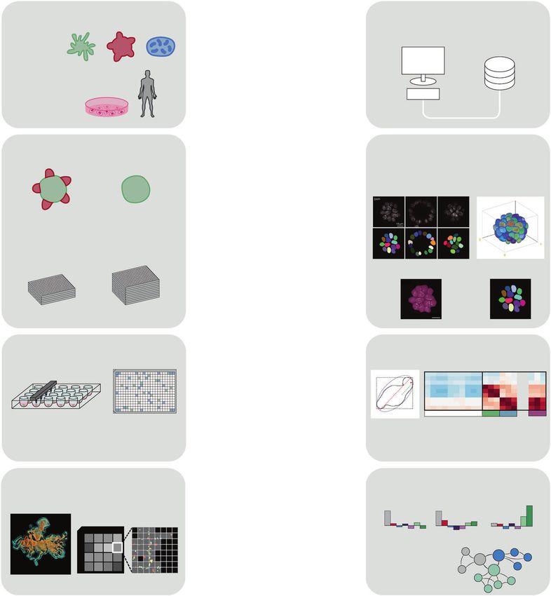
Fig. 1 Overview of the screening process in organoids. A screen consists of experimental setup and performance and subsequently the
analysis of the generated data. Both aspects include several steps with specific options or challenges. For a successful screen, these need to be
evaluated and optimized in advance. Importantly, every decision in the assay setup, including organoid system, marker selection, type of
controls, number of replicates, and imaging resolution, needs to be reconciled with steps in the data analysis process, including data handling,
object segmentation, feature extraction approach, data normalization and interpretation, and vice versa. Figures are adapted from refs. 41,52,57.
Furthermore, the larger the scale is, the greater are the considered carefully. Treatment throughout the formation of the
requirements for specialized equipment for handling large organoid allows assessment of the influence of the perturbation
number of plates, time for sample preparation, imaging and data on its complete development or the onset of a disease phenotype.
storage space. If a specific and defined question needs to be However, additional validation experiments are usually necessary
addressed, one may consider the advantages of a small-scale to understand which specific step is affected. To study a particular
approach. A defined library targeting genes expressed at a certain event during organoid formation, such as the emergence of a cell
developmental stage, in a specific organ, in a disease of interest or type or morphogenetic change, perturbation should be applied
belonging to a relevant group of signaling pathways might be around the time when this event occurs.
sufficient to shed light on the biological process while reducing Finally, if the screen is not read out continuously with time-
costs and workload. lapse imaging44, an endpoint needs to be determined. Screens
can be terminated when the organoid reaches a mature state, yet
Treatment time frame and screen endpoint. The decision on when this also goes hand-in-hand with increased costs and might not be
and for how long to apply screened conditions and at what point necessary when an intermediate step is to be studied.
to read out results depends largely on the scientific question and
the prior knowledge of the biological system. Especially for PSC- Controls, plate arrangement, and replicates. For every scientific
derived organoids, which follow the development of the organ experiment, measures need to be taken to ensure the quality and
from an early stage, treatment at an early progenitor stage statistical relevance of the screen, particularly when considering a
compared to a more mature stage elicits drastically different larger scale. Controls must always be included in the assay, ideally
responses. Moreover, ASC-derived organoids usually undergo in every plate, for quality control and the determination of the
different phases recapitulating regenerative and homeostatic phenotypic feature space. These should preferably include both
conditions. In addition, the treatment duration needs to be passive and active controls, whereby passive controls, which
Experimental & Molecular Medicine (2021) 53:1495 – 1502I. Lukonin et al.
1498
Assay development Screen Imaging Phenotypic analysis and hit selection
Miniaturize Sample prep Image quantification Multivariate data analysis
Library design Compound treatment Low-res scan Feature extraction Phenotypic clustering
cell-level
Profile compounds
- Segment Control
Organoid system organoid-level
- Identify objects CpdA
Suspension or - Rescan at high res Per-plate normalisation CpdB
Replicate 2 Replicate 1
matrix-embedded
Miniaturized CpdC
z-step
culture
organoid
Unperturbed 1 2 3 n culture z
Identify hits,
“wild-type” ... cluster by similarity
y
1-n: steps of
automated x A B
Confocal imaging C
organoid culture
Active controls,
known phenotype Feature1 Featuren
Fig. 2 Design of the assay and analysis pipeline. For any organoid system, the culture protocol must be miniaturized to 96-, 384-, or 1536-
well plate format and automated. At the same time, active controls that induce a measurable phenotype must be identified and incorporated
into the screening library. The measurement relies on efficient yet high-quality imaging, for instance, employing iterative imaging approaches
and generating images that are then used to extract cell- and organoid-level features. To make the data cross-comparable, plate normalization
should be applied to minimize systematic variance between individual plates. The normalized features can then be used to cluster individual
organoids by phenotypic similarity and profile screened conditions, assessing, for instance, the frequencies at which the identified
phenotypes occur. Ultimately, multivariate analysis serves to select hits from the screen that can, in turn, be used to infer system-level
properties, such as functional interactions. Figures are adapted from refs. 41,83.
include no active substance (for instance, treatment with the depends largely on the phenotypic consistency of the organoid
vehicle alone), are used to assess reproducibility and to normalize system, which can be determined using positive controls.
the data, compensating for potential batch effects. Furthermore,
one needs to determine a priori if and how the passive control Culture system setup. HTS and HCS employ automated culture,
might affect organoid formation and growth. Active controls, on sample preparation, and readout to deal with the large number of
the other hand, elicit a desired phenotype and are used to assess samples. Most original organoid protocols, however, are manual
the validity of the screen and serve as comparison to identified and do not employ multiwell plates. Protocol adaptation and
hits, but they are not always readily available. automation include simple transfer into the automation system48,
The number of passive control wells to include depends largely upscaling41,49, transfer of already formed organoids into the
on the downstream statistical analysis: if normalizing results to screening plates45, or changes to the protocol that allow simpler
passive controls alone (when a phenotypic effect is expected from automation50. The use of automation systems brings additional
most treatment conditions), more replicates of the control benefits, such as reduction of human bias and, thus, improved
condition, usually ~10%, need to be included than if the whole reproducibility and consistency of organoid cultures. It should,
plate will be used for normalization (when a phenotypic effect is however, be assessed whether changes in the culture and assay
expected from only a small number of treatment conditions). In format alter the organoid behavior.
general, as many control wells as possible and practical should be
included to facilitate downstream analysis. Optimization and pilot screen. For the success of a screen,
When using multiwell plates, plate arrangement of the controls optimization and preparation are key. Each step—from organoid
is crucial to avoid the introduction of bias45. Experiments in culture51 to perturbation administration, sample preparation, and,
multiwell plates are susceptible to plate effects46 that cause ultimately, screen readout—needs to be thoroughly tested. It is
different behaviors in organoids in the edge wells due to faster advisable to perform either a small pilot screen or a dry run, such
evaporation of the medium. If a potential plate effect cannot be as performing all steps with water and a small number of plates.
excluded, edge wells may be avoided. Controls should be This allows to test for an error-free process and creates a test
distributed across the plate in a randomized order to detect and dataset that can be used to set up the analysis pipeline.
normalize artifacts such as edge effects, liquid dispensation
differences, or thermal gradients47. Data acquisition
Replicates are used to rule out false positives and false The power of screening assays lies in systematic comparison of
negatives and to determine the variability within a perturbation. numerous conditions, minimizing experimental and technical
They can range from individual organoids within a well to variance. High-content imaging offers an unmatched combination
replicate wells within the same screening run or independent of throughput and information density: organoids cultured in
screening runs or can stem from parallel screening of different cell thousands of arrayed conditions41 can be imaged at high
lines, mouse, or patient origins. Ideally, a screen has two or more resolution to extract single-cell features52, describing observed
independent replicates (two screens conducted after each other phenotypes in depth.
or with different material) and several dependent replicates To date, a variety of organoid systems have been studied with
(organoids treated with the same compound within a run). HCS to extract different kinds of information, relying mostly on
However, the number of the latter depends strongly on how easily immunofluorescence or reporter expression at the terminal time
organoids can be obtained and might range from ten to several point41,42,49,51. For practical reasons, however, the number of
hundred. The statistical benefit of a larger number of replicates readouts is often limited, and one of the crucial limiting factors is
needs to be balanced against increased costs and workload and imaging. Most imaging systems offer up to five independent
Experimental & Molecular Medicine (2021) 53:1495 – 1502I. Lukonin et al.
1499
fluorophores per acquisition, effectively setting a cap on the systems due to a plethora of technical and biological factors. In
number of readouts. Multiplexed imaging offers a cost-efficient the last decade, 2D single-cell segmentation has become a
solution to increase the information density by repeated imaging widespread and almost trivial task, aided by an arsenal of
of the same sample probed for different readouts. One of the automated algorithms. Most commonly used methods of
methods best fitted for this application is antibody elution: state- foreground–background thresholding, however, perform poorly
of-the-art protocols offer means to image up to forty antibody- on organoid datasets due to signal inhomogeneity in the third
bound epitopes53 in the same sample using common imaging dimension. Whereas the correction of illumination in 2D images is
devices and fluorophores. The benefits of multiplexed imaging for widespread and well established, compensating for signal decay
organoid systems include not only more thorough phenotypic in 3D is far less trivial: light scattering occurring due to deeper
profiling but also improved cost efficiency, as additional informa- penetration into the sample can be highly nonlinear. This problem
tion can be extracted from the same sample. Another method can be addressed experimentally, as discussed above, but
making use of multiparametric profiling and multivariate pheno- nonetheless often requires adapting segmentation algorithms.
types is cell painting54, a profiling assay describing every single Deep convolutional neural networks (DCNNs) offer an efficient and
cell with eight subcellular components. For poorly defined targets, robust solution to this problem. The DCNN workflow requires a
this could be the only option: hits from such a screen would curated ground truth dataset used for training the artificial neural
include conditions where for instance, a wild-type phenotype is network to recognize desired objects, such as nuclei, in images of
restored in an organoid disease model. While the original varying quality57,58. Given the unreliable results of threshold-
protocol55 uses generic cell features such as cell morphology based segmentation in organoid images, the overhead of creating
and nuclear and organelle structure, for organoids, this could be a training dataset is overshadowed by the performance of neural
integrated with or replaced by cell-type composition. networks. Another field of application for deep learning methods
As described above, imaging can deliver a comprehensive is image interpolation: as discussed above, generating isotropic
description of the phenotypes observed under the screened images for a high number of organoids is not technically feasible
conditions. However, to obtain high-quality imaging data, one needs but can be achieved by DCNN-assisted image interpolation.
to consider the challenges presented by 3D model systems. The Algorithms such as CARE59 allow interpolating planes in sub-
most appropriate imaging method for organoids is confocal sampled 3D datasets, paving the way for single-cell-level organoid
microscopy, as it allows optical sectioning of the sample. For a screens at a large scale.
screen consisting of several multiwell plates, however, confocal
microscopy can result in extremely large amounts of imaging data: Enhancing the depth of phenotypic profiling. A non-image-based
imaging just a single well in a 384-well plate with ten confocal planes method that delivers a wealth of information at single-cell
and four readouts at high magnification can result in up to 3000 resolution is single-cell RNA sequencing. It is, however, technically
images, making data collection, storage, and processing challenging. and financially infeasible to profile thousands of arrayed condi-
This can be circumvented by selecting acquisition regions with tions. Furthermore, despite providing a comprehensive picture of
iterative imaging: the sample is first scanned at low resolution to gene expression, single-cell sequencing does not deliver informa-
identify the positions of organoids, which are then reimaged at the tion on individual organoid phenotypes and lacks the organoid
desired higher magnification. This approach can increase the context of the sequenced cells. Recent protocols try to fill this gap:
imaging throughput manifold while simultaneously reducing the single organoids can be first imaged to extract phenotypes and
amount of imaging data produced without any fidelity loss41. then sequenced to profile gene expression changes60,61. Similarly,
An important factor influencing the confocal imaging setup is in situ sequencing approaches can be used in organoids, enabling
the resolution in the third dimension, the z-step: it requires an the transcriptomic profiling of intact organoid structures62. While
initial decision on whether the images produced will be used to still in its infancy, this methodology opens exciting new
reconstruct the organoid in 3D. For medium- and high-throughput possibilities for organoid research.
assays, this approach would likely be impractical due to the
imaging time per sample and amount of data generated per Feature extraction
condition. An alternative solution is to subsample the third In multivariate image analysis, every object, such as a single cell or
dimension of the organoid, setting the z-step to approximate cell an organoid, is described with extracted features relating to either
size to ensure that all cells comprising the organoids are sampled. the morphology (e.g., size and shape) or the intensity of the
A further step in simplifying the readout and reducing the data measured channels. To quantitatively describe the observed
amount is the generation of intensity projections, effectively phenotypes, one should select features with high information
reducing every 3D organoid to a 2D projected image that still content and low technical variance. The first can be achieved by
contains the information necessary for phenotypic profiling. calculating the amount of information encoded by a given feature,
Another challenge related to the third dimension is the sheer whereas the latter can be done systematically by computing
size of organoids: they can consist of thousands of individual cells feature covariance matrices and eliminating highly redundant and
spanning several hundreds of microns. To ensure imaging quality noisy features11. An alternative approach to feature selection is the
in the upper z-planes, it is often necessary to use sample clearing generation of metafeatures, such as principal components (PC),
and/or refractive index matching, which increase the penetration allowing the unbiased selection and integration of most
depth and ultimately facilitate imaging of the entire organoid52,56. informative readouts. It should be noted that metafeatures have
Producing a high number of images places a strain on the limitations; for instance, PCs are not able to encode nonlinear
computation infrastructure and requires a well-planned data interactions. Furthermore, metafeatures often lack interpretability,
lifecycle: before starting an image-based screen, one should try to as they do not represent a single, intuitively understandable
estimate the storage space required for the dataset, ideally using a parameter. Recent work also proposes the use of DCNNs for
pilot assay with an equivalent sample. Furthermore, once unbiased image parametrization, delegating feature selection to
acquired, images need to be transferred to storage that are the neural network algorithm63. This can be achieved by training
accessible by the servers running the image analysis software. DCNNs on images coming from positive and negative controls,
learning image-level features and using the resulting network for
Object segmentation. Image segmentation is the foundation of the detection of non-wild-type phenotypes64. Similarly, machine
quantitative image analysis, converting intensity images into learning methods, particularly deep learning, can be applied to
labeled maps used for downstream feature extraction. As simple detect disease phenotypes in patient-derived organoids, as
as the task might sound, it can become increasingly difficult for 3D reviewed extensively in refs. 65,66.
Experimental & Molecular Medicine (2021) 53:1495 – 1502I. Lukonin et al.
1500
Cell- and organoid-level information. A key difference between phenotypic signatures for every condition, describing the
data from 2D cell assays and data from organoids is in the levels of abundance of all detected phenotypic classes. In this approach,
imaged object hierarchy: in the single-cell quantification of 2D cell differences in phenotypic distributions describe every condition,
assays, every cell is directly linked to the condition, i.e., the well, making it straightforward to identify conditions that differ from
but the organization of organoid data is more complicated. In fact, the controls41. Furthermore, comparing phenotypic signatures
organoid data feature an extra level of complexity: every cell can between replicates of the same condition can be used to assess
be assigned to a parent organoid, which in turn is assigned to the reproducibility.
treatment condition67. The properties of individual cells compos-
ing the organoids give rise to emergent properties of the system, What can we learn?
and cell-to-cell variability is a crucial driving factor for organoid Since the first organoid system was established more than a
development57. This reflects the complexity of the organoid decade ago70, a multitude of systems have followed and enabled
system and requires a paradigm shift in addressing quantitative scientific discoveries that otherwise would have been challenging
data at single-cell resolution. Depending on the question at hand or even unattainable. As scientists start to employ organoids in
and the number of organoids sampled per condition, single-cell chemical screening, an even greater potential becomes accessible.
data can either be used as-is or be converted to organoid-level Screening in organoids is dominated by disease- and drug
descriptors, such as cell-type composition. discovery-driven research. Recently, however, it has also been
leveraged by basic science to further our understanding of
Data normalization. In a screening assay, data points typically fundamental biological processes. A factorial screening approach
come from individual plates, making well plates the experimental of small molecules in embryoid bodies led to the formulation of an
units. As outlined above, every plate should contain a sufficient ideal culture medium composition for the robust derivation of
number of controls to allow per-plate normalization. A frequently several embryonic lineages in in vitro structures, facilitating the
used normalization strategy is z-scoring, normalizing the differ- study of early embryonic development71. A similar approach was
ence between the measured value and the mean of the reference taken to improve and increase the throughput of kidney organoid
condition by the standard deviation of the reference. Z-score culture, which was subsequently used to screen the drug response
normalization results in cross-comparable values that also reflect of organoids modeling a kidney disease51. A deep understanding
the strength of the effect and whether the feature is increased or of how organoids and their organs of origin develop can also
decreased in the observed condition. Due to the nature of the directly benefit the identification of disease treatments: an
method, the appropriate reference population should always be inhibitor of the retinoic acid signaling pathway identified in a
identified. When normalizing to a control condition, an insufficient high-content screen of intestinal organoid development improved
number of control replicates or high variance in a given feature tissue regeneration after irradiation in mice, providing a lead for
would result in poor z-score values, masking the true extent of the the treatment of tissue damage caused by radiation cancer
phenotype. This is especially true for organoid models that therapy41.
contain several cell types and show a high degree of morpholo- Undoubtedly, organoid screening provides immense potential
gical variability. If normalization is performed to the mean of the for the drug development process, and the peak of this
entire plate, the majority of the objects should not exhibit a development will be when the first organoid-developed treatment
phenotype; similarly, there should be no bias in condition receives approval. The possibility of generating organ-like
distribution between plates. For example, conditions resulting in structures from human cells not only reduces the amount of
reduced organoid size and viability should not be grouped on a animal testing but also enables the modeling of human-specific
single plate of a multiplate experiment. It should be noted that an diseases with cells originating from patients. Drug screens in
entire arsenal of statistical tools is available for addressing cases organoids are currently used to identify potential leads, test their
where frequently used algorithms perform poorly46,68. Overall, efficacy, and assess their toxicity72,73. The advantages of using
normalization is crucial to enable cross-comparison of data points organoids instead of a single cell type grown in a monolayer lie in
from individual batches of the assay, and the normalization their diverse cell-type composition, which unmasks potential side
strategy should be chosen according to the assay design and the effects in cell types not specifically targeted by the drug, as well as
data distributions obtained. in their 3D architecture, which recapitulates the correct drug
uptake74. However, the greatest potential of organoids lies in their
Quantitative phenotype description. Quality control is well application in personalized medicine. The main challenge in
defined for single-parameter HCS workflows through metrics such finding a cure for malignancies such as cancer is their hetero-
as z-prime46, which describes the dynamic range between positive geneities, meaning that tumors in the same organ in two patients
and negative controls. For phenotypic screens, however, no do not necessarily share the same genetic mutations, and even
universally applicable parameters exist. As phenotypes are often cells within a single tumor differ in their genotype75. This makes
complex and described by differences in several readouts, treatment choice challenging and often results in relapse if not all
clustering in multivariate feature space can be used to assign tumor cells are eliminated by the treatment. Cancer organoids
similarity classes. Depending on the a priori knowledge of the grown from a patient biopsy reflect the full scope of tumor
system, phenotypes can be classified either in a supervised or in diversity and are used to identify the most efficient drug or drug
an unsupervised, data-driven manner. For an assay with known combination to treat a particular patient76–78. This significantly
phenotypic effects, for instance, rescue of the wild-type organoid increases positive treatment outcomes and decreases potential
phenotype, machine learning algorithms such as support vector adverse effects.
machines or random forest classifiers can be used to distinguish In summary, although chemical screening in organoids is still in
between individual data points. Alternatively, phenotypic classifi- its infancy, it has already been leveraged for diverse use cases
cation can be performed by unsupervised clustering of the entire from basic to applied sciences, enabling profound scientific
dataset using software packages such as PhenoGraph69. Once insight.
classified, conditions can be ranked by the presence of
phenotypes not observed in the controls and by depletion of
the “wild-type” phenotype. Due to the variability observed in OUTLOOK
organoid systems, however, control conditions can present Organoids have opened vast possibilities to study biological
pleiotropic phenotypes, meaning that no single control pheno- processes in health and disease in an organotypic setting. By
type can be assigned. To address this, one can generate combining this potential with chemical screening, scientists have
Experimental & Molecular Medicine (2021) 53:1495 – 1502I. Lukonin et al.
1501
a powerful tool at hand to increase both the speed and reliability 21. Lancaster, M. A. & Knoblich, J. A. Organogenesis in a dish: modeling development
of scientific discoveries. Since organoids capture much of the and disease using organoid technologies. Science 345, 1247125 (2014).
complexity of tissues and organs, including structural and 22. Clevers, H. Modeling development and disease with organoids. Cell 165,
functional composition, but are still amenable to experimental 1586–1597 (2016).
23. Rossi, G., Manfrin, A. & Lutolf, M. P. Progress and potential in organoid research.
interference, they are a suitable system to study development and
Nat. Rev. Genet 19, 671–687 (2018).
homeostasis as well as the manifestation of diseases. Further 24. Fatehullah, A., Tan, S. H. & Barker, N. Organoids as an in vitro model of human
developments in the organoid field, such as cocultures with development and disease. Nat. Cell Biol. 18, 246–254 (2016).
immune cells or different tissue types, will surely improve the 25. Eiraku, M. et al. Self-organizing optic-cup morphogenesis in three-dimensional
biological relevance of organoid models and bring them closer to culture. Nature 472, 51–56 (2011).
their in vivo counterparts. Additional technical advances in culture 26. Spence, J. R. et al. Directed differentiation of human pluripotent stem cells into
conditions, liquid handling machinery, and image analysis intestinal tissue in vitro. Nature 469, 105–109 (2011).
algorithms will allow us to increase the homogeneity and 27. Lancaster, M. A. et al. Cerebral organoids model human brain development and
reproducibility of organoid protocols and screening approaches, microcephaly. Nature 501, 373–379 (2013).
28. Takasato, M. et al. Directing human embryonic stem cell differentiation towards a
enabling reliable and advanced conclusions from organoid
renal lineage generates a self-organizing kidney. Nat. Cell Biol. 16, 118–126
chemical screening79,80. Furthermore, since they can be derived (2014).
from iPSCs or ASCs of patient origin, organoids pave the way for 29. Kurmann, A. A. et al. Regeneration of thyroid function by transplantation of
precision and personalized medicine. The establishment of differentiated pluripotent stem cells. Cell Stem Cell 17, 527–542 (2015).
organoid biobanks facilitates the use of organoids in screening 30. Dye, B. R. et al. In vitro generation of human pluripotent stem cell derived lung
to better understand disease and for drug discovery, develop- organoids. eLife 4, 1–25 (2015).
ment, safety, and efficacy81,82. Simultaneously, as both high- 31. Shirai, H. et al. Transplantation of human embryonic stem cell-derived retinal
throughput organoid culture and HCS approaches also become tissue in two primate models of retinal degeneration. Proc. Natl Acad. Sci. U. S. A.
more accessible to scientists in academia, the insights we can gain 113, E81–E90 (2016).
32. McCracken, K. W. et al. Modelling human development and disease in pluripotent
in basic research by these means will certainly increase.
stem-cell-derived gastric organoids. Nature 516, 400–404 (2014).
In summary, screening in organoids introduces a multitude of 33. Sato, T. et al. Single Lgr5 stem cells build crypt-villus structures in vitro without a
possibilities for translating scientific discoveries to patients’ quality mesenchymal niche. Nature 459, 262–265 (2009).
of life. With the continuous improvement of organoid culture, a 34. Yui, S. et al. Functional engraftment of colon epithelium expanded in vitro from a
growing list of organoid models and technical advances in HCS, single adult Lgr5(+) stem cell. Nat. Med. 18, 618–623 (2012).
there is still more to come. 35. Butler, C. R. et al. Rapid expansion of human epithelial stem cells suitable for
airway tissue engineering. Am. J. Respir. Crit. Care Med. 194, 156–168 (2016).
36. Olabi, S., Ucar, A., Brennan, K. & Streuli, C. H. Integrin-Rac signalling for mammary
REFERENCES epithelial stem cell self-renewal. Breast Cancer Res. 20, 128 (2018).
1. Ghosh, R. N. et al. Cell-based, high-content screen for receptor internalization, 37. Tanaka, J. et al. Generation of orthotopically functional salivary gland from
recycling and intracellular trafficking. Biotechniques 29, 170–175 (2000). embryonic stem cells. Nat. Commun. 1–13, https://doi.org/10.1038/s41467-018-
2. Carpenter, A. E. Image-based chemical screening. Nat. Chem. Biol. 3, 461–465 06469-7 (2018).
(2007). 38. Loomans, C. J. M. et al. Expansion of adult human pancreatic tissue yields
3. Stockwell, B. R. Exploring biology with small organic molecules. Nature 432, organoids harboring progenitor cells with endocrine differentiation potential.
846–854 (2004). Stem Cell Rep. 10, 712–724 (2018).
4. Inglese, J., Shamu, C. E. & Guy, R. K. Reporting data from high-throughput 39. Stoddart, L. A., White, C. W., Nguyen, K., Hill, S. J. & Pfleger, K. D. Fluorescence- and
screening of small-molecule libraries. Nat. Chem. Biol. 3, 438–441 (2007). bioluminescence-based approaches to study GPCR ligand binding. Br. J. Pharm.
5. Blay, V., Tolani, B., Ho, S. P. & Arkin, M. R. High-throughput screening: today’s 173, 3028–3037 (2016).
biochemical and cell-based approaches. Drug Discov. Today 25, 1807–1821 40. Lin, Y. et al. Identification of antituberculosis agents that target ribosomal protein
(2020). interactions using a yeast two-hybrid system. Proc. Natl Acad. Sci. USA. 109,
6. Yuan, H. et al. Use of reprogrammed cells to identify therapy for respiratory 17412–17417 (2012).
papillomatosis. N. Engl. J. Med. 367, 1220–1227 (2012). 41. Lukonin, I. et al. Phenotypic landscape of intestinal organoid regeneration. Nature
7. Adan, A., Kiraz, Y. & Baran, Y. Cell proliferation and cytotoxicity assays. Curr. 1–34, https://doi.org/10.1038/s41586-020-2776-9 (2020).
Pharm. Biotechnol. 17, 1213–1221 (2016). 42. Zhou, T. et al. High-content screening in hPSC-neural progenitors identifies drug
8. Ma, Q., Ye, L., Liu, H., Shi, Y. & Zhou, N. An overview of Ca(2+) mobilization assays candidates that inhibit Zika virus infection in fetal-like organoids and adult brain.
in GPCR drug discovery. Expert Opin. Drug Discov. 12, 511–523 (2017). Cell Stem Cell 21, 274–283.e5 (2017).
9. Boutros, M., Heigwer, F. & Laufer, C. Microscopy-based high-content screening. 43. Boj, S. F. et al. Forskolin-induced swelling in intestinal organoids: an in vitro assay
Cell 163, 1314–1325 (2015). for assessing drug response in cystic fibrosis patients. J. Vis. Exp. https://doi.org/
10. Liberali, P., Snijder, B. & Pelkmans, L. Single-cell and multivariate approaches in 10.3791/55159 (2017).
genetic perturbation screens. Nat. Rev. Genet. 16, 18–32 (2015). 44. Ostrop, J. et al. A semi-automated organoid screening method demonstrates
11. Loo, L. H., Wu, L. F. & Altschuler, S. J. Image-based multivariate profiling of drug epigenetic control of intestinal epithelial maturation. bioRxiv 1–25, https://doi.
responses from single cells. Nat. Methods 4, 445–453 (2007). org/10.1101/2020.07.23.217414 (2020).
12. Liberali, P., Snijder, B. & Pelkmans, L. A hierarchical map of regulatory genetic 45. Francies, H. E., Barthorpe, A., McLaren-Douglas, A., Barendt, W. J. & Garnett, M. J.
interactions in membrane trafficking. Cell 157, 1473–1487 (2014). Drug sensitivity assays of human cancer organoid cultures. Methods Mol. Biol.
13. Collinet, C. et al. Systems survey of endocytosis by multiparametric image ana- 1576, 339–351 (2019).
lysis. Nature 464, 243–249 (2010). 46. Malo, N., Hanley, J. A., Cerquozzi, S., Pelletier, J. & Nadon, R. Statistical practice in
14. Pelkmans, L. et al. Genome-wide analysis of human kinases in clathrin- and high-throughput screening data analysis. Nat. Biotechnol. 24, 167–175 (2006).
caveolae/raft-mediated endocytosis. Nature 436, 78–86 (2005). 47. Mazoure, B., Nadon, R. & Makarenkov, V. Identification and correction of spatial
15. Bickle, M. The beautiful cell: high-content screening in drug discovery. Anal. bias are essential for obtaining quality data in high-throughput screening tech-
Bioanal. Chem. 398, 219–226 (2010). nologies. Sci. Rep. 7, 11921 (2017).
16. Clemons, P. A. Complex phenotypic assays in high-throughput screening. Curr. 48. Benning, L., Peintner, A., Finkenzeller, G. & Peintner, L. Automated spheroid
Opin. Chem. Biol. 8, 334–338 (2004). generation, drug application and efficacy screening using a deep learning clas-
17. Garvey, C. M. et al. A high-content image-based method for quantitatively sification: a feasibility study. Sci. Rep. 1–11, https://doi.org/10.1038/s41598-020-
studying context-dependent cell population dynamics. Sci. Rep. 6, 29752 (2016). 67960-0 (2020).
18. Snijder, B. et al. Single-cell analysis of population context advances RNAi 49. Du, Y. et al. Development of a miniaturized 3D organoid culture platform for
screening at multiple levels. Mol. Syst. Biol. 8, 579 (2012). ultra-high throughput screening. J. Mol. Cell Biol. 1–35, https://doi.org/10.1093/
19. Zheng, W., Thorne, N. & McKew, J. C. Phenotypic screens as a renewed approach jmcb/mjaa036/5873160 (2020).
for drug discovery. Drug Discov. Today 18, 1067–1073 (2013). 50. Mead, B. E. et al. High-throughput organoid screening enables engineering of
20. Kretzschmar, K. & Clevers, H. Organoids: modeling development and the stem intestinal epithelial composition. bioRxiv, 1–60, https://doi.org/10.1101/
cell niche in a dish. Dev. Cell 38, 590–600 (2016). 2020.04.27.063727 (2020).
Experimental & Molecular Medicine (2021) 53:1495 – 1502I. Lukonin et al.
1502
51. Czerniecki, S. M. et al. High-throughput screening enhances kidney organoid 75. Kopper, O. et al. An organoid platform for ovarian cancer captures intra- and
differentiation from human pluripotent stem cells and enables automated mul- interpatient heterogeneity. Nat. Med. 1–27, https://doi.org/10.1038/s41591-019-
tidimensional phenotyping. Cell Stem Cell 22, 929–940.e4 (2018). 0422-6 (2019).
52. Dekkers, J. F. et al. High-resolution 3D imaging of fixed and cleared organoids. 76. Vlachogiannis, G. et al. Patient-derived organoids model treatment response of
Nat. Protoc. 14, 1–19 (2019). metastatic gastrointestinal cancers. Science 359, 920–926 (2018).
53. Gut, G., Herrmann, M. D. & Pelkmans, L. Multiplexed protein maps link subcellular 77. Verissimo, C. S. et al. Targeting mutant RAS in patient-derived colorectal cancer
organization to cellular states. Science 361, https://doi.org/10.1126/science. organoids by combinatorial drug screening. eLife 5, 1–26 (2016).
aar7042 (2018). 78. Broutier, L. et al. Human primary liver cancer–derived organoid cultures for dis-
54. Bray, M.-A. & Carpenter, A. Advanced assay development guidelines for image- ease modeling and drug screening. Nature, 1–19, https://doi.org/10.1038/
based high content screening and analysis. Nat. Protoc. 1–30 (2016). nm.4438 (2017).
55. Bray, M. A. et al. Cell Painting, a high-content image-based assay for morpho- 79. Huch, M., Knoblich, J. A., Lutolf, M. P. & Martinez Arias, A. The hope and the hype
logical profiling using multiplexed fluorescent dyes. Nat. Protoc. 11, 1757–1774 of organoid research. Development 144, 938–941 (2017).
(2016). 80. Hofer, M. & Lutolf, M. P. Engineering organoids. Nat. Rev. Mater. https://doi.org/
56. Masselink, W. et al. Broad applicability of a streamlined ethyl cinnamate-based 10.1038/s41578-021-00279-y (2021).
clearing procedure. Development 146, dev166884–166829 (2019). 81. van de Wetering, M. et al. Prospective derivation of a living organoid biobank of
57. Serra, D. et al. Self-organization and symmetry breaking in intestinal organoid colorectal cancer patients. Cell 161, 933–945 (2015).
development. Nature 569, 66–72 (2019). 82. Sachs, N. et al. A living biobank of breast cancer organoids captures disease
58. Serra, D. et al. glib-nature2018-materials, (2019). heterogeneity. Cell 172, 373–386.e10 (2018).
59. Weigert, M. et al. Content-aware image restoration: pushing the limits of fluor- 83. Lukonin, I. Intestinal Regeneration: Lessons From Organoids. (Natue Research
escence microscopy. Nat. Methods, 1–12, https://doi.org/10.1038/s41592-018- Bioengineering Community, 2020).
0216-7 (2018).
60. Tirier, S. M. et al. Pheno-seq—linking visual features and gene expression in 3D
cell culture systems. Sci. Rep. 9, 12367 (2019). ACKNOWLEDGEMENTS
61. Bues, J. et al. Deterministic scRNA-seq of individual intestinal organoids reveals We acknowledge the members of the Liberali laboratory for discussions and
new subtypes and coexisting distinct stem cell pools. bioRxiv 38, 35–22 (2020). feedback on the manuscript. This work received funding from the ERC under the
62. Lee, J. H. et al. Fluorescent in situ sequencing (FISSEQ) of RNA for gene expression European Union’s Horizon 2020 research and innovation program (grant agreement
profiling in intact cells and tissues. Nat. Protoc. 10, 442–458 (2015). no. 758617 to P.L.) and from the Swiss National Foundation (POOP3_157531 to P.L.).
63. Kandaswamy, C., Silva, L. M., Alexandre, L. A. & Santos, J. M. High-content analysis
of breast cancer using single-cell deep transfer learning. J. Biomol. Screen. 21,
252–259 (2016).
64. Kensert, A., Harrison, P. J. & Spjuth, O. Transfer learning with deep convolutional COMPETING INTERESTS
neural networks for classifying cellular morphological changes. SLAS Discov. 24, The authors declare no competing interests.
466–475 (2019).
65. Scheeder, C., Heigwer, F. & Boutros, M. Machine learning and image-based pro-
filing in drug discovery. Curr. Opin. Syst. Biol. 10, 43–52 (2018). ADDITIONAL INFORMATION
66. Kraus, O. Z. et al. Automated analysis of high‐content microscopy data with deep Correspondence and requests for materials should be addressed to P.L.
learning. Mol. Syst. Biol. 13, 924–915 (2017).
67. Velasco, S. et al. Individual brain organoids reproducibly form cell diversity of the Reprints and permission information is available at http://www.nature.com/
human cerebral cortex. Nature 570, 523–527 (2019). reprints
68. Bray, M. A. & Carpenter, A. Advanced Assay Development Guidelines for Image-
based High Content Screening and Analysis. In: Markossian S, Sittampalam GS, Publisher’s note Springer Nature remains neutral with regard to jurisdictional claims
Grossman A, et al., editors. Assay Guidance Manual [Internet] Bethesda (MD): Eli Lilly in published maps and institutional affiliations.
& Company and the National Center for Advancing Translational Sciences (2017).
69. Levine, J. H. et al. Data-driven phenotypic dissection of AML reveals progenitor-
like cells that correlate with prognosis. Cell 162, 184–197 (2015).
70. Eiraku, M. et al. Self-organized formation of polarized cortical tissues from ESCs Open Access This article is licensed under a Creative Commons
and its active manipulation by extrinsic signals. Stem Cell 3, 519–532 (2008). Attribution 4.0 International License, which permits use, sharing,
71. Vrij, E. J. et al. Chemically-defined induction of a primitive endoderm and adaptation, distribution and reproduction in any medium or format, as long as you give
epiblast-like niche supports post-implantation progression from blastoids. bioRxiv appropriate credit to the original author(s) and the source, provide a link to the Creative
120, 173–123 (2019). Commons license, and indicate if changes were made. The images or other third party
72. Driehuis, E. et al. Patient-derived oral mucosa organoids as an in vitro model for material in this article are included in the article’s Creative Commons license, unless
methotrexate induced toxicity in pediatric acute lymphoblastic leukemia. PLoS indicated otherwise in a credit line to the material. If material is not included in the
ONE 15, e0231588 (2020). article’s Creative Commons license and your intended use is not permitted by statutory
73. Watanabe, M. et al. Self-organized cerebral organoids with human- specific fea- regulation or exceeds the permitted use, you will need to obtain permission directly
tures predict effective drugs to combat Zika Virus Infection. CellReports 21, from the copyright holder. To view a copy of this license, visit http://creativecommons.
517–532 (2017). org/licenses/by/4.0/.
74. Zietek, T. et al. Organoids to study intestinal nutrient transport, drug uptake and
metabolism—update to the human model and expansion of applications. Front.
Bioeng. Biotechnol. 8, 1–14 (2020). © The Author(s) 2021
Experimental & Molecular Medicine (2021) 53:1495 – 1502You can also read