Linking Oxidative Stress and DNA Damage to Changes in the Expression of Extracellular Matrix Components
←
→
Page content transcription
If your browser does not render page correctly, please read the page content below
REVIEW
                                                                                                                                                published: 29 July 2021
                                                                                                                                      doi: 10.3389/fgene.2021.673002

                                              Linking Oxidative Stress and DNA
                                              Damage to Changes in the
                                              Expression of Extracellular Matrix
                                              Components
                                              Susana G. Martins 1,2, Rita Zilhão 1,3, Sólveig Thorsteinsdóttir 1,2 and Ana Rita Carlos 1,2*
                                              1
                                               Centro de Ecologia, Evolução e Alterações Ambientais, Faculdade de Ciências, Universidade de Lisboa, Lisboa, Portugal,
                                              2
                                               Departamento de Biologia Animal, Faculdade de Ciências, Universidade de Lisboa, Lisboa, Portugal, 3 Departamento de
                                              Biologia Vegetal, Faculdade de Ciências, Universidade de Lisboa, Lisboa, Portugal

                                              Cells are subjected to endogenous [e.g., reactive oxygen species (ROS), replication stress]
                                              and exogenous insults (e.g., UV light, ionizing radiation, and certain chemicals), which
                                              can affect the synthesis and/or stability of different macromolecules required for cell and
                                              tissue function. Oxidative stress, caused by excess ROS, and DNA damage, triggered in
                          Edited by:          response to different sources, are countered and resolved by specific mechanisms,
                Sebastian M. Waszak,
        Centre for Molecular Medicine
                                              allowing the normal physiological equilibrium of cells and tissues to be restored. One
           Norway (NCMM), Norway              process that is affected by oxidative stress and DNA damage is extracellular matrix (ECM)
                      Reviewed by:            remodeling, which is a continuous and highly controlled mechanism that allows tissues
        Laura Mondragón Martínez,
                                              to readjust in reaction to different challenges. The crosstalk between oxidative stress/DNA
Josep Carreras Leukaemia Research
                Institute (IJC), Spain        damage and ECM remodeling is not unidirectional. Quite on the contrary, mutations in
            Ezhilarasi Chendamarai,           ECM genes have a strong impact on tissue homeostasis and are characterized by
  Washington University in St. Louis,
                        United States
                                              increased oxidative stress and potentially also accumulation of DNA damage. In this
                  *Correspondence:
                                              review, we will discuss how oxidative stress and DNA damage affect the expression and
                      Ana Rita Carlos         deposition of ECM molecules and conversely how mutations in genes encoding ECM
                     arcarlos@fc.ul.pt
                                              components trigger accumulation of oxidative stress and DNA damage. Both situations
                  Specialty section:          hamper the reestablishment of cell and tissue homeostasis, with negative impacts on
        This article was submitted to         tissue and organ function, which can be a driver for severe pathological conditions.
Cancer Genetics and Oncogenomics,
              a section of the journal        Keywords: oxidative stress, DNA damage, ECM remodeling, tissue homeostasis, ECM gene expression
                 Frontiers in Genetics

        Received: 26 February 2021
          Accepted: 23 June 2021              INTRODUCTION
           Published: 29 July 2021
                            Citation:         Redox reactions are central to ensure correct cell function, allowing a dynamic transfer of
                Martins SG, Zilhão R,         electrons across different molecules (McCord, 2000). This occurs, for example, during molecular
               Thorsteinsdóttir S and         oxygen metabolism in the mitochondria, which are the central metabolic machinery of cells.
   Carlos AR (2021) Linking Oxidative
                                              Molecular oxygen metabolism is the basis of oxidative phosphorylation, and promotes the
Stress and DNA Damage to Changes
     in the Expression of Extracellular
                                              production of reactive oxygen species (ROS) (Sabharwal and Schumacker, 2014; Shadel and
                 Matrix Components.           Horvath, 2015). ROS also play an important role in immune defenses, where innate immune
            Front. Genet. 12:673002.          cells produce ROS as part of their pathogen killing strategies (Khan et al., 2018). In fact,
    doi: 10.3389/fgene.2021.673002            neutrophils and some macrophages express myeloperoxidase (MPO), which leads to the generation

Frontiers in Genetics | www.frontiersin.org                                         1                                              July 2021 | Volume 12 | Article 673002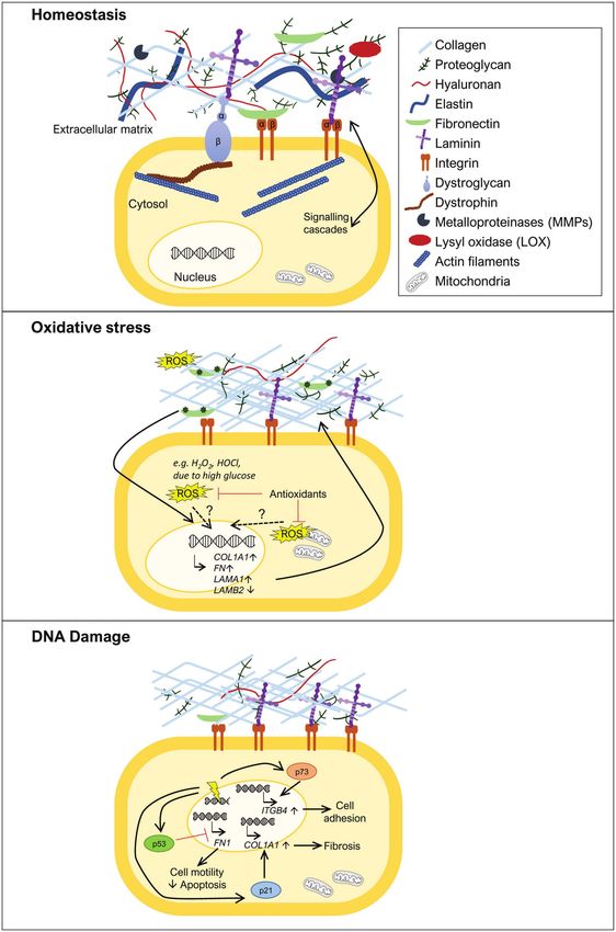
Martins et al.                                                                                                           ROS/DNA Damage and ECM

of different ROS, including hypochlorous acid (HOCl), a potent               (Moore and Winder, 2012; Afratis et al., 2017; Bachmann
ROS that is involved in pathogen killing mechanisms                          et al., 2019). These transmembrane proteins bind directly to
(Khan et al., 2018).                                                         specific domains of ECM molecules on the extracellular side,
    However, when in excess, ROS may cause serious damage                    and to cytoskeletal proteins and different kinases on the
to different molecules promoting DNA damage, protein oxidation,              intracellular side, thus allowing the transmission of signaling
and lipid peroxidation (McCord, 2000). The deleterious                       cascades. In addition, the ECM can also act as a route or
accumulation of ROS may have multiple origins, including                     reservoir for different molecules with important autocrine and
the excessive ROS production by the mitochondria, which                      paracrine functions that include cytokines, chemokines, and
drives mitochondrial dysfunction, an important trait of a variety            growth factors, which modulate a variety of different cellular
of diseases. Mitochondrial dysfunction can be due to a metabolic             processes, such as cell adhesion, migration, differentiation,
imbalance (Bhatti et al., 2017), like, for example, in diabetes,             proliferation, survival, and apoptosis (Bowers et al., 2010;
where high levels of glucose promote overproduction of ROS                   Bonnans et al., 2014).
(Yaribeygi et al., 2019), or it may also arise as a consequence                  Extracellular matrix remodeling is a normal process that
of mutations in mitochondrial DNA, defective OXPHOS, and                     occurs during embryonic development, allowing the progressive
alterations in ROS production, which are common phenomena                    formation of tissues and organs, and continues throughout
in cancer (Hsu et al., 2016; Porporato et al., 2018). As mentioned           adulthood in order to ensure the balance of organismal
above, another example of excess ROS generation is through                   homeostasis (Rozario and DeSimone, 2010; Bonnans et al.,
MPO in neutrophils, which can have a detrimental outcome                     2014). It is also an important player in tissue healing and
if it leads to chronic inflammation, a driver of pathology in                repair but, when not properly regulated, it can result in the
a variety of diseases (Khan et al., 2018).                                   loss of normal tissue structure and the development of different
    DNA damage can be caused by several factors, including                   pathological conditions. Examples of such conditions are (i)
by exogenous agents, e.g., UV light, ionizing radiation, as well             fibrosis, where extensive ECM deposition and scar formation
as by endogenous triggers, such as errors in DNA replication                 hamper normal tissue and organ function (Herrera et al., 2018;
and oxidative stress. DNA replication is a central process during            Li et al., 2018), (ii) diabetes, where ECM remodeling is negatively
cell division, where accuracy is essential to maintain the genetic           affected in several aspects of the disease, including in the
information intact (Sancar et al., 2004; Zeman and Cimprich,                 impaired healing of chronic diabetic wounds and in diabetes-
2014). However, DNA replication can also be a source of                      associated fibrosis (Falanga, 2005; Law et al., 2012; Ahmad
deleterious events leading to DNA damage, in particular, due                 et al., 2020), and (iii) cancer, where several components involved
to aberrant replication fork structures such as breaks associated            in ECM remodeling have their specific functions hijacked by
with replication fork stalling and collapse (Zeman and Cimprich,             cancer cells, enabling their proliferation, epithelial-to-
2014). Oxidative stress is also an important source of DNA                   mesenchymal transition, and migration (Bonnans et al., 2014;
damage (Whitaker et al., 2017), since increased levels of ROS                Winkler et al., 2020). Interestingly, diseases characterized by
can result in DNA oxidation, particularly in oxidized DNA                    extensive ECM remodeling often involve oxidative stress and
bases. Depending on the source of DNA damage, a specific                     DNA damage, either as the triggers or as a consequence of
machinery is activated to promote successful repair (Sancar                  their pathophysiology. This is the case of the aforementioned
et al., 2004; Zeman and Cimprich, 2014). When DNA damage                     pathologies: fibrosis, a pathological condition marked by high
cannot be repaired, cells may undergo programmed cell death                  levels of oxidative stress and a direct implication of DNA
or accumulate mutations that drive genomic instability, which                damage (Cheresh et al., 2013; Li et al., 2018); diabetes, which
can be a trigger for tumorigenesis (Hanahan and Weinberg, 2011).             is associated with high levels of blood glucose that in turn
    Even though, dedicated machineries exist to cope with both               lead to increased ROS levels (Yaribeygi et al., 2019); and cancer
oxidative stress and DNA damage, when these insults occur,                   in which both DNA damage and oxidative stress play an
they often lead to a variety of cellular and extracellular changes,          important role in disease progression (Luo et al., 2009; Reuter
which are important to ensure that cells return to their normal              et al., 2010; Hanahan and Weinberg, 2011). On the other hand,
physiological equilibrium. One of these changes is extracellular             several diseases due to mutations in ECM components or
matrix (ECM) remodeling. The ECM provides support to tissues                 proteins linking the ECM to the cell cytoskeleton have been
and contributes to maintain their appropriate physiological                  shown to display increased oxidative stress and DNA damage
environment (Frantz et al., 2010; Bonnans et al., 2014). It                  in the affected tissues (Rando, 2002; Irwin et al., 2003; Rodriguez
consists of a network of extracellular glycoproteins (e.g., collagens,       and Tarnopolsky, 2003; Millay et al., 2008; Shkryl et al., 2009;
fibronectin, and laminins), elastins, proteoglycans, hyaluronan,             Menazza et al., 2010; de Oliveira et al., 2014; Sorato et al.,
and a number of other less well characterized ECM components,                2014; Fontes-Oliveira et al., 2017; Gremminger et al., 2019;
collectively termed matrisome (Hynes and Naba, 2012; Naba                    Kölbel et al., 2019; Moore et al., 2020). This is particularly
et al., 2016; Figure 1A). ECM components can be directly                     striking when considering the muscular dystrophies, but other
linked to the cell surface and the underlying cytoskeleton,                  examples also exist.
allowing a rapid and precise communication between the cells                     It is essential to point out that ECM remodeling does not
and the ECM. The communication between the extracellular                     only involve the synthesis and deposition of ECM molecules,
and intracellular environments is mediated by transmembrane                  which transmit signals to cells through cell surface receptors.
proteins, such as integrins, syndecans, and dystroglycan                     The action of specific proteins that degrade ECM components,

Frontiers in Genetics | www.frontiersin.org                              2                                       July 2021 | Volume 12 | Article 673002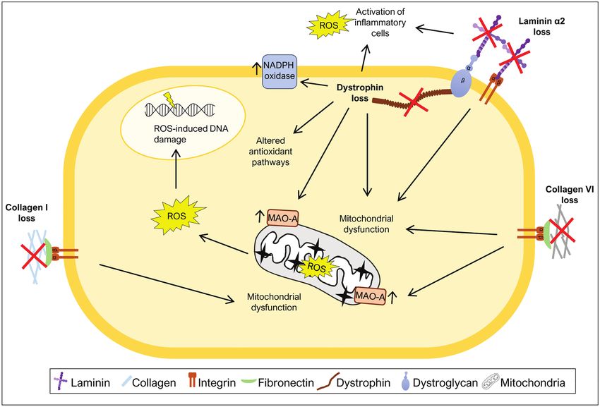
Martins et al. ROS/DNA Damage and ECM
A
B
C
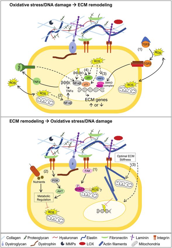
FIGURE 1 | Extracellular matrix (ECM) remodeling upon oxidative stress and DNA damage. (A) The ECM is composed of various molecules, such as extracellular
glycoproteins (e.g., collagens, fibronectin, and laminins), elastins, proteoglycans, and hyaluronan. These components are in close communication with the cell via
transmembrane proteins (e.g., integrins, syndecans, and dystroglycan) that anchor different ECM components to the cell, activating various signal transduction
cascades. Specific tissues have specialized ECMs and cell surface receptors, as can be observed for skeletal muscle, where the dystrophin-glycoprotein complex
anchors laminins and links them to the actin cytoskeleton via dystrophin. ECM remodeling is carried out through the action of metalloproteinases (MMPs) and
(Continued)
Frontiers in Genetics | www.frontiersin.org 3 July 2021 | Volume 12 | Article 673002Martins et al. ROS/DNA Damage and ECM
FIGURE 1 | lysyl oxidase (LOX). (B) ECM remodeling can be promoted by oxidative stress. In the presence of increased reactive oxygen species (ROS) levels [e.g.,
diabetes, high H2O2, hypochlorous acid (HOCl), or HOCl-modified fibronectin], some ECM genes become more highly expressed, such as fibronectin (FN1), collagen
α1 chain (COL1A1), and laminin α1 chain (LAMA1), while others show decreased expression, as observed for laminin β2 chain (LAMB2). The exact mechanism(s) by
which oxidative stress induces expression of ECM genes remains to be fully uncovered. The role of oxidative stress is also revealed by treatment with antioxidants
where reduction in the levels of ROS normalizes ECM composition. (C) DNA damage (represented as a yellow electric ray symbol on top of double stranded DNA) is
another insult that triggers changes in the ECM. The DNA damage responsive protein p53 has been implicated in the downregulation of FN1 expression, promoting
cell motility and suppressing apoptosis. Another important player in this context is p21. This negative regulator of the cell cycle has been shown to induce COL1A1
expression and consequently lead to collagen deposition and fibrosis. The transcription factor p73, a p53 family member, is also a key protein in ECM remodeling in
the response to DNA damage. Upon activation, p73 has been demonstrated to directly promote the transcription of ITGB4, encoding integrin β4, and consequently
promoting cell adhesion. For simplicity, not all ECM components are shown in the figures, just the ones that are relevant to illustrate the phenomenon being
highlighted.
including metalloproteinases (MMPs), and enzymes that shape (Figure 1B). However, the exact role played by oxidative stress
ECM structure, such as lysyl oxidase (LOX), also play a crucial in the regulation of collagen synthesis is still not fully understood,
role (Bonnans et al., 2014; Shimoda and Khokha, 2017; Laczko and apparent contradictory results have been described. Siwik
and Csiszar, 2020; Figure 1A). Indeed, the importance of MMPs et al. (2001) showed that oxidative stress can inhibit collagen
and their inhibitors, tissue inhibitors of metalloproteinases I and IV mRNA expression, while other studies have shown that
(TIMPs), in the response to oxidative stress and DNA damage oxidative stress can also lead to increased collagen expression
has been extensively addressed elsewhere (Mehta et al., 2006; (Iglesias-De La Cruz et al., 2001; Zhao et al., 2008; Guo et al.,
Kandasamy et al., 2010; Reuter et al., 2010; Sanchez-Valle et al., 2018; Kazakov et al., 2018). This apparent discrepancy might
2012; Broustas and Lieberman, 2014; Kumari et al., 2018) and be explained by the amount of oxidative stress in the tissue, as
are beyond the scope of this review. suggested by Liu et al. (2016). In this work, human uterosacral
Considering the body of work showing the deleterious effects ligament-derived fibroblasts were treated with a low concentration
of oxidative stress and DNA damage in the context of a variety of H2O2, an important ROS, which led to a decrease in COL1A1
of diseases on the one hand, and on the other hand, the (gene codifying for collagen type I α1 chain) expression. In
impact of changes in the ECM for disease onset and progression, contrast, when these cells were exposed to a higher concentration
this review aims to examine the potential links between oxidative of H2O2, their COL1A1 expression increased (Liu et al., 2016).
stress/DNA damage and the ECM in the context of disease This suggests that mild oxidative stress, associated with normal
development. We explore studies showing how oxidative stress metabolism or small insults, may promote the reduction of collagen
and DNA damage affect the synthesis of ECM molecules, levels, whereas high levels of oxidative stress may favor collagen
focusing on the major, and best studied, glycoproteins of the accumulation. Collagen deposition is required for wound healing,
ECM, namely collagens, fibronectin, and laminins (Hynes and but if in excess, can also account for a tissue injury status like
Naba, 2012; Naba et al., 2016). We also examine the inverse fibrosis. In support of this observation, several studies have shown
situation, where mutations in genes encoding some of these that the presence of elevated levels of oxidative stress lead to
key ECM glycoproteins or proteins that link the ECM to the increased collagen I and III levels contributing to myocardial
cell cytoskeleton trigger an accumulation of oxidative stress fibrosis (Zhao et al., 2008; Sriramula and Francis, 2015; Kazakov
and DNA damage. This dual approach aims to address to et al., 2018). In addition, treatment with antioxidants in the
what extent oxidative stress and DNA damage are part of the context of cardiac fibrosis has been shown to reduce collagen
molecular processes that control, and are controlled by, the levels (Zhao et al., 2008; Guo et al., 2018), further suggesting a
ECM, and through this perspective, we may deepen our direct effect between increased oxidative stress and collagen
understanding of how they contribute to disease development deposition. Glucose deregulation is another mechanism known
and progression. to cause oxidative stress, as observed for diabetes (Yaribeygi et al.,
2019). Kidney glomerular mesangial cells cultured with high levels
of glucose show increased collagen IV synthesis (Ayo et al., 1991).
THE IMPACT OF OXIDATIVE STRESS Moreover, the synthesis of collagen I and III is increased in
ON ECM REMODELING hearts of experimental diabetic rats, a condition that was countered
by antioxidant treatment (Guo et al., 2018). The regulation of
Analysis of NCBI Gene Expression Omnibus database for stress collagen synthesis by ROS may not only occur at the transcriptional
responses in HeLa cells showed that several different forms level. It is well established that the antioxidant vitamin C is a
of stress, including oxidative stress, lead to changes in the cofactor of prolyl hydroxylase and lysyl hydroxylase, required for
expression of genes encoding structural and signaling components post translational modifications of procollagen and consequently
of the ECM (Chovatiya and Medzhitov, 2014). Here, we review in the correct formation of the mature collagen triple helix (Frantz
the literature reporting when oxidative stress affects the synthesis et al., 2010; Bonnans et al., 2014). To corroborate the relationship
or structure of the best studied ECM glycoproteins, namely between collagen levels and oxidative stress, it has recently been
collagens, fibronectin, and laminins. suggested that vitamin C, due to its role as an antioxidant, could
Several studies have shown that the synthesis and/or the stability improve healing associated with musculoskeletal diseases, by
of several collagens, including collagen I, the most abundant reducing the levels of ROS associated with the inflammatory
fibrous protein of the ECM, are regulated by oxidative stress response (DePhillipo et al., 2018). Even though, pre-clinical studies
Frontiers in Genetics | www.frontiersin.org 4 July 2021 | Volume 12 | Article 673002Martins et al. ROS/DNA Damage and ECM
have shown that vitamin C can reduce ROS and promote tissue to increased cell motility and decreased apoptosis (Yokoi et al., 2020;
healing via collagen I synthesis during bone fracture recovery Figure 1C), while treatment with the p53 activator RITA caused
and in ruptured tendons, more studies in a clinical setting are a decrease in fibronectin levels (You et al., 2017). These studies
needed to assess its therapeutic benefit (DePhillipo et al., 2018). thus indicate p53 as a negative regulator of FN1 expression. In
The expression or stability of fibronectin has also been linked accordance with this notion, overexpression of wild type p53 in
to oxidative stress. Fibronectin (FN1) expression has been shown ovarian carcinoma cells was able to repress the activity of the
to be positively regulated in the presence of different sources of FN1 promoter, while a mutant p53 failed to do so (Yokoi et al.,
oxidative stress (Iglesias-De La Cruz et al., 2001; Siwik et al., 2020), showing that p53 can act as a transcription factor directly
2001; Lee et al., 2004; Nybo et al., 2018; Figure 1B). Oxidative regulating FN1 transcription. Another indication of an effect of
stress, due to high glucose levels, increases the expression of DNA damage on the expression of ECM components came from
FN1 in vitro (Ayo et al., 1991; Lee et al., 2004; Lin et al., 2006), a study on the CDK inhibitor p21 (Yosef et al., 2017), a negative
as well as in vivo, in the context of diabetes (Lin et al., 2006). regulator of the cell cycle, which can be induced by p53 and
ROS reduction by treatment with the ROS scavenging enzyme drive cell cycle arrest (Williams and Schumacher, 2016). This study
superoxide dismutase (SOD) led to a reduction in fibronectin revealed that in p21 knockout mice, senescent cells were eliminated,
levels in diabetic rats and to less kidney damage (Lin et al., and liver fibrosis was alleviated mainly via transcriptional
2006). Furthermore, HOCl generated through MPO activity in downregulation of collagen type I α1 (COL1A1; Yosef et al., 2017;
neutrophils has been shown to induce fibronectin modifications, Figure 1C). In fact, p21 knockout reduced Col1a1 expression,
such as tyrosine chlorination and dichlorination and oxidation prevented collagen deposition and consequently fibrosis, in mice
of different residues, which reduced cell adhesion and increased treated with carbon tetrachloride, a fibrogenic agent known to
proliferation of human coronary artery smooth muscle cells in cause DNA damage (Yosef et al., 2017). These observations suggest
vitro (Nybo et al., 2018). The HOCl-induced modifications of that one of the actions of p21 is to increase COL1A1 expression
fibronectin also led to changes in the expression of ECM genes in response to DNA damage. However, further studies are needed
by these smooth muscle cells, including a significant upregulation to assess whether these described effects of p53 and p21 on FN1
of FN1 and LAMA1 and downregulation of LAMB2 expression and COL1A1 expression, respectively, are cell type specific and
(Nybo et al., 2018; Figure 1B). LAMA1 codifies the laminin α1 exactly how the pathways involved control cell survival, senescence,
chain, which is normally not present in the basement membrane and apoptosis.
of smooth muscle cells, while downregulation of LAMB2 codifying Integrins provide yet another link between ECM structure
the laminin β2 chain indicates that cells are prevented from and DNA damage-induced apoptosis (Hoyt et al., 1996; Sethi
maintaining β2 laminins, characteristic of mature smooth muscle et al., 1999; Hodkinson et al., 2006; Jinushi et al., 2012; Ahmed
basement membranes (Yousif et al., 2013). These data indicate et al., 2018; Kowalski-Chauvel et al., 2018; Hou et al., 2019;
that cells attempt to remodel their ECM in response to their Jung et al., 2019; Naci et al., 2019; Li et al., 2021). Nevertheless,
oxidant-altered fibronectin substrate and associated effects on cell the connection between adhesion and apoptosis is not linear
adhesion and proliferation. and integrin binding to the ECM can either promote or prevent
Altogether, these various studies show that oxidative stress cells, in particular cancer cells, from undergoing apoptosis. It
causes changes in both ECM gene expression and in ECM is possible that regulation mediated by factors involved in the
structure which correlate with tissue damage. DNA damage response, such as p53 and p21, control the
expression of integrins or ECM components and therefore
modulate adhesion. Indeed, it has been shown that p73, a
THE IMPACT OF DNA DAMAGE ON p53 family member that can also be activated in response to
ECM REMODELING DNA damage, directly promotes transcription of ITGB4, encoding
integrin β4, therefore acting as a positive regulator of cell
It is well established that DNA integrity is essential for gene adhesion (Xie et al., 2018; Figure 1C).
expression and for the correct transmission of genetic information Collectively, this suggests that central DNA damage response
to daughter cells during mitosis. It is not surprising that DNA factors might be able to directly control the expression of
damage is one of the most deleterious forms of injury inflicted ECM components and integrins, hence being able to regulate
on cells (Sancar et al., 2004; Branzei and Foiani, 2008). Here, cell survival and apoptosis.
we will consider how DNA damage, independently of the
source (e.g., ROS, radiation, and mutagens), can directly or
indirectly affect the expression of genes encoding either ECM MUTATIONS IN GENES ENCODING ECM
glycoproteins or proteins linking the ECM to the cell cytoskeleton. COMPONENTS TRIGGER OXIDATIVE
A central protein involved in the DNA damage response is STRESS AND DNA DAMAGE
the tumor suppressor p53, which, when activated, either induces
cell cycle arrest to allow for DNA repair or, if the damage inflicted As discussed above, the notion that oxidative stress and DNA
is too large, promotes, for example, apoptosis (Sancar et al., 2004; damage result in alterations in the ECM in the context of different
Branzei and Foiani, 2008; Williams and Schumacher, 2016). p53 diseases has been gaining ground. However, does the inverse
has been implicated in the regulation of FN1 expression. Depletion occur? Does a dysfunctional ECM induce oxidative stress and
of p53 was shown to increase FN1 mRNA expression, leading DNA damage in the cells it harbors? Indeed, some observations
Frontiers in Genetics | www.frontiersin.org 5 July 2021 | Volume 12 | Article 673002Martins et al. ROS/DNA Damage and ECM
suggest that this is the case. For example, culture of human in Col6a1−/− mice and patients with collagen VI deficiency.
dermal fibroblasts in fragmented collagen matrices, resulted in Altogether, these studies reinforce the idea that the absence of
elevated levels of ROS in these cells compared to fibroblasts collagen VI drives excess ROS production in the mitochondria,
cultured in intact collagen matrices (Fisher et al., 2009). Here, potentially contributing to the observed muscle fiber damage.
we explore the literature on diseases caused by mutations in
genes encoding ECM components and proteins that link the LAMA2 Deficiency
ECM to the cytoskeleton, more specifically collagens, laminin The LAMA2 gene encodes the laminin α2 chain of laminin
211, and dystrophin, to assess whether they are linked to oxidative 211. Mutations in LAMA2 lead to a congenital muscular dystrophy
stress and/or DNA damage. (LAMA2-CMD; also known as merosin-deficient congenital
muscular dystrophy type 1A, MDC1A) which is characterized
Collagen Deficiency by muscle weakness, fibrosis, and chronic inflammation (Gawlik
Collagen I is present in a variety of connective tissues (Frantz and Durbeej, 2011; Yurchenco et al., 2018). Recently, different
et al., 2010; Bonnans et al., 2014). Mutations in COL1A1 and mouse models of LAMA2-CMD (the dy2J/dy2J and dy3K/dy3K
COL1A2 genes, encoding the α1 and α2 chains of collagen I, mouse models) and biopsies from LAMA2-CMD patients were
respectively, cause osteogenesis imperfecta, a disease characterized used to address the impact of oxidative stress on this condition
by brittle bones, short stature, and muscle weakness (van Dijk (de Oliveira et al., 2014; Fontes-Oliveira et al., 2017; Gawlik
et al., 2011; Marini et al., 2017). Recently, a study using the et al., 2019; Kölbel et al., 2019; Harandi et al., 2020). These
mouse model of osteogenesis imperfecta, the Col1a2oim mouse, studies indicate that the muscles of Lama2-deficient mice and
also known as oim/oim mouse, revealed that the muscles of mice LAMA2-CMD patients display an increase in oxidative stress
homozygous for the mutation display a mitochondrial dysfunction both in early and more advanced stages of the disease, pointing
in that they have decreased levels of electron transport chain to an important role of oxidative stress throughout disease
complex IV, mitochondrial encoded cytochrome oxidase I, energy progression (Gawlik et al., 2019; Harandi et al., 2020). In fact,
production, citrate synthase activity and respiration, and elevated dy3K/dy3K mice show high levels of oxidative stress in quadriceps
levels of PGC1α (Gremminger et al., 2019), all markers of at least from post-natal day 7 and increased Gclm and Sqstm1
mitochondrial dysfunction (Adhihetty et al., 2009; Li et al., 2020; expression, markers of oxidative stress, at post-natal day 21
Melouane et al., 2020; Figure 2). These results suggest that (Gawlik et al., 2019). Analysis of muscles from 3-week-old dy2J/
mitochondrial dysfunction is an important feature of the muscles dy2J mice and muscle biopsies from LAMA2-CMD patients (age
of oim/oim mice, but the exact mechanism that links collagen ranging between 22 days and 29 years) also confirmed the
I deficiency to mitochondrial dysfunction remains to be determined. presence of elevated levels of ROS in both cases (Harandi et al.,
Moreover, excessive ROS production in the context of collagen 2020). Moreover, the treatment of dy2J/dy2J mouse quadriceps
I deficiency may also contribute to the exacerbation of osteogenesis and triceps muscles with the antioxidant N-acetylcysteine (NAC)
imperfecta pathology in bone. Indeed, high ROS generation is partially rescued the motor function of dy2J/dy2J mice, reduced
associated with inhibition of new bone formation, suggesting that the number of apoptotic fibers, lowered the levels of inflammation
antioxidant treatment may be useful to treat diseases with bone markers and prevented fibrosis (Harandi et al., 2020). Vitamin
loss (Wauquier et al., 2009). E, another antioxidant, applied to the same mouse model reduced
Collagen VI is a type of collagen mainly found in muscles, the number of apoptotic fibers and attenuated inflammation in
tendons, and skin (Bönnemann, 2011). Mutations in the COL6A1, quadriceps muscles, although, it did not improve motor function
COL6A2, and COL6A3 genes, encoding chains of collagen VI, or reduce fibrosis (Harandi et al., 2020). Nevertheless, both
are associated with Ullrich congenital muscular dystrophy and antioxidants were able to improve dy2J/dy2J skeletal muscle
Bethlem myopathy, a severe and mild form of collagen VI morphology, in particular, by normalizing the proportion of
deficiency, respectively (Bönnemann, 2011; Bernardi and Bonaldo, small fibers, decreasing the number of fibers with centrally located
2013). Collagen VI deficiency has also been correlated with nuclei, and decreasing ROS production (Harandi et al., 2020).
oxidative stress. Studies using a mouse model of collagen VI Mitochondrial dysfunction, closely associated with elevated
deficiency, Col6a1−/− mice, revealed elevated levels of ROS, oxidative stress, also plays a central role in LAMA2-CMD
mitochondrial dysfunction and elevated levels of monoamine pathology. In fact, it was observed that Lama2-deficient mice
oxidase A (MAO-A) (Irwin et al., 2003; Menazza et al., 2010; present signs of mitochondrial swollenness (Millay et al., 2008)
Sorato et al., 2014; Figure 2), one of the major contributors to and downregulation of a variety of mitochondria specific genes
ROS production in the mitochondria (Canton et al., 2014). After encoding components of the electron transport chain/oxidative
treatment with MAO inhibitor, muscles from Col6a1−/− mice phosphorylation (de Oliveira et al., 2014; Fontes-Oliveira et al.,
showed a decrease in ROS levels when compared to untreated 2017; Figure 2). Likewise, muscles or muscle-derived cells from
mice (Menazza et al., 2010; Sorato et al., 2014), highlighting LAMA2-CMD patients were described as having an abnormal
the enhanced production of ROS by these enzymes when collagen membrane potential (Fontes-Oliveira et al., 2017), impaired
VI is absent. Consistent with this, treatment with a MAO inhibitor mitochondrial function and bioenergetic status and
(Menazza et al., 2010; Sorato et al., 2014) and treatment with downregulation of a variety of mitochondrial related genes
a mitochondrial permeability transition pore inhibitor (Irwin (Fontes-Oliveira et al., 2017; Kölbel et al., 2019). Altogether,
et al., 2003; Merlini et al., 2008; Palma et al., 2009), reduced these results support the hypothesis that oxidative stress and
the apoptotic phenotype and improved mitochondrial function mitochondrial dysfunction are hallmarks of LAMA2-CMD.
Frontiers in Genetics | www.frontiersin.org 6 July 2021 | Volume 12 | Article 673002Martins et al. ROS/DNA Damage and ECM FIGURE 2 | Mutations in ECM components increase oxidative stress. Many diseases caused by mutations in ECM components or components that link ECM to the cytoskeleton have been associated with generation of excess ROS levels. The loss of collagen I, collagen VI, laminin α2, or dystrophin was shown to induce mitochondrial dysfunction, a major source of ROS. Additionally, high ROS levels due to either increased activity of monoamine oxidase A (MAO-A) or NADPH oxidase have also been associated with the loss of specific ECM or ECM-related components. Loss of laminin α2 or dystrophin leads to increased levels of ROS as a consequence of the activation of inflammatory cells. Dystrophin loss was also shown to promote changes in the activity of antioxidant pathways and ROS-induced DNA damage. Dystrophin Mutations ROS in DMD patients and mdx mice may have multiple sources, Dystrophin is part of the dystrophin-glycoprotein complex which including activation of inflammatory cells (Whitehead et al., 2008; links cytoplasmic dystrophin to laminin 211 in the muscle fiber Grounds et al., 2020), increased activity of NADPH oxidase basement membrane, a link that is responsible for conferring (Williams and Allen, 2007; Whitehead et al., 2010), mitochondrial cellular stability during skeletal muscle contractions (Ervasti and dysfunction (Shkryl et al., 2009; Menazza et al., 2010), and Campbell, 1993). Mutations in the DMD gene, encoding dystrophin, alterations in antioxidant pathways, including, for example, the can cause Duchenne muscular dystrophy (DMD), characterized elevated levels of oxidative stress sensor nuclear factor erythroid by muscle weakness, inflammation, and fibrosis (Grounds et al., 2-related factor 2 (Nrf2) (Petrillo et al., 2017) and overactivation 2020). Several mechanisms are known to contribute to DMD of the nuclear factor κB (NF-κB) pathway (Kumar and Boriek, pathology, including oxidative stress (Rando, 2002; Grounds et al., 2003; Monici et al., 2003; Figure 2). Using antioxidizing drugs, 2020). Early studies of the disease showed that DMD muscles such as vitamin C or SOD, and treatment with the antioxidant display oxidative stress features, such as increased lipid peroxidation NAC reduced the levels of ROS in cardiac (Williams and Allen, (Hunter and Mohamed, 1986) and increased levels of oxidative 2007) and skeletal muscles (Whitehead et al., 2008) of mdx mice. stress responsive enzymes (Austin et al., 1992). Moreover, in vivo It also reduced the expression of markers of inflammation and and in vitro analyzes of mdx mice, the most widely used DMD fibrosis (Williams and Allen, 2007), as well as, the number of model, showed an increased susceptibility to muscle injury caused apoptotic fibers (Whitehead et al., 2008). This observed rescue by free radicals (Disatnik et al., 1998; Rando et al., 1998), suggesting of the DMD phenotype using antioxidant treatments suggests a a deficiency in oxidative stress responses in mdx mice. During possible role of ROS in the inflammation, fibrosis, and apoptosis the last decades, the oxidative stress hypothesis has been gathering characteristic of DMD. support with several studies showing high levels of ROS in muscles Elevated levels of NADPH oxidase and its regulator caveolin-3 of mdx mice (Ragusa et al., 1997; Williams and Allen, 2007; is also a hallmark of mdx muscles (Williams and Allen, 2007; Segatto et al., 2020) and DMD patients (Haycock et al., 1997; Whitehead et al., 2010; Petrillo et al., 2017; Segatto et al., 2020). Rodriguez and Tarnopolsky, 2003). The excessive production of NADPH oxidase produced in skeletal muscles fibers has been Frontiers in Genetics | www.frontiersin.org 7 July 2021 | Volume 12 | Article 673002
Martins et al.                                                                                                            ROS/DNA Damage and ECM

shown to be a major source of stretch-induced ROS in mdx                     triggering a domino effect, which severely compromises tissue
mice (Shkryl et al., 2009; Whitehead et al., 2010), since it allows          integrity. In this review, we examined several examples, where
the generation of the free radical superoxide. Furthermore, NAC              increased oxidative stress and DNA damage alter the expression
treatment reduced caveolin-3 expression and NF-κB activation                 of genes encoding key ECM components, and conversely, where
in skeletal muscles from mdx mice (Whitehead et al., 2008).                  mutations in genes encoding some of the best studied ECM
Another important player in DMD pathology is mitochondrial                   glycoproteins and proteins linking the ECM to the cell
dysfunction (Menazza et al., 2010; Moore et al., 2020). Elevated             cytoskeleton elicit an increase in oxidative stress and DNA
levels of MAO were reported in mdx mice (Menazza et al., 2010),              damage. The crosstalk between these two processes, and their
as well as, reduced mitochondrial DNA copy number (Moore                     major impact on tissue integrity and function, highlights the
et al., 2020). Inhibition of MAO reduced tropomyosin oxidation               necessity to study the molecular nature of this relationship
(an oxidation marker), normalized fiber size, and reduced tissue             further, opening the possibility of identifying new candidate
inflammation and apoptosis (Menazza et al., 2010).                           pathways as targets for therapy.
    Reactive oxygen species pose various risks for the integrity                 In response to oxidative stress and DNA damage, regulation
of different macromolecules including DNA. In keeping with this              of ECM remodeling may occur at different levels, including
notion, DMD patients present high levels of DNA damage associated            (i) transcriptional, via modulation of different transcription
with increased oxidative stress (Rodriguez and Tarnopolsky, 2003)            factors, (ii) posttranslational, by the action of MMPs, TIMPs,
and cells derived from DMD patients are more sensitive to                    and LOX, which control the degradation of ECM components,
DNA-damaging agents than control cells (Robbins et al., 1984).               and (iii) at the network level, where changes in an ECM
More recently, Jelinkova et al. (2019) generated dystrophin-deficient        components or molecules that bridge the ECM to the cell
human pluripotent stem cell (DMD hPSC) and showed that these                 cytoskeleton, may trigger a chain reaction that can compromise
cells have high levels of ROS, which leads to increased levels of            tissue integrity. Here, we discuss how oxidative stress and DNA
ROS-induced DNA damage.                                                      damage could influence or be influenced by the expression of
    Although, the precise mechanisms and direct or indirect actions          genes encoding ECM components or proteins linking to the
and targets are not yet clarified, all these studies sustain the             cell cytoskeleton and dissect out which pathways may be required
notion that mutations in the genes encoding ECM components,                  for this regulation.
or associated proteins, may trigger oxidative stress and DNA                     TGF-β signaling is one of the key regulators of ECM production
damage. Importantly, some studies, especially those focusing on              (Hinz, 2015; Tu and Quan, 2016), via the activation of Smad
muscular dystrophies, have raised the question whether oxidative             transcription factors, which regulate the transcription of ECM
stress constitutes a primary or a secondary event in disease                 genes (Figure 3A), for example, collagens and fibronectin, but
progression (Rando, 2002; Moore et al., 2020). In most cases,                also ECM proteolytic enzymes, such as MMPs (Tu and Quan,
studies are conducted during the active phase of the disease,                2016). In particular, induction of TGF-β signaling by oxidative
making it impossible to distinguish between these two events.                stress has been associated with increased transcription of collagens
Thus, in the context of mutations in genes encoding ECM                      and fibronectin (Iglesias-De La Cruz et al., 2001; Zhao et al.,
components or proteins that link the ECM to cytoskeleton, it is              2008; Krstić et al., 2015; Tu and Quan, 2016). Another study,
unclear whether oxidative stress acts as a cause, a consequence              points to the activation of the TNF pathway as important for
or both, of disease progression. Hence, more studies are needed              the transcription of collagen I and III genes, in response to
to fully understand how oxidative stress contributes to each phase           angiotensin II-induced oxidative stress (Sriramula and Francis,
of the disease. Taking into account the link between oxidative               2015), possibly via the activation of the NF-κβ transcription factor
stress and DNA damage, it is likely that oxidative stress induced            (Morgan and Liu, 2011). NF-κβ is also activated in response to
DNA damage may not be limited to DMD (Robbins et al., 1984;                  DNA damage (Janssens and Tschopp, 2006), leading to the
Rodriguez and Tarnopolsky, 2003; Jelinkova et al., 2019), but                transcription of several targets, including ECM related genes
may also be a feature of the other diseases discussed here. The              (Janssens and Tschopp, 2006; Guo et al., 2021; Figure 3A). NRF2
concomitant presence of oxidative stress and DNA damage can                  is another important orchestrator of the oxidative stress response
act as an important driving force for disease progression. Therefore,        and which acts as a transcription factor (He et al., 2020). This
understanding the exact mechanisms behind the accumulation                   important stress sensor has been shown to be required for the
of oxidative stress in each phase of these diseases and determining          transcriptional regulation of several matrisome genes, including
the cellular consequences of increased ROS in every phase is                 collagen I, III, and laminin α1 chain genes (Hiebert et al., 2018;
crucial to design targeted therapies for these devastating conditions.       Hiebert, 2021), raising the possibility that in the presence of
                                                                             oxidative stress, NRF2 may control ECM remodeling (Figure 3A).
                                                                             As mentioned above, the transcriptional regulation of ECM
DISCUSSION                                                                   components may also be mediated directly or indirectly via the
                                                                             transcription factors p53 and p73, both playing important roles
The ECM is an intricate network of different molecules that                  sensing oxidative stress and DNA damage (Holmstrom et al.,
communicate directly to the intracellular space through cell                 2014; Pflaum et al., 2014; Williams and Schumacher, 2016;
surface receptors and downstream signaling cascades. It is                   Figure 3A). Whether these candidate pathways and transcription
becoming increasingly clear that altered expression of one of                factors, known to regulate the expression of ECM genes, act in
these molecules may perturb this fine-tuned communication,                   a tissue or disease specific manner, remains to be further explored.

Frontiers in Genetics | www.frontiersin.org                              8                                        July 2021 | Volume 12 | Article 673002
Martins et al. ROS/DNA Damage and ECM
A
B
FIGURE 3 | Oxidative stress and DNA damage links to ECM remodeling. (A) Oxidative stress and DNA damage may trigger ECM remodeling by different
mechanisms. One possible mechanism is associated with ROS-mediated release of TGFβ from the large latent complex, allowing it to bind to the TGFβ receptor (1).
This promotes the activation of the SMAD transcriptional complex, known to promote transcription of ECM genes. Another possible mechanism involves the activation
of the NF-κβ transcription factor (2). This may occur in the presence of oxidative stress leading to NF-κβ mediated transcription of TNFα, which in turn further
promotes NF-κβ activation and transcriptional regulation of ECM genes. NF-κβ can also be activated by DNA damage and control the transcription of ECM target
genes. The oxidative stress sensor nuclear factor erythroid 2-related factor 2 (NRF2; 3), may also have a crucial role in the transcriptional regulation of ECM genes.
The transcription factors p53 and p73, which regulate ECM gene expression, are both known targets of oxidative stress and DNA damage (4). (B) ECM stability is
important to prevent the generation of oxidative stress and DNA damage. The integrin-focal adhesion kinase (FAK) pathway promotes mitochondrial function, via
(Continued)
Frontiers in Genetics | www.frontiersin.org 9 July 2021 | Volume 12 | Article 673002Martins et al. ROS/DNA Damage and ECM
FIGURE 3 | phosphorylation of STAT3, which is implicated in the reduction of ROS and maintenance of mitochondrial membrane potential (1). Maintaining ECM
integrity and cell adhesion contributes to sustain nutrient uptake and phosphatidyl-inositol-3-kinase (PI3K)/AKT signaling, together reinforcing metabolic regulation,
and preventing accumulation of ROS (2). Biomechanical properties of the ECM are also critical to maintain cell structure (3). An appropriate ECM stiffness maintains
mitochondrial dynamics, possibly preventing ROS production, and nuclear structure, supporting genomic integrity and avoiding DNA damage. Dashed lines represent
pathways that may involve one or more intermediate players.
As detailed earlier, several studies point to oxidative stress cells more sensitive to DNA damaging agents (Deng et al., 2020),
and DNA damage being a consequence, rather than a cause, of while high ECM stiffness promotes nuclear rupture also leading
changes in the ECM. In line with this, mutations in genes encoding to increased DNA damage (Cho et al., 2019; Figure 3B). Altogether,
ECM components or molecules that bridge the ECM to the cell these studies suggest that countering mitochondrial dysfunction,
cytoskeleton have been described to lead to mitochondrial and possibly preventing DNA damage, may be a promising strategy
dysfunction (Irwin et al., 2003; Millay et al., 2008; Shkryl et al., to revert the pathology caused by some mutations in genes
2009; Menazza et al., 2010; de Oliveira et al., 2014; Sorato et al., encoding ECM components, their receptors or molecules that
2014; Fontes-Oliveira et al., 2017; Gremminger et al., 2019; Kölbel bridge ECM receptors to the cell cytoskeleton.
et al., 2019), a driving force for ROS accumulation. There are In conclusion, future studies are needed to characterize all
several different mechanisms that can explain why mitochondria the players and intermediates involved in the link between
may function as an important sensor for ECM changes (De oxidative stress/DNA damage and ECM remodeling. Only with
Cavanagh et al., 2009), and therefore, why mitochondrial dysfunction this increased knowledge can new candidate pathways and
is a hallmark of muscular dystrophies (Cantó-Santos et al., 2020). targets be identified. This will be instrumental to establish
ECM glycoproteins, including laminins and fibronectin, have been novel lines of therapy, for diseases where ROS and DNA damage
shown to promote mitochondrial function via integrin-focal drive ECM remodeling, or where mutations in ECM-related
adhesion kinase (FAK) signaling, which culminates in the genes elicit an over-accumulation of ROS and DNA damage.
translocation of phosphorylated STAT3 to the mitochondria
(Visavadiya et al., 2016; Figure 3B). Other possible mechanisms
are related to the loss of adhesion to the ECM, which changes AUTHOR CONTRIBUTIONS
the cellular uptake of nutrients, namely glucose (Schafer et al.,
2009; Eble and De Rezende, 2014). These effects together with SGM and ARC performed the literature review and wrote the
the reduction in the activity of phosphatidyl-inositol-3-kinase manuscript. RZ and ST contributed with active discussion and
(PI3K) and Akt, also associated with altered ECM composition, revision. All authors contributed to the article and approved
elicits dramatic metabolic changes including decreased activity the submitted version.
of the pentose phosphate pathway, which represents an important
source of NADPH, a powerful antioxidant molecule (Eble and
De Rezende, 2014; Figure 3B). In addition to signaling pathways FUNDING
linking ECM to mitochondria, biomechanical properties of the
ECM, such as stiffness (i.e., how rigid the ECM structure is), This work is supported by Association Française contre
can impact mitochondrial function (Chen et al., 2021; Figure 3B). les Myopathies (AFM) Téléthon (contract no. 23049) to ST,
High ECM stiffness has been shown to promote mitochondrial Fundação para a Ciência e Tecnologia (FCT, Portugal;
fusion and suppress mitochondrial fission downstream of integrin CEECIND/01589/2017), and L’Oréal Portugal Medals of Honor
signaling (Chen et al., 2021). The importance of ECM biomechanical for Women in Science 2019 to ARC. Fundação para a Ciência
properties goes beyond maintaining mitochondrial stability. Low e a Tecnologia Project (Ref. PTDC/BTM-ORG/1383/2020) and
ECM stiffness compromises the DNA damage response, making Unit Funding (Ref. UIDB/00329/2020) supports all authors.
REFERENCES Austin, L., de Niese, M., McGregor, A., Arthur, H., Gurusinghe, A., and
Gould, M. K. (1992). Potential oxyradical damage and energy status in
Adhihetty, P. J., Uguccioni, G., Leick, L., Hidalgo, J., Pilegaard, H., and Hood, D. A. individual muscle fibres from degenerating muscle diseases. Neuromuscul.
(2009). The role of PGC-1α on mitochondrial function and apoptotic Disord. 2, 27–33. doi: 10.1016/0960-8966(92)90023-Y
susceptibility in muscle. Am. J. Physiol. Cell Physiol. 297, C217–C225. doi: Ayo, S. H., Radnik, R. A., Glass, W. F., Garoni, J. A., Rampt, E. R., Appling, D. R.,
10.1152/ajpcell.00070.2009 et al. (1991). Increased extracellular matrix synthesis and mRNA in mesangial
Afratis, N. A., Nikitovic, D., Multhaupt, H. A. B., Theocharis, A. D., Couchman, J. R., cells grown in high-glucose medium. Am. J. Phys. 260, F185–F191. doi:
and Karamanos, N. K. (2017). Syndecans – key regulators of cell signaling 10.1152/ajprenal.1991.260.2.F185
and biological functions. FEBS J. 284, 27–41. doi: 10.1111/febs.13940 Bachmann, M., Kukkurainen, S., Hytönen, V. P., and Wehrle-Haller, B. (2019).
Ahmad, K., Choi, I., and Lee, Y. H. (2020). Implications of skeletal muscle Cell adhesion by integrins. Physiol. Rev. 99, 1655–1699. doi: 10.1152/
extracellular matrix remodeling in metabolic disorders: diabetes perspective. physrev.00036.2018
Int. J. Mol. Sci. 21:3845. doi: 10.3390/ijms21113845 Bernardi, P., and Bonaldo, P. (2013). Mitochondrial dysfunction and defective
Ahmed, K. M., Pandita, R. K., Singh, D. K., Hunt, C. R., and Pandita, T. K. autophagy in the pathogenesis of collagen VI muscular dystrophies. Cold
(2018). β1-integrin impacts Rad51 stability and DNA double-strand break Spring Harb. Perspect. Biol. 5:a011387. doi: 10.1101/cshperspect.a011387
repair by homologous recombination. Mol. Cell. Biol. 38:e00672-17. doi: Bhatti, J. S., Bhatti, G. K., and Reddy, P. H. (2017). Mitochondrial dysfunction
10.1128/MCB.00672-17 and oxidative stress in metabolic disorders – a step towards mitochondria
Frontiers in Genetics | www.frontiersin.org 10 July 2021 | Volume 12 | Article 673002Martins et al. ROS/DNA Damage and ECM based therapeutic strategies. Biochim. Biophys. Acta Mol. basis Dis. 1863, Fontes-Oliveira, C. C., Steinz, M., Schneiderat, P., Mulder, H., and Durbeej, M. 1066–1077. doi: 10.1016/j.bbadis.2016.11.010 (2017). Bioenergetic impairment in congenital muscular dystrophy type 1A Bonnans, C., Chou, J., and Werb, Z. (2014). Remodelling the extracellular and leigh syndrome muscle cells. Sci. Rep. 7:45272. doi: 10.1038/srep45272 matrix in development and disease. Nat. Rev. Mol. Cell Biol. 15, 786–801. Frantz, C., Stewart, K. M. M., and Weaver, V. M. M. (2010). The extracellular doi: 10.1038/nrm3904 matrix at a glance. J. Cell Sci. 123, 4195–4200. doi: 10.1242/jcs.023820 Bönnemann, C. G. (2011). The collagen VI-related myopathies: muscle meets Gawlik, K. I., and Durbeej, M. (2011). Skeletal muscle laminin and MDC1A: its matrix. Nat. Rev. Neurol. 7, 379–390. doi: 10.1038/nrneurol.2011.81 pathogenesis and treatment strategies. Skelet. Muscle 1:9. doi: 10.1186/2044-5040-1-9 Bowers, S. L. K., Banerjee, I., and Baudino, T. A. (2010). The extracellular Gawlik, K. I., Körner, Z., Oliveira, B. M., and Durbeej, M. (2019). Early skeletal matrix: at the center of it all. J. Mol. Cell. Cardiol. 48, 474–482. doi: 10.1016/j. muscle pathology and disease progress in the dy3K/dy3K mouse model of yjmcc.2009.08.024 congenital muscular dystrophy with laminin α2 chain-deficiency. Sci. Rep. Branzei, D., and Foiani, M. (2008). Regulation of DNA repair throughout the 9:14324. doi: 10.1038/s41598-019-50550-0 cell cycle. Nat. Rev. Mol. Cell Biol. 9, 297–308. doi: 10.1038/nrm2351 Gremminger, V. L., Jeong, Y., Cunningham, R. P., Meers, G. M., Rector, R. S., Broustas, C. G., and Lieberman, H. B. (2014). DNA damage response genes and Phillips, C. L. (2019). Compromised exercise capacity and mitochondrial and the development of cancer metastasis. Radiat. Res. 181, 111–130. doi: dysfunction in the osteogenesis imperfecta murine (oim) mouse model. 10.1667/RR13515.1 J. Bone Miner. Res. 34, 1646–1659. doi: 10.1002/jbmr.3732 Canton, M., Menazza, S., and Di Lisa, F. (2014). Oxidative stress in muscular Grounds, M. D., Terrill, J. R., Al-Mshhdani, B. A., Duong, M. N., dystrophy: from generic evidence to specific sources and targets. J. Muscle Radley-Crabb, H. G., and Arthur, P. G. (2020). Biomarkers for Duchenne Res. Cell Motil. 35, 23–36. doi: 10.1007/s10974-014-9380-2 muscular dystrophy: myonecrosis, inflammation and oxidative stress. DMM Cantó-Santos, J., Grau-Junyent, J. M., and Garrabou, G. (2020). The impact Dis. Model. Mech. 13:dmm043638. doi: 10.1242/dmm.043638 of mitochondrial deficiencies in neuromuscular diseases. Antioxidants 9:964. Guo, Q., Chen, X., Chen, J., Zheng, G., Xie, C., Wu, H., et al. (2021). STING doi: 10.3390/antiox9100964 promotes senescence, apoptosis, and extracellular matrix degradation in Chen, K., Wang, Y., Deng, X., Guo, L., and Wu, C. (2021). Extracellular matrix osteoarthritis via the NF-κB signaling pathway. Cell Death Dis. 12:13. doi: stiffness regulates mitochondrial dynamics through PINCH-1- and kindlin-2- 10.1038/s41419-020-03341-9 mediated signalling. Curr. Res. Cell Biol. 2:100008. doi: 10.1016/j. Guo, S., Meng, X. W., Yang, X. S., Liu, X. F., Ou-Yang, C. H., and Liu, C. crcbio.2021.100008 (2018). Curcumin administration suppresses collagen synthesis in the hearts Cheresh, P., Kim, S. J., Tulasiram, S., and Kamp, D. W. (2013). Oxidative stress of rats with experimental diabetes. Acta Pharmacol. Sin. 39, 195–204. doi: and pulmonary fibrosis. Biochim. Biophys. Acta Mol. basis Dis. 1832, 1028–1040. 10.1038/aps.2017.92 doi: 10.1016/j.bbadis.2012.11.021 Hanahan, D., and Weinberg, R. A. (2011). Hallmarks of cancer: the next Cho, S., Vashisth, M., Abbas, A., Majkut, S., Vogel, K., Xia, Y., et al. (2019). generation. Cell 144, 646–674. doi: 10.1016/j.cell.2011.02.013 Mechanosensing by the lamina protects against nuclear rupture, DNA damage, Harandi, V. M., Oliveira, B. M. S., Allamand, V., Friberg, A., Fontes-Oliveira, C. C., and cell-cycle arrest. Dev. Cell 49, 920.e5–935.e5. doi: 10.1016/j. and Durbeej, M. (2020). Antioxidants reduce muscular dystrophy in the devcel.2019.04.020 dy2J/dy2J mouse model of laminin α2 chain-deficient muscular dystrophy. Chovatiya, R., and Medzhitov, R. (2014). Stress, inflammation, and defense of Antioxidants 9:244. doi: 10.3390/antiox9030244 homeostasis. Mol. Cell 54, 281–288. doi: 10.1016/j.molcel.2014.03.030 Haycock, J. W., Mac Neil, S., Jones, P., Harris, J. B., and Mantle, D. (1997). De Cavanagh, E. M., Ferder, M., Inserra, F., and Ferder, L. (2009). Angiotensin Oxidative damage to muscle protein in Duchenne muscular dystrophy. II, mitochondria, cytoskeletal, and extracellular matrix connections: an Neuroreport 8, 357–361. doi: 10.1097/00001756-199612200-00070 integrating viewpoint. Am. J. Phys. 296, H550–H558. doi: 10.1152/ He, F., Ru, X., and Wen, T. (2020). NRF2, a transcription factor for stress ajpheart.01176.2008 response and beyond. Int. J. Mol. Sci. 21:4777. doi: 10.3390/ijms21134777 de Oliveira, B. M., Matsumura, C. Y., Fontes-Oliveira, C. C., Gawlik, K. I., Herrera, J., Henke, C. A., and Bitterman, P. B. (2018). Extracellular matrix as Acosta, H., Wernhoff, P., et al. (2014). Quantitative proteomic analysis a driver of progressive fibrosis. J. Clin. Invest. 128, 45–53. doi: 10.1172/ reveals metabolic alterations, calcium dysregulation, and increased JCI93557 expression of extracellular matrix proteins in Laminin α2 Chain-deficient Hiebert, P. (2021). The Nrf2 transcription factor: a multifaceted regulator of muscle. Mol. Cell. Proteomics 13, 3001–3013. doi: 10.1074/mcp.M113. the extracellular matrix. Matrix Biol. Plus 10:100057. doi: 10.1016/j. 032276 mbplus.2021.100057 Deng, M., Lin, J., Nowsheen, S., Liu, T., Zhao, Y., Villalta, P. W., et al. (2020). Hiebert, P., Wietecha, M. S., Cangkrama, M., Haertel, E., Mavrogonatou, E., Extracellular matrix stiffness determines DNA repair efficiency and cellular Stumpe, M., et al. (2018). Nrf2-mediated fibroblast reprogramming drives sensitivity to genotoxic agents. Sci. Adv. 6:eabb2630. doi: 10.1126/sciadv. cellular senescence by targeting the matrisome. Dev. Cell 46, 145.e10–161. abb2630 e10. doi: 10.1016/j.devcel.2018.06.012 DePhillipo, N. N., Aman, Z. S., Kennedy, M. I., Begley, J. P., Moatshe, G., Hinz, B. (2015). The extracellular matrix and transforming growth factor-β1: and LaPrade, R. F. (2018). Efficacy of vitamin C supplementation on collagen tale of a strained relationship. Matrix Biol. 47, 54–65. doi: 10.1016/j. synthesis and oxidative stress after musculoskeletal injuries: a systematic matbio.2015.05.006 review. Orthop. J. Sports Med. 6:2325967118804544. doi: 10.1177/232596711 Hodkinson, P. S., Elliott, T., Wong, W. S., Rintoul, R. C., Mackinnon, A. C., 8804544 Haslett, C., et al. (2006). ECM overrides DNA damage-induced cell cycle Disatnik, M. H., Dhawan, J., Yu, Y., Beal, M. F., Whirl, M. M., Franco, A. A., arrest and apoptosis in small-cell lung cancer cells through β1 integrin- et al. (1998). Evidence of oxidative stress in mdx mouse muscle: studies dependent activation of PI3-kinase. Cell Death Differ. 13, 1776–1788. doi: of the pre- necrotic state. J. Neurol. Sci. 161, 77–84. doi: 10.1016/ 10.1038/sj.cdd.4401849 S0022-510X(98)00258-5 Holmstrom, K. M., Finkel, T., Holmström, K. M., and Finkel, T. (2014). Cellular Eble, J. A., and De Rezende, F. F. (2014). Redox-relevant aspects of the extracellular mechanisms and physiological consequences of redox-dependent signalling. matrix and its cellular contacts via integrins. Antioxid. Redox Signal. 20, Nat. Rev. Mol. Cell Biol. 15, 411–421. doi: 10.1038/nrm3801 1977–1993. doi: 10.1089/ars.2013.5294 Hou, S., Jin, W., Xiao, W., Deng, B., Wu, D., Zhi, J., et al. (2019). Integrin Ervasti, J. M., and Campbell, K. P. (1993). A role for the dystrophin-glycoprotein α5 promotes migration and cisplatin resistance in esophageal squamous cell complex as a transmembrane linker between laminin and actin. J. Cell Biol. carcinoma cells. Am. J. Cancer Res. 9, 2774–2788. 122, 809–823. doi: 10.1083/jcb.122.4.809 Hoyt, D. G., Mannix, R. J., Gerritsen, M. E., Watkins, S. C., Lazo, J. S., and Falanga, V. (2005). Wound healing and its impairment in the diabetic foot. Pitt, B. R. (1996). Integrins inhibit LPS-induced DNA strand breakage in Lancet 366, 1736–1743. doi: 10.1016/S0140-6736(05)67700-8 cultured lung endothelial cells. Am. J. Physiol. Lung Cell. Mol. Physiol. 270, Fisher, G. J., Quan, T., Purohit, T., Shao, Y., Moon, K. C., He, T., et al. (2009). L689–L694. doi: 10.1152/ajplung.1996.270.4.L689 Collagen fragmentation promotes oxidative stress and elevates matrix Hsu, C. C., Tseng, L. M., and Lee, H. C. (2016). Role of mitochondrial metalloproteinase-1 in fibroblasts in aged human skin. Am. J. Pathol. 174, dysfunction in cancer progression. Exp. Biol. Med. 241, 1281–1295. doi: 101–114. doi: 10.2353/ajpath.2009.080599 10.1177/1535370216641787 Frontiers in Genetics | www.frontiersin.org 11 July 2021 | Volume 12 | Article 673002
You can also read