Invitroselection of L-DNA aptamers that bind a structured D-RNA molecule
←
→
Page content transcription
If your browser does not render page correctly, please read the page content below
Published online 17 January 2020                                                          Nucleic Acids Research, 2020, Vol. 48, No. 4 1669–1680
                                                                                                                         doi: 10.1093/nar/gkz1236

In vitro selection of L-DNA aptamers that bind a
structured D-RNA molecule
                                                                               *
Sougata Dey and Jonathan T. Sczepanski

Department of Chemistry, Texas A&M University, College Station, TX 77843, USA

Received November 20, 2019; Revised December 25, 2019; Editorial Decision December 28, 2019; Accepted January 03, 2020

                                                                                                                                                                  Downloaded from https://academic.oup.com/nar/article/48/4/1669/5707588 by guest on 19 September 2020
ABSTRACT                                                                               tured RNA elements have been discovered to play crit-
                                                                                       ical roles in a variety of diseases, including viral infec-
The development of structure-specific RNA bind-                                        tions, neurological disorders and cancer (2–4). Given the
ing reagents remains a central challenge in RNA                                        importance of RNA in human health and disease, target-
biochemistry and drug discovery. Previously, we                                        ing RNAs based on their unique structures is a poten-
showed in vitro selection techniques could be used                                     tially transformative diagnostic and therapeutic strategy.
to evolve L-RNA aptamers that bind tightly to struc-                                   Furthermore, structure-specific reagents open the door to
tured D-RNAs. However, whether similar RNA-binding                                     develop novel tools to study RNA function. However, dis-
properties can be achieved using aptamers com-                                         covery of molecules that are capable of binding RNA struc-
posed of L-DNA, which has several practical ad-                                        tures with high affinity and specificity has proven challeng-
vantages compared to L-RNA, remains unknown.                                           ing using current approaches. For example, RNAs with ex-
Here, we report the discovery and characterization                                     tensive secondary structures are often inaccessible to tra-
                                                                                       ditional hybridization-based reagents (5,6). Furthermore,
of the first L-DNA aptamers against a structured
                                                                                       RNA-binding small molecules often suffer from poor se-
RNA molecule, precursor microRNA-155, thereby es-                                      lectivity due to RNA’s negatively charged backbone and
tablishing the capacity of DNA and RNA molecules                                       structural redundancy (7–9). Therefore, development of
of the opposite handedness to form tight and spe-                                      structure-specific RNA binding reagents remains a central
cific ‘cross-chiral’ interactions with each other. L-                                  challenge.
DNA aptamers bind pre-miR-155 with low nanomo-                                            A potential solution to this problem is the use of ap-
lar affinity and high selectivity despite the inability                                tamers (10,11), which are short single-stranded nucleic acid
of L-DNA to interact with native D-RNA via Watson–                                     molecules capable of binding to a specific target. In the-
Crick base pairing. Furthermore, L-DNA aptamers in-                                    ory, it should be possible to evolve an aptamer against
hibit Dicer-mediated processing of pre-miRNA-155.                                      any RNA target using in vitro selection methods. Indeed,
The sequence and structure of L-DNA aptamers are                                       several groups have successfully isolated aptamers capa-
                                                                                       ble of binding structured RNA molecules (12–16). More
distinct from previously reported L-RNA aptamers
                                                                                       recently, we demonstrated that aptamers composed of L-
against pre-miR-155, indicating that L-DNA and L-                                      ribose-based nucleic acids (L-RNA), which are the synthetic
RNA interact with the same RNA sequence through                                        enantiomers of natural D-nucleotides, can also be evolved
unique modes of recognition. Overall, this work                                        to bind tightly to RNA structures, and may be particu-
demonstrates that L-DNA may be pursued as an al-                                       larly well suited for this purpose (17–19). This is because D-
ternative to L-RNA for the generation of RNA-binding                                   and L-oligonucleotides are incapable of forming contiguous
aptamers, providing a robust and practical approach                                    WC base pairs with each other (20–22), instead forcing L-
for targeting structured RNAs.                                                         aptamers to interact with native D-RNA through tertiary in-
                                                                                       teractions. This unique mode of nucleic acid recognition, re-
                                                                                       ferred to as ‘cross-chiral’ recognition, is expected to be more
INTRODUCTION                                                                           specific than simple WC pairing as it depends on both the
                                                                                       sequence and structure (i.e. shape) of the target RNA. De-
Base-pairing properties enable RNA to fold into intricate
                                                                                       spite the inability of L-aptamers to form WC base pairs with
three-dimensional structures that result in its diverse cel-
                                                                                       RNA, high-affinity binding can still be achieved. For exam-
lular functions. RNA structural motifs can influence tran-
                                                                                       ple, we previously isolated a cross-chiral L-RNA aptamer
scription, splicing, cellular localization, stability and trans-
                                                                                       capable of binding the hairpin structure of precursor mi-
lation of RNA (1). Thus, RNA structure plays a role in al-
                                                                                       croRNA (pre-miR)-155 with a Kd of 11 nM, making it one
most every facet of gene expression. Furthermore, struc-

* To   whom correspondence should be addressed. Email: jon.sczepanski@chem.tamu.edu
Disclaimer: The content is solely the responsibility of the authors and does not necessarily represent the official views of the National Institutes of Health.


C The Author(s) 2020. Published by Oxford University Press on behalf of Nucleic Acids Research.
This is an Open Access article distributed under the terms of the Creative Commons Attribution License (http://creativecommons.org/licenses/by/4.0/), which
permits unrestricted reuse, distribution, and reproduction in any medium, provided the original work is properly cited.
1670 Nucleic Acids Research, 2020, Vol. 48, No. 4
of the tightest binding, non-hybridization-based reagents microRNA with clinical implications. A parallel selection
ever developed against a structured RNA target (18). This experiment was also carried out employing the cationic
aptamer was also shown to inhibit Dicer-mediated process- nucleotide 5-aminoallyl-2 -deoxyuridine (5AdU), yielding
ing of pre-miR-155 in vitro. Therefore, in addition to high cross-chiral L-DNA aptamers harbouring the same modi-
affinity and selectivity, cross-chiral aptamers are capable of fication. Both modified and unmodified L-DNA aptamers
modulating RNA function by blocking RNA–protein in- bound pre-miR-155 with low nanomolar affinity, similar
teractions. Importantly, because L-oligonucleotides are or- to what previously reported for L-RNA aptamers for the
thogonal to the stereospecific environment of native biol- same target. In the case of the modified L-DNA aptamer, the
ogy, cross-chiral aptamers are completely resistant to nucle- cationic moiety was critical for binding. The sequence and
ase degradation (22,23) and expected to be less susceptible structure of cross-chiral L-DNA aptamers isolated herein
to off-target interactions with native biomacromolecules, are distinct from their RNA counterparts despite binding to
including other nucleic acids. Taken together, these proper- the same site (i.e. nucleotide sequence) on the target RNA.
ties suggest that cross-chiral aptamers hold great promise as At last, we showed that formation of L-aptamer–pre-miR-
Downloaded from https://academic.oup.com/nar/article/48/4/1669/5707588 by guest on 19 September 2020
structure-specific RNA affinity reagents, especially for ap- 155 complexes inhibit Dicer cleavage in vitro, thereby block-
plication in biological environments. ing formation of the mature miR-155. Overall, our results
All cross-chiral aptamers reported to date have been com- establish the capacity of L-DNA aptamers to bind native D-
posed of L-RNA. Despite their attractive properties, how- RNA structures (and vice versa), and show that the absence
ever, the use of L-RNA places potential limits on the prac- of the 2 -hydroxyl group is not a disadvantage for cross-
tical utility of these aptamers. In particular, the inefficiency chiral nucleic acid recognition.
of solid-phase RNA synthesis and the relative high cost of
L-nucleoside phosphoramidites, makes it difficult to obtain MATERIALS AND METHODS
the needed quantity of L-RNA cross-chiral aptamers for
many downstream applications. In addition, although L- Materials
RNA is nuclease resistant, it is still vulnerable to hydrolytic Oligonucleotides were either purchased from Integrated
cleavage of the sugar-phosphate backbone due to the pres- DNA Technologies (Coralville, IA, USA) or prepared by
ence of the 2 -OH group. This chemical instability may pre- solid-phase synthesis on an Expedite 8909 DNA/RNA
vent the use of cross-chiral L-RNA aptamers within certain synthesizer. Synthesizer reagents and D-nucleoside phos-
biological/chemical environments and limit their ability to phoramidites were purchased from Glen Research (Ster-
be stored long-term, especially in low-resource settings. In ling, VA, USA). D-5-aminoallyl-dUTP was purchased
contrast, the use of L-DNA may offer a more practical ap- from TriLink Biotechnologies (San Diego, CA, USA). L-
proach. Compared to RNA, solid-phase synthesis of DNA nucleoside phosphoramidites were purchased from Chem-
oligonucleotides is more rapid, higher yielding and does not Genes (Wilmington, MA, USA). All oligonucleotides were
require arduous 2 -OH deprotection procedures. As a re- purified by polyacrylamide gel electrophoresis (PAGE)
sult, the use of a DNA backbone is expected to greatly sim- and desalted by ethanol precipitation. Histidine-tagged
plify the post-selection synthesis of cross-chiral L-aptamers, KOD mutant (exo-) polymerase was expressed and puri-
while substantially increasing the amount of material that fied as previously reported (27). Streptavidin coated mag-
can be obtained. Furthermore, the lack of a 2 -OH group netic beads (MyOne Streptavidin C1 Dynabeads) were
makes L-DNA considerably more stable than L-RNA, and purchased from Life Technologies (Carlsbad, CA, USA)
thus, compatible with a more diverse range of environments. and high capacity streptavidin agarose beads (50% slurry)
While both DNA and RNA libraries (and more recently were purchased from Thermo Fischer Scientific (Rockford,
XNA libraries) have yielded high-affinity aptamers against IL, USA). [␥ -32 P]ATP was purchased from Perkin Elmer
a diverse range of targets (24), and in some cases the same (Waltham, MA, USA) and dNTPs were purchased from
target, cross-chiral recognition between DNA and RNA Sigma Aldrich (St Louis, MO, USA). Mass spectrometry
cannot be assumed on this basis. Early work into D/L- analysis of purified oligonucleotides was performed by No-
hybrids showed that RNA had a greater propensity for vatia, LLC (Newtown, PA, USA). All the other chemicals
binding the enantiomer of its complement than did DNA, were purchased from either Sigma-Aldrich (St Louis, MO,
although these interactions did not take place through WC USA) or Alfa Aesar (Haverhill, VA, USA) and used without
base pairing (22,25). The conformational rigidity of the further purification.
sugar backbone has also been found to correlate positively
with formation of stable heterochiral complexes (26). Fur-
thermore, in the absence of typical WC base pairing be- Methods
tween L-aptamers and D-RNA, the 2 -OH group present on Preparation of the L-5-aminoallyldeoxyuridine phospho-
L-RNA aptamers may provide critical interactions needed ramidites (L-5AdU-CEP) and aptamer L-AdU5t is de-
to form stable cross-chiral complexes. scribed in detail in the ‘Supplementary Methods’ section.
With the above considerations in mind, the goal of the
current study was to determine whether cross-chiral ap-
Preparation of ssDNA libraries
tamers could be isolated from libraries of DNA rather
than RNA, and if so, can similar RNA-binding properties A single-stranded DNA (ssDNA) library (Lib.86) contain-
be achieved. Using mirror-image in vitro selection meth- ing a 45 nt random domain flanked by two primer binding
ods, we generated several L-DNA aptamers against the regions was amplified in a 10 ml polymerase chain reaction
hairpin structure of pre-miR-155, a prototypical oncogenic (PCR) reaction containing 0.5 M of each primers Fwd.86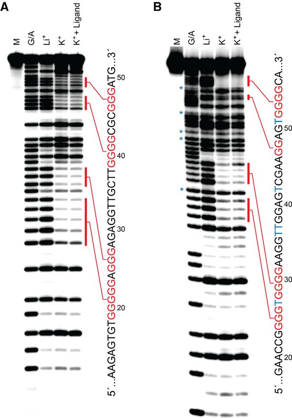
Nucleic Acids Research, 2020, Vol. 48, No. 4 1671
and Rev.86, 50 mM KCl, 1.5 mM MgCl2 , 0.1% TRITON- mg/ml, Roche Diagnostics, Indianapolis, IN, USA). The
X, 10 mM Tris (pH 9.0) and 0.5 mM each of the four DNA was then ethanol precipitated and used directly in a
dNTPs (see Supplementary Table S1 for sequences). The ds- 100 l PCR amplification reaction containing 1 U KOD
DNA product was ethanol precipitated, redissolved in 500 Dash DNA polymerase, 0.5 M primer Fwd.86, 0.5 M
l water and mixed with 1.5 ml of a ∼20% slurry of high- primer Rev.86, 120 mM Tris, pH 8.0, 10 mM KCl, 15 mM
capacity streptavidin coated agarose beads in Buffer WB (50 MgSO4 , 6 mM (NH4 )2 SO4 , 0.01% BSA, 0.01% TRITON-
mM NaCl, 10 mM Tris, pH 7.6). The bead-binding reac- X and 0.5 mM each of the four dNTPs. For the first cycle of
tion was allowed to occur for 30 min at room temperature amplification, the reaction mixture was heated to 95◦ C for
with shaking and complete immobilization of the DNA was 2 min, annealed at 46◦ C for 30 s and extended at 70◦ C for
confirmed by agarose gel electrophoresis. The supernatant 30 min. All subsequence cycles were carried out using the
was removed via filtration, and the beads were washed two following program: 95◦ C for 30 s, 46◦ C for 30 s and 70◦ C
times with 500 l Buffer WB and once with water. The for 1 min. The amplified DNA was ethanol precipitated and
non-biotinylated strand (Lib.dT) was then eluted from the used to generate a new DNA library for the next round of
Downloaded from https://academic.oup.com/nar/article/48/4/1669/5707588 by guest on 19 September 2020
beads using 200 l ice cold Buffer EB (50 mM NaOH, 1 in vitro selection. Supplementary Figure S2 summarizes se-
mM ethylenediaminetetraacetic acid (EDTA)) and the elu- lection conditions for each round. Following round 8, the
ent immediately neutralized with 20 l of Buffer NB1 (1 enriched dsDNA pool was cloned into Escherichia coli us-
M Tris, pH 7.6 and 3 M NaOAc) and ethanol precipitated. ing the TOPO TA cloning kit (Life Technologies, Carls-
The beads were washed three times with 1 ml Buffer WB bad, CA, USA). Bacteria were grown for 16 h at 37◦ C on
and resuspended in a reaction mixture (3.6 ml) consist- LB agar plates containing 50 g/mL kanamycin. Individ-
ing of 4 M primer Fwd.86, 500 M each dNTP (dATP, ual colonies were amplified by PCR and sequenced by Eton
dCTP, dGTP and 5-aminoallyl-dUTP), 36 U KOD Dash Biosciences Inc. (San Diego, CA, USA).
DNA Polymerase (40:1 mixture of KOD mutant exo- and
KOD DNA polymerase) (27), 10 mM KCl, 15 mM MgSO4 ,
6 mM (NH4 )2 SO4 , 0.01% bovine serum albumin (BSA),
Transcription of RNA
0.01% TRITON-X and 120 mM Tris, pH 8.0. The reac-
tion was then heated to 95◦ C for 1 min, annealed at 46◦ C DNA templates used to prepare the various versions of
for 30 s and incubated at 70◦ C for 30 min. The super- D-pre-miR-155 were generated by cross-extension of two
natant was removed by filtration and the beads washed two overlapping synthetic oligonucleotides (Supplementary Ta-
times with 500 l Buffer WB. The modified ssDNA strand ble S6). The oligonucleotides (2 nmol each) where annealed
(Lib.AdU) was eluted from the beads using 200 l ice cold in a 500 l reaction mixture containing 6 mM MgCl2 , 150
Buffer EB and the eluent immediately neutralized with 20 mM KCl, 20 mM DTT and 100 mM Tris (pH 8.3), which
l of Buffer NB1 and ethanol precipitated. The beads were was heated at 90◦ C for 1 min and then cooled slowly to room
quickly washed three times 1 ml Buffer WB and the above temperature. Reverse Transcriptase (16 U/l) was added
extension reaction repeated. Overall, three extension reac- and the reaction incubated at 42◦ C for 45 min. Following
tions were carried out on the same portion of beads. All ethanol precipitation, the resulting dsDNA was added to a
eluted DNA libraries were purified by denaturing PAGE transcription reaction mixture containing 10 U/l T7 RNA
(10%, 19:1 acrylamide:bis-acrylamide) prior to use. polymerase, 0.001 U/l inorganic pyrophosphatase (IPP),
25 mM MgCl2 , 2 mM spermidine, 10 mM DTT, 40 mM
Tris pH 7.9 and 5 mM of each of the four NTPs. After in-
In vitro selection
cubating for 2 h at 37◦ C, reaction mixture was ethanol pre-
Separate in vitro selection experiments were carried out for cipitated and the RNA purified by denaturing PAGE (10%,
Lib.dT and Lib.5AdU. A 500 l reaction mixture contain- 19:1 acrylamide:bis-acrylamide).
ing 1 nmol of ssDNA library (∼1014 unique sequences) in
Buffer SB (5 mM MgCl2 , 50 mM KCl, 20 mM NaCl, 25 mM
Tris, pH 7.6) was heated at 90◦ C for 3 min and allowed to
Electrophoretic mobility shift assay
cool slowly to room temperature. The reaction mixture was
incubated with 100 l of streptavidin-coated Dynabeads Dissociation constants for aptamer–pre-miR complexes
(previously blocked with 1 mg/ml yeast tRNA in Buffer SB) were determined as described previously (18). Briefly, ei-
and incubated at room temperature for 1 h. The supernatant ther 5 nM of 5 -fluoroscein (FAM)-labeled L-pre-miR-155t
was removed and the beads discarded to remove any bead- or 1 nM 5 -[32 P]-labeled D-pre-miR-155(t) was incubated
binding oligonucleotides (i.e negative selection step). To the with various concentrations (0–3000 nM) of the indicated
retained supernatant was added 100 pmol of L-pre-miR- DNA aptamer in a 20 l reaction mixture containing ei-
155t and the solution was incubated at room temperature ther 2 or 5 mM MgCl2 , 50 mM KCl, 20 mM NaCl, 0.01
for 30 min. The reaction mixture was then mixed with 100 mg/ml tRNA and 25 mM Tris (pH 7.6). After 30 min, the
l streptavidin-coated Dynabeads and incubated at room samples were analysed by non-denaturing PAGE (8%, 19:1
temperature for an additional 15 min. The beads were then acrylamide:bis-acrylamide), which contained either 2 or 5
washed five times with 1 mL Buffer SB in order to remove mM MgCl2 , 50 mM KOAc, 20 mM NaOAc and 25 mM
weakly bound ssDNA molecules and the retained ssDNA Tris (pH 7.6). Electrophoresis was carried out at 30 mA for
was eluted using 300 l Buffer EB. The elute ssDNA was ∼3 h. The gel was imaged by either autoradiography or by
quickly neutralized with a solution containing 30 l 3 M fluorescence emission using a Typhoon FLA-9500 Molecu-
NaOAc, 30 l of 1 M Tris (pH 7.6) and 4 l glycogen (1 lar Imager (General Electric Co., Boston, MA, USA).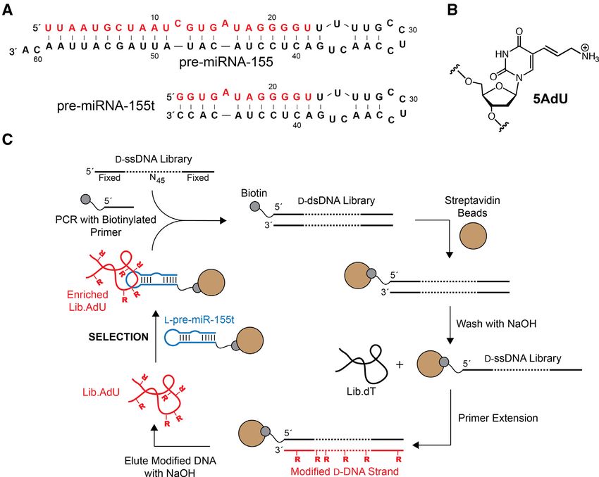
1672 Nucleic Acids Research, 2020, Vol. 48, No. 4
In-line probing analysis of D-pre-miR-155t 19:1 acrylamide:bis-acrylamide). The gel was imaged by au-
32 toradiography using a Typhoon FLA-9500 Molecular Im-
A 20 l reaction mixture containing 100 nM of 5 -[ P]-
ager.
labeled D-premiR-155t, either 0 or 20 M L-aptamer, 5
mM MgCl2 , 50 mM KCl, 20 mM NaCl and 25 mM
RESULTS AND DISCUSSION
Tris (pH 7.6) was incubated at room temperature for 48
h. The cleavage products were resolved by denaturing Discovery of cross-chiral DNA aptamers for pre-miR-155
PAGE (15%, 19:1 acrylamide:bis-acrylamide) and imaged
For this study, we chose precursor microRNA-155 (pre-
by autoradiography using a Typhoon FLA-9500 Molecular
miR-155) as the model target (Figure 1A). The mature form
Imager.
of pre-miR-155, microRNA-155 (miR-155), is a prototyp-
ical oncogenic miR whose overexpression has been linked
Circular dichroism (CD) spectroscopy with the development and invasiveness of several human
cancers, making it an attractive therapeutic target (29). Fur-
Downloaded from https://academic.oup.com/nar/article/48/4/1669/5707588 by guest on 19 September 2020
The indicated DNA aptamer (9 M) in Buffer CD (20 mM thermore, two L-RNA aptamers were previously selected
NaCl, 25 mM Tris, pH 7.6) supplemented with 50 mM of against pre-miR-155 (18), suggesting that this hairpin struc-
either KCl or LiCl was folded by heating at 90◦ C for 3 ture favors cross-chiral recognition. These L-RNA aptamers
min before being cooled slowly to room temperature. Data also serve as a benchmark for evaluating the cross-chiral
were obtained from a 450 l sample in a quartz cuvette us- binding properties of L-DNA. L-Aptamers are initially se-
ing an Applied Photophysics Chirascan spectrophotome- lected as D-aptamers against the enantiomer of the tar-
ter (Leatherhead, England) at 1 nm intervals with time per get ligand, which enables enzymatic amplification of the
point 1.5 s from 220 to 310 nm. All data were collected at a D-oligonucleotides during the process of in vitro selection.
constant temperature of 24◦ C. Therefore, we chemically synthesized a 38 nt truncated ver-
sion of pre-miR-155 (L-pre-miR-155t) using commercially
DMS footprinting available L-nucleoside phosphoramidites (Figure 1A). L-
pre-miR-155t was biotinylated at its 3 end allowing it to
Dimethyl sulphate (DMS) footprinting was carried out ac- be immobilized on streptavidin-coated beads during the se-
cording to the protocol described by Laurence et al. (28). lection step.
Briefly, 1 M of the indicated [32 P]-labeled D-DNA ap- We prepared two single-stranded D-DNA libraries: one
tamer was refolded by heating at 90◦ C for 5 min and then comprised of canonical deoxynucleotides (Lib.dT) and one
cooling slowly to room temperature in reaction mixture (20 containing 5AdU in place of dT (Lib.AdU) (Figure 1B). We
l) containing 5 mM MgCl2 , 20 mM NaCl, either 50 mM hypothesize that merging the shape-based recognition of L-
KCl or 50 mM LiCl, and 25 mM Tris (pH 7.6). For each aptamers with functional groups absent in natural nucleic
aptamer, a parallel reaction mixture (20 l) was prepared acids will generate RNA-binding properties that are un-
that also contained 1 M L-pre-miR-155t. Each reaction achievable using current modalities. Indeed, the use of mod-
mixture was treated with 2 l DMS (10% dimethyl sul- ified nucleotides is a well-proven strategy for increasing the
phate by volume in 1:1 ethanol:water), mixed well and al- success rate of in vitro selection experiments against more
lowed to react for 2 min at room temperature. The reaction demanding targets, and has been shown to facilitate evo-
was quenched with 80 l of a solution containing 0.5 M lution of D-aptamers with significantly improved activities
NaOAc (pH 5.6) and 1 mg/ml tRNA, and ethanol precip- over their unmodified counterparts (30–32). Importantly,
itated. The DNA was pelleted by centrifugation and sub- incorporation of cationic modifications into selection li-
sequently dissolved in 100 l of a 1 M piperidine solution braries has previously led to identification of aptamers that
(in water). After heating at 90◦ C for 30 min, the solution bind tightly to negatively charged ligands, including RNA
was cooled to room temperature and concentrated by vac- (17,33–35). The 5AdU-containing DNA library was gener-
uum. The resulting pellets were dissolved in Buffer LB (4 ated using a modified version of the procedure originally
M urea, 10 mM EDTA) and resolved by denaturing PAGE reported by Eaton et al. (Figure 1C) (36). Briefly, a ssDNA
(15%, 19:1 acrylamide:bis-acrylamide). The gel was imaged library (∼1014 unique sequences) containing a 45 nt random
by autoradiography using a Typhoon FLA-9500 Molecular domain flanked by fixed primer binding sites (Supplemen-
Imager. tary Table S1) was amplified by PCR in order to generate
multiple copies of each DNA molecule. One of the primers
used during PCR was biotinylated at its 5 end, allowing
Dicer inhibition assay for the immobilization of the double-stranded product on
The indicated concentration of L-DNA aptamer was added streptavidin coated agarose beads following amplification.
to a reaction mixture (20 l) containing 1 nM 5 -[32 P]- The non-biotinylated strand was subsequently removed via
labeled D-pre-miR-155, 5 mM MgCl2 , 20 mM NaCl, 50 NaOH treatment (0.1 M) and became the starting pool for
mM KCl, 0.01 mg/ml tRNA, 10 mM DTT and 25 mM Tris the unmodified in vitro selection experiment (i.e. Lib.dT).
(pH 7.6), and the reaction was initiated by the addition of The bead-bound DNA strand was then used as a template
10 nM human Dicer protein. After 30 min, an aliquot was for a primer extension reaction employing 5-aminoallyl-
removed and quenched with a two volumes of formamide dUTP in place of dTTP. The extension was carried out using
loading buffer (95% formamide, 10 mM EDTA) and the KOD Dash DNA polymerase, which was previously shown
cleavage products were resolved by denaturing PAGE (10%, to be highly tolerant of dUTP derivatives modified at the
C5 position (36). Following the extension step, the newly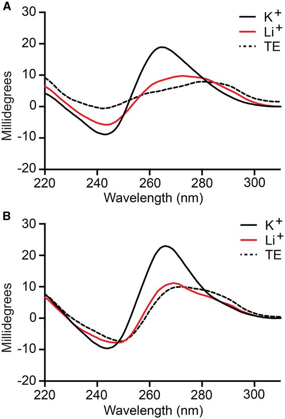
Nucleic Acids Research, 2020, Vol. 48, No. 4 1673

                                                                                                                                                       Downloaded from https://academic.oup.com/nar/article/48/4/1669/5707588 by guest on 19 September 2020
Figure 1. Mirror image in vitro selection of cross-chiral DNA aptamers for pre-miR-155. (A) Sequences and secondary structures of pre-miR-155 and
truncated variant pre-miR-155t. For consistency, numbering of nucleotides is maintained upon truncation. (B) Chemical structure of 5-aminoallyl-2 -
deoxyuridine (5AdU). (C) Schematic representation of the in vitro selection method used to isolate modified DNA aptamers. For unmodified aptamers,
Lib.dT is directly applied to the selection step.

generated 5AdU-modified DNA strand was separated from                       DNA library from 2000 to 100 nm, and by increasing the
the bead-bound template via NaOH treatment and further                      duration of the bead-washing steps (see Supplementary Fig-
purified by gel electrophoresis. Multiple extension reactions               ure S2 for details). The temperature at which the selection
were typically carried out on the same portion of template-                 was carried out was also raised from 23◦ C to 37◦ C during
bound beads in order to generate a sufficient quantity of                   the final two rounds. Following the final round of in vitro
Lib.AdU for the selection experiment (Supplementary Fig-                    selection, the enriched Lib.T and Lib.AdU libraries were
ure S1).                                                                    cloned and sequenced (Supplementary Tables S2 and 3, re-
  Separate in-vitro selection experiments were carried out                  spectively).
for each of the two libraries (Lib.T and Lib.AdU) (Fig-                        Five clones from both the unmodified (Lib.T) and mod-
ure 1C). Each D-DNA library was incubated together with                     ified (Lib.AdU) DNA libraries, each representing a unique
the biotinylated L-pre-miR-155t target in a reaction mix-                   aptamer sequence, were screened for binding L-pre-miR-
ture containing 5 mM MgCl2 , 20 mM NaCl, 50 mM KCl,                         155 using an electrophoretic mobility shift assay (EMSA)
0.1%TWEEN-20 and 25 mM Tris (pH 7.6) at room temper-                        (Supplementary Figure S3). Interestingly, one sequence (D-
ature. D-DNA molecules that bound L-pre-miR-155t were                       AdU2 and D-T1) appeared within both enriched popula-
captured using streptavidin-coated magnetic beads, which                    tions and was capable of binding L-pre-miR-155t regard-
were subsequently washed with the same binding solution.                    less of its modification state. Nevertheless, clones D-T9 and
Aptamers that remained bound to the beads were eluted                       D-AdU5 from Lib.T and Lib.AdU, respectively, emerged
using NaOH (0.1 M) and amplified by PCR using KOD                           as the tightest binders, and thus, all further studies were
Dash DNA polymerase. As before, a biotinylated primer                       based on these two sequences (Figure 2A). D-T9 and D-
was used during the amplification step and the resulting                    AdU5 bound L-pre-miR-155t with Kd values of 60 ± 4
double-stranded PCR product was used to generate the cor-                   and 36 ± 2 nM, respectively. To the best of our knowl-
responding pool of single-stranded DNA for the next round                   edge, these aptamers represent the first example of a stable
of in vitro selection.                                                      interaction between DNA and RNA of the opposite chi-
  A total eight rounds of in vitro selections were carried out              rality. Importantly, the Kd values for D-T9 and D-AdU5
for both unmodified (Lib.dT) and modified (Lib.AdU) D-                      were similar to the previously reported RNA aptamer for
DNA libraries. The selection pressure was increased during                  the same target (Kd = 19 nM) (18), suggesting that the ab-
successive rounds by reducing the concentration of the D-                   sence of the 2 -hydroxyl group is not a disadvantage for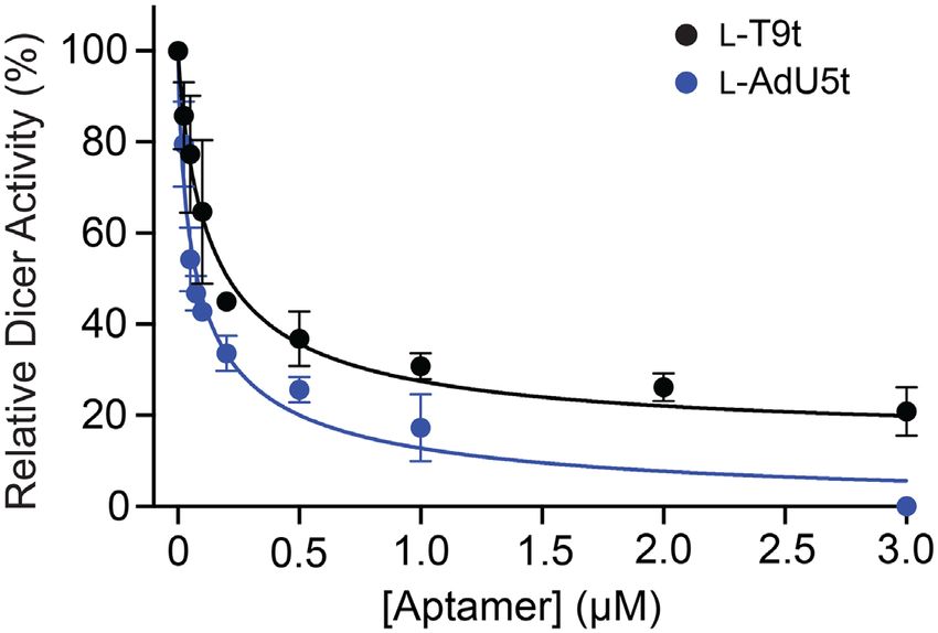
1674 Nucleic Acids Research, 2020, Vol. 48, No. 4
Downloaded from https://academic.oup.com/nar/article/48/4/1669/5707588 by guest on 19 September 2020
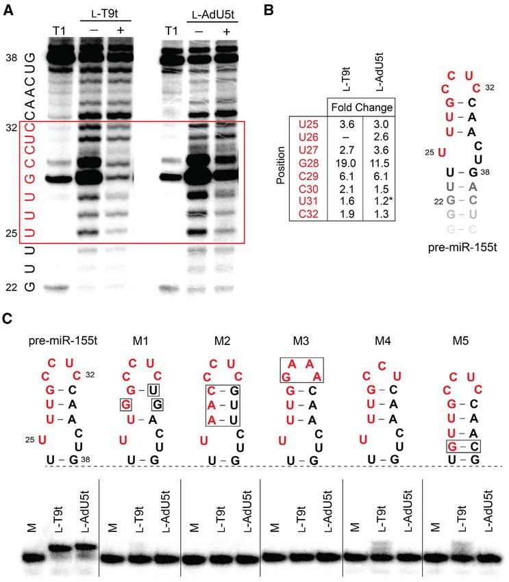
Figure 2. Sequence and binding affinity of cross-chiral DNA aptamers. (A) Sequences of full-length aptamers T9 and AdU5 and their truncated variants.
Blue Ts indicate the position of 5AdU and primer-binding sites are underlined. Guanosine residues shown to be involved in G-quadruplex formation
are colored red (see Figure 4). (B) Saturation plots for binding of T9t to either pre-miR-155t or pre-miR-155 (asterisk) having the opposite chirality. (C)
Saturation plots for binding of AdU5t to either pre-miR-155t or pre-miR-155 (asterisk) having the opposite chirality. Kd values reported as mean ± S.D.
(n = 3). (D) Chemical structure of 5-aminoallyl-dU phosphoramidite (L-5AdU-CEP).
cross-chiral nucleic acid recognition. A version of D-AdU5 2A), indicating that the majority of cationic modifications
containing dT rather than 5AdU was unable to bind L- within D-AdU5 were dispensable for binding L-pre-miR-
pre-miR-155t, demonstrating that binding is dependent on 155.
the cationic modification (Supplementary Figure S4a). Sim- Next, we prepared L-DNA versions of both truncated ap-
ilarly, replacing dT with 5AdU within the unmodified D- tamers via solid-phase DNA synthesis using L-nucleoside
T9 aptamer significantly impaired its ability to bind L-pre- phosphoramidites (Supplementary Figures S5 and 6). The
miR-155t. This observation shows that simply appending a 5AdU modification was incorporated into L-AdU5t using
cationic moiety to an aptamer does not itself improve bind- L-5AdU-CEP (Figure 2D), which was synthesized accord-
ing, but rather precise positioning of the modification is re- ing to published procedures (see Supplementary Data) (37–
quired and must be achieved through in vitro selection. 39). In general, a standard 1 M scale synthesis yielded
∼100 nmol of purified L-DNA aptamer. This is compared to
∼10–30 nmols for the previously reported L-RNA aptamer
Preparation of truncated L-DNA aptamers with affinity for against pre-miR-155 (data not shown). As is dictated by
D-pre-miR-155
the principal of reciprocal chiral substrate specificity (40),
We next sought to determine the minimal binding do- the affinity of both L-DNA aptamers for D-pre-miR-155t
main for aptamers D-T9 and D-AdU5, both of which were was very similar to their corresponding D-DNA aptamers
>80 nt in length. Our primary goal was to identify trun- for L-pre-miR-155t (Figure 2B and C). L-T9t and L-AdU5t
cated sequences that could be prepared efficiently by chem- bound D-pre-miR-155t with Kd values of 53 ± 2 and 37 ±
ical oligonucleotide synthesis using L-nucleoside phospho- 5 nM, respectively, when measured under in vitro selections
ramidites. The minimal binding domains of D-T9 and D- conditions. The somewhat lower affinity of L-AdU5t rela-
AdU5 were determined by generating variants with a suc- tive to D-AdU5t (∼3-fold) likely reflects the poorer qual-
cession of 5 and 3 truncations and screening for binding by ity of synthetic L-DNA compared to D-DNA generated by
EMSA (Supplementary Tables S4 and 5). This iterative pro- enzymatic polymerization, as evident by mass spectrom-
cess resulted in the reduction of D-T9 into a 56 nt sequence etry (Supplementary Figure S6). For example, acryloni-
(D-T9t) and D-AdU5 into a 60 nt sequence (D-AdU5t) hav- trile adducts were observed on L-AdU5t but not D-AdU5t,
ing Kd values of 51 ± 2 and 12 ± 1 nM, respectively, when which are a result of the deprotection step during solid-
measured under the in vitro selection conditions (i.e. 5 mM phase oligonucleotide synthesis. The affinity of L-T9t and
Mg2+ ) (Figure 2B and C). It was fortuitous, but not uncom- L-AdU5t for D-pre-miR-155t decreased ∼3- to 4-fold when
mon, that both truncated aptamers showed higher binding the concentration of Mg2+ in the binding buffer was reduced
affinity to pre-miR-155t than either full-length parent ap- from 5 to 2 mM, indicating that both aptamers were de-
tamer. Scrambled versions of both truncated D-aptamers pendent on Mg2+ (Supplementary Figure S7). Importantly,
failed to bind L-pre-miR-155 (Supplementary Figure S4b). both L-DNA aptamers bound the full-length D-pre-miR-
It is worth noting that truncation of aptamer D-AdU5 re- 155 RNA hairpin (which is the intended target) with sim-
duced the number of 5AdU residues from 14 to 6 (Figure ilar Kd values as the truncated target used during in vitroNucleic Acids Research, 2020, Vol. 48, No. 4 1675
Downloaded from https://academic.oup.com/nar/article/48/4/1669/5707588 by guest on 19 September 2020
Figure 4. Characterization of G-quadruplex formation in D-T9t (A) and
Figure 3. CD spectrum of D-T9t (A) and D-AdU5t (B) aptamers. All CD D-AdU5t (B) aptamers. Each aptamer was annealed in the presence of
spectrum were obtained with 9 M aptamer in the presence of a buffer LiCl, KCl or KCl plus L-pre-miR-155t and subjected to DMS footprinting.
containing 20 mM NaCl, 25 mM Tris (pH 7.6) and either 50 mM KCl The nucleotide sequence of each aptamer is shown to the right of the gel.
(K+ , shown in black solid line) or LiCl (Li+ , shown in red solid line) at Protected guanosine residues are colored red and 5AdU is represented by
23◦ C. CD spectra recorded under no salt condition were prepared in TE blue Ts. Treatment of D-AdU5t with formic acid (G/A) and DMS resulted
buffer (shown in dashed black line). in cleavage at 5AdU residues (asterisks). M = untreated control.
selection (Figure 2B and C). Not surprisingly, neither T9t used during the in vitro selection experiment (20 mM Na+
nor AdU5t aptamers were able to bind pre-miR-155 of the and 50 mM K+ ) showed a negative peak at ∼240 and a pos-
same chirality (Supplementary Figure S8). itive peak at ∼260 nm (Figure 3), which are characteristic of
a parallel-stranded G-quadruplex structure (44,45). Replac-
ing K+ with Li+ , which does not facilitate G-quadruplex
Aptamers T9t and AdU5t form G-quadruplex structures
formation (43), resulted in the loss of these features (Fig-
The DNA structure prediction software Mfold (41) failed to ure 3), instead giving CD spectrum that more closely re-
identify stable secondary structures within T9t and AdU5t sembled that of D-T9t and D-AdU5t obtained in the ab-
aptamer sequences (Supplementary Figure S9). However, sence of any ions (i.e. TE buffer). As expected, similar re-
both minimal aptamers contained at least four clusters sults were obtained using the L-DNA versions of both ap-
of consecutive guanine residues that are indicative of G- tamers, except that their CD spectrum were inverted (Sup-
quadruplexes (Figure 2A), which is not uncommon for plementary Figure S10b and c) (46). Taken together, these
DNA aptamers (42). Therefore, we wished to determine data strongly suggest that T9t and AdU5t aptamers contain
whether T9t and AdU5t form G-quadruplex structures a potassium-dependent, parallel-stranded G-quadruplex
and if such structures are important for binding pre-miR- structure, the formation of which is required for binding to
155. In the absence of K+ , which is known to stabilize G- pre-miR-155.
quadruplexes more effectively than other monovalent ions In order to confirm the above results and gain additional
(43), both D-DNA aptamers failed to bind L-pre-mir-155t, information regarding the guanine residues involved in G-
even at concentrations of aptamer far exceeding the ob- quadruplex formation, we carried out a dimethyl sulphate
served Kd (Supplementary Figure S10a). The CD spectrum (DMS) footprinting assay (28). The availability of guanine
of D-T9t and D-AdU5t in the presence of ion concentrations N7 for methylation by DMS in single-stranded and duplex1676 Nucleic Acids Research, 2020, Vol. 48, No. 4
Downloaded from https://academic.oup.com/nar/article/48/4/1669/5707588 by guest on 19 September 2020
Figure 5. Cross-chiral DNA aptamers bind selectively to the loop domain of pre-miR-155. (A) In-line probing analysis of pre-miR-155t in the presence
and absence of the indicated L-aptamer. Residues that underwent significant conformational changes (i.e. protection from hydrolysis) in the presence of
excess L-aptamer are boxed in red. Partial digestion by ribonuclease T1 (cleavage after G residues) is shown to the left (T1). Uncropped gel images are
shown in Supplementary Figures S11a and b. (B) The extent of pre-miR-155 protection differs between aptamers. Fold change was determined by dividing
the fraction cleaved in the absence of the aptamer by that in its presence (i.e. greater fold change corresponds to greater protection). Asterisk indicates
increased hydrolysis in the presence of the aptamer. (C) Binding of excess L-T9t and L-AdU5t to 5 -[32 P]-pre-miR-155t or modified variants. All binding
reactions contain 3 M aptamer, 1 nM 5 -[32 P]-pre-miR-155t (or variant), 5 mM MgCl2 , 50 mM KCl, 20 mM NaCl, and 25 mM Tris (pH 7.6). Variant
M1 is mouse pre-miR-155. M = 5 -[32 P]-pre-miR-155t (or variant) control.
DNA, but not in G-quadruplexes, is a diagnostic indicator be interesting to determine whether eliminating this degen-
of G-quadruplex structures. The DMS cleavage patterns of eracy leads to improved binding properties. G-quadruplex
both D-T9t and D-AdU5t revealed four G-rich tracts that formation was not dependent on ligand binding, since in-
were strongly protected from cleavage in the presence of K+ , clusion of L-pre-miR-155t during the DMS assay did not
implying that these residues are indeed involved in forma- significantly alter the cleavage pattern for either aptamer.
tion of a G-quadruplex (Figure 4). Interestingly, for both This also suggests that the protected guanine bases are un-
aptamers, the number of protected guanines varied between likely to be involved in hydrogen bonding to pre-miR-155.
G-tracks. For example, D-T9t had two tracks of three gua- Interestingly, a previously identified cross-chiral RNA ap-
nines (G25–G27 and G46–48), one track of four guanines tamer for pre-miR-155 (aptamiR-155.2) does not contain a
(G39–G42), and one track of five guanines (G19–G23) that G-quadruplex, as evident by the lack of monovalent ion de-
were protected from DMS cleavage (Figure 4A). This ob- pendency in its CD spectrum (Supplementary Figure S10d).
servation is consistent with the presence of redundant gua- Thus, despite binding to the same RNA target (i.e. the same
nines within the longer G-tracks, and indicates that the ob- sequence of nucleotides), DNA aptamers T9t and AdU5t
served DMS cleavage patterns represent a mixture of sev- form entirely different structural motifs compared to their
eral G-quadruplex structures each consisting of a different RNA counterpart. In addition to T9t and AdU5t, several
configuration of guanine residues (47). In the future, it will other DNA aptamer sequences identified following in vitroNucleic Acids Research, 2020, Vol. 48, No. 4 1677
ysis were distinct (e.g. residue G28; Figure 5B). Because the
rate of in-line hydrolysis is highly dependent on linkage ge-
ometry, which is dictated by the overall tertiary structure
of the RNA, these distinct cleavage patterns likely represent
two unique heterochiral interfaces that perturb the structure
of pre-miR-155 in different ways. This observation is con-
sistent with the sequence and structural differences between
T9t and AdU5t. In order to validate the in-line hydroly-
sis data, we also showed that both aptamers failed to bind
several versions of D-pre-miR-155t containing mutations
and/or deletions that perturbed the structure of the loop
domain (Figure 5C). Both aptamers also failed to bind sev-
eral other pre-miR hairpin structures, including the mouse
Downloaded from https://academic.oup.com/nar/article/48/4/1669/5707588 by guest on 19 September 2020
homologue of pre-miR-155 (M1) (Supplementary Figure
S11c). Together, the above results indicate that L-T9t and
Figure 6. Cross-chiral L-DNA aptamers inhibit Dicer-mediated cleavage L-AdU5t are selective for binding the loop domain of pre-
of pre-miR-155. Percent Dicer activity (relative to a no L-aptamer control) miR-155.
was plotted as a function of L-aptamer concentration. Each data point
represents the mean ± S.D. (n = 3). The data were fit to a four-parameter
sigmoidal dose-response model and IC50 values were determined. L-DNA cross-chiral aptamers inhibit Dicer-mediated cleav-
age of pre-miR-155
selection have at least four clusters of consecutive guanine During microRNA biogenesis, the distal stem-loop domain
residues that are indicative of G-quadruplexes (Supplemen- of pre-miRs is excised by the ribonuclease Dicer prior to
tary Tables S2 and 3), suggesting that this could be a com- being loaded onto an Argonuate protein, where it is eventu-
mon structural motif for cross-chiral recognition between ally used to guide sequence-specific silencing of target mR-
DNA and RNA. It may also indicate that G-quadruplexes NAs (49). Previous studies have shown that Dicer-catalyzed
provide a stable structural scaffold when divalent cations cleavage of specific pre-miRs can be blocked using reagents
(e.g. magnesium) are limited. It will be interesting to select that bind the corresponding distal stem-loop domain (50–
L-DNA aptamers against a diverse range of RNA structures 53), and this strategy is currently being pursued for thera-
in order to determine whether G-quadruplex formation is a peutic purposes (54). Binding to the loop domain of pre-
general phenomenon. miRs by our previously reported cross-chiral L-RNA ap-
tamers also inhibited Dicer. Therefore, we predicted that
L-T9t and L-AdU5t would be effective at inhibiting Dicer-
L-DNA aptamers bind selectively to the distal loop of pre-
mediated cleavage of pre-miR-155. To test this, D-pre-miR-
miR-155
155 was incubated in the presence of human Dicer pro-
The L-pre-miR-155t target used during in vitro selection was tein, together with various concentrations of either L-T9t
truncated relative to the native pre-miR-155 RNA. This was or L-AdU5t. We observed a concentration-dependent inhi-
done intentionally in order to encourage isolation of ap- bition of pre-miR-155 processing for both aptamers (Figure
tamers capable of binding to the loop domain, which has 6). The IC50 values calculated for L-T9t and L-AdU5t were
been shown to inhibit Dicer-mediated miR biogenesis in 139 and 80 nM, respectively, which are in good agreement
vitro (17,18). In order to determine whether L-T9t and L- with their corresponding Kd values (Figure 2B and C). As
AdU5t bound the loop of pre-miR-155, we performed an a control for non-specific inhibition of Dicer, we found that
in-line hydrolysis analysis of pre-miR-155t in the presence neither D-T9t nor D-AdU5t inhibited Dicer-mediated cleav-
and absence of saturating concentrations of L-DNA ap- age to a significant degree (Supplementary Figure S12),
tamer (Figure 5A) (48). Uncatalyzed in-line hydrolysis (i.e. which is consistent with their inability to bind D-pre-miR-
cleavage) of phosphodiester linkages in RNA requires an 155.
in-line geometry (180◦ ) between the 2 -oxygen nucleophile,
the phosphorus electrophile and 5 -oxygen leaving group.
CONCLUSION
Binding of the L-aptamer to D-pre-miR-155t is expected to
restrict the conformational freedom of residues at the bind- In summary, we have demonstrated in vitro selection of the
ing site, thereby hindering their ability to achieve the in-line first cross-chiral aptamers comprised of L-DNA, thereby
geometry needed for cleavage. Incubating D-pre-miR-155t establishing the capacity of DNA and RNA molecules of
with either L-T9t or L-AdU5t led to the protection of the the opposite handedness to form tight and specific inter-
same 8-nucleotide region from cleavage (U25 –C32 ), strongly actions with each other. Thus, this work greatly expands
suggesting that both L-aptamers bound to this site within our current definition of nucleic acid molecule recognition.
the loop domain (Figure 5A). Interestingly, previously re- Our best L-DNA aptamers had low nM affinities for pre-
ported L-RNA aptamers for pre-miR-155 also bound these miR-155, similar to previously identified L-RNA aptamers
same residues, suggesting that this binding sight is particu- against the same target. Furthermore, like their RNA coun-
larly well suited for cross-chiral recognition (18). Despite terparts, these L-DNA aptamers bound selectively to the
their similar footprints, however, the extent to which L- loop domain of pre-miR-155 and inhibited Dicer-mediated
T9t and L-AdU5t protected D-pre-miR-155t from hydrol- processing in vitro. Thus, despite prior evidence suggesting1678 Nucleic Acids Research, 2020, Vol. 48, No. 4
that DNA may be less adept than RNA at forming het- DNA pool, the modified ssDNA library (Lib.AdU) yielded
erochiral interactions (22,25–26), our results clearly show aptamer sequences that were distinct from its native DNA
that the lack of a 2 -OH group is not a disadvantage for counterpart (Lib.dT) and were dependent on the 5AdU
cross-chiral recognition of RNA. The question of whether modification for binding. This suggests that the modified
DNA or RNA will yield better cross-chiral aptamers in DNA pool was able to sample unique structural spaces
terms of RNA-binding requires further investigation, and or distinct interactions with pre-miR-155. Other functional
may ultimately depend on the identity of the target. Nev- group modifications will likely have different properties that
ertheless, DNA is more stable, easier to produce and less make them more or less amenable toward binding RNA. In
expensive than RNA, making it a more practical choice for contrast to protein targets, for which the influence of unnat-
the generation of cross-chiral aptamers having applications ural nucleobase modifications on aptamer binding has been
in biotechnology. studied extensively (30,31), almost nothing is known about
Despite binding to the same site (i.e. nucleotide sequence) what type of modifications are most apt for RNA binding
on pre-miR-155, the sequences and structures of L-DNA by aptamers. This study now provides a robust platform for
Downloaded from https://academic.oup.com/nar/article/48/4/1669/5707588 by guest on 19 September 2020
and L-RNA aptamers were found to be distinct from each screening a diverse set of modified nucleotides in order to
other. This was somewhat unexpected, as both the aptamer identify functional groups capable of enhancing the binding
and target are comprised of nucleic acids, providing the op- properties of cross-chiral aptamers. It is worth noting that,
portunity for DNA and RNA aptamers to form similar in general, modified deoxyribose triphosphates and their
sequence-specific interactions with the target. Indeed, D- corresponding phosphoramidites are easier to obtain syn-
DNA and D-RNA aptamers have been found to bind the thetically than their ribose counterparts due to the absence
same D-RNA target through similar sequence-specific in- of the 2 -hydroxyl group, representing another advantage of
teractions (WC base pairing in this case) (12,55), giving rise generating cross-chiral aptamers from libraries of DNA.
to DNA and RNA aptamers with closely related sequences Inhibition of miR biogenesis using reagents that target
and structures. Even in the absence of WC base pairing be- the unique structures of pre-miRs represents a promising
tween D- and L-oligonucleotides, it is possible that similar therapeutic strategy for a variety of diseases, including can-
sequence-specific interactions could emerge from libraries cer (54). Such inhibitors also represent new biochemical
of both DNA and RNA, although these interactions would tools for interrogating pre-miR function, and can poten-
likely be more idiosyncratic in nature than WC base pair- tially be utilized as affinity reagents for imaging and di-
ing. Therefore, the lack of sequence and structural similari- agnostic applications. Although several challenges remain,
ties between cross-chiral DNA and RNA aptamers, as well our results demonstrate that cross-chiral L-DNA aptamers
as between T9t and AdU5t, is significant because it rules have properties well-suited for these purposes, including
out this possibility and provides further support for the hy- high affinity and selectivity, nuclease resistance and potent
pothesis that cross-chiral nucleic acid recognition occurs in- inhibition of Dicer-mediated cleavage. While pre-miRNA-
dependent of sequence-specific interaction, instead relying 155 served as a convenient proof-of-concept target for this
on structure-specific tertiary interactions. study, it represents one of the most common RNA struc-
During the course of this study, we explored the potential tures: a hairpin. Therefore, we anticipate that the methods
for cationic modifications (i.e. 5AdU) to facilitate and/or developed herein can be readily applied to the isolation of
promote cross-chiral interactions between DNA and RNA cross-chiral L-DNA aptamers for many clinically relevant
of the opposite chirality. A previous in vitro selection exper- RNAs, including long noncoding RNAs and viral RNA el-
iment using an RNA library containing the same cationic ements.
modification (5-aminoallyluridine) yielded cross-chiral L-
RNA aptamers for pre-miR-19a having low nanomolar Kd
value (17). However, a parallel selection experiment using
SUPPLEMENTARY DATA
an unmodified RNA library was not carried out, making it
difficult to determine what advantages, if any, the cationic Supplementary Data are available at NAR Online.
modification bestowed. Herein, we found that both modi-
fied and unmodified ssDNA libraries readily yielded cross-
chiral aptamers against L-pre-miR-155. The Kd value for
ACKNOWLEDGEMENTS
the best modified aptamer (AdU5t) was only slightly better
(∼2- to 3-fold) than the best unmodified aptamer (T9t) un- The authors would like to thank Prof. Tadhg Begley and his
der all conditions tested (Figure 2 and Supplementary Fig- laboratory for assistance obtaining CD data.
ure S6). Based on these observations, we conclude that the
cationic aminoallyl modification did not provide a signifi-
cant advantage for cross-chiral recognition of pre-miR-155
FUNDING
by DNA. This result was unexpected based on the nature
of the target, and is significant because it suggests that the National Institute of General Medical Sciences at the Na-
use of cationic modifications may not benefit future cross- tional Institutes of Health [R35GM124974]; J.T.S is a
chiral aptamer selections. However, we acknowledge these CPRIT Scholar of Cancer Research supported by the Can-
experiments represent a very small sample size and we can- cer Prevention and Research Institute of Texas [RR150038].
not rule out that different results may be obtained under al- Funding for open access charge: National Institutes of
ternative in vitro selection condition and/or for other RNA Health [R35GM124974].
targets. Importantly, despite originating from the same ds- Conflict of interest statement. None declared.Nucleic Acids Research, 2020, Vol. 48, No. 4 1679
REFERENCES control of gene expression. The case of L-DNAs and-RNAs.
Nucleosides Nucleotides, 17, 1275–1287.
1. Wan,Y., Kertesz,M., Spitale,R.C., Segal,E. and Chang,H.Y. (2011) 26. D’Alonzo,D., Guaragna,A. and Palumbo,G. (2011) Exploring the
understanding the transcriptome through RNA structure. Nat. Rev.
role of chirality in nucleic acid recognition. Chem. Biodivers., 8,
Genet., 12, 641–655. 373–413.
2. Bernat,V. and Disney,M.D. (2015) RNA structures as mediators of 27. Nishioka,M., Mizuguchi,H., Fujiwara,S., Komatsubara,S.,
neurological diseases and as drug targets. Neuron, 87, 28–46.
Kitabayashi,M., Uemura,H., Takagi,M. and Imanaka,T. (2001) Long
3. Croce,C.M. (2009) Causes and consequences of microRNA
and accurate PCR with a mixture of KOD DNA polymerase and its
dysregulation in cancer. Nat. Rev. Genet., 10, 704–714. exonuclease deficient mutant enzyme. J. Biotechnol., 88, 141–149.
4. Engelman,A. and Cherepanov,P. (2012) The structural biology of 28. Sun,D. and Hurley,L.H. (2010) Biochemical techniques for the
HIV-1: mechanistic and therapeutic insights. Nat. Rev. Microbiol., 10,
characterization of G-quadruplex structures: EMSA, DMS
279–290. footprinting, and DNA polymerase stop assay. Methods Mol. Biol.,
5. Freier,S.M., Kierzek,R., Jaeger,J.A., Sugimoto,N., Caruthers,M.H.,
608, 65–79.
Neilson,T. and Turner,D.H. (1986) Improved free-energy parameters
29. Higgs,G. and Slack,F. (2013) The multiple roles of microRNA-155 in
for predictings of RNA duplex stability. Proc. Natl. Acad. Sci.
oncogenesis. J. Clin. Bioinform., 3, 17.
U.S.A., 83, 9373–9377.
30. Meek,K.N., Rangel,A.E. and Heemstra,J.M. (2016) Enhancing
Downloaded from https://academic.oup.com/nar/article/48/4/1669/5707588 by guest on 19 September 2020
6. Li,P.T.X., Vieregg,J. and Tinoco,I.J. (2008) How RNA unfolds and
aptamer function and stability via in vitro selection using modified
refolds. Annu. Rev. Biochem., 77, 77–100.
nucleic acids. Methods, 106, 29–36.
7. Guan,L. and Disney,M.D. (2012) Recent advances in developing
31. Diafa,S. and Hollenstein,M. (2015) Generation of aptamers with an
small molecules targeting RNA. ACS Chem. Biol., 7, 73–86.
expanded chemical repertoire. Molecules, 20, 16643–16671.
8. Shortridge,M.D. and Varani,G. (2015) Structure based approaches
32. Rohloff,J.C., Gelinas,A.D., Jarvis,T.C., Ochsner,U.A., Schneider,D.J.,
for targeting non-coding RNAs with small molecules. Curr. Opin.
Gold,L. and Janjic,N. (2014) Nucleic acid ligands with protein-like
Struct. Biol., 30, 79–88.
side chains: modified aptamers and their use as diagnostic and
9. Thomas,J.R. and Hergenrother,P.J. (2008) Targeting RNA with small
therapeutic agents. Mol. Therapy, 3, e201.
molecules. Chem. Rev., 108, 1171–1224.
33. Battersby,T.R., Ang,D.N., Burgstaller,P., Jurczyk,S.C., Bowser,M.T.,
10. Ellington,A.D. and Szostak,J.W. (1990) In vitro selection of RNA
Buchanan,D.D., Kennedy,R.T. and Benner,S.A. (1999) Quantitative
molecules that bind specific ligands. Nature, 346, 818–822.
analysis of receptors for adenosine nucleotides obtained via in vitro
11. Tuerk,C. and Gold,L. (1990) Systematic evolution of ligands by
selection from a library incorporating a cationic nucleotide analog. J.
exponential enrichment: RNA ligands to bacteriophage T4 DNA
Am. Chem. Soc., 121, 9781–9789.
polymerase. Science, 249, 505–510.
34. Vaish,N.K., Larralde,R., Fraley,A.W., Szostak,J.W. and
12. Duconge,F. and Toulmé,J.-J. (1999) In vitro selection identifies key
McLaughlin,L.W. (2003) A novel, modification-dependent
determinants for loop-loop interactions: RNA aptamers selective for
ATP-binding aptamer selected from an RNA library incorporating a
the TAR RNA element of HIV-1. RNA, 5, 1605–1614.
cationic functionality. Biochemistry, 42, 8842–8851.
13. Sánchez-Luque,F.J., Stich,M., Manrubia,S., Briones,C. and
35. Ohsawa,K., Kasamatsu,T., Nagashima,J.I, Hanawa,K.,
Berzal-Herranz,A. (2014) Efficient HIV-1 inhibition by a 16 nt-long
Kuwahara,M., Ozaki,H. and Sawai,H. (2008) Arginine-modified
RNA aptamer designed by combining in vitro selection and insilico
DNA aptamers that show enantioselective recognition of the
optimization strategies. Sci. Rep., 4, doi:10.1038/srep06242.
dicarboxylic acid moiety of glutamic acid. Anal. Sci., 24, 167–172.
14. Mayer,G., Raddatz,M.-S.L., Grunwald,J.D. and Famulok,M. (2007)
36. Vaught,J.D., Bock,C., Carter,J., Fitzwater,T., Otis,M., Schneider,D.,
RNA ligands that distinguish metabolite-induced conformations in
Rolando,J., Waugh,S., Wilcox,S.K. and Eaton,B.E. (2010) Expanding
the TPP riboswitch. Angew. Chem. Int. Ed., 46, 557–560.
the chemistry of DNA for in vitro selection. J. Am. Chem. Soc., 132,
15. Lünse,C., Michlewski,G., Hopp,C.S., Rentmeister,A., Cáceres,J.F.,
4141–4151.
Famulok,M. and Mayer,G. (2010) An aptamer targeting the
37. Sakthivel,K. and Barbas,C.F. III (1998) Expanding the potential of
apical-loop domain modulates pri-miRNA processing. Angew. Chem. DNA for binding and catalysis: highly functionalized dUTP
Int. Ed., 49, 4674–4677. derivatives that are substrates for thermostable DNA polymerases.
16. Scarabino,D., Crisari,A., Lorenzini,S., Williams,K. and
Angew. Chem. Int. Ed., 37, 2872–2875.
Tocchini-Valentini,G.P. (1999) tRNA prefers to kiss. EMBO J., 18, 38. Lermer,L., Roupioz,Y., Ting,R. and Perrin,D.M. (2002) Toward an
4571–4578. RNaseA mimic: a DNAzyme with imidazoles and cationic amines. J.
17. Kabza,A.M. and Sczepanski,J.T. (2017) An L-RNA aptamer with Am. Chem. Soc., 124, 9960–9961.
expanded chemical functionality that inhibits microRNA biogenesis.
39. Asakura,J. and Robins,M.J. (1990) Cerium(IV)-mediated
Chem. Bio. Chem., 18, 1824–1827. halogenation at C-5 of uracil derivatives. J. Org. Chem., 55,
18. Sczepanski,J.T. and Joyce,G.F. (2015) Specific inhibition of 4928–4933.
microRNA processing using L-RNA aptamers. J. Am. Chem. Soc., 40. Milton,R.C., Milton,S.C. and Kent,S.B. (1992) Total chemical
137, 16032–16037.
synthesis of a D-enzyme: the enantiomers of HIV-1 protease show
19. Sczepanski,J.T. and Joyce,G.F. (2013) Binding of a structured D-RNA reciprocal chiral substrate specificity. Science, 256, 1445–1448.
by an L-RNA Aptamer. J. Am. Chem. Soc., 135, 13290–13293. 41. Zuker,M. (2003) Mfold web server for nucleic acid folding and
20. Garbesi,A., Capobianco,M.L., Colonna,F.P., Tondelli,L., hybridization prediction. Nucleic Acids Res., 31, 3406–3415.
Arcamone,F., Manzini,G., Hilbers,C.W., Aelen,J.M.E. and
42. Tucker,W.O., Shum,K.T. and Tanner,J.A. (2012) G-quadruplex DNA
Blommers,M.J.J. (1993) L-DNAs as potential antimessenger aptamers and their ligands: structure, function and application. Curr.
oligonucleotides: a reassessment. Nucleic Acids Res., 21, 4159–4165. Pharm. Des., 18, 2014–2026.
21. Hoehlig,K., Bethge,L. and Klussmann,S. (2015) Stereospecificity of
43. Bhattacharyya,D., Mirihana Arachchilage,G. and Basu,S. (2016)
oligonucleotide interactions revisited: no evidence for heterochiral
Metal cations in G-Quadruplex folding and stability. Front. Chem., 4,
hybridization and ribozyme/DNAzyme activity. PLoS One, 10,
38.
e0115328.
44. Vorlı́čková,M., Kejnovská,I., Sagi,J., Renčiuk,D., Bednářová,K.,
22. Ashley,G.W. (1992) Modeling, synthesis, and hybridization properties
Motlová,J. and Kypr,J. (2012) Circular dichroism and guanine
of (L)-ribonucleic acid. J. Am. Chem. Soc., 114, 9732–9736.
quadruplexes. Methods, 57, 64–75.
23. Hauser,N.C., Martinez,R., Jacob,A., Rupp,S., Hoheisel,J.D. and
45. Karsisiotis,A.I., Hessari,N.M.a., Novellino,E., Spada,G.P.,
Matysiak,S. (2006) Utilising the left-helical conformation of L-DNA
Randazzo,A. and Webba da Silva,M. (2011) Topological
for analysing different marker types on a single universal microarray
characterization of nucleic acid G-quadruplexes by UV absorption
platform. Nucleic Acids Res., 34, 5101–5111.
and circular dichroism. Angew. Chem. Int. Ed., 50, 10645–10648.
24. Dunn,M.R., Jimenez,R.M. and Chaput,J.C. (2017) Analysis of
46. Deckard,C.E. and Sczepanski,J.T. (2018) Polycomb repressive
aptamer discovery and technology. Nat. Rev. Chem., 1,
complex 2 binds RNA irrespective of stereochemistry. Chem.
doi:10.1038/s41570-017-0076.
Commun., 54, 12061–12064.
25. Garbesi,A., Capobianco,M.L., Colonna,F.P., Maflini,M., Niccoiai,D.
47. Seenisamy,J., Rezler,E.M., Powell,T.J., Tye,D., Gokhale,V.,
and Tondelli,L. (1998) Chirally-modifiedoligonucleotides and the
Joshi,C.S., Siddiqui-Jain,A. and Hurley,L.H. (2004) The dynamicYou can also read