Insights into xanthine riboswitch structure and metal ion-mediated ligand recognition
←
→
Page content transcription
If your browser does not render page correctly, please read the page content below

Published online 14 June 2021                                                             Nucleic Acids Research, 2021, Vol. 49, No. 12 7139–7153
                                                                                                                https://doi.org/10.1093/nar/gkab486

Insights into xanthine riboswitch structure and metal
ion-mediated ligand recognition
Xiaochen Xu1,† , Michaela Egger2,† , Hao Chen1,† , Karolina Bartosik2 , Ronald Micura2,* and
Aiming Ren1,*
1
 Life Sciences Institute, Zhejiang University, Hangzhou, Zhejiang 310058, China and 2 Institute of Organic Chemistry,
Center for Molecular Biosciences Innsbruck, University of Innsbruck, Innsbruck 6020, Austria

                                                                                                                                                              Downloaded from https://academic.oup.com/nar/article/49/12/7139/6298625 by guest on 30 September 2021
Received March 19, 2021; Revised April 29, 2021; Editorial Decision May 17, 2021; Accepted May 27, 2021

ABSTRACT                                                                                 one forming the aptamer for specific recognition of a small
                                                                                         ligand, and the other forming the so-called expression plat-
Riboswitches are conserved functional domains in                                         form which becomes restructured upon ligand binding to
mRNA that mostly exist in bacteria. They regulate                                        trigger ON or OFF for gene expression. In most cases,
gene expression in response to varying concen-                                           the expression platform utilizes an interplay between ter-
trations of metabolites or metal ions. Recently, the                                     minator and anti-terminator stems (transcriptional regula-
NMT1 RNA motif has been identified to selectively                                        tion) or between repressor and anti-repressor stems (trans-
bind xanthine and uric acid, respectively, both are in-                                  lational regulation) as signal to induce or shut down the
volved in the metabolic pathway of purine degrada-                                       expression of genes coding for proteins involved in the
tion. Here, we report a crystal structure of this RNA                                    biosynthesis or transport of the cognate ligand. There-
bound to xanthine. Overall, the riboswitch exhibits                                      fore, riboswitch regulation works as a feed-back mechanism
a rod-like, continuously stacked fold composed of                                        (2,7,8).
                                                                                             The first riboswitches were discovered in 2002 (9–12),
three stems and two internal junctions. The binding-
                                                                                         since then >45 distinct riboswitch-ligand systems have
pocket is determined by the highly conserved junc-                                       been identified in all three domains of life (2,13). Some
tional sequence J1 between stem P1 and P2a, and                                          of them are specific for metal ions while most of them
engages a long-distance Watson–Crick base pair to                                        specifically recognize metabolites (13). Purines and purine
junction J2. Xanthine inserts between a G–U pair                                         derivatives are one of the most abundant classes of
from the major groove side and is sandwiched be-                                         metabolites (14). Purine nucleotides such as adenosine 5 -
tween base triples. Strikingly, a Mg2+ ion is inner-                                     triphosphate (ATP) and guanosine 5 -triphosphate (GTP)
sphere coordinated to O6 of xanthine and a non-                                          are not only essential precursors of RNA and DNA, but
bridging oxygen of a backbone phosphate. Two fur-                                        also the crucial carriers for supplying cellular energy in
ther hydrated Mg2+ ions participate in extensive in-                                     biological systems. Other purine nucleotides such as c-
teractions between xanthine and the pocket. Our                                          di-GMP, c-di-AMP and c-AMP-GMP serve as important
                                                                                         signalling molecules for signal transduction (15). In ad-
structure model is verified by ligand binding analy-
                                                                                         dition, purines also constitute an integral part of sev-
sis to selected riboswitch mutants using isothermal                                      eral co-factors, such as nicotinamide adenine dinucleotide
titration calorimetry, and by fluorescence spectro-                                      (NAD), flavin adenine dinucleotide (FAD), coenzyme A
scopic analysis of RNA folding using 2-aminopurine-                                      (CoA) and S-adenosyl methionine (SAM) that enable
modified variants. Together, our study highlights the                                    specific chemical reactions catalysed by protein enzymes
principles of metal ion-mediated ligand recognition                                      (16,17). The intracellular purine levels are maintained in
by the xanthine riboswitch.                                                              purine metabolism, and deregulations of these processes
                                                                                         can cause severe cellular disorders (14). Several important
                                                                                         riboswitches were previously identified to regulate the trans-
INTRODUCTION                                                                             port and biosynthesis of purines and purine nucleotides in
Riboswitches are non-coding functional RNA domains                                       bacteria, including the guanine riboswitch (18), adenine ri-
that are frequently located in the 5 -end regions of bacte-                             boswitch (19), 5-amino-4-imidazole carboxamide riboside
rial mRNAs (1–6). They contain two overlapping zones,                                    5 -triphosphate (ZTP) riboswitch (20), 2 -deoxyguanosine-

* To
   whom correspondence should be addressed. Tel: +86 571 88981227; Fax: +86 571 88981336; Email: aimingren@zju.edu.cn
Correspondence may also be addressed to Ronald Micura. Email: ronald.micura@uibk.ac.at
†
    The authors wish it to be known that, in their opinion, the first three authors should be regarded as Joint First Authors.


C The Author(s) 2021. Published by Oxford University Press on behalf of Nucleic Acids Research.
This is an Open Access article distributed under the terms of the Creative Commons Attribution License (http://creativecommons.org/licenses/by/4.0/), which
permits unrestricted reuse, distribution, and reproduction in any medium, provided the original work is properly cited.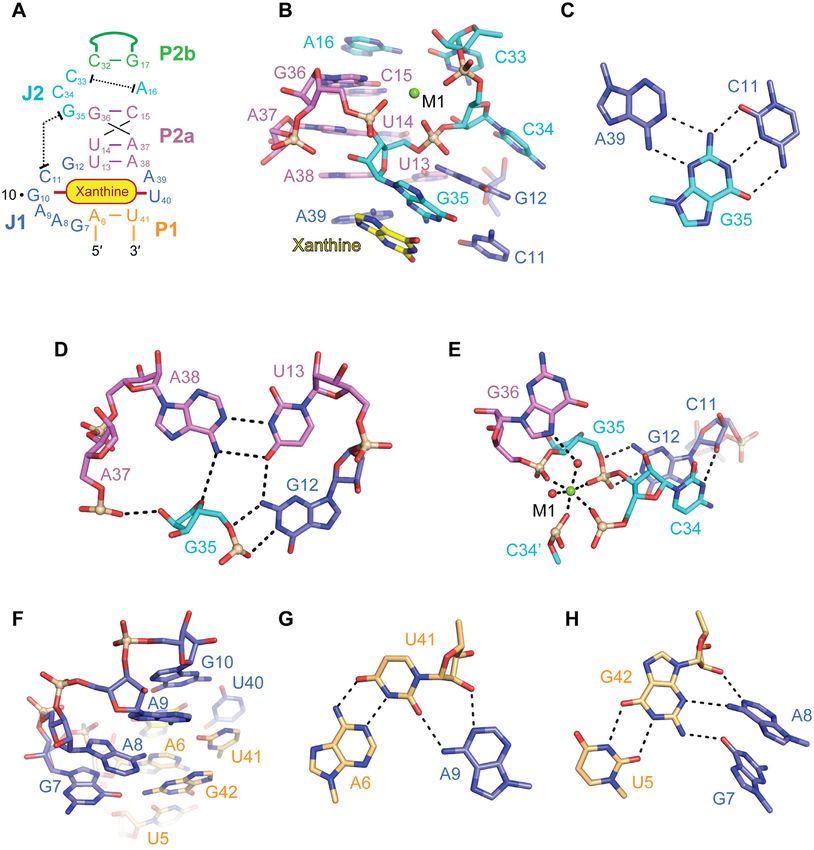
7140 Nucleic Acids Research, 2021, Vol. 49, No. 12
5 -monophosphate (dGMP) riboswitch (21,22), phospho- Ligands
ribosylpyrophosphate (PRPP) riboswitch (23) and others
Xanthine, uric acid, hypoxanthine, adenosine, adenine
(24,25).
and inosine were purchased from Yuanye Bio-Technology
Very recently, an orphan RNA motif called NMT1
Co. Ltd. (Shanghai). Xanthine sodium salt and 8-
present in proteobacteria was identified to bind xanthine
azaxanthine monohydrate were purchased from Sigma-
(25, 26) which is an oxidization product generated during
Aldrich.
purine degradation in purine metabolism. The consensus
sequence model of the NMT1 motif was derived from phy-
logenetic analysis and essentially suggested two stems ad- Crystallization
joined to a large junction (26). Almost all nucleotides that Crystals were obtained with a 46 nucleotide long RNA of
reside in this junction are highly conserved. In-line prob- the sequence 5 -GGAGUAGAAGCGUUCAGCGGCC-
ing experiments revealed that the NMT1 motif binds xan- GAAA-GGCCGCCCGGAAAUUGCUCC-3 (Ideonella
thine with low micromolar affinity in 1:1 stoichiometry,
Downloaded from https://academic.oup.com/nar/article/49/12/7139/6298625 by guest on 30 September 2021
sp. B508-1 (NZ BADL01000600.1) (Supplementary Figure
and alternatively, uric acid with 7-fold lower affinity (26). S1C) in the presence of xanthine: The RNA was diluted
In addition, it was found that the NMT1 motif binds 8- to a concentration of 0.4 mM and annealed at 65 ◦ C for
azaxanthine, which is known as inhibitor of uric acid/urate 5 min in buffer containing 50 mM HEPES, pH 7.0, 50 mM
oxidase (27). KCl, 5 mM MgCl2 before cooling on ice for half an hour.
To reveal the three-dimensional fold of the xanthine ri- The ligand xanthine was dissolved in 100 mM sodium hy-
boswitch, we set out for crystallographic experiments. Here, droxide solution and was added to the RNA solution to
we describe the 2.7 Å resolution structure of NMT1 RNA reach a final xanthine concentration of 6 mM. The RNA-
in complex with xanthine. The structure reveals a unique xanthine complex was incubated on ice for another half
RNA architecture adopting a rod-like fold with an intrigu- an hour, followed by centrifugation at 13 000 rpm for 10
ingly structured binding pocket that is critically depend- min at 4◦ C before crystallization. For crystallization, 0.18
ing on divalent metal ions. Most fascinating is the obser- l RNA-ligand complex was mixed with the reservoir so-
vation of a Mg2+ ion that bridges the xanthine’s O6 and lution at an equimolar ratio using the drop vapor diffu-
a backbone phosphate oxygen atom through inner-sphere sion method at 16◦ C. High-resolution crystals grew from
coordination. We verified the novel RNA fold by muta- the condition containing 0.1 M MES, pH 6.0, 0.2 M cal-
tional analysis and ligand binding assays based on isother- cium acetate and 10% (v/v) isopropanol after ∼1 week.
mal titration calorimetry (ITC). Furthermore, insights into The crystals were transferred in reservoir solution supple-
the Mg2+ /xanthine-induced folding path and into local mented with 30% glycerol and flash frozen in liquid ni-
structural dynamics were obtained by fluorescence spec- trogen. For Ir(NH3 )6 3+ soaking experiments, crystals were
troscopy using synthetic riboswitch variants containing transferred in reservoir solution supplemented with 50 mM
2-aminopurine fluorophores. On top, we complemented Ir(NH3 )6 3+ and allowed to equilibrate for 4 h at 4◦ C prior to
the study with a second structure of the NMT1 RNA flash freezing. For Mn2+ soaking experiments, crystals were
bound to the ligand congener 8-azaxanthine. Our stud- transferred in reservoir solution supplemented with 66 mM
ies highlight the principles underlying RNA-based recog- MnCl2 and allowed to equilibrate for 2 h at 4◦ C prior to
nition of xanthine, a key degradation product in purine flash freezing.
metabolism.
Structure determination and refinement
MATERIALS AND METHODS All X-ray diffraction data were collected on beam line
BL19U1 at the Shanghai Synchrotron Radiation Facility
Preparation of RNA for crystallization
(SSRF) and processed with HKL3000 (HKL Research).
Based on the consensus sequence of NMT1 RNA motif The crystals belonged to space group P21 21 2 and the struc-
(26), we introduced GNRA tetra-loop motifs and/or the ture was determined at 2.7 Å resolution. There were two
U1A-protein recognition loop into the variable loop of stem RNA molecules in each asymmetric unit as predicted by
P2b in crystallization (Supplementary Figure S1). The se- Matthews coefficient in CCP4 suite (30). The phase problem
quence of the NMT1 riboswitch, followed by the sequence was solved with the single-wavelength anomalous diffrac-
of the self-cleaving HDV ribozyme was cloned into pUT7 tion (SAD) method using the anomalous signal collected
plasmids with a T7 RNA polymerase promoter, which was from the Ir(NH3 )6 3+ -soaked crystals using the AutoSol pro-
amplified in E. coli and linearized by endonuclease Hind III gram in Phenix suite (29) (Supplementary Figure S2). The
delivering the template for transcription (28). In vitro tran- model was further built in COOT (31) and refined using
scription was carried out with T7 RNA polymerase at 37◦ C, Refmac5 program in CCP4 (30) and phenix.refine program
followed by denatured polyacrylamide gel electrophoresis in Phenix suite (29).
(PAGE) purification. The product RNA was visualized us- The native and Mn2+ -soaked crystal structures of the
ing ultraviolet light at a wavelength of 365 nm, excised and xanthine riboswitch in complex with xanthine, as well as
soaked in 0.5× TAE buffer at 4◦ C. The leach solution was the native structure of the xanthine riboswitch in com-
precipitated by the iso-propanol method and washed by plex with 8-azaxanthine were determined with the molec-
80% ethanol. Then, the lyophilized RNA was dissolved in ular replacement (MR) method using the Phaser MR pro-
diethyl pyrocarbonate (DEPC) treated, double-distilled wa- gram in the CCP4 suite (30) (Supplementary Table S1). The
ter for next-step experiments. Ir(NH3 )6 3+ -soaked structure solved with SAD method was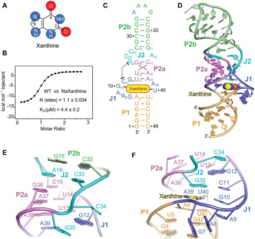
Nucleic Acids Research, 2021, Vol. 49, No. 12 7141
used as the initial model. To minimize the model bias, 5% scan rate, 120 nm/min; slit widths, 10 nm. Thermodynamic
diffractions were selected randomly as the test set in each and kinetic parameters Kd and kobs were obtained as de-
structure refinement. The ligand xanthine was added to scribed in reference (44).
structure at last step in the model building and refinement.
The crystallographic statistics of all X-ray data collection RESULTS AND DISCUSSION
and refinement are listed in Supplementary Table S1.
The two molecules (Mol A and Mol B) in each asymmet- Design of NMT1 riboswitch constructs for crystallization
ric unit stack on each other in an end-to-end way (Supple- and structure solution
mentary Figure S3A, B). We further note that the junction The secondary structure model of the aptamer of the NMT1
J1 of each RNA molecule in the asymmetric unit also forms riboswitch conforms to a large junction with two stems (P1
stacking interaction with the stem-loop of P2 from another and P2), wherein stem P2 contains a small internal bulge
symmetry-related RNA molecule, in which Mol A contacts splitting it into two segments (P2a and P2b) (Supplemen-
Mol B’ and Mol B contacts Mol A” (Supplementary Figure
Downloaded from https://academic.oup.com/nar/article/49/12/7139/6298625 by guest on 30 September 2021
tary Figure S1A) (26). We screened a large number of in vitro
S3). Since Mol A had better electron density than Mol B, transcribed RNA constructs for crystallization in the pres-
our structural analysis is predominantly based on the coor- ence of xanthine and obtained crystals for a 46 nucleotide
dinates of Mol A. long RNA with the sequence from Ideonella species that
diffracted to 2.7 Å (Figure 1A, C). In this construct, the
Isothermal titration calorimetry variable loop that closes stem P2b was replaced by the extra-
Isothermal titration calorimetry (ITC) experiments stable tetranucleotide loop GAAA to facilitate crystalliza-
were performed at 25◦ C on a Microcal PEAQ-ITC tion (Supplementary Figure S1B, C).
microcalorimeter at the National Center for Protein We solved the structure with single-wavelength anoma-
Science·Shanghai (NCPSS). A final concentration of 1.4 lous diffraction (SAD) phasing by collecting the anoma-
mM wild-type and mutant RNAs were refolded at 65◦ C lous signal from Ir(NH3 )6 3+ -soaked crystals (Supplemen-
for 5 min and incubated on ice for an hour after dialysis at tary Figure S2). The X-ray crystallographic statistics are
4◦ C overnight against a buffer containing 50 mM Tris pH provided in Supplementary Table S1.
8.0, 50 mM KCl and 10 mM MgCl2 . To test the impact of The binding affinity of the 46 nt RNA (GAAA loop) to
MgCl2 on binding activity between RNA and xanthine, xanthine was determined by isothermal titration calorime-
wild-type RNAs were dialyzed against buffer containing 50 try (ITC) and gave a dissociation constant of Kd = 4.4 ±
mM Tris pH 8.0, 50 mM KCl, supplemented with different 0.2 M in aqueous buffer containing 10 mM Mg2+ cations
concentration of MgCl2 from 0 mM to 20 mM. The sample (Figure 1B, Supplementary Figure S4 and Supplementary
cell was filled with 200 l 0.1 mM ligand dissolved in each Table S2), which was comparable to the binding affinity
dialysis buffer. The prepared RNA sample titrated into the of the original sequence (GCCC loop) of the xanthine ri-
ligand solution with an initial 0.4 l injection, followed by boswitch (Kd = 3.6 ± 0.3 M, Supplementary Figure S1B,
18 serial 2 l injections, with 2 min interval between each S4 and Supplementary Table S2). The stoichiometry of
injection. The reference power was set as 5 cal s−1 . Inte- binding approached 1:1, with estimated thermodynamic pa-
grated heat data were analyzed via MicroCal PEAQ-ITC rameters of H = –15.1 ± 0.1 kcal mol–1 and S = 0.026
Analysis Software provided by the manufacturer using a kcal mol–1 K–1 (Figure 1B, Supplementary Figure S4 and
‘one set of sites’ binding model. All the binding constants Supplementary Table S2).
and thermodynamic values are listed in Supplementary
Table S2. Overall tertiary fold of the NMT1 riboswitch in complex with
xanthine
Fluorescence spectroscopy
The tertiary structure of the NMT1 xanthine riboswitch is
All steady-state fluorescence spectroscopic experiments schematically shown in Figure 1C and in cartoon represen-
were measured on a Cary Eclipse spectrometer (Varian, tation in Figure 1D. Three stems named P1 (in orange), P2a
Australia) equipped with a peltier block, a magnetic stir- (in violet) and P2b (in green) are formed, which is consis-
ring device and a RX2000 stopped-flow apparatus (Ap- tent with the predicted secondary structure model for the
plied Photophysics Ltd., UK). The data obtained were pro- NMT1 motif. The stems are connected by a large junction
cessed with OriginPro 2018 software (OriginLab, USA). J1 (in blue) and a small bulge J2 (in cyan) (Figure 1C,D
Aminopurine-modified RNA samples were prepared in 0.5 and Supplementary Figure S1A). The RNA folds in a rod-
M concentration in a total volume of 1 ml of buffer (100 like compact helical scaffold (‘I-shape’), in which the three
mM Tris–HCl, 100 mM KCl, pH 8.4). The samples were stems exhibit co-axial stacking mediated by the junctional
heated to 90◦ C for 2 min, allowed to cool to room temper- segments (Figure 1C,D). The ligand xanthine is intercalat-
ature, transferred to quartz cuvettes equipped with a small ing and becomes an integrated part of the continuous base
stir bar and held at 20◦ C in the Peltier controlled sample staple (Figure 1C,D). A view on the overall base stacking
holder. Then, ligands were manually pipetted in a way not alignment of ligand-bound NMT1 RNA is highlighted in
to exceed a total volume increase of 3%. The solution was Supplementary Figure S5A. Notably, stem P1 is extended
stirred after ligand addition and allowed to equilibrate for at by two base pairs from the originally assigned junctional
least 15 min before data collection. Spectra were recorded region J1 by forming one wobble base pair (U5–G42) and
from 320 to 500 nm using the following instrumental pa- one canonical base pair (A6-U41) (Figure 1C, D and Sup-
rameters: excitation wavelength, 308 nm; increments, 1 nm; plementary Figure S1C).
7142 Nucleic Acids Research, 2021, Vol. 49, No. 12

                                                                                                                                                              Downloaded from https://academic.oup.com/nar/article/49/12/7139/6298625 by guest on 30 September 2021
Figure 1. Secondary and tertiary structure of the NMT1 riboswitch bound to xanthine. (A) Chemical structure of xanthine (oxygen atoms are highlighted
with red shadows, nitrogen atoms are highlighted with blue shadow). (B) Exemplary isothermal titration calorimetry (ITC) experiment of wild-type (WT)
NMT1 riboswitch binding with xanthine. Three replicates are shown in Supplementary Figure S4 and the corresponding thermodynamic parameters are
listed in Supplementary Table S2. (C) Schematic secondary structure of the folding topology based on the NMT1 riboswitch crystal structure, same colour
code as in the cartoon representation in panel (D) is used. (D) Cartoon representation of the tertiary structure of the NMT1 riboswitch bound to xanthine
(shown in spheres). (E) Close-up view of the long-distance interaction focusing on junction J2. (F) Close-up view of the long-distance interaction focusing
on junction J1.

   Between stem P2a and P2b, a non-canonical base pair                          pair U13–A38 of stem P2a (Figure 1F and Supplementary
A16-C33 from bulge J2 is formed and interconnects the ter-                      Figure S6C,D), and hence, they further stabilize the strict
minal base pair G36–C15 of stem P2a and the terminal base                       coaxial alignment of stem P2a, P1 and junction J1 (Supple-
pair C32–G17 of stem P2b, therefore a continuous helical                        mentary Figure S5C).
structure is generated from stem P2a to stem P2b (Figure                           Xanthine is bound at the intersection of J1, J2 and stem
1C–E and Supplementary Figure S5A, B). Consequently,                            P1, where it is bracketed by the two strands of J1 and sand-
the two nucleotides C34 and G35 of J2 are pushed aside                          wiched by the terminal base pair A6-U41 of stem P1 and the
from the main helix axis and fold in parallel to stem P2a.                      base tiers formed with the nucleotides from J1 and J2 (Fig-
Thereby, C34 and G35 reach out to the north terminal of                         ure 1C, D and Supplementary Figure S1A, S5C–E). The fi-
J1 (Figure 1E) and form long-distance interactions with the                     nal 2Fo – Fc map of the native xanthine riboswitch structure
nucleotides therein (Figure 1C–E). More precisely, C34 and                      in complex with xanthine is shown in Supplementary Fig-
G35 sandwich G12, the north 1st nucleotide from J1 (Fig-                        ure S6 with emphasis on the J1–J2 junctional region (Sup-
ure 1E), and additionally, G35 forms a canonical Watson–                        plementary Figure S6A) and the binding pocket (Supple-
Crick base pair with C11, the north 2nd nucleotide from J1                      mentary Figure S6B). We point out that the highly con-
(Figure 1F). C34, G12, C11–G35 and the four-nucleotide                          served nucleotides (shown in red in Supplementary Figure
line G10–A9–A8–G7 from J1 align continuously stacked,                           S1B) are brought into close proximity around the bound
thereby generating the cavity for the ligand (Figure 1F). A39                   xanthine. Their pairing and stacking interactions define the
and U40 located in the opposite strand of J1 also form con-                     overall tertiary structure of NMT1 riboswitch and shape the
secutive stacking interactions between the north terminal                       binding pocket specific for xanthine (Supplementary Fig-
base pair A6–U41 of stem P1 and the south terminal base                         ures S1A and S7).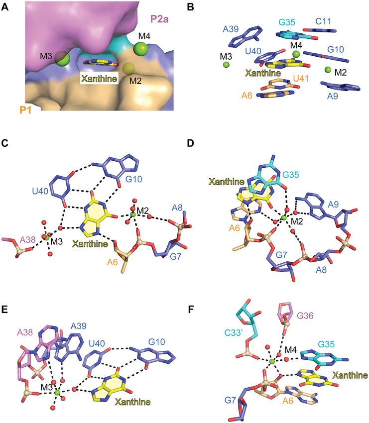
Nucleic Acids Research, 2021, Vol. 49, No. 12 7143

Structural organization of the junctional regions                minor interactions with A6-U41, the terminal base pair in
                                                                 stem P1, in which the Watson-Crick edge of this base forms
The tertiary structure of the xanthine riboswitch is cru-
                                                                 two hydrogen bonds with O2 and 2 -OH of U41 (Figure
cially determined by the structural organization of junc-
                                                                 2G). A8 stacks on G7 and forms two hydrogen bonds with
tions J1 and J2. In an expanded view, Figure 2A shows the
                                                                 N3 and 2 -OH of G42, while G7 forms another hydrogen
secondary structure of this region in the riboswitch cen-
                                                                 bond with 2-NH2 of G42 (Figure 2H). Together, this stack-
ter, with all nucleotides numbered and their tertiary inter-
                                                                 ing and base pairing network helps to define the conforma-
actions highlighted. One of the most impressive features of
                                                                 tion and crosstalk between junction J1 and J2, and hence
the structure is the formation of long-distance interactions
                                                                 contributes to the overall stability of the xanthine-bound
between J1 and J2, which are separated by stem P2a in the
                                                                 NMT1 RNA.
secondary structure (Figures 1B–E, 2A and Supplementary
Figure S1).
   Four nucleotides A16, C33, C34 and G35 constitute junc-
                                                                 Pocket architecture and divalent metal ion-mediated binding

                                                                                                                                 Downloaded from https://academic.oup.com/nar/article/49/12/7139/6298625 by guest on 30 September 2021
tion J2, in which A16 and C33 form a non-cognate base pair
                                                                 to xanthine
with only one H-bond. This pair is tightly stacked between
stems P2a and P2b, while C34 and G35 point outwards to           The surface representation of the xanthine riboswitch
reach out for the long-range interaction (Figures 1B-E, 2A       ligand-binding pocket makes clear that xanthine (depicted
and Supplementary Figure S5C). G35 (J2) forms a canoni-          in sticks) intercalates between stem P1 and P2a and becomes
cal Watson-Crick base pair with C11 (J1) (Figure 1F). C34        almost completely buried with the help of junctional nu-
(J2) and the base pair G35-C11 sandwich the base of G12          cleotides (Figure 3A and Supplementary Figure S9A). The
(J1) and stack on it from both sides (Figure 1F). Further        bound xanthine is stacked between two base triples, namely
detailed inspection revealed that the base of G35 (J2) not       A39–G35–C11 (Figures 2C and 3B) and A6–U41–A9 (Fig-
only forms Watson–Crick pairing interaction with C11 (J1),       ures 2G and 3B). Xanthine itself is also engaged in a triple
but also pairs with the Watson–Crick edge of A39 (J1) using      furnished together with G10 and U40 (Figure 3C). The ar-
the sugar edge, thereby a base triple A39 (J1)–G35 (J2)–C11      rangement is additionally strengthened by the three metal
(J1) is formed as the first tier stacking above the bound xan-   ions M2, M3 and M4 that are located along three sides of
thine (Figure 2B,C). The main chain (ribose and backbone)        the bound xanthine (Figure 3C and Supplementary Figure
of G35 is positioned in the intersection of J1 and stem P2a      S9B). The metal ions were assigned based on the 2Fo – Fc
and forms additional hydrogen bonding interaction with           and Fo – Fc maps guided by the coordination geometries
nucleotides from J1 and stem P2a (Figure 2B–E). The ri-          (Supplementary Figure S6C), and the divalent character of
bose of G35 (J2) forms one hydrogen bond with the 6-NH2          the ions was suggested by the anomalous signal collected
of A38 and an additional one with the phosphate oxygen of        from the Mn2+ -soaked crystals (Supplementary Figure S8).
A37, while the phosphate oxygen of G35 (J2) forms two hy-        All three metal ions are critically involved in the composi-
drogen bonds with the Watson-Crick edge of G12 from J1           tion of the binding pocket as described below (Figure 3 and
(Figure 2D). G12 (J1) is located in the major groove side of     Supplementary Figure S9).
the terminal base pair A38–U13 of stem P2a and is further           Inside the binding pocket, O2 of U40 forms one hydro-
stabilized by one hydrogen bond formed between 2-NH2 of          gen bond with the 2-NH2 of G10. Further, both U40 and
G12 (J1) and O4 of U13 (P2a) (Figure 2D). The ribose of          G10 use their Watson-Crick faces to form two hydrogen
G12 (J1) adopts 2 -endo conformation and forms one addi-        bonds with the minor groove edge of xanthine and two hy-
tional hydrogen bond with N3 of C34 (J2) beside the base         drogen bonds with the Watson-Crick edge of xanthine re-
stacking interaction between G12 (J1) and C34 (J1) (Figure       spectively (Figure 3C). Therefore, a base triple is formed by
2E).                                                             clamping the 2-oxo pyrimidine moiety of xanthine (Figure
   As briefly addressed before, C33 from J2 participates in      3C). The final 2Fo – Fc map of G10, U40 and the bound
the long helical base staple, while C34 and G35 are directed     xanthine is shown in Supplementary Figure S6D. M2 is lo-
outwards to form the long-distance interaction. It is notable    cated in the corner between the Hoogsteen edge and the
that the main chain of C33–C34–G35 (J2) and G36 (P2a)            Watson-Crick edge of xanthine, and importantly, coordi-
form a three-side rectangle with two successive right angle      nates directly to the O6 atom of xanthine (Figure 3D). Be-
turns at the sugar positions of C34 and G35 in the bulged        sides, M2 forms an additional inner-sphere coordination
region (Figure 2B). One Mg2+ cation (M1) is located in the       with the phosphate of G7 and several outer-sphere coordi-
center of the rectangle, which was suggested by the anoma-       nations with other residues including the phosphate of A8,
lous signal collected from crystals soaked with Mn2+ (Sup-       the Hoogsteen edge of A9, O6 of G35, and N3 of A6 (Fig-
plementary Figure S8). M1 forms inner-sphere coordina-           ure 3C, D). It is notable that xanthine intercalates between
tion with the non-bridging phosphate-oxygen of C34, C34         A6 and G35 with the purine rings stacked in parallel, thus
(C34 from a symmetric molecule in crystal) G35 and G36,          enabling an additional hydrogen bond between 2 -OH of A6
and outer-sphere coordination with N7 of G36 (Figure 2E).        (in 2 -endo ribose conformation) and N7 of xanthine (Fig-
With the assistance of M1, the base of G36 from stem P2a         ure 3C, D). These interactions are selective for recognizing
appears to be pulled into the main helical scaffold (Figure      the Hoogsteen edge of xanthine. M3 is located along the
2B).                                                             minor groove edge of xanthine and forms inner-sphere co-
   The four consecutive purine nucleotides G10–A9–A8–            ordination with the phosphate of A38 (the terminal residue
G7 from J1 employ a successive stacking mode against the         of stem P2a). It further displays outer-sphere coordination
minor groove side of stem P1 (Figure 2F). A9 exhibits A-         with N7 of A38, N7 of A39, O4 of U40 and N9 of xanthine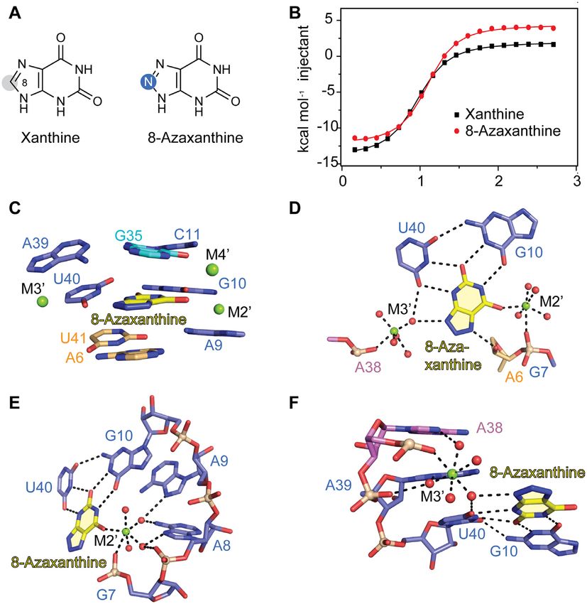
7144 Nucleic Acids Research, 2021, Vol. 49, No. 12

                                                                                                                                                         Downloaded from https://academic.oup.com/nar/article/49/12/7139/6298625 by guest on 30 September 2021
Figure 2. Long-distance interactions between J1 and J2 in the NMT1 riboswitch. (A) Schematic secondary structure of NMT1 RNA core. (B) Structural
details of the J1–J2 long-distance interaction shown in sticks representation. A16 (J2) and C33 (J2) form a wobble base pair within J2, while G35 (J2)
and C34 (J2) stretch out to form the long-distance interaction with J1. The metal ion M1 (Mg2+ ) (shown as green ball) stabilizes the conformational
turn of the J2 chain C33-C34-G35. (C) Base triple formed by C11 (J1), G35 (J2) and A39 (J1), with C11 (J1) and G35 (J2) Watson-Crick paired and its
minor-groove reccognizing the Watson–Crick edge of A39 (J1). (D) The Watson–Crick edge of G12 forms two hydrogen bonds with the phosphate of G35,
which together interact with the major groove side of A38-U13 (the terminal base pair of stem P2a). Besides, the 2 -OH of G35 also forms one hydrogen
bond with the phosphate of A37. (E) The metal ion M1 forms inner-sphere coordination with the phosphates of C34, G35, G36 and a symmetry-related
C34 , and outer-sphere coordination with N7 of G36. G12 adopts 2 -endo ribose conformation and the 2 -OH forms one hydrogen bond with N3 of C34.
(F) G7, A8, A9 and G10 continuously stack and interact with the minor groove side of stem P1. (G) A9 forms A-minor interaction with A6-U41 (the first
north terminal base pair of stem P1). (H) G7 and A8 hydrogen bond with U5-G42 (the second north terminal base pair of stem P1).

(Figure 3E). Thereby, the sequence A38–A39–U40 forms a                       teractions are key for shaping the binding pocket to guar-
characteristic continuous base staple (Figure 3E and Sup-                    antee high ligand specificity.
plementary Figure S5C). M4 is facing the Hoogsteen edges                        Since divalent metal ions are crucially involved in xan-
of G35 and xanthine but is not engaged in direct interac-                    thine riboswitch tertiary structure formation, we performed
tions with xanthine. However, M4 forms inner-sphere co-                      ITC titration experiments at varying Mg2+ concentration,
ordination to A6 (2 -OH) and outer-sphere coordination to                   ranging from 0 to 20 mM in order to find out the concentra-
G35 (N7), whose nucleobases sandwich the ligand (Figure                      tions of divalent cations needed for efficient ligand binding.
3F). The phosphate of G7 and the phosphate of C33 from a                    As shown in Supplementary Figure S10 and S11, binding
symmetrical molecule in the crystal both form inner-sphere                   of xanthine to the NMT1 RNA requires a Mg2+ concen-
coordination with M4, while the phosphate of G36 forms                       tration of at least 0.5 mM, and becomes optimal for Mg2+
outer-sphere coordination. The three metal ion-relying in-                   concentrations around 5 mM or higher.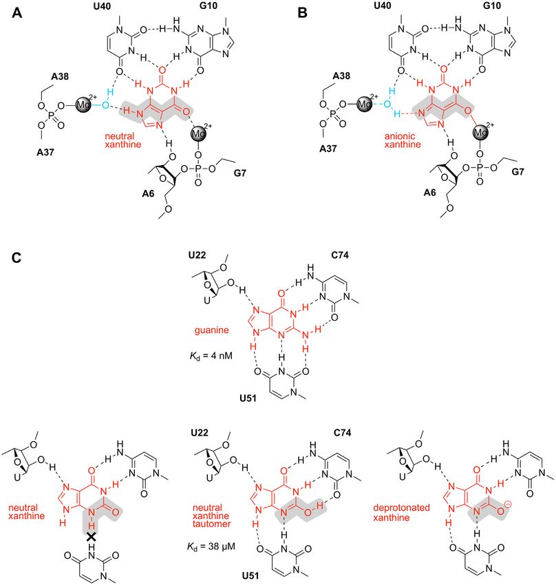
Nucleic Acids Research, 2021, Vol. 49, No. 12 7145

                                                                                                                                                           Downloaded from https://academic.oup.com/nar/article/49/12/7139/6298625 by guest on 30 September 2021
Figure 3. Ligand interaction between xanthine and the NMT1 riboswitch. (A) Xanthine (shown in sticks) is almost completely encapsulated by the RNA
binding pocket (shown in surface representation). Three metal ions M2, M3 and M4 are involved in interactions with the ligand. (B) Xanthine pairs with
G10 and U40, and together are stacked between two base triples, A39–G35–C11 and A9–A6–U41. (C) G10 and U40 form one direct hydrogen bond
between them and use the rest of their Watson-Crick faces to recognize the Watson–Crick and the minor groove edges of xanthine (i.e. the N3H–C2 =
O–N1H urea sub-structure). M2 and M3 are located on opposite sides of xanthine, where M2 forms a direct coordination and M3 forms a water-mediated
coordination with xanthine. The 2 -OH of A6 in equatorial position (2 -endo ribose pucker) forms one hydrogen bond with N7 of xanthine. (D) M2 forms
direct coordinations with xanthine and the phosphate of G7, while it forms indirect coordinations with the bases of A6, A9 and G35, and the phosphate
of A8. Xanthine is sandwiched between A6 and G35. The coordination of M2 with xanthine, G35 and A6 strengthens xanthine binding. (E) M3 is directly
coordinated to the phosphate of A38, while it forms water-mediated coordination with the bound xanthine, A38, A39 and U40. (F) M4 forms direct
interactions with the 2 -OH of A6, the phosphates of G7 and a symmetry-related C33 . Additional indirect coordination is observed with the base of G35
and the phosphate of G36. M4 coordinates to both G35 and A6 that stack on the two sides of xanthine.

Structure-based mutation analysis of the NMT1 riboswitch                      via its minor groove face to the Watson-Crick face of A39
                                                                              (J1) (Figure 2C). To assess the impact of these pairing in-
The junctional nucleotides are involved in tertiary interac-
                                                                              teractions on ligand binding, we made the mutations C11G
tions that appear critical for shaping the binding pocket of
                                                                              (J1) and G35C (J2), respectively, to impair their specific in-
the riboswitch (Figure 2A, B). These interactions became
                                                                              teractions. Both mutants resulted in loss of xanthine bind-
visible in the crystal structure and were then evaluated by
                                                                              ing activity (Figure 4A). Then, we prepared constructs
examining the binding capacity of base mutants of the RNA
                                                                              with the compensatory mutations C11U–G35A and C11G–
that had been used for crystallization, applying isothermal
                                                                              G35C, respectively, to retain the possibility for Watson–
titration calorimetry (ITC) as readout (Figure 4 and Sup-
                                                                              Crick pairing between nucleotide-11 (J1) and –35 (J2) but to
plementary Figure S12).
                                                                              break the hydrogen-bond interactions to A39 (J1) and the
   G35 (J2) forms a long-distance canonical Watson–Crick
                                                                              divalent metal ion coordination of G35 (J2) (Figure 3C–E);
base pair with C11 (J1) and is additionally hydrogen bonded
                                                                              the mutants had no binding activity (Figure 4A). Together,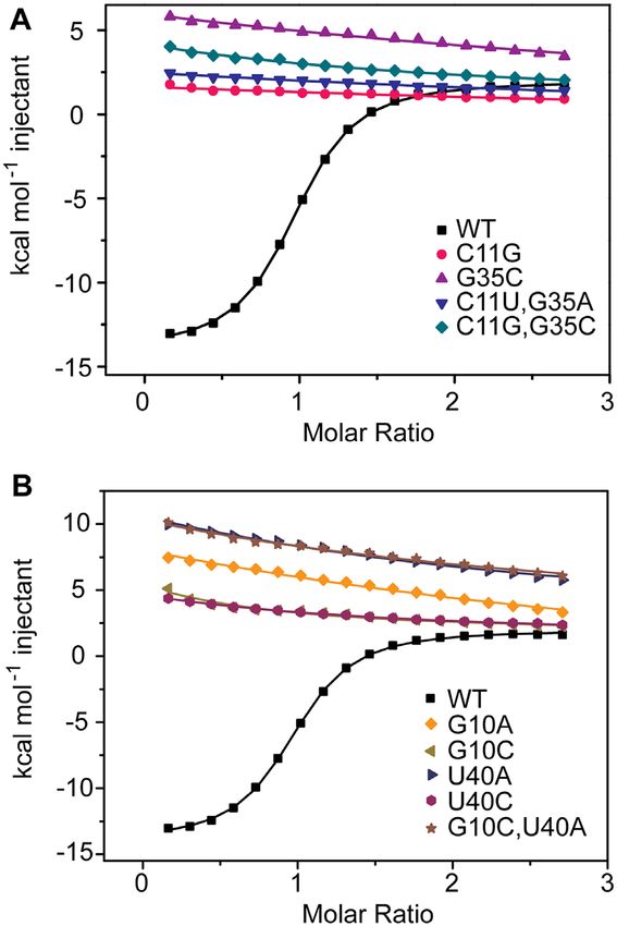
7146 Nucleic Acids Research, 2021, Vol. 49, No. 12
tant binds xanthine with only 4-fold less affinity, as does
the A16G/C33U double mutant (Supplementary Table S2,
Supplementary Figure S12C). This is consistent with re-
tained stacking interactions and the continuation of the
stem P2b base staple as observed in the WT RNA.
In junction J1, A9 forms an A-minor interaction with the
terminal base pair A6-U41 of stem P1 (Figure 2F and G).
To test the significance of the A9 base identity for ligand
binding, it was mutated to C. Not surprisingly, the A9C mu-
tant did no longer bind to xanthine (Supplementary Figure
S12D). As shown in Figure 2H, A8 and G7 form hydrogen
bonds with the minor groove side of the second terminal
base pair of stem P1, a wobble base pair (U5-G42). When
Downloaded from https://academic.oup.com/nar/article/49/12/7139/6298625 by guest on 30 September 2021
we mutated A8 to C or U individually, both RNAs lost
capacity for ligand binding (Supplementary Figure S12D).
Additionally, the mutant G7A-A8G was prepared; for the
G/A switched RNA, ligand binding was not detectable any
more (Figure 2F, H, and Supplementary Figure S12D).
Inside the pocket, G10 and U40 clamp the 2-oxo pyrim-
idine moiety of xanthine from both sides by forming a
base triple (Figure 3C). Individual mutations of these nucle-
obases (G10A, G10C, U40A, and U40C) were not tolerated
(Figure 4B). The double mutant G10C-U40A did neither
bind xanthine (Figure 4B). In summary, our ITC-based mu-
tation study is consistent with the three-dimensional struc-
ture model of the NMT1 riboswitch obtained by X-ray crys-
tallography.
Figure 4. Isothermal titration calorimetry of the NMT1 riboswitch and
structure-based mutants binding with xanthine. (A) Overlay of integrated
fitted heat plots obtained from ITC experiments of wildtype (WT) in com- Binding capacity of the NMT1 riboswitch to xanthine related
parison with mutants concerning nucleotides in the long-distance J1/J2 compounds
interaction. (B) Same as (A) but with mutants concerning nucleotides in
the ligand binding site. It has been reported in the original study on xanthine ri-
boswitch discovery (26) that 8-azaxanthine and uric acid
bind to the NMT1 motif, while other compounds involved
these findings highlight the importance of the base triple in purine degradation pathways including hypoxanthine,
A39 (J1)–G35 (J2)–C11 (J1) and conforms with their high adenine, inosine and adenosine (for chemical structures see
conservation in the sequence (26) (Supplementary Figure Figure 5A and Supplementary Figure S13A) have no or
S1A and B). significantly lower binding affinities. Here, we again used
G12 residing in J1 is sandwiched between G35 and C34, isothermal titration calorimetry (ITC) assays to evaluate
and additionally forms hydrogen bonds with the phosphate these ligand-RNA interactions and to confirm the earlier
of G35 and the terminal base U13 of stem P2a (Figure 2B observations from the Breaker lab (Supplementary Figure
and D). Here, mutation of G12 to A resulted in somewhat S13B, S14 and S15). More precisely, 8-azaxanthine gave a
lower ligand affinity (7-fold decrease), consistent with the similar binding affinity compared to xanthine (Kd = 3.0 ±
loss of a hydrogen bond but retainment of stacking interac- 0.1 M), while uric acid gave an about 7-fold lower binding
tions (Supplementary Figure S12A). Additional mutations affinity (Kd = 20.4 ± 1.7 M) (Figure 5B, Supplementary
were tested for C34, which is participating in the J1-J2 long- Figure S4 and S14, and Supplementary Table S2).
range interaction by intense base stacking onto G12 and by We were successful in co-crystallization and structure de-
forming a single hydrogen bond with the 2 -OH of G12 ri- termination of the NMT1 riboswitch in complex with 8-
bose (Figure 2B,E and Supplementary Figure S12B). While azaxanthine. The overall 3D-folds of the riboswitch bound
the C34A mutant exhibited binding affinity comparable to to xanthine and 8-azaxanthine, respectively, are nearly iden-
the wild-type RNA, the C34U and C34G mutants, respec- tical (Figure 1D and Supplementary Figure S16A) as are
tively, resulted in a slight decrease (3- to 4-fold) in binding the recognition modes for xanthine and 8-azaxanthine in
affinity only (Supplementary Figure S12B). Both G12 and the binding pocket (Figure 5C–E). The three characteristic
C34 are not highly conserved (Supplementary Figure S1A metal ions M2 , M3 and M4 , assigned to Mg2+ accord-
and B), and this is consistent with our observation that their ing to 2Fo – Fc and Fo – Fc maps guided by the coordination
mutations were largely tolerated. geometries are present as well (Figure 5D–F and Supple-
With respect to junction 2, we tested the internal one- mentary Figure S16B) and the interactions of the hydrated
hydrogen bonded A16•C33 pair which stacks on the termi- metal ions with neighboring nucleotides are also retained
nal base pair of stem P2b. While A16 is largely conserved for the 8-azaxanthine-riboswitch complex (Figure 3, 5 and
in nucleobase identity, nucleotide-33 is not. The A16G mu- Supplementary Figures S9B, S16B).Nucleic Acids Research, 2021, Vol. 49, No. 12 7147
Downloaded from https://academic.oup.com/nar/article/49/12/7139/6298625 by guest on 30 September 2021
Figure 5. Structure of the NMT1 riboswitch bound to 8-azaxanthine. (A) Chemical structures of xanthine and 8-azaxanthine (different atom identities
are highlighted with grey (xanthine) and blue (8-azaxathine) shadows. (B) ITC heat plot of the NMT1 riboswitch binding with xanthine (in black) or 8-
azaxathine (in red). (C) 8-Azaxathine hydrogen bonds with G10 and U40, and together are stacked between two base triples A39-G35-C11 and A9-A6-U41
as observed in the xanthine-bound structure. Three metal ions M2 , M3 and M4 are also retained in the 8-azaxathine-bound structure. (D) Hydrogen
bonding interactions and metal ion-involved coordination of 8-azaxathine highlighted for the NMT1 riboswitch pocket. (E, F) Metal ion-coordinations of
M2 (E) and M3 (F) observed in the NMT1 riboswitch pocket occupied by 8-azaxanthine.
Both structures reveal that the ligands xanthine and 8- Some insights into Mg2+ and xanthine dependent riboswitch
azaxanthine are almost completely encapsulated. The only folding
position that is accessible from the solvent is ligand posi-
tion C8 (Figure 3C and 5D). Therefore, the replacement of Ligand-induced RNA folding can usually be investigated
C8-H by a nitrogen atom (as in 8-azaxanthine) or the addi- by applying 2-aminopurine (Ap) modified RNA variants
tion of a carbonyl oxygen at C8 (as in uric acid) is consis- in a fluorescence spectroscopic approach termed 2ApFold
tent with our observation that 8-azaxanthine and uric acid (Figure 6A) (8,32,33). One has to be aware that the UV
are well accepted by the riboswitch scaffold (Supplementary absorbance of xanthine is depending on the pH value of
Figures S13B and S14). In contrast, the related purines hy- the solution. To underline this, we determined the pKa1 and
poxanthine and adenine do not satisfy the hydrogen donor- pKa2 values for xanthine (which were 7.7 and 11.7, respec-
acceptor pattern provided by the pocket. In particular, G10 tively) by UV-spectroscopic detection of pH titration ex-
and U40 which are perfectly complementary to the 2-oxo periments (Figure 6B). Moreover, because of the spectral
pyrimidine moiety of xanthine (Figure 3C), miss this recog- overlap of xanthine and 2-aminopurine, limitations are en-
nition possibilities if confronted with the pyrimidine moiety counter for quantitative 2ApFold measurements. However,
of hypoxanthine or adenine, both lacking the 2-oxo group. on a qualitative level, we were confident that the analysis
Hence, the inability to bind any of these compounds is con- of riboswitch variants with Ap replacements at selected po-
sistent with the H-donor/H-acceptor profile provided in the sitions should allow to detect regions of xanthine-induced
tight pocket (Supplementary Figures S13 and S15). Fur- compaction of the riboswitch fold (Figure 6C, D). More-
thermore, inosine and adenosine do not bind to the NMT1 over, Mg2+ -induced local structural rearrangements that
RNA and steric hindrance of the large ribose group at N9 take place in the course of RNA pre-folding (in the absence
is the likely reason for this observation (Figure 3C, Supple- of xanthine) should be detectable, even on a quantitative
mentary Figures S13 and S15). level.7148 Nucleic Acids Research, 2021, Vol. 49, No. 12
Downloaded from https://academic.oup.com/nar/article/49/12/7139/6298625 by guest on 30 September 2021
Figure 6. Fluorescence spectroscopic assessment of NMT1 RNA folding and ligand binding. (A) Chemical structure of 2-aminopurine in an RNA chain.
(B) Protonation equilibria of xanthine (sites of deprotonation are shown according to reference 46) and corresponding UV-spectra obtained by pH titration
experiments used for pKa determination. (C) Secondary structure of NMT1 RNA with color-coded nucleotides that were individually replaced by Ap. (D)
Cartoon representation of NMT1 RNA highlighting Ap substitutions by using the same color code as in (C). (E) Real time fluorescence traces for five
different Ap riboswitch variants (c = 0.5 M) upon Mg2+ (20 mM) and ligand (3 mM) additions; buffer conditions: 100 mM Tris–HCl, pH 8.4, 100 mM
KCl. (F) Fluorescence changes upon titration of A8Ap RNA (c = 0.5 M) with increasing concentrations of Mg2+ ions; normalized fluorescence intensity
of the A8Ap variant plotted as a function of Mg2+ concentration. The graph shows the best fit of the two parametric quadratic fit. Changes in fluorescence
(F – F0 ) were normalized to the maximum fluorescence measured in saturating concentrations of Mg2+ ligand. The obtained Kd value for Mg2+ in 100
mM Tris–HCl, pH 8.4, 100 mM KCl, at 293 K, is indicated; inset: fluorescence emission spectra (ex = 308 nm) from 320 to 450 nm of the A8Ap variant
for each Mg2+ concentration. (G) Monitoring the kinetics of Mg2+ -induced NMT1 RNA folding using the A8Ap labeled RNA. Exemplary fluorescence
traces for Mg2+ additions are depicted; conditions: 0.5 M RNA, 100 mM Tris–HCl, 100 mM KCl, pH 8.4, at 293 K. Final MgCl2 concentrations as
indicated.Nucleic Acids Research, 2021, Vol. 49, No. 12 7149
The xanthine-bound riboswitch fold does not provide Concluding remarks
any nucleobases that are unstacked and completely ex-
Bacterial RNAs containing the NMT1 motif turn off gene
posed to the solvent. Such nucleotide positions are usu-
expression upon ligand sensing, likely by regulating trans-
ally used for replacements by Ap, because they are struc-
lation initiation (26). Members of this riboswitch class re-
turally non-invasive and the corresponding RNA variants
spond to high concentrations of oxidized purines and they
respond by a pronounced fluorescence increase upon lig-
regulate genes predominantly associated to purine trans-
and binding. Nevertheless, the xanthine riboswitch fold of-
port and oxidation, thus avoiding the deleterious effects
fers several internal positions where the aminopurine re-
of accumulation of purine degradation products in the cell
placement retains the hydrogen bond and/or stacking pat-
(26).
tern. These are A8Ap, G7Ap, A6Ap, G12Ap. Moreover, by
There is currently only one other known example of a
considering the double mutation C11U-G35Ap, the long-
riboswitch that binds a nucleobase via inner-sphere Mg2+
range Watson-Crick base pair that stacks on top of xan-
coordination, namely the ZMP (5-amino-4-imidazole car-
thine is retained, and its formation should also be trace-
Downloaded from https://academic.oup.com/nar/article/49/12/7139/6298625 by guest on 30 September 2021
boxamide ribose-5 -monophosphate) riboswitch. This ri-
able by a putative xanthine-induced fluorescence response.
boswitch is widespread and regulates de novo purine syn-
We synthesized all of these variants and the qualitative
thesis (20). It recognizes the carboxamide oxygen of ZMP
fluorescence responses of the riboswitch variants (0.5 M
through coordination with a Mg2+ cation, which is si-
each) upon addition of saturating concentrations of Mg2+
multaneous coordinated to two backbone phosphates (34–
ions (20 mM), and subsequently, of xanthine (3 mM) were
36) (Figure 7A). More frequently, Mg2+ -mediated bind-
recorded and are depicted in Figure 6E. Due to solubility
ing of small molecules to RNA concerns the phosphate
of xanthine, we use buffer conditions with a slightly ba-
moieties of these ligands, as observed e.g. for the thi-
sic pH of 8.4. Interestingly for the majority of Ap variants
amine pyrophosphate (TPP) riboswitch (37–39), the flavin
the Mg2+ addition resulted in no (A6Ap, G7Ap, G12Ap)
mononucleotide (FMN) riboswitch (40), guanosine-3 ,5 -
or only a minor (C11U-G35Ap) decrease in fluorescence,
bispyrophosphate (ppGpp) and ␣-5 -phosphoribosyl-1 -
suggesting a rather pre-organized RNA fold that largely re-
pyrophosphat (PRPP) riboswitches (41,42), and the nicoti-
sembles the final xanthine-bound fold. From this subset,
namide adenine dinucleotide (NAD-I) riboswitch (43,44)
G6Ap, G7Ap and G12Ap experienced a small (onefold) flu-
(Figure 7B–F). The fluoride riboswitch also uses the con-
orescence increase when xanthine became available, indicat-
cept of Mg2+ mediated ligand binding. The tiny anion is
ing that these nucleobases are slightly more exposed in the
anchored to the RNA through direct coordination to three
ligand-bound fold compared to their positions in the Mg2+ -
Mg2+ ions, which in turn are octahedrally coordinated to
induced pre-folded state. Remarkably, the nucleotide at po-
water molecules and five inwardly pointing backbone phos-
sition 8 responded significantly to both ligands and this be-
phates (45) (Figure 7G).
havior identifies this region to be the conformationally most
Our structural study reveals the molecular basis for the
flexible one. While during Mg2+ -triggered preorganization
high specificity of this RNA to bind xanthine and only near
the nucleotide-8 becomes exposed (fluorescence increase)
cognate ligands such as uric acid and 8-azaxanthine. The
it slides into its finally stacked position between G7 and
key recognition feature is insertion of the xanthine’s pyrim-
A9 when xanthine becomes bound (fluorescence decrease).
idine moiety between an opened G–U base pair, thus en-
This is consistent with the Mg2+ (M2) ‘bridge’ between the
abling shape complementarity and simultaneous recogni-
backbone of G7-A8 and the O6 of xanthine as seen in the
tion of the C2 urea substructure by a maximum of four
crystal structure.
hydrogen bonds. The 2-oxo group is lacking in hypoxan-
The pronounced fluorescence increase for Mg2+ -induced
thine and hence the pocket strongly discriminates against
folding of the A8Ap riboswitch variant allowed for quantifi-
it. The second key feature is Mg2+ -mediated recognition of
cation. The concentration-dependent fluorescence response
the xanthine-O6 atom. This crucial interaction cannot be
data of Mg2+ titrations (Figure 6F) were fit using a two-
compensated by the isosteric N6 group of adenine related
parametric quadratic equation and gave an apparent dis-
derivatives that cannot bind. Our structure also explains the
sociation constant, Kd (Mg2+ ) of 10.8 ± 2.3 mM (Figure
similar affinity for 8-azaxanthine and the only slightly lower
6F). Clearly, this Kd is well above physiologically encoun-
one for uric acid, because the purine-8 position is solvent-
tered Mg2+ concentrations, however, it is compatible with
accessible and the only position that is not engaged in the
the assumption that a small population of RNA with ex-
tight hydrogen bonding and stacking network generated by
posed A8 is characteristic and likely supportive for ligand
the riboswitch pocket.
recognition in the natural environment (pre-folding). Ad-
For a deeper understanding of xanthine recognition, its
ditionally, we investigated the kinetics of Mg2+ binding for
pH dependence has to be taken into account. The pKa
this riboswitch region. The rates (kobs ) were determined in
of xanthine N3-H is 7.7 and therefore at physiological
Mg2+ concentration-dependent manner by fitting the indi-
pH, a significant population of xanthine is deprotonated
vidual fluorescence traces to a single exponential equation,
(monoanion). We therefore analyzed the potential H-bond
obtained from manually performed measurements (Figure
patterns for neutral as well as deprotonated xanthine in the
6G). The rates were in the order of 0.13 s–1 and turned out
pocket (Figure 8). It becomes obvious that a tautomeric
to be independent of Mg2+ concentration. This observation
form of deprotonated xanthine can retain G10-U40 recog-
indicates that not Mg2+ binding itself is rate limiting but
nition (Figure 8B); thereby, the negative charge at the O6
rather a change in RNA conformation which is consistent
atom is compensated by the Mg2+ ion (M2) that is bound
with the conformational rearrangement of A8 as proposed
to the phosphate between A6 and G7 (Figure 8B). Simul-
above.7150 Nucleic Acids Research, 2021, Vol. 49, No. 12
Downloaded from https://academic.oup.com/nar/article/49/12/7139/6298625 by guest on 30 September 2021
Figure 7. Comparison of Mg2+ -mediated ligand recognition by various riboswitches. (A) The carboxamide oxygen of ZMP applies one inner-sphere
coordination with a Mg2+ cation in the binding pocket of the ZMP riboswitch (PDB code: 4ZNP). (B) The phosphates of TPP form direct coordinations
with two Mg2+ cations in the binding pocket of the TPP riboswitch (PDB code: 2GDI). (C) The phosphate of FMN is recognized by forming a direct
and an indirect coordination with the Mg2+ cation in the binding pocket of the FMN riboswitch (PDB code: 3F2Q). (D) Four Mg2+ cations are involved
in interactions between ppGpp and the ppGpp riboswitch pocket (PDB code: 6DMC). (E) One Mg2+ cation mediates the interaction of the phosphate
of PRPP and the PRPP riboswitch pocket (PDB code: 6DLT). (F) The phosphates of NAD+ forms inner-sphere and outer-sphere coordination with two
Mg2+ cations in the pocket of the NAD-I riboswitch (PDB code: 7D7X). (G) Fluoride directly contacts three Mg2+ cations, which in turn form direct
interactions with five inwardly pointing phosphates of the fluoride riboswitch pocket (PDB code: 4ENC).
taneously, the switch from N9-H (neutral xanthine) to the = 2.2 ± 0.2 M. (Supplementary Table S2, Supplementary
N9 imine (deprotonated xanthine) is compensated by the Figure S17).
altered hydration mode of the second Mg2+ ion (M3) lo- In this context, we note that 8-azaxanthine has a pKa of
cated at the backbone of A38 (Figure 8A, B). The chemical 4.8 and at physiological pH, the monoanion is dominantly
structure analysis suggests that the binding pocket fulfils the populated (47). Also for this system, the binding pocket of
requirements for binding of both neutral and anionic xan- the xanthine riboswitch can in principle tolerate both, the
thine without an obvious preference for one or the other neutral ligand (either the 8H or 9H tautomer) and the de-
form. This also suggests that both (neutral and anionic form protonated form (Supplementary Figure S18). This obser-
of xanthine) likely bind with rather comparable affinities. vation is consistent with its unaltered affinity compared to
We therefore performed additional ITC experiments at pH xanthine.
6.0, pH 7.0, and pH 8.0 in the same buffer system (HEPES), Finally, we juxtapose xanthine recognition by the xan-
and indeed, we found comparable affinities (Kd (pH 6.0) = thine riboswitch to xanthine recognition by the G ri-
2.6 ± 0.8 M, Kd (pH 7.0) = 0.9 ± 0.1 M, and Kd (pH 8.0) boswitch. In an earlier crystallographic study, the G ri-Nucleic Acids Research, 2021, Vol. 49, No. 12 7151
Downloaded from https://academic.oup.com/nar/article/49/12/7139/6298625 by guest on 30 September 2021
Figure 8. Chemical analysis of xanthine recognition by RNA taking tautomeric and deprotonated ligand forms into account. (A) Model for recognition
of neutral xanthine by the NMT1 xanthine riboswitch (this study). (B) Model for recognition of deprotonated xanthine by the NMT1 xanthine riboswitch
(this study). (C) Models for recognition of guanine versus xanthine by the guanine riboswitch based on an earlier study (PDB code: 3GAO) (48). The
Mg2+ -mediated binding mode of the xanthine riboswitch tolerates both the neutral and the deprotonated form of xanthine due to the easily adaptable
hydration mode of Mg2+ ions that allows for charge compensation.
boswitch was crystalized with a series of guanine analogs DATA AVAILABILITY
including xanthine (48). Although xanthine binds signif-
Atomic coordinates and structure factors for the re-
icantly weaker to the G riboswitch (Kd of 38 M) com-
ported crystal structures have been deposited with the Pro-
pared to its native ligand guanine (Kd of 4 nM), the recog-
tein Data bank under accession number 7ELR (xanthine
nition pattern by the pocket turned out to be identical.
bound), 7ELP (Ir(NH3 )6 3+ -soaked), 7ELQ (Mn2+ -soaked)
Thereby, the mode of recognition likely involves the N3-
and 7ELS (8-azaxanthine bound).
imino tautomer or the corresponding deprotonated xan-
thine (Figure 8C). No metal ion is involved in xanthine
recognition by the G riboswitch. This lack of charge com- SUPPLEMENTARY DATA
pensation in the G riboswitch scaffold compared to the xan- Supplementary Data are available at NAR Online.
thine riboswitch might be the reason for the lower ligand
affinity.
Taken together, our study provides deep insights into the ACKNOWLEDGEMENTS
molecular principles of ligand recognition of the NMT1 We thank the staff members of the Large-scale Pro-
RNA motif that defines the xanthine riboswitch. tein Preparation System, BL-17B, BL18U1, and BL-19U17152 Nucleic Acids Research, 2021, Vol. 49, No. 12
beamlines at the National Facility for Protein Science in 15. Yoon,S.H. and Waters,C.M. (2021) The ever-expanding world of
Shanghai (NFPS), Zhangjiang Lab, China for providing bacterial cyclic oligonucleotide second messengers. Curr. Opin.
Microbiol., 60, 96–103.
technical support and assistance in data collection and anal- 16. Denessiouk,K.A., Rantanen,V.V. and Johnson,M.S. (2001) Adenine
ysis. We thank the staff of the BL-17U1 beamline at SSRF recognition: a motif present in ATP-, CoA-, NAD-, NADP-, and
for their assistance in X-ray data collection. We thank the FAD-dependent proteins. Proteins, 44, 282–291.
technical assistance from the core facility of the Life Sci- 17. Fontecave,M., Atta,M. and Mulliez,E. (2004) S-Adenosylmethionine:
ences Institute (LSI), Zhejiang University. We thank Olga nothing goes to waste. Trends Biochem. Sci., 29, 243–249.
18 Mandal,M., Boese,B., Barrick,J.E., Winkler,W.C. and Breaker,R.R.
Krasheninina (Innsbruck) for discussions on fluorescence (2003) Riboswitches control fundamental biochemical pathways in
spectroscopic data fitting. Bacillus subtilis and other bacteria. Cell, 113, 577–586.
19. Mandal,M. and Breaker,R.R. (2004) Adenine riboswitches and gene
activation by disruption of a transcription terminator. Nat. Struct.
Mol. Biol., 11, 29–35.
FUNDING 20. Kim,P.B., Nelson,J.W. and Breaker,R.R. (2015) An ancient
Downloaded from https://academic.oup.com/nar/article/49/12/7139/6298625 by guest on 30 September 2021
riboswitch class in bacteria regulates purine biosynthesis and
National Natural Science Foundation of China [32022039, one-carbon metabolism. Mol. Cell, 57, 317–328.
31870810, 91940302, 91640104, 31670826 to A.R.]; Zhe- 21. Kim,J.N., Roth,A. and Breaker,R.R. (2007) Guanine riboswitch
jiang Province [LR19C050003 to A.R.]; Fundamental Re- variants from Mesoplasma florum selectively recognize
search Funds for the Central Universities [2017QN81010 2 -deoxyguanosine. Proc. Natl. Acad. Sci. U.S.A., 104, 16092–16097.
22. Weinberg,Z., Nelson,J.W., Lunse,C.E., Sherlock,M.E. and
to A.R.]; Zhejiang University (to A.R.); Austrian Science Breaker,R.R. (2017) Bioinformatic analysis of riboswitch structures
Fund FWF [P31691, F8011-B to R.M.]; Vienna Science uncovers variant classes with altered ligand specificity. Proc. Natl.
and Technology Fund WWTF [LS17-003 to R.M.]; Aus- Acad. Sci. U.S.A., 114, E2077–E2085.
trian Research Promotion Agency FFG [West Austrian 23. Sherlock,M.E., Sudarsan,N., Stav,S. and Breaker,R.R. (2018)
Tandem riboswitches form a natural Boolean logic gate to control
BioNMR 858017 to R.M]. Funding for open access charge: purine metabolism in bacteria. Elife, 7, e33908.
National Natural Science Foundation of China [31870810]. 24. Sherlock,M.E., Sadeeshkumar,H. and Breaker,R.R. (2019) Variant
Conflict of interest statement. None declared. bacterial riboswitches associated with nucleotide hydrolase genes
sense nucleoside diphosphates. Biochemistry, 58, 401–410.
25. Sherlock,M.E. and Breaker,R.R. (2020) Former orphan riboswitches
reveal unexplored areas of bacterial metabolism, signaling, and gene
REFERENCES control processes. RNA, 26, 675–693.
1. Breaker,R.R. (2012) Riboswitches and the RNA world. Cold Spring 26. Yu,D. and Breaker,R.R. (2020) A bacterial riboswitch class senses
Harb. Perspect. Biol., 4, a003566. xanthine and uric acid to regulate genes associated with purine
2. Serganov,A. and Nudler,E. (2013) A decade of riboswitches. Cell, oxidation. RNA, 26, 960–968.
152, 17–24. 27. Colloćh,N., El Hajji,M., Bachet,B., L’Hermite,G., Schiltz,M.,
3. Bédard,A.V., Hien,E.D.M. and Lafontaine,D.A. (2020) Riboswitch Prangé,T., Castro,B. and Mornon,J.P. (1997) Crystal Structure of the
regulation mechanisms: RNA, metabolites and regulatory proteins. protein drug urate oxidase-inhibitor complex at 2.05 Å resolution.
Biochim. Biophys. Acta Gene Regul. Mech., 1863, 194501. Nat. Struct. Biol., 4, 947–952.
4. Jones,C.P. and Ferré-D’Amaré,A.R. (2017) Long-range interactions 28. Pikovskaya,O., Serganov,A.A., Polonskaia,A., Serganov,A. and
in riboswitch control of gene expression. Annu. Rev. Biophys., 46, Patel,D.J. (2009) Preparation and crystallization of riboswitch-ligand
455–481. complexes. Methods Mol. Biol., 540, 115–128.
5. Garst,A.D., Edwards,A.L. and Batey,R.T. (2011) Riboswitches: 29. Adams,P.D., Afonine,P.V., Bunkóczi,G., Chen,V.B., Davis,I.W.,
structures and mechanisms. Cold Spring Harb. Perspect. Biol., 3, Echols,N., Headd,J.J., Hung,L.W., Kapral,G.J.,
a003533. Grosse-Kunstleve,R.W. et al. (2010) PHENIX: a comprehensive
6. Micura,R. and Höbartner,C. (2020) Fundamental studies of Python-based system for macromolecular structure solution. Acta
functional nucleic acids: aptamers, riboswitches, ribozymes and Crystallogr. D Biol. Crystallogr., 66, 213–221.
DNAzymes. Chem. Soc. Rev., 49, 7331–7353. 30. Emsley,P. and Cowtan,K. (2004) Coot: model-building tools for
7. Fürtig,B., Nozinovic,S., Reining,A. and Schwalbe,H. (2015) Multiple molecular graphics. Acta Crystallogr. D. Biol. Crystallogr., 60,
conformational states of riboswitches fine-tune gene regulation. Curr. 2126–2132.
Opin. Struct. Biol., 30, 112–124 31. Murshudov,G.N., Vagin,A.A. and Dodson,E.J. (1997) Refinement of
8. Haller,A., Soulière,M.F. and Micura,R. (2011) The dynamic nature macromolecular structures by the maximum-likelihood method. Acta
of RNA as key to understanding riboswitch mechanisms. Acc. Chem. Crystallogr. D. Biol. Crystallogr., 53, 240–255.
Res., 44, 1339–1348. 32. Rieder,R., Lang,K., Graber,D. and Micura,R. (2007) Ligand-induced
9. Nahvi,A., Sudarsan,N., Ebert,M.S., Zou,X., Brown,K.L. and folding of the adenosine deaminase A-riboswitch and implications on
Breaker,R.R. (2002) Genetic control by a metabolite binding mRNA. riboswitch translational control. Chembiochem, 8, 896–902.
Chem. Biol., 9, 1043–1049. 33. Soulière,M.F., Haller,A., Rieder,R. and Micura,R. (2011) A powerful
10. Winkler,W., Nahvi,A. and Breaker,R.R. (2002) Thiamine derivatives approach for the selection of 2-Aminopurine substitution sites to
bind messenger RNAs directly to regulate bacterial gene expression. investigate RNA folding. J. Am. Chem. Soc., 133, 16161–16167.
Nature, 419, 952–956. 34. Ren,A., Rajashankar,K.R. and Patel,D.J. (2015) Global RNA fold
11. Mironov,A.S., Gusarov,I., Rafikov,R., Lopez,L.E., Shatalin,K., and molecular recognition for a pfl riboswitch bound to ZMP, a
Kreneva,R.A., Perumov,D.A. and Nudler,E. (2002) Sensing small master regulator of One-Carbon metabolism. Structure, 23,
molecules by nascent RNA: a mechanism to control transcription in 1375–1381.
bacteria. Cell, 111, 747–756. 35. Trausch,J.J., Marcano-Velázquez,J.G., Matyjasik,M.M. and
12. Winkler,W.C., Cohen-Chalamish,S. and Breaker,R.R. (2002) An Batey,R.T. (2015) Metal ion-mediated nucleobase recognition by the
mRNA structure that controls gene expression by binding FMN. ZTP riboswitch. Chem. Biol., 22, 829–837.
Proc. Natl. Acad. Sci. U.S.A., 99, 15908–15913. 36. Jones,C.P. and Ferré-D’Amaré,A.R. (2015) Recognition of the
13. McCown,P.J., Corbino,K.A., Stav,S., Sherlock,M.E. and bacterial alarmone ZMP through long-distance association of two
Breaker,R.R. (2017) Riboswitch diversity and distribution. RNA, 23, RNA subdomains. Nat. Struct. Mol. Biol., 22, 679–685.
995–1011. 37. Serganov,A., Polonskaia,A., Phan,A.T., Breaker,R.R. and Patel,D.J.
14. Pedley,A.M. and Benkovic,S.J. (2017) A new view into the regulation (2006). Structural basis for gene regulation by a thiamine
of purine metabolism: the purinosome. Trends Biochem. Sci., 42, pyrophosphate-sensing riboswitch. Nature, 441, 1167–1171.
141–154.You can also read