Chromatographic methods for protein purification - My Hedhammar, Amelie Eriksson Karlström, Sophia Hober Royal Institute of Technology, AlbaNova ...
←
→
Page content transcription
If your browser does not render page correctly, please read the page content below
Chromatographic methods
              for
      protein purification

My Hedhammar, Amelie Eriksson Karlström, Sophia Hober

Royal Institute of Technology, AlbaNova University Center, Dept. of Biotechnology,

SE-106 91 Stockholm, Sweden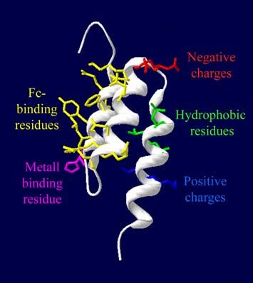
Protein Purification .................................................................................................................... 3
Chromatography......................................................................................................................... 3
1. Ion Exchange Chromatography.............................................................................................. 5
   1.1. Charge Properties of Proteins.......................................................................................... 5
   1.2. The Stationary Phase....................................................................................................... 6
   1.3. The Mobile Phase............................................................................................................ 7
      1.3.1 Elution Strategies ...................................................................................................... 7
   1.4. Features ........................................................................................................................... 8
2. Chromatographic Methods Based on Hydrophobicity........................................................... 9
   2.1. Hydrophobicity................................................................................................................ 9
   2.2. Hydrophobic Interaction Chromatography ................................................................... 10
      2.2.1 The Stationary Phase............................................................................................... 10
      2.2.2 The Mobile Phase.................................................................................................... 11
         2.2.2.1. Elution Strategies ............................................................................................ 12
      2.2.3 Features ................................................................................................................... 12
   2.3. Reversed Phase Chromatography.................................................................................. 13
      2.3.1 The Stationary Phase............................................................................................... 13
      2.3.2 The Mobile Phase.................................................................................................... 14
         2.3.2.1. Ion Suppression ............................................................................................... 14
         2.3.2.2. Ion Pairing ....................................................................................................... 15
         2.3.2.3. Elution Strategies ............................................................................................ 15
      2.3.3 Features and Applications ....................................................................................... 15
         2.3.3.1. Reversed Phase High-Performance Liquid Chromatography ......................... 15
3. Affinity Chromatography..................................................................................................... 16
   3.1. The Stationary Phase..................................................................................................... 16
      3.1.1 Ligands .................................................................................................................... 17
      3.1.2 Spacer Arms ............................................................................................................ 17
      3.1.3 Ligand Coupling...................................................................................................... 17
   3.2. The Mobile Phase.......................................................................................................... 19
      3.2.1 Elution Strategies .................................................................................................... 20
   3.3. Features ......................................................................................................................... 21
   3.4. Applications .................................................................................................................. 21
      3.4.1 Immunoaffinity........................................................................................................ 21
      3.4.2 Purification of Immunoglobulins ............................................................................ 22
      3.4.3 Purification of Glycoconjugates.............................................................................. 22
      3.4.4 Purification of DNA-Binding Proteins.................................................................... 22
      3.4.5 Purification of Receptor Proteins ............................................................................ 23
      3.4.6 Purification of Enzymes .......................................................................................... 23
      3.4.7 Purification by the use of Synthetic Dye Ligands................................................... 23
      3.4.8 Isolation of Cells ..................................................................................................... 23
      3.4.9 Isolation of Nucleotides .......................................................................................... 23
   3.5. Affinity Tags ................................................................................................................. 23
4. Immobilized Metal Affinity Chromatography ..................................................................... 24
   4.1. Metal Ion Affinities ....................................................................................................... 24
   4.2. The Stationary Phase..................................................................................................... 24
   4.3. The Mobile Phase.......................................................................................................... 25
      4.3.1 Elution Strategies .................................................................................................... 25
   4.4. Features ......................................................................................................................... 26
   4.5. Applications .................................................................................................................. 26
5. Size Exclusion Chromatography.......................................................................................... 27
5.1. Molecular Sieving ......................................................................................................... 27
5.2. The Stationary Phase..................................................................................................... 27
5.3. The Mobile Phase.......................................................................................................... 28
5.3.1 Elution Strategies .................................................................................................... 28
5.4. Features ......................................................................................................................... 28
5.5. Applications .................................................................................................................. 29
5.5.1 Buffer Exchange...................................................................................................... 29
5.5.2 Protein Fractionation ............................................................................................... 29
5.5.3 Determination of Molecular Size ............................................................................ 29
Further Reading........................................................................................................................ 30
2Protein Purification
To be able to isolate a specific protein from a crude mixture the physical and/or chemical
properties of the individual protein must be utilized. There is no single or simple way to
purify all kinds of proteins. Procedures and conditions used in the purification process of one
protein may result in the inactivation of another. The final goal also has to be considered
when choosing purification method. The purity required depends on the purpose for which the
protein is needed. For an enzyme that is to be used in a washing powder, a relatively impure
sample is sufficient, provided it does not contain any inhibiting activities. However, if the
protein is aimed for therapeutic use it must be extremely pure and purification must then be
done in several subsequent steps.
The aim of a purification process is not only removal of unwanted contaminants, but also the
concentration of the desired protein and the transfer to an environment where it is stable and
in a form ready for the intended application.
In the early days of protein chemistry, the only practical way to separate different types of
proteins was by taking advantage of their relative solubility. Part of a mixture was caused to
precipitate through alteration of some properties of the solvent e.g. addition of salts, organic
solvents or polymers, or varying the pH or temperature. Fractional precipitation is still
frequently used for separation of gross impurities, membrane proteins and nucleic acids.
Under certain conditions, proteins adsorb to a variety of solid phases, preferably in a selective
manner. Calcium phosphate gels have frequently been used to specifically adsorb proteins
from heterogeneous mixtures. The adsorption principle is further explored in column
chromatography. Due to their high resolving power, different chromatography techniques
have become dominant for protein purification.
Chromatography
Chromatography refers to a group of separation techniques that involves a retardation of
molecules with respect to the solvent front that progresses through the material. The name
literally means “color drawing” and was originally used to describe the separation of natural
pigments on filter papers by differential retardation. The same principle is now commonly
used for protein separation. Column chromatography is the most common physical
configuration, in which the stationary phase is packed into a tube, a column, through which
the mobile phase, the eluent, is pumped. The degree to which the molecule adsorbs or
interacts with the stationary phase will determine how fast it will be carried by the mobile
phase. Chromatographic separation of protein mixtures has become one of the most effective
and widely used means of purifying individual proteins.
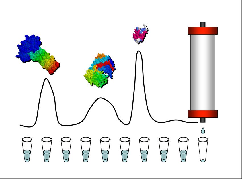
3General properties of proteins are used to isolate them from other, non-protein contaminants.
Minor differences between various proteins, such as size, charge, hydrophobicity and
biospecific interaction (Fig. 1) are used to purify one protein from another.
Figure 1. Selective protein properties
Examples of properties that are used to separate
one protein from another.
In a typical chromatography process the first step is a capturing step, where the product binds
to the adsorbent while the impurities do not. Further, weakly bound proteins are washed away
before the conditions are changed so that the target protein is eluted. Several versions of
liquid chromatography, differing mainly in the types of stationary phase, are used for protein
purification. The process of Size exclusion Chromatography is a bit different since separation
is based on sieving properties of the stationary phase and not on adsorption. Two variants of
column chromatography, Ion Exchange Chromatography and Size Exclusion
Chromatography, are illustrated in figure 2.
Figure 2. Illustrations of two classical chromatographic methods
A. Ion Exchange Chromatography. The charges of a protein are used for purification.
B. Size Exclusion Chromatography. Protein size is used for fractionation.
41. ION EXCHANGE CHROMATOGRAPHY
Ionic interactions are the basis for purification of proteins by Ion Exchange Chromatography
(IEXC) (Fig. 2A). The separation is due to competition between proteins with different
surface charges for oppositely charged groups on an ion exchanger adsorbent.
The fundamental theory of IEXC has a very long history. One of the first examples of ion
exchange purification is attributed to Moses, who purified acrid water with the aid of a special
type of wood (2 Mos. 15:25). The first synthetic ion exchangers consisted of hydrophobic
polymer matrices, highly substituted with ionic groups. Due to their low permeability these
matrices had low capacities for larger molecules such as proteins. In addition, the
hydrophobic nature of the matrix denatured the proteins. It was not until hydrophilic materials
of macroporous structure were introduced, in the late 1950s, that ion exchange
chromatography of biological macromolecules became a useful separation tool.
1.1. CHARGE PROPERTIES OF PROTEINS
Proteins are complex ampholytes that have both positive and negative charges. The isoelectric
point (pI) of a protein (the pH at which the net charge is zero) depends on the proportions of
ionizable amino acid residues in its structure. Positive charges are usually provided by
arginines, lysines and histidines, depending of the pH of the surrounding buffer. Any free N-
terminal amine will also contribute with a positive charge below pH 8. Negative charges are
principally provided by aspartate and glutamate residues and the C-terminal carboxyl group.
Virtually all these residues are ionized above pH 6. At higher pH values (>8) cysteines may
become ionized too.
The charged groups nearly always reside on the protein surface. Exceptions are mainly
metallo-proteins, where an internal metal ion often is coordinated by charged residues.
Influences from neighboring groups and the position in the tertiary structure will affect the
pKa for the side-chain groups. The combined influence of all of the charged side chains will
give the protein a varying net charge depending on the pH of the solute.
Therefore, it is possible to separate proteins using either fixed positive charges on the
stationary phase, anion exchanger, or fixed negative charges, cation exchanger. A protein
must displace the counterions to become attached and consequently the net charge on the
protein will be the same as that of the counterions displaced, thereof the term “ion exchange”.
Generally, anion exchange chromatography (AIEXC) is carried out at pH values above the
isoelectric point of the protein of interest, while cation exchange chromatography (CIEXC) is
carried out below the isoelectric point. The pH interval in which ion exchange
chromatography is carried out is restricted by the pH range in which the protein is stable. To
achieve good adsorption, the pH of the buffer chosen should be at least one pH unit above or
below the isoelectric point of the analytes to be separated. The pH in the microenvironment of
an ion exchanger is not exactly the same as that of the applied buffer. This is due to the so
called Donnan effect, that protons are attracted or expelled (depending on charge of the
matrix) from the microenvironment close to the matrix. In general, the pH close to the matrix
is up to 1 unit higher than that in the surrounding buffer in anion exchangers, and 1 unit lower
in cation exchangers. Consequently, if a protein is adsorbed on a cation exchanger at pH 5, it
will be exposed to pH 4, and if it has poor stability at that pH, it may be denatured.
Most proteins are negatively charged at physiological pH values (pH 6 – 8) and therefore, in
many applications of IEXC a first approach is to use an anion exchanger. However, for
optimal separation the selection of pH should aim to, if possible, introduce as large charge
5difference as possible between the target protein and the contaminants. A distribution profile
of the isoelectric points of a wild-type E. coli proteome showed that 95% of the intracellular
proteins were negatively charged at basic pH and should therefore most easily be removed
using CIEXC.
It is possible to use IEXC more than once in a purification strategy since the pH of the
separation can be modified to alter the charge characteristics of the sample components.
Typically IEXC is used to bind the target molecule and then wash away non-bound
contaminants. However, the technique can also be used to bind the impurities if required. In
that case the protein of interest should be found in the flow through.
The interaction between a protein and an ion exchanger depends not only on the net charge
and the ionic strength, but also on the surface charge distribution of the protein. If there are
surface regions with a high concentration of charged groups on the protein, binding can occur
even when the overall charge of the protein is zero. This phenomenon is exemplified by yeast
phosphoglycerate kinase that binds to a cation exchanger at a pH when its net charge is
slightly negative. Moreover, since the chromatographic behavior also depends on protein
conformation, some structural changes can affect the separation by IEXC.
1.2. THE STATIONARY PHASE
The properties of the ion exchanger will also influence the separation. Ion exchangers are
usually classified as weak or strong (Fig. 3). The name refers to the pKa values of their
charged groups (by analogy with weak and strong acids or bases) and does not say anything
about the strength of the interaction. Strong ion exchangers have functional groups, e.g.
sulphonate and quaternary ammonium, with pKa values outside the pH range in which it is
usual to work with proteins (i.e. pH 4-10) and pH changes will therefore not change the
charge of the ion exchanger. Contrary, weak ion exchangers have functional ionic groups, e.g.
carboxylate and diethyl ammonium, with a limited pH range for their use. Thus, weakly
ionizable proteins, requiring a very high pH or very low pH for ionization, can be separated
only on a strong exchanger. On the other hand, for more highly charged proteins, weak ion
exchangers are advantageous for a number of reasons, including a reduced tendency to sample
denaturation, their inability to bind weakly charged impurities and the enhanced elution
resolution.
Figure 3. Ionization curves of the most common types of ion exchangers
The strong exchangers Q and S are fully charged at all pH values usable for protein purification.
61.3. THE MOBILE PHASE
The pH and the conductivity of the running buffer are of outermost importance since the
grade of adsorption is determined by these factors. Low conductivity and a pH that gives the
protein an optimal charge have to be chosen.
The choice of buffering ions also influences the separation. For example, the electrostatic
attraction between two oppositely charged groups is higher in a hydrophobic environment. In
several instances, ammonium acetate buffers have increased the resolution. This buffer has
also the advantage of being volatile and can thus easily be removed by lyophilization.
1.3.1 Elution Strategies
Normally, proteins with the same charge as the resin will pass through the column without
adsorbing while proteins with the opposite charge will be bound. If the sample components
only are differentially retarded, and thereby separated under constant solvent composition, no
changes in buffer composition are required. This is termed isocratic elution. In this way only
sample volumes much smaller than the total bed volume can be applied and the resolution
increases as the square root of the column length.
More often, however, a decrease in affinity is mediated by a change in the buffer, in order to
selectively release the proteins from the column. Two general methods are available,
changing the pH of the eluting buffer or increasing the ionic strength by addition of NaCl.
If a pH change is used, anion exchangers should be eluted by a decrease in pH to make the
adsorbed proteins less negative, whereas cation exchangers are eluted by an increase in pH to
make the proteins less positively charged. However, in practice pH-elution is generally not
very successful. Since many proteins show minimum solubility in the vicinity of their
isoelectric point care and precautions must be taken to avoid precipitation on the column.
Moreover, unless having a very high buffering capacity, large pH changes can occur when
proteins become eluted. This leads to a less good separation of individual components.
However, by employing special systems that maximize buffering capacity, it has been
possible to achieve high resolution of proteins by pH elution. This technique is referred to as
chromatofocusing.
A more common strategy to achieve elution is to increase the concentration of a non-buffering
salt, such as NaCl. These ions compete with the protein for binding sites on the resin. More
weakly charged proteins are eluted at lower salt concentrations while the more strongly
charged proteins are eluted at higher salt concentrations.
Buffer changes required for elution can be added either in stages, by stepwise elution or
continuously, by gradient elution. Stepwise elution is a serial application of several isocratic
elution steps, each step consisting of about one bed volume of eluent. Stepwise elution is
often used for recovery of a concentrated protein in the breakthrough peak of the displacing
buffer. In gradient elution (Fig. 4) the concentration of the eluting buffer is changed
continuously. First the least strongly adsorbed protein is desorbed and at somewhat higher
concentration the second protein and so on. Gradient elution generally leads to improved
resolution since zone sharpening occurs during elution. The separation obtained is similar to
isocratic elution, but because of the continuously increasing elution power, the peaks do not
become much broader as the gradient develops. Decreasing the slope of the gradient will lead
to a greater separation of the solutes. However, as the slope decreases, the proteins will be
more diluted. Due to extended equipment requirements, gradient elution may sometimes not
be feasible for large-scale processes. The optimized gradient conditions need, in this case, to
be transferred to a series of steps to first elute less retained contaminants, then elute the
protein product and finally, release all tightly bound solutes in a wash step.
7Figure 4. Ion Exchange Chromatogram
A typical chromatogram from an ion exchange purification using salt gradient elution.
1.4. FEATURES
IEXC is one of the more powerful protein purification techniques available and probably the
most frequently used chromatographic technique for the separation of proteins, polypeptides,
nucleic acids, polynucleotides, and other charged biomolecules. An advantage of this
technique is that the elution normally takes place under mild conditions, so that the protein
can maintain its native conformation during the chromatographic process.
In general, ion exchangers are more densely substituted than other adsorbents used in protein
chromatography and its capacity for protein binding is very high. Its broad specificity also
allows for removal of significant impurities such as deamidated forms, endotoxins and
unwanted glycoforms. Still, non-specific interactions with proteins due to hydrophobic or
other non-ionic interactions are low.
Additional reasons for the success of IEXC are the straightforward separation principle and
ease of performance and controllability of the method. Moreover, ion exchanger resins are
very robust and can be sanitized in place and used for hundreds of cycles. The main
disadvantage of IEXC is its limitations in selectivity.
82. CHROMATOGRAPHIC METHODS BASED ON
HYDROPHOBICITY
Proteins can be separated by differences in their hydrophobicity using two different methods;
Hydrophobic Interaction Chromatography (HIC) and Revered Phase Chromatography (RPC).
In both methods the proteins are retained differently by a hydrophobic support depending on
their hydrophobicity. The actual nature of the hydrophobic interaction itself is a matter of
debate but the conventional wisdom assumes the interaction to be the result of a favorable
entropy effect.
2.1. HYDROPHOBICITY
The word hydrophob literally means “afraid of water” and refers to the physical property of a
molecule that is repelled by water. At present, no universally agreed single measurement
exists for hydrophobicity of proteins, although many approaches have been used for the
estimation of hydrophobicity of the individual amino acids. Different hydrophobicity scales
have been constructed on the basis of free energy transfer for the amino acids from organic
solvents to water. Generally, hydrophobic amino acids are those with side chains that lack
active groups for formation of hydrogen-bonds with water (e.g. isoleucine, valine, leucine,
and phenylalanine) and thus do not like to reside in an aqueous environment. For this reason,
one generally finds these amino acids buried within the hydrophobic core of the native
protein, or within the lipid portion of the membrane. However, since only a small fraction of
the amino acids can be buried, some hydrophobic amino acids will also appear on the surface.
The hydrophobicity of native proteins is thus the sum of the hydrophobicities of the exposed
side chains and part of the protein backbone.
Figure 5. Hydrophobic interaction
A. Around hydrophobic surfaces the water molecules are highly ordered.
B. When two hydrophobic surfaces interact and shield each other the water molecules are released into the bulk.
In aqueous solution, hydrophobic regions on the protein are covered with an ordered film of
water molecules that effectively masks the hydrophobic groups (Fig. 5A). This increased
order of the water molecules leads to a decrease in entropy (∆SHydrophobic interactions are now commonly accepted to be of prime importance in many
biological systems. It is responsible for the self-association of phospholipids and other lipids
to form the biological membrane bilayer and the binding of integral membrane proteins.
Moreover, it is a major driving force behind the folding of globular proteins, in the binding of
many small molecules to proteins and also in the dynamics of protein motion in solution.
Hydrophobic interactions have also been exploited in techniques for separations of proteins.
2.2. HYDROPHOBIC INTERACTION CHROMATOGRAPHY
Hydrophobic Interaction Chromatography (HIC) is based on the reversible interaction
between a protein surface and a chromatographic sorbents of hydrophobic nature. The
proteins are separated according to differences in the amount of exposed hydrophobic amino
acids. To facilitate hydrophobic interactions, the protein mixture is loaded on the column in a
buffer with a high concentration of salt.
The concept of protein separation under HIC-conditions was outlined by Tiselius already in
1948, when he first reported that proteins are retarded in a buffer containing salts in a so-
called salting out chromatography. He noted that “…proteins and other substances which are
precipitated at high concentrations of neutral salts (salting out), often are adsorbed quite
strongly already in salt solutions of lower concentration than is required for their
precipitation, and that some adsorbents which in salt-free solutions show no or only slight
affinity for proteins, at moderately high salt concentrations become excellent adsorbents”.
Since then, great improvements have been made in developing the technique. The first
matrices of practical use were of a mixed hydrophobic-ionic character. Later, charge-free
hydrophobic adsorbents were synthesized and thereby the hydrophobic character of the
adsorption could be proved. This led to that Hjertén in 1973 suggested the now generally
accepted name of the technique: Hydrophobic Interaction Chromatography. It was also
demonstrated that the binding of proteins was enhanced by high concentrations of neutral
salts, as previously observed by Tiselius, and that elution of the bound proteins was achieved
simply by washing the column with salt-free buffer or by decreasing the polarity of the eluent.
2.2.1 The Stationary Phase
Many types of matrices are suitable for preparing adsorbents for HIC, but the most
extensively used has been agarose. When the technique has been adapted to High-
Performance Liquid Chromatography (HPLC), silica and organic polymer resins have also
been employed. Since elution is performed at low ionic strengths, the adsorbents should
preferably be charge free to avoid ionic interaction between the protein and the matrix.
The most widely used ligands for HIC are linear chain alkanes, with or without a terminal
amino group. In general, alkyl (e.g. butyl, octyl) ligands show pure hydrophobic character.
Sometimes it can be advantageous to use aryl ligands (e.g. phenyl) which also provide some
aromatic (π-π) interactions. A phenyl group has about the same hydrophobicity as a pentyl
group, although a phenyl ligand can have a quite different selectivity compared to a pentyl
ligand, since aromatic groups on protein surfaces can interact specifically with the aromatic
ligands.
The strength of the interaction between a protein and hydrophobic ligands increases with the
increase in length of the carbon chain. Ligands containing between 4 and 10 carbon atoms are
suitable for most separation problems. However, for proteins with poor solubility in buffers of
high salt concentration (e.g., membrane proteins) HIC adsorbents with rather long ligands are
recommended.
10The protein binding capacities will of course also increase with the amount of immobilized
ligands. However, at a sufficiently high degree of ligand substitution the apparent binding
capacity of the adsorbent remains constant whereas the strength of the interaction increases.
Solutes bound under such circumstances are difficult to elute due to multi-point absorbance.
Thus, a rather low ligand density (e.g. in the range of 10-50 µmol/ml gel) is advantageous for
preserving protein structure.
2.2.2 The Mobile Phase
HIC requires the presence of certain salt ions, which preferentially take up the ordered water
molecules and thereby promote hydrophobic interactions. Both anions and cations can be
sorted in a list, called the Hofmeister (lyotropic) series, starting with those that highly favor
the interaction to those that will reduce hydrophobic forces.
For anions, the series is: PO43- > SO42- > CH3COO- > Cl- > Br- > NO3- > ClO4- > I- > SCN-
The cations series is: NH4+ > Rb+ > K+ > Na+ > Li+ > Mg2+ > Ca2+ > Ba2+
The ions at the beginning of this series, called cosmotropes or anti-chaotropes, are considered
to exhibit stronger interactions with water molecules and thereby also be water structuring
and promote hydrophobic interactions.
The retention mechanism of proteins on HIC matrices has been widely studied but none of the
proposed theories has enjoyed general acceptance. The surface increment of the salt has been
considered as one of the most important parameters. Depending on its components, different
salts affect the surface tension of water differently. Generally, a salt that increases the tension
of water will also give rise to an increase in the strength of interaction between proteins and
the HIC adsorbent. The strength of molal surface tension follows the series:
MgCl2 > Na2SO4 > K2SO4 > (NH4)2SO4 > MgSO4 > Na2HPO4 > NaCl > LiCl > KSCN
However, the effect of salt composition on protein retention is very complex and appears to
include other factors such as specific interactions between the protein and the salt, which may
change the protein structure and the hydration of the protein. For example, MgCl2 do not
enhance the protein binding to hydrophobic stationary phases as much as expected from the
surface tension increment.
Ammonium sulphate ((NH4)2SO4) and sodium sulphate (Na2SO4) are the most utilized salts in
HIC. These salts are also known to have a stabilizing influence on protein structure.
Unfortunately, ammonium sulphate is instable and forms ammonia gas under basic conditions
and therefore the pH should be below 8 when using this salt. Sodium sulphate is suitable as a
salting-out agent, but it often causes solubility problems at high concentrations. Since HIC is
carried out at high ionic strength the risk for protein precipitation in the system or on the
column must be considered. The concentration of salt used for adsorption should therefore
always be kept below the concentration that precipitates any protein in the sample. 1 M
ammonium sulphate is typically a good starting point for screening experiments. If the
substance does not bind then a more hydrophobic medium should be chosen. It is also
important that the bound protein can be eluted from the column in a salt-free buffer and with
high recovery. If non-polar solvents are required for its elution, a less hydrophobic medium
should be employed.
The pH of the buffers used in HIC experiments has a decisive influence on the adsorption of
proteins to the adsorbent. Usually, an increase in the pH value (up to 9-10) decreases the
11hydrophobic interaction between proteins and the hydrophobic ligands, due to the increased
hydrophilicity promoted by the change in the charge of the protein. However, some proteins
with high pI values bind strongly to HIC matrices at elevated pH values. Since the change in
retardation with pH is large for most proteins, it can be worthwhile to test different pH values
for adsorption. The only limitation is the stability of the protein to be purified and the stability
of the chromatographic matrix (e.g., silica is not stable at high pH).
The temperature dependence of HIC is not simple, although, generally, increasing the
temperature enhances protein retention and lowering the temperature generally promotes the
protein elution. However, labile proteins should be separated at low temperatures.
2.2.2.1. Elution Strategies
The favorability of hydrophobic interactions can be weakened if the salt concentration is
lowered, and, thereby, the protein can be eluted. Desorption occurs in the order of increasing
surface hydrophobicity. Many proteins elute only when the salt concentration is very low. In
some cases a decrease of the solvent polarity is also needed. The addition of polarity-reducing
compound, such as ethylene glycol, can be made after the salt has been removed from the
column, or concomitantly with the decrease of salt concentration. For purification of
membrane proteins the addition of detergents (usually 1%), which work as displacers of the
proteins, might also be necessary. In some cases the binding is too strong to be useful in a
chromatographic process and may be practically irreversible. If organic solvents, detergents or
chaotropic agents are required to elute a strongly bound protein, it may lead to protein
denaturation.
Simple linear gradients are the first choice for screening experiments, but for optimal
separation it might be advantageous to make a gradient shallower in areas where resolution is
inadequate. Step-wise elution is often preferred in large scale preparative applications since it
is technically more simple and reproducible than gradient elution. It can sometimes also be
advantageous in small scale applications since the compound of interest can be eluted in a
more concentrated form if the eluting strength of the buffer can be kept high enough without
causing co-elution of more strongly bound compounds.
2.2.3 Features
Separations by HIC are often designed using nearly opposite conditions to those used in
IEXC. The sample is loaded in a buffer containing a high concentration of salt which makes
this method very useful as a subsequent step after proteins are eluted from ionic exchange
columns with high salt. The proteins are then eluted from the HIC resin as the concentration
of the salt in the buffer is decreased and are then ready for the next purification step without
further buffer exchange.
Selectivity is also orthogonal to IEXC as separation is done by a different principle that adds a
new dimension to the separation process and does not merely repeat the selectivity of the
other. In HIC the proteins are separated primarily by hydrophobic regions on the structure,
while differences in charge have little impact on selectivity.
Hydrophobic interaction chromatography is, in general, a very mild method. The structural
damage to the biomolecules is minimal, certainly due to the stabilizing influence of salts and
also thanks to the rather weak interaction with the matrix. Still, recoveries are often high.
Thus, HIC combines the non-denaturing characteristics of salt precipitation and the precision
of chromatography to yield excellent activity recoveries.
122.3. REVERSED PHASE CHROMATOGRAPHY
For biochemists the name Reversed Phase Chromatography (RPC) can be a bit confusing
since it relates to an older technique used in organic chemistry, denoted normal (or polar)
phase chromatography, in which the adsorbent is hydrophilic and the liquid in the column is
an organic solvent. RPC, as well as the closely related technique HIC, are both based upon
interactions between hydrophobic ligands covalently attached to the adsorbent and the
hydrophobic patches of molecules that are applied in the aqueous mobile phase. The
adsorbents used in the two techniques differ in the way that adsorbents for RPC are an order
of magnitude more highly substituted with hydrophobic ligands than in those used for HIC.
This leads to that in RPC the hydrophobic interaction is strong enough to adsorb proteins in
pure water. However, the very strong interactions that thereby are provided usually require the
use of organic solvents and other additives to desorb the protein. This will most often have a
denaturing effect on the protein. Still, the basic molecular interactions are very similar to HIC,
and RPC may conceptually be regarded as a strong type of HIC or vice versa.
The technique behind RPC was originally developed for the separation of relatively small
organic molecules which more or less dissolved in the hydrocarbon phase. It was first in the
late 1970s that the use of RPC was applied to purification of polypeptides and it has since
then achieved considerable interest due to the high resolving power of the technique.
However, the mechanism of the interaction for peptides and proteins deviates distinctly from
that of the typical organic molecule. Small molecules are probably subject to partitioning
while peptides and proteins, that are rather large in comparison to the traditional organic
target molecule, are probably mainly retained by adsorption to the stationary phase, often by
multi-point attachment.
2.3.1 The Stationary Phase
The most common base matrix for RPC gels is porous silica beads with modified Si-OH
groups to attach the ligand. Silica beads are mechanically strong and also chemically stable in
the organic solvents typically used for RPC. However, the coupling between the ligand and
the Si-OH group is chemically unstable at high pH values (>7.5). The thereby released Si-OH
groups can then be deprotonated which will lead to a mixed chromatography. The aimed
hydrophobic interactions between the protein and the ligand will then be combined with ionic
interactions between negatively charged silanol groups exposed on the support and the
positively charged amino groups on the protein. The effect of this mixed mode retention is
increased retention times with significant peak broadening and should thus be avoided.
In order to obtain the strong hydrophobic interaction that is wanted in RPC, the silica particles
should be almost completely covered (several hundred µmol/ml gel) with chemically bonded
hydrocarbon chains that represent the hydrophobic phase. Any residual silanol groups is
believed to contribute to deleterious mixed mode ionic retentions and are therefore reacted
with smaller alkylsilane reagents in a process referred to as end-capping.
Small organic molecules behave as if they were dissolved in the hydrocarbon phase and are
therefore sensitive to the chain length of the bonded phase. Proteins and peptides, on the other
hand, behave as if they were adsorbed to the stationary phase and are much less sensitive in
this respect. Shorter carbon chains (C2-C8) are typically lengths used for protein separations,
in order to avoid too strong interactions that require higher concentrations of organic modifier
for elution. However, longer aliphatic chains (C8-C18) can preferably be used for separation
of smaller peptides.
13Synthetic organic polymers, e.g. beaded polystyrene, are also available as reversed phase gels.
Unlike silica gels, organic polymer packings are stable at pH values up to 12. However, they
are usually not as mechanically stable and tend to shrink or swell when exposed to different
solvents. These beads have a surface that is itself strongly hydrophobic and, therefore, left
underivatised.
2.3.2 The Mobile Phase
The initial mobile phase binding conditions used in RPC are primarily aqueous with a high
degree of organized water structure surrounding the column support which is very
hydrophobic in nature. This leads to a protein binding that is usually very strong and requires
the use of organic solvents in the mobile phase for elution. The organic solvent will lower the
polarity and the lower the polarity of the mobile phase, the greater is its eluting strength.
Although a large variety of organic solvents can be used, in practice only a few are routinely
employed. The two most widely used are acetonitrile and methanol, although acetonitrile is
the more popular choice. Isopropanol can be employed because of its strong eluting
properties, but is limited by its high viscosity which results in lower column efficiencies and
higher backpressures. The relative retention of a particular polypeptide or protein decreases in
order of the following series of solvent modifiers at the same volume percentage:
methanol < ethanol < acetonitrile < isopropanol
UV transparency is a crucial property for RPC, since column elution is typically monitored
using UV detectors. Peptide bonds only absorb at low wavelengths in the ultra-violet
spectrum. Acetonitrile is therefore the almost exclusively choice when separating peptides
lacking aromatic amino acids, since it provides much lower background absorbance at low
wavelengths.
Addition of an organic solvent modifier to a protein will, in general, have a denaturing effect
due to regional disruption of the hydrophobic interactions between nonpolar side chains in the
protein and perturbation of the hydrogen bonding characteristics of the protein through
disruption of peptide backbone dipoles. Where mobile phase induced denaturation occurs, the
three-dimensional structure is disrupted and an increase in surface contact with the stationary
phase is anticipated. Consequently, the retention value becomes larger for proteins in the
denatured form. Separation in RPC is thus according to differences in the total
hydrophobicity, since almost all of the amino acid residues are available for interaction with
the stationary phase.
2.3.2.1. Ion Suppression
As mentioned above, the stability of silica-based reversed phase gels dictates that the
operating pH of the mobile phase should be below pH 7.5. Thus, a common trick employed
routinely with RPC is to prepare the mobile phase with strong acids such as trifluoroacetic
acid (TFA) or ortho-phosphoric acid that reduces the pH to between 2 and 3. Low pH
conditions will through ion suppression result in the elimination of mixed mode retention
effects due to ionizable silanol groups remaining on the silica gel surface. However, there are
some further advantages of using a low pH in the mobile phase. Proteins also contain
ionizable groups and the degree of ionization will affect their retention in RPC. At low pH
conditions, suppression of carboxyl group ionization occurs and the amino groups are
essentially fully protonated. Thus, the solute can be considered as a single, averaged ionized
species. The isoelectric points of most polypeptides and proteins are above these low pH
values. For reasons not yet fully clarified, higher selectivity can be obtained when running
14below the pI value of a polypeptide or protein in reversed-phase systems. Moreover, low ionic
strengths can be used at a low pH value, which facilitates recovery and usually gives a better
peak shape and more reproducible retention.
2.3.2.2. Ion Pairing
Trifluoroacetic acid (TFA) in the mobile phase can also be attributed the effect of an ion
pairing agent. Ion pairing agents are thought to bind to the sample molecules by ionic
interactions, which results in the modification of the net hydrophobicities. Thereby the
retention times of proteins can be modified and the selectivity changed. In some cases the
addition of ion pairing agents to the mobile phase is an absolute requirement for binding of
the solute to the reversed phase gel. However, their greatest advantage is in affecting
selectivity and thereby improving the chances for complete resolution of sample components.
The retention behavior of the sample components may be affected by both the type and
concentration of ion. Many other alkanoic acids have been proven effective mobile-phase
additives, although TFA is the most commonly used. It is preferable to use a volatile acid that
can be removed readily by lyophilization.
2.3.2.3. Elution Strategies
While proteins strongly adsorb to the surface of a reversed phase matrix under aqueous
conditions, they desorb from the matrix within a very narrow window of organic modifier
concentration. A typical biological sample usually contains a broad mixture of biomolecules
with a correspondingly diverse range of adsorption affinities. The only practical method for
reversed phase separation of complex biological samples, therefore, is gradient elution. This
is usually done by decreasing the polarity of the mobile phase by increasing the percentage of
organic modifier in the mobile phase.
2.3.3 Features and Applications
The strong adsorption and the organic modifiers needed for desorption in RPC usually leads
to protein denaturation. For successful preparative purification, either an inactive protein must
be adequate for the purpose or, alternatively, the protein must be sturdy enough to withstand
the rigors of the environment during chromatography, or, if not, a return to the correct tertiary
structure must be easily attained. For proteins, this technique is therefore mostly used for
purity check and quality control analyses, when recovery of activity and tertiary structure are
not essential. For smaller proteins (Mw3. AFFINITY CHROMATOGRAPHY
The biological function of proteins often involves specific interactions with other molecules,
called ligands. These interactions might occur with low molecular weight substances such as
substrates or inhibitors but do particularly occur with other proteins. An interacting protein
has binding sites with complementary surfaces to its ligand. The binding can involve a
combination of electrostatic or hydrophobic interactions as well as short-range molecular
interactions such as van der Waals forces and hydrogen bonds. Affinity chromatography owes
its name to the exploitation of these various biological affinities for laboratory purification of
proteins. A specific ligand is then covalently attached to an inert chromatographic matrix
(Fig. 6). The sample is applied under conditions that favor specific and reversible binding of
the target protein to the ligand. Since only the intended protein is adsorbed from the extract
passing through the column, other substances will be washed away. To elute the target
molecule the experimental conditions are changed so that the protein-ligand interaction is
broken.
Figure 6. An affinity matrix binding to its target protein
A. The bead. B. The Spacer arm. C. The ligand. D. The target protein.
The technique was originally developed by Cuatrecasas, Wilchek and Anfinsen in 1968 for
the purification of enzymes but it has since been extended to receptor proteins,
immunoglobulins, glycoconjugates, nucleotides and even to whole cells and cell fragments.
Applications of the technique are limited only by the availability of immobilized ligands.
The term affinity chromatography referred originally to the use of an immobilized natural
ligand, which specifically interacts with the desired protein, but has then been given quite
different connotations by different authors. Sometimes it is very broad and includes all kinds
of adsorption chromatography techniques based on non-traditional ligands, and is thus used in
a more general sense of attraction. In other cases it refers only to specific interactions between
biologically functional pairs which interact at natural binding sites.
3.1. THE STATIONARY PHASE
For affinity chromatography applications, the ideal gel material should have certain
characteristics. First of all, it must possess suitable chemical groups to which the ligand can
be covalently coupled and have a relatively large surface area available for attachment. The
harsh conditions that are used during derivatization demand that the matrix must be both
chemically and mechanically stable. It must also be inert in the solvents and buffers that are
employed in the process, which can be rather harsh, especially during elution of the protein.
Hydrophilic and neutral matrices are preferred, to prevent the proteins from interacting non-
specifically with the gel matrix itself. The matrix should be macroporous to accommodate
free interaction of large proteins with the ligands but it must also exhibit good flow properties.
Ever since its introduction, agarose has been the most popular base for affinity matrices. A
contributing reason for this popularity is that there were early simple and convenient coupling
methods developed for agarose, and even commercially available preactivated matrices.
16A number of synthetic organic and inorganic porous bead matrices are also available, e.g.
cross-linked dextrans, polystyrene, polyacrylamide, cellulose, porous glass and silica.
3.1.1 Ligands
Successful affinity purification requires a biospecific ligand that can be covalently attached to
the chromatographic matrix. Ligands can be extremely selective and bind to only a single or a
very small number of proteins. Examples are antibodies, protein receptors, steroid hormones,
vitamins, and certain enzyme inhibitors. Some ligands are less selective and bind to a group
of closely related compounds with similar chemical characteristics. However, these
interactions have also proved to be extremely helpful in solving many separation problems.
Good examples are staphylococcal protein A and G ligands that are group selective for
immunoglobulins.
The coupled ligand must be able to form reversible complexes with the protein to be isolated.
The stability of the complex should be high enough for the formation of complexes at least
sufficient for retardation in the chromatographic procedure. However, after washing away
unbound material it is important that it is easy to dissociate the complex by a simple change in
the medium, without irreversibly affecting the protein to be isolated or the ligand.
For the preparation of the affinity adsorbent the ligand should be compatible with the solvents
used during the coupling procedure. In this sense the best type of ligand for affinity
chromatography would be a synthetic molecule that is stable, safe and inexpensive. Some
dyes have been used in this way, but the problem so far has been a lack of specificity. Protein
ligands usually provide higher selectivity but are not ideal for production, since they can be
irreversibly denatured by cleaning solutions. Moreover, proteins are expensive and must be
pharmaceutically pure before being bound to the column.
It is essential that the ligand possesses at least one functional chemical group by which it can
be immobilized to the matrix. The most common of such groups are amines, thiols,
carbohydroxides and hydroxyl groups. It is also important that the functional group that is
used for coupling is nonessential for its binding properties to assure that the ligand retains its
specific binding affinity for the target molecules.
3.1.2 Spacer Arms
To prevent that the attachment of the ligand to the matrix interferes with its ability to bind the
target molecule, it is generally advantageous to interpose a spacer arm between the ligand and
the matrix (Fig. 6). In this way the ligand is distanced from the surface of the matrix which
reduces steric hindrance of the binding that can occur when the ligand is bound directly to the
bead. Spacers are most important for small immobilized ligands and are generally not
necessary for macromolecular ligands. The optimum length of a spacer arm is 6-10 carbon
atoms or their equivalent. Spacer arms should neither chemically or structurally affect the
sample or the ligand.
3.1.3 Ligand Coupling
In general, the procedure for immobilization of a ligand consists of three steps. First the
matrix is activated to make it reactive toward the functional group of the ligand. Thereafter
the ligand is covalently coupled through some chemical reaction. Finally, residual unreacted
groups are blocked by a large excess of a suitable low molecular weight substance such as
ethanolamine. This provides a higher degree of certainty that all binding will be between the
ligand and the sample.
17Methods for activation of an affinity adsorbent are varied and dependent on the chemistry of
the ligand and the adsorbent itself, and also whether a spacer arm is required. Some
commonly used immobilization procedures are shown in table 1. The matrix must be
activated in such a way that quite gentle chemistry allows covalent attachment of a ligand.
The activation normally consists of the introduction of an electrophilic group into the matrix.
During ligand coupling this group reacts with nucleophilic groups, such as amino, thiol and
hydroxyl groups on the ligand. Alternatively, a matrix activated with nucleophilic groups, e.g.
thiol, can be used to immobilize a ligand containing an electrophilic group, although such an
approach is less common.
Table 1. Overview of some commonly used immobilization procedures
The most common method of attachment involves treatment with cyanogen bromide (CNBr)
which reacts with hydroxyl groups in polysaccharide matrices to produce a reactive support.
CNBr-activated matrices are suitable for ligands such as polypeptides and proteins, since they
react swiftly in weakly alkaline conditions (pH 9-10) with primary amines to give principally
an isourea derivative. Unfortunately, the isourea bond is positively charged at physiological
pH (pKa ~ 9.5) and is thus imparting anion exchange properties to the adsorbent. On the other
hand, many of the ligands that are attached are negatively charged, so the isourea derivative
may cancel possible cation exchange effects. However, the CNBr technique is not ideal for
single point attached ligands since the isourea bond can be cleaved rather easily. Since CNBr
is extremely toxic and releases HCN on acidification, commercially activated matrices are
recommended.
The matrix can also be activated using other chemical reactions. For example, polysaccharide
matrices can be activated by epichlorohydrins or bisepoxiranes which introduce an epoxy
group as the active electrophile. Except reaction with primary amines, epoxy groups react
rapidly with sulfhydryls and also slowly with hydroxyls. This is especially useful since is
provides the possibility of coupling sugar ligands. An interesting characteristic when using
18bisoxirane is that the ligands will be provided automatically with a 12 atom long hydrophilic
spacer arm which may be desirable in certain applications.
After attachment of for example an alkanoic acid as a spacer arm to the matrix, the terminal
carboxyl group of this moiety can be further activated to form active N-hydroxysuccinimide
(NHS) esters. NHS-activated matrices are now commonly used, since ligands containing
primary amino groups can react directly with this active ester to form a chemically very stable
amide linkage.
Polysaccharide matrices can also be activated using less noxious carbonylating agents such as
N,N’ carbonyldiimidazole (CDI) to give reactive imidazole carbonate derivatives. These in
turn will at alkaline pH readily react with ligands containing primary amines under the
formation of carbamates. The carbamate bond is very stable and non-charged under
conditions usually employed for affinity chromatography. However, this linkage gives
minimal spacing from the matrix.
A method that does not introduce any spacer at all uses organic sulfonyl halides e.g. tosyl
chloride or the more reactive tresyl chloride. These agents react with hydroxyl groups to form
sulfonates, which are themselves good leaving groups, that allow binding of nucleophiles on
the ligands directly to the hydroxyl carbon.
Other adsorbents containing hydroxyl groups, e.g. some cellulose and silica matrices, can also
be activated successfully using organic sulfonyl halides. Silica gels require generally more
considerations before activation, since the negatively charged silanol groups can be changed
by chemical modification. Matrices having amide groups, such as polyacrylamide, can also be
activated using glutaraldehyde and hydrazine methods that do not work directly on
polysaccharide matrices.
Many pre-activated matrices, prepared using these coupling reagents to facilitate the coupling
of specific types of ligand, are available commercially. Several supports of the agarose,
dextran and polyacrylamide type are also commercially available with a variety of spacer
arms and ligands pre-attached ready for immediate use.
3.2. THE MOBILE PHASE
In affinity chromatography, a certain degree of care must be used to make binding between
target molecule and ligand as opportune as possible. The ideal binding buffer conditions are
optimized to ensure that the target molecules interact effectively with the ligand and are
retained by the affinity medium while nonspecific interactions are minimized. In most cases
the binding buffer is also used to wash unbound substances from the column without eluting
the target molecules. Variations in flow rate can exhibit monumental effects on the success of
affinity chromatography. If the sample is pumped too quickly proper binding may not take
place.
In the elution phase buffer conditions are changed to break the interaction between the target
molecules and the ligand and thereby eluting target molecules from the column. A property
that needs special consideration is the association strength between ligand and counterligand.
If it is too weak there will be no adsorption, whereas if it is too strong it will be difficult to
elute the adsorbed protein. It is always important to find conditions that promote the
dissociation of the complex without at the same time destroy the active protein and degrade
the matrix. This is often the major difficulty with affinity chromatography.
19You can also read