The Mechanism of Proton Exclusion in the Aquaporin-1 Water Channel
←
→
Page content transcription
If your browser does not render page correctly, please read the page content below
doi:10.1016/j.jmb.2003.08.003                                                                 J. Mol. Biol. (2003) 333, 279–293

The Mechanism of Proton Exclusion in the
Aquaporin-1 Water Channel
Bert L. de Groot1*, Tomaso Frigato2, Volkhard Helms2 and
Helmut Grubmüller1
1
 Theoretical Molecular                  Aquaporins are efficient, yet strictly selective water channels. Remarkably,
Biophysics Group                        proton permeation is fully blocked, in contrast to most other water-filled
Max-Planck-Institute for                pores which are known to conduct protons well. Blocking of protons by
Biophysical Chemistry, Am               aquaporins is essential to maintain the electrochemical gradient across
Fassberg 11, 37077 Göttingen           cellular and subcellular membranes. We studied the mechanism of proton
Germany                                 exclusion in aquaporin-1 by multiple non-equilibrium molecular
2                                       dynamics simulations that also allow proton transfer reactions. From the
 Theoretical Biophysics Group           simulations, an effective free energy profile for the proton motion along
Max-Planck-Institute for
                                        the channel was determined with a maximum-likelihood approach. The
Biophysics, Kennedyallee 70
                                        results indicate that the main barrier is not, as had previously been
60596 Frankfurt am Main
                                        speculated, caused by the interruption of the hydrogen-bonded water
Germany
                                        chain, but rather by an electrostatic field centered around the fingerprint
                                        Asn-Pro-Ala (NPA) motif. Hydrogen bond interruption only forms a
                                        secondary barrier located at the ar/R constriction region. The calculated
                                        main barrier height of 25 –30 kJ mol21 matches the barrier height for the
                                        passage of protons across pure lipid bilayers and, therefore, suffices to
                                        prevent major leakage of protons through aquaporins. Conventional
                                        molecular dynamics simulations additionally showed that negatively
                                        charged hydroxide ions are prevented from being trapped within the
                                        NPA region by two adjacent electrostatic barriers of opposite polarity.
                                                                                    q 2003 Elsevier Ltd. All rights reserved.
                                        Keywords: Q-HOP; proton transfer; molecular dynamics simulation; proton
*Corresponding author                   gradient; membrane permeability

Introduction                                                         for the cell to maintain the water balance under
                                                                     different conditions, and thus to allow water
   Maintaining proton gradients across cellular                      molecules to efficiently permeate their bilayer
membranes is essential for the bioenergetics of                      membranes. Specialized channels, so-called aqua-
any living cell, as the resulting proton motive                      porins, have therefore evolved for that purpose.2
force drives numerous transport processes, mem-                         Aquaporins constitute a large and ubiquitous
brane fusion, and ATP synthesis. In particular, the                  family of integral membrane proteins that facilitate
synthesis of the universal biological energy carrier                 highly efficient, yet strictly selective passive
ATP is driven by the electro-chemical trans-                         permeation of water and other small neutral
membrane potential associated with the proton                        solutes across biological membranes3 – 7 by allowing
gradient, either across the inner mitochondrial                      water molecules to form hydrogen-bonded chains
membrane in eukaryotes, the thylakoid membrane                       through their channels. Despite the fact that hydro-
of chloroplasts in plants, or across the cell mem-                   gen-bonded water chains are generally known to
brane in bacteria.1 Accordingly, leakage of protons                  conduct protons well,8 – 11 aquaporins are unusual
across biological membranes would therefore be                       water-filled pores, in that they restrict proton flux
fatal to the cell. However, it is equally important                  through the pore to a rate that is at least 1000-fold
                                                                     slower than the water flux. Moreover, not only the
  Abbreviations used: MD, molecular dynamics; bAQP1,                 hopping of protons between water molecules in
bovine aquaporin-1.                                                  the pore region should be hindered, but also the
  E-mail address of the corresponding author:                        co-diffusion of hydronium (or the negatively
bgroot@gwdg.de                                                       charged hydroxide) ions together with water

0022-2836/$ - see front matter q 2003 Elsevier Ltd. All rights reserved.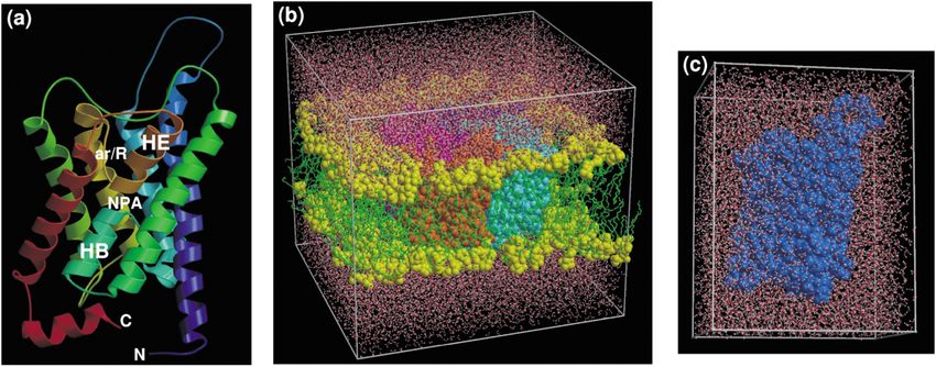
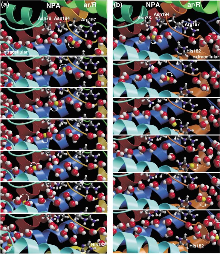
280                                                            The Mechanism of Proton Exclusion in Aquaporins

molecules must be prevented. The question of how          proton blockage in aquaglyceroporins came from
aquaporins prevent proton permeation through              conventional molecular dynamics (MD) simu-
their water-filled pores, although controversially        lations of water permeation through the aqua-
discussed for many years, is still unresolved.            porin-1 and GlpF pores.35,36 We have found that
   Almost 200 years ago, von Grotthuss proposed           water molecules are strongly oriented in the pore,
the idea of “conducting wires” in water.12                and that the hydrogen-bonded water network is
Although he was not aware of the underlying               severely interrupted within the ar/R constriction
mechanism that caused the conductance of water,           region of the pore which is located approximately
the basic concept later proved to be valid, when          10 Å towards the extracellular side from the NPA
further refinement of the mechanism became                region, and have proposed that this interruption
feasible through experiments and quantum chemi-           forms a proton barrier.35 In MD simulations of
cal calculations.10,13,14 In bulk water, protons can      GlpF, strong alignment of water molecules was
efficiently “hop” from one water molecule to the          also observed.35,36 Accordingly, this alignment was
next along a network of hydrogen bonds. After             found to be weakened when the charges on the B
each proton transfer, the changed electrostatic           and E helices were switched off.36 The local
charge distribution forces the involved water mol-        orientational restriction of water molecules in the
ecules to rearrange and thus to optimize the local        NPA region was proposed to play a major role in
hydrogen network, which would suggest that a              the     proton      exclusion     mechanism        in
reduced rotational water mobility hinders proton          aquaglyceroporins.36 Although the details of the
conduction. However, also in other water conduct-         proposed mechanisms differ, they have in common
ing pores, like the model pore gramicidin A,              that an interruption of the hydrogen-bonded water
proton conduction along a chain of water                  chain, either at the ar/R constriction site or at the
molecules has been observed,15 despite consider-          NPA motif, forms the main barrier for protons to
able restriction of the rotation of water molecules.8,9   traverse across the channel.
In carbon nanotubes, an even larger proton                   However, all proton exclusion mechanisms that
hopping rate along one-dimensional water chains           have been proposed so far are based on indirect
than in bulk water has been described.16 Proton-          evidence only, either on detailed analyses of the
conducting water chains also underlie the function        aquaporin structure, or on the behavior of water
of the proton pump bacteriorhodopsin.17 – 20 Simi-        molecules in the aquaporin pore during conven-
larly, proton leakage across biological lipid bilayer     tional force field based MD simulations, which
membranes has been proposed to occur via                  cannot describe intrinsically quantum mechanical
transient water chains.21,22                              transfer reactions of protons. Here, we report a
   In humans alone, more than ten different aqua-         direct approach by analyzing the dynamics of
porins with specialized functionality are known           protons in the pore using both Q-HOP37 molecular
and expressed in tissues as diverse as kidney, red        dynamics simulations, which explicitly describe
blood cells, and the brain. A number of severe            the intrinsically quantum mechanical proton
diseases have been identified that are caused by          transfer processes, as well as conventional force
dysfunctional aquaporins.5,23,24 Not only the water-      field-based MD simulations to study the diffusion
specific aquaporin channels belong to the aqua-           of H3Oþ ions. Finally, because the permeation of a
glyceroporin superfamily, but also glycerol               hydroxyl ðOH2 Þ ion across the membrane in one
facilitators, like the bacterial glycerol facilitator     direction effectively translocates a proton in the
GlpF.25 Structural analyses of human26,27 and             opposite direction, we have also analyzed the
bovine28 aquaporin-1 as well as of the homologous         dynamics of hydroxyl ions in the aquaporin-1 pore.
GlpF,29 provided the first insight into the mechan-
ism of solute permeation through their pores.
Aquaglyceroporins fold into homo-tetramers of             Results and Discussion
independent monomeric channels,30,31 with each
monomer consisting of six transmembrane helices           Behavior of protons, H3O1 and OH2 in the pore
(Figure 1(a)). Two highly conserved loops, each
containing the fingerprint Asn-Pro-Ala (NPA)                 As described in detail in Methods, the dynamic
motif, fold back into the protein and meet in the         behaviour of aquaporin monomers or tetramers
center of the channel.32 These loops leave the            with inserted protons, H3Oþ and OH2 was charac-
channel on either side, each forming a short              terized extensively using MD simulations. We first
a-helix33 (helices B and E). The positive sides of        present the main findings of the simulations and
the macro-dipoles associated with these short             their biological relevance.
helices34 meet in the center of the channel. At this         Figure 2 shows the motion of protons along the
position, the side-chains of both asparagine residues     pore direction in Q-HOP MD simulations that
of the NPA motifs have been proposed to isolate a         started from configurations with protons at differ-
passing water molecule from its neighbors by              ent positions in the pore. As can be seen, protons
exclusively forming hydrogen bonds to the water           initially placed near the center of the pore in the
oxygen, thereby putatively impeding the hopping           vicinity of the NPA region quickly start leaving
of a proton to and from this water molecule.26            the pore in either the extracellular (upwards) or
   Other indirect evidence on the mechanism of            intracellular (downwards) direction. Towards the
The Mechanism of Proton Exclusion in Aquaporins                                                                 281

  Figure 1. (a) The monomeric structure of bovine AQP1, showing the six-transmembrane helices and the short helices
formed by loops B and E (HB and HE) that both contain the fingerprint NPA motif, which meet in the center of the
pore. The ar/R constriction region is located 10 Å extracellular from the NPA region. The simulation systems for
the conventional MD simulations, in which an AQP1 tetramer was embedded in a solvated POPE bilayer, and for the
Q-HOP simulations, in which an AQP1 monomer in water was simulated are shown in (b) and (c), respectively.

intracellular side, protons reach the channel entry/        nel (NPA) towards the extracellular side. No hops
exit rather efficiently, whereas those that travel          were observed that resulted in a translocation of a
towards the extracellular side typically get stuck          proton from the extracellular side of the channel
for some time at the ar/R constriction region               towards the NPA region. These findings clearly
before they leave the channel (see also Figure 3).          indicate a strong barrier for proton conduction,
Eventually, all protons that started inside the pore        located near the NPA region, as well as a smaller
leave the channel on either side. Few protons               one located near the ar/R constriction region.
cross the NPA region during passage; most leave                Conventional      MD     simulations   including
the NPA region at the same side at which they               excess H3Oþ phenomenologically show the same
were placed initially. All crossings of the ar/R            behavior as the proton hopping Q-HOP simu-
constriction region are from the center of the chan-        lations, albeit at a much lower rate (Figure 4, top).

   Figure 2. Motion of individual protons along the pore axis during all Q-HOP MD simulations (left). The protons’
starting positions for the 48 simulations are marked at the left (horizontal line sections). To facilitate structural
interpretation of the pore axis coordinate, the bAQP1 X-ray structure is shown, together with all proton positions at
which a hop was observed (right).
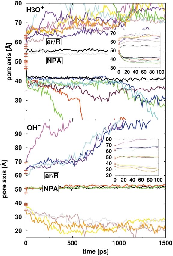
282                                                              The Mechanism of Proton Exclusion in Aquaporins

 Figure 3. Snapshots of Q-HOP simulations during hop events along a typical path of a proton on its way out of the
AQP1 pore towards (a) the intracellular side and (b) the extracellular side.

Whereas in the Q-HOP simulations, protons typi-            (bottom panel) shows that excess OH2 display a
cally leave the pore within 1 ps, H3Oþ take about          behavior nearly opposite to that of the positive
20 ps. Despite this difference in time-scales, the         charges. In particular, those OH2 that started from
barrier seems to be capable of blocking both proton        positions near the NPA region are attracted
transfer reaction and diffusion of H3Oþ (to dis-           towards the NPA region and are trapped there.
criminate between these two cases, we will sub-            However, those OH2 that were started farther
sequently use the term proton to refer to the first,       away from the center of the pore (which is the
and H3Oþ or hydronium ion to refer to the latter           physiologically relevant situation) either avoid the
proton motion process). However, although proton           pore region or are expelled from the pore just like
hops across the ar/R constriction region are               protons. This observation suggests secondary
observed (Figure 2), no diffusion of H3Oþ across           barriers for hydroxide ions at both sides of the
this barrier is seen (Figure 4, top), an observation       main NPA proton barrier.
that also has been made for the M2 channel of the             Using the maximum likelihood approach
influenza A virus.38                                       described in the Appendix, we constructed an
   One must expect that the proton barrier acts as a       effective free energy profile for the proton as a
sink for the oppositely charged hydroxide ðOH2 Þ           function of the pore axis from the collection of
ions, which therefore would block water per-               non-equilibrium Q-HOP simulations with an
meation through the pore. Indeed, Figure 4                 excess proton. The obtained profile (Figure 5)
The Mechanism of Proton Exclusion in Aquaporins                                                              283

                                                           the pore center. As can also be seen in Figure 5,
                                                           the non-equilibrium distributions of both the pro-
                                                           tons that were allowed to undergo quantum mech-
                                                           anical proton transfer reactions, as well as those
                                                           described by diffusion of H3Oþ, qualitatively
                                                           match the free energy profile. Note that a similar
                                                           qualitative match is seen for the OH2 between
                                                           (the negated) free energy profile and their non-
                                                           equilibrium distribution.
                                                              Near the free energy barrier at the NPA region,
                                                           relatively low proton and H3Oþ densities are
                                                           observed whereas relatively high densities are
                                                           observed in the regions with lower free energy
                                                           (around 40 Å and 70 Å, respectively). The density
                                                           profile for hydroxide ions is largely comple-
                                                           mentary to that of protons and H3Oþ. Highest
                                                           hydroxide densities are observed near the NPA
                                                           region and near the intracellular face of the pore,
                                                           whereas relatively low densities are observed near
                                                           the proton free energy minima near 40 Å and
                                                           70 Å. Note that the high proton and H3Oþ densities
                                                           near the ar/R constriction region are non-equi-
                                                           librium effects, reflecting protons and H3Oþ that
                                                           started in the pore near the NPA region and got
                                                           stuck in the ar/R region before being able to leave
                                                           the channel on the extracellular side. The equi-
                                                           librium density of protons and H3Oþ can therefore
                                                           be expected to be much lower in this region,
                                                           which is consistent with the free energy profile,
                                                           where the minimum just intracellular from the ar/
   Figure 4. Diffusion of H3Oþ (top) and OH2 (bottom) as   R region is only a secondary one.
observed in conventional molecular dynamics simu-             The proton minima near 40 Å and 70 Å are only
lations. The initial positions are marked in red.
                                                           transiently occupied by protons and H3Oþ. These
                                                           wells are too shallow to trap a proton (or H3Oþ)
                                                           tightly and, as can be seen from the longer conven-
shows a clear maximum near the NPA region, as              tional MD simulations (Figure 4, top), H3Oþ indeed
was expected from the observed motion of protons           leave these wells on both sides at intermediate
within the pore. Also the rest of the profile matches      timescales. Note that, because of the relatively low
the observed qualitative behavior of protons in the        bulk proton concentration at physiological pH,
pore. Closer inspection reveals a secondary barrier        there is an entropic cost due to the confinement of
near the ar/R constriction region and two minima           the proton’s available configurational volume to
at both the intracellular and extracellular sides of       part of the aquaporin pore as compared to the

                                                                                 Figure 5. Maximum likelihood
                                                                              free energy profile for protons
                                                                              along the pore axis in Q-HOP simu-
                                                                              lations (black curve). For compari-
                                                                              son, the relative non-equilibrium
                                                                              distributions of protons (red), H3Oþ
                                                                              ions (blue), and OH2 (green) are
                                                                              shown.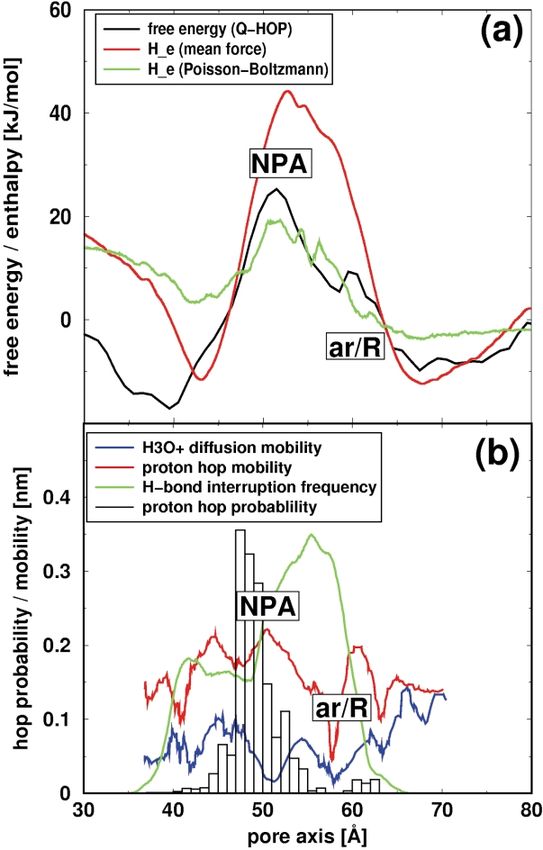
284                                                              The Mechanism of Proton Exclusion in Aquaporins

mean volume available in bulk water, which                  involving a local isolation of the water oxygen
explains why protons are not trapped within these           atom by both asparagine residues of the NPA
minima.                                                     motifs, which would thus prevent a proton from
                                                            hopping onto this water molecule,26 now seems
Structural determinants of proton exclusion                 unlikely, since no such exclusive isolation of water
                                                            molecules from other water molecules was
  The location of the main barrier for protons and          observed in our simulations. Moreover, a recent
H3Oþ near the NPA region is somewhat                        refinement of this mechanism,36 involving an
unexpected, because the interruption of the hydro-          additional barrier caused by the putative reorienta-
gen bonded water network inside the pore, which             tion step of the Grotthuss mechanism against the
was generally assumed to form the main barrier              local electrostatic field also seems unlikely, since
for protons, is most pronounced within the ar/R             we actually more frequently observed proton hops
constriction region35 (see also Figure 6(b)). Also a        in this region than in others (cf. Figure 6(b)). Even
previously proposed alternative mechanism                   a number of hop events across the NPA region are
                                                            observed.
                                                               This finding puts even more relevance on the
                                                            question of what are the underlying structural and
                                                            energetic determinants that cause proton exclusion
                                                            and the main proton barrier in the NPA region as
                                                            well as the secondary barrier in the ar/R constric-
                                                            tion region. In particular, one would like to know
                                                            to what extent the barrier is electrostatic in nature,
                                                            as opposed to being caused by interruption of
                                                            hydrogen-bonded water chains.
                                                               A comparison of the proton free energy profile to
                                                            an estimate for the mean electrostatic potential in
                                                            the pore (Figure 6(a)) shows a qualitative corre-
                                                            spondence between the two profiles. In particular,
                                                            the location of the main maximum agrees very
                                                            well, as does the presence of the two minima. Not
                                                            seen in the mean electrostatic potential profile is
                                                            the secondary maximum, and also the location of
                                                            the two minima appears to be somewhat shifted.
                                                            This qualitative agreement suggests that the main
                                                            determinants for the proton exclusion mechanism
                                                            in aquaporins are electrostatic interactions. The
                                                            most prominent electrostatic features of the pore
                                                            are the macro-dipoles of the B and E loop helices
                                                            formed by their positive N-terminal ends, which
                                                            meet in the NPA region in the central part of the
                                                            pore. These results suggest that the strong electric
                                                            field blocks proton permeation not indirectly by
                                                            perturbation of the Grotthuss mechanism, but
                                                            rather mainly directly by creating an electrostatic
                                                            barrier for protons as well as for H3Oþ and other
                                                            cations.
                                                               As can be seen in Figure 6(a), the electrostatic
                                                            potential estimate monotonously decreases from
   Figure 6. Determinants of proton exclusion. (a) The      the NPA region towards the extracellular side of
effective electrostatic potential for a proton inside the   the channel, and can therefore not explain the
pore, based on the mean force on a probe charge (red)       presence of the secondary maximum in the proton
together with the maximum likelihood proton free            free energy profile near the ar/R constriction
energy profile (black), and the continuum Poisson –         region. The contiguous decrease of the electrostatic
Boltzmann electrostatic profile (green). (b) The role of    potential within the ar/R region is unexpected,
hydrogen bond intactness between water molecules on         because the side-chain of a positively charged
the hopping efficiency in Q-HOP simulations. Shown          arginine residue (Arg197 for bAQP1, Arg195 for
are hop probabilities (for a Q-HOP to a neighboring         human AQP1) faces the pore in this region. Inter-
water molecule) as a function of the pore axis (black)
                                                            estingly, this is the region where a frequent inter-
and the distances traveled along the pore direction due
to proton hopping events (red) and H3Oþ diffusion           ruption of the hydrogen-bonded water network
(blue). For comparison, the frequency of interruption of    was previously observed which was predicted to
contiguous hydrogen bonded water chains as observed         disfavor proton hopping events between water
in a conventional MD simulation of human AQP135 is          molecules.35 Indeed, when the hop probability to
shown in green.                                             neighboring water molecules is calculated as a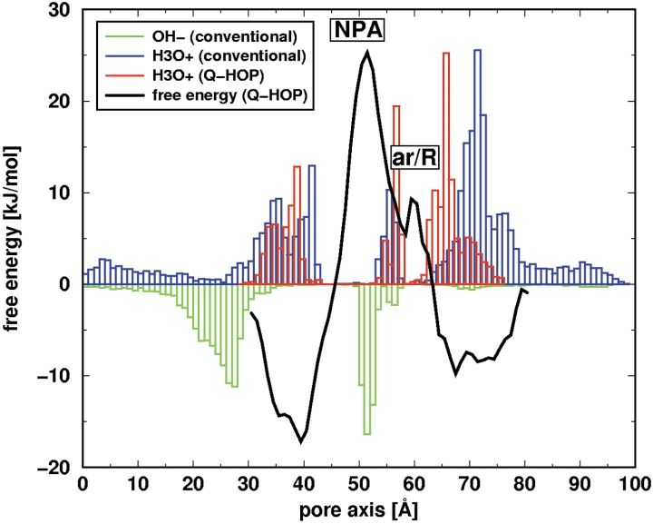
The Mechanism of Proton Exclusion in Aquaporins                                                           285

function of the pore position, the ar/R constriction    (Figure 5). Is this barrier high enough to prevent
region displays an extremely low hop probability        protons from leaking into the cell, which would
of less than 0.01%, whereas near the NPA region,        destroy the electro-chemical gradient across the
values up to 30% are observed (cf. Figure 6(b)).        membrane? In this respect, it should be noted that
Moreover, 15 of the 20 proton trajectories              the experimentally determined proton perme-
that ended at the extracellular side, hopped via        abilities of phospholipid bilayers39 – 42 range from
His182 in the ar/R region due to the lack of inter-     1 £ 1025 to 5 £ 1024 cm s21 ; and thus are only
water hydrogen bonds in this region (see also           slightly lower than permeability values typically
Figure 3). These observations strongly suggest          reported for water.43,44 Since the estimated barrier
the secondary, local maximum in the ar/R con-           height for protons through the AQP1 pore of
striction region to be caused by disruption of          approximately 25 – 30 kJ mol21 is very close to the
the hydrogen bond network between water                 barrier height of typical membranes for water,45
molecules.                                              the permeability of AQP1 for protons can be
   Additionally, the ar/R constriction region also      expected to be in the same range as the water and
forms a barrier for H3Oþ (Figure 4, top panel and       proton permeabilities of lipid membranes.
Figure 6(b)). An analysis of hydrogen bond              Apparently, aquaporins, or any other membrane
enthalpies of water molecules in the pore showed        protein, need not have a lower proton permeability
a maximum in the hydrogen bond energy among             than the intrinsic membrane permeability for
water molecules in this region.35 This indicates        protons.
that also the H3Oþ barrier in this region is hydro-        Assuming a pH gradient of 0.75 pH unit (the
gen-bond mediated.                                      typical proton gradient across the mitochondrial
   Note that the hop probabilities shown in             membrane during respiring conditions) and a
Figure 6(b) not only result from the local intactness   membrane potential of 170 mV, the free energy
of the hydrogen bonded water chain, but may also        required to pump a proton across the membrane
be affected by an uneven distribution of reorienta-     (or that is released by proton diffusion in the
tion energies connected to the turn of water            opposite direction) is approximately 20 kJ mol21,
molecules that follows a hop in the Grotthuss12 or      which in a simple approximation would corre-
“hop-and-turn” mechanism11 to optimally align           spond to a shift of þ 10 kJ mol21 on one side and
their dipoles with the new charge distribution.         of 2 10 kJ mol21 on the other side of the membrane
Unfavorable alignment of water dipoles with the         in the bulk regions of the free energy profile
macro-dipoles of the B and E helices in the NPA         (Figure 5). Adequate blocking of protons, therefore,
region during such reorientation events had pre-        can be expected also in this situation.
viously been proposed to form the main proton              From an evolutionary viewpoint it is interesting
exclusion barrier in aquaporins.36 The fact that        to note that, apparently, the proton barrier is not
hop events are frequently observed in the NPA           larger than absolutely necessary. This suggests
region suggests, however, that the reorientation        that a compromise between efficient water chan-
barriers exist in this region are generally small       nels and proton filters has been achieved during
and, therefore, are not a main determinant for          evolution, resulting from a tradeoff between, on
proton blockage.                                        one hand, the optimization of the water per-
   Our prediction that electrostatic effects dominate   meability and, on the other hand, the blocking of
the mechanism of proton exclusion could be              protons and other ions.
tested by a number of experimental techniques.             Errors in the barrier height, as estimated from
First, point mutations near the NPA region that         the effective maximum-likelihood free energy pro-
would reduce the positive electrostatic potential       file (Figure 5, see also Appendix), might arise
locally are expected to reduce the barrier for          from limited statistics (number of hops near the
protons to pass the pore, e.g. be Phe24Asp or           top of the barrier) and from the assumption that
Phe24Glu. Phe24 is located across the channel           the observed hop rates are proportional to the
from both Asn residues from the NPA motif.              Boltzmann factor of the free energy difference
A negatively charged residue at this location can       between adjacent slices in the pore. The latter rests
be expected to reduce the local positive electro-       on the assumption that the proton’s vicinity is
static potential caused by the macro-dipoles of the     close to equilibrium at all times, as is also generally
B and E helices. Second, with a voltage clamp           assumed in Kramers’ theory. However, although
experiment it might be possible to measure the          we do expect fast equilibration to occur, it may in
voltage dependence of proton conduction across a        some instances not be fast enough in light of the
membrane with embedded aquaporins, in                   partially fast motion of the proton. In that sense,
comparison to pure lipid membranes, which               the obtained energy profile should not be inter-
could yield information on the electrostatic barrier    preted as an equilibrium free energy, but rather as
width.                                                  one that is specifically adapted to the time-scale
                                                        set by the motion of the proton. Since this motion,
The barrier height                                      as the process of interest, dictates the relevant
                                                        time-scale, we feel that our non-equilibrium
  The estimated barrier height for protons through      approach is more appropriate than a full equi-
the AQP1 pore is approximately 25 –30 kJ mol21          librium treatment would be.
286 The Mechanism of Proton Exclusion in Aquaporins
Hydroxide exclusion (and the derived proton free energy profile) are
affected not only by the classical free energy deter-
Like protons, OH2 also must be excluded from minants (predominantly electrostatics and entropic
the pores of aquaglyceroporins, since permeation effects), but also by additional factors that specifi-
of hydroxide ions in one direction would be equiv- cally affect proton transfer, most notably the
alent to the leakage of a proton in the opposite intactness of a hydrogen-bonded chain. Indeed,
direction. Furthermore, the proton main electro- although the proton free energy profile is dominated
static barrier at the NPA region is a deep well for by electrostatic effects (Figure 6(a)), the secondary
OH2 or other anions, and accumulation of OH2 proton barrier in the ar/R constriction region is
within the well would block water permeation. It found to be caused by a frequent interruption of the
is thus of particular interest to investigate how the hydrogen-bonded chain (Figure 6(b)).
aquaporin prevents OH2 or other negatively To further decompose the proton free energy
charged ions from being attracted to the NPA profile into individual components, the next step
region of the pore. Indeed, when placed near the is to estimate the electrostatic contribution. The
center of the pore, hydroxide ions are indeed determination of the electrostatic potential that
attracted towards the NPA region of the pore protons (in the form of H3Oþ) experience on their
(Figure 4, bottom). When placed farther away way across the pore, however, is complicated by
from the central part of the pore, however, OH2 the fact that the electrostatic component of the
are no longer attracted by the NPA region but, force (Figure 7(b), red curve) is, on average, an
instead, are expelled from the pore. order of magnitude larger than the total net force
Figure 5 also suggests that the mechanism of that these ions experience (blue curve and green
hydroxide exclusion is mainly electrostatic in circles). Closer inspection showed that this effect
nature, caused by two barriers on both sides of is caused mainly by strong short-range electrostatic
the NPA region (near 30 Å and 70 Å, respectively), interactions (typically hydrogen bonds) that are
which appear as local minima in the proton free compensated largely by repulsive Pauli inter-
energy and electrostatic profile (Figure 6(a)). Note, actions. Therefore, the net force on an ion in the
however, that the free energy profile for hydroxide pore is typically much smaller than the electro-
ions is not necessarily the negated proton free static component of the force, even in the case
energy profile, as the mechanisms for proton and when the underlying potential was mainly electro-
hydroxide conduction are very different.46 How- static in nature. This situation makes it problematic
ever, if the OH2 barrier is dominated by electro- to calculate the effective electrostatic potential pro-
statics as is the proton barrier, then the (negated) file across the pore. However, one may assume
proton free energy profile can be interpreted as an that this short-range compensation varies only
approximate free energy profile for hydroxide slightly in strength along the pore (as will be ana-
ions. In this case, one would estimate an OH2 bar- lyzed and justified further below). In this case, the
rier height of approximately 10 kJ mol21, which at net force acting on an (H3Oþ) ion will indeed
first sight seems too low to prevent hydroxide allow us to estimate the effective electrostatic
ions from getting trapped in the NPA region of potential felt by the proton.
the pore. Note, however, that there is an entropic This mean force acting on H3Oþ was determined
cost connected to moving from bulk into the in a number of ways. The method of choice would
narrow channel, just like for protons, as described be to calculate a true PMF over a sufficient number
above, which leads to the observed exclusion of of constrained or umbrella positions of the ion in
OH2 from the pore (Figure 4, bottom). the pore. Because of the observed slow conver-
gence, however, we used the calculations using
Control calculations probe charges at suitable water positions instead,
which requires a careful estimate of the error
We have carried out a number of additional associated with it (see also Methods). The obvious
calculations. First, to be able to compare the way to do this is to select a subset for comparison
purely diffusive motion of H3Oþ and OH2, as with the computationally expensive method of
described by conventional MD simulations with choice. To this end, umbrella sampling simulations
the Q-HOP MD simulations. Second, to be able to were started in which H3Oþ were kept fixed with
estimate the amount of statistical error in each of a harmonic potential at 20 different positions
the presented energy profiles as well as their along the pore (see Methods). As can be seen in
reproducibility. Figure 7(b), the mean umbrella forces correlate
As can be seen from the non-equilibrium well with the mean electrostatic force profile. Note
trajectories (Figures 2 and 4) as well as from the that despite simulation times of up to 5 ns for
distributions of protons, H3Oþ, and OH2 positions each single umbrella sampling simulation, these
along the pore (Figure 5), there is good qualitative forces scatter considerably, which underscores the
agreement between the Q-HOP and conventional indeed slow convergence that made the direct
MD simulations, albeit that the proton kinetics is approach untractable. The slow convergence is
much faster in the Q-HOP simulations due to the mainly due to the slow equilibration of neighbor-
ability of protons to be involved in proton transfer ing water molecules and protein side-chains in the
reactions. Because of this, the Q-HOP simulations pore.The Mechanism of Proton Exclusion in Aquaporins 287
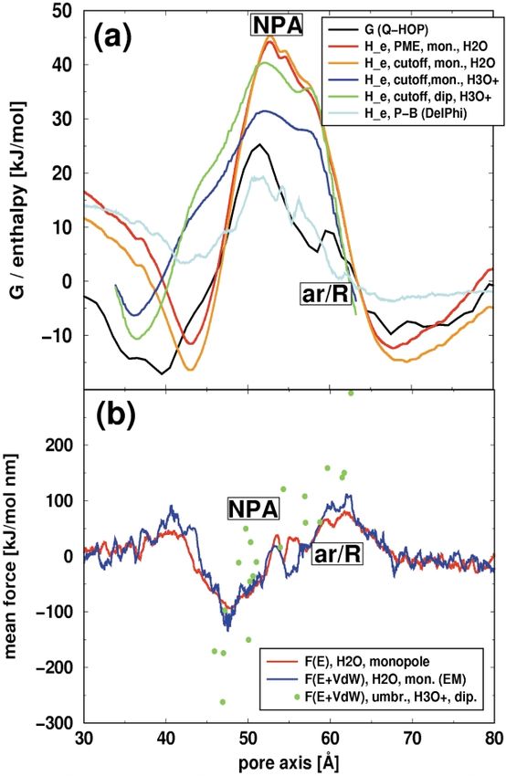
In full agreement with the forces obtained for the
probe point charge (Figure 7(b)) the electrostatic
component of the force acting on the H3Oþ in the
umbrella simulations is larger by a factor of about
12 than the total force acting on the H3Oþ, as
obtained from a least-squares fit between the two
quantities. A similar ratio was obtained when the
force profile from the water positions was recalcu-
lated not only including the electrostatic, but also
the Lennard –Jones components (Figure 7(b)), after
energy minimization. All electrostatics profiles
that were solely calculated based on the electro-
static component of the mean force were therefore
scaled (see also Methods).
As discussed above, the electrostatics profile as
determined by integrating the mean force on a
probe charge on water positions is generally
expected to deviate from a PMF. Although for
AQP1 the above control calculations suggest that
these deviations are relatively small, a number of
specific potential sources of deviation deserve par-
ticular attention: (a) the fact that water positions
instead of true proton positions are taken as probe
positions; (b) the approximation of the H3Oþ
charge by a point charge (whereas H3Oþ is a
dipole); and (c) the lack of relaxation (most notably
the alignment of neighboring dipoles) of the sur-
roundings due to the presence of a positive charge
in the pore.
To check to what extent the calculated profile is
affected by the chosen set of positions as well as
Figure 7. Control calculations. (a) The maximum likeli- by the pore environment, we have compared the
hood proton free energy profile (black) together with the profile obtained with water positions with a profile
electrostatic potential profiles obtained with the different obtained from snapshots of the Q-HOP simu-
methods described in the text. The red and orange lations, i.e. including true H3Oþ positions
curves show the electrostatic potential calculated from (Figure 7). Although the statistics for the latter is
the mean force on a positive unit charge, determined at worse than for the former (493 versus 17,255 pos-
different water positions along the pore, using PME and itions, respectively), the profiles are qualitatively
a cut-off of 1.2 nm, respectively. Electrostatic profiles similar, especially near the main NPA barrier. The
calculated from the mean force at actual H3Oþ positions only significant deviations are seen near the first
taken from Q-HOP simulations are shown in blue and
green, respectively. The blue curve was determined, like minimum at 40 Å. Interestingly, this minimum is
the red and orange curves, with a positive probe unit shifted by about 6 Å to the intracellular side with
charge on the oxygen position, whereas the green curve respect to the profile calculated from the water
is based on the actual dipole charges of the H3Oþ. The positions, and now better agrees with the mini-
cyan curve shows a continuum Poisson – Boltzmann mum in the free energy profile obtained from the
electrostatic profile calculated with DelPhi,47 averaged Q-HOP simulations. This suggests that the paths
over multiple MD snapshots, using water oxygen taken by protons and water molecules are
positions from passing water molecules as probe relatively similar to each other, except for the ar/R
positions. (b) Mean force profiles as a function of the constriction region, where the proton was found
pore axis. Shown (in red) is the mean electrostatic force
to hop across His182 in 15 of the 20 cases where
on a positive probe unit charge on the oxygen position
of water molecules that passed the pore in an equi- the proton left the pore on the extracellular side.
librium MD simulation (integration of this curve yields Moreover, it shows that the electrostatic field inside
the red curve in (a)), and (in blue) the total non-bonded the pore, that causes water molecules to strongly
(electrostatic and Lennard– Jones) force (after energy- align inside the pore, is caused mainly by the
minimization) at the same positions. The red curve was protein, and is perturbed only weakly by an excess
scaled (by 0.127) to fit the blue curve. The green points positive charge. Additionally, this result shows that
depict mean forces on H3Oþ in the pore direction the neglect of a more physiological protein
obtained from multi-nanosecond umbrella sampling environment (tetramer, membrane), as well as the
simulations during which the oxygen atom of the H3Oþ applied position restraints on Ca atoms in the
was fixed by an umbrella potential along the z direction
Q-HOP simulations does not cause significant
at 20 different positions along the pore.
artefacts. This is also true for the use of a cut-off
(12 Å radius) in the Q-HOP simulations, which, as
shown in in Figure 7(a), yields energy profiles that288 The Mechanism of Proton Exclusion in Aquaporins
are similar to those calculated with full electro- of electrostatic barriers and entropic effects, which
statics (particle-mesh Ewald, PME). is essential not only for preventing the dissipation
We note that the Q-HOP method in its current of the electrochenical gradient, but also for the
implementation cannot explicitly account for con- function of water channel because it avoids
certed proton hops, and currently only hops to hydroxide ions from being trapped within the
water and to histidine are implemented. Although positively charged NPA region, which would
we do not expect these limitations to significantly otherwise block the channel.
affect our results (see also Methods), we note that
if these features were implemented, this would
lead to an enhancement of the proton transfer
rates, and a corresponding lowering of the Methods
hydrogen bond-mediated proton transfer barriers,
rendering the role of electrostatics even more Conventional MD simulations
pronounced.
We compared the electrostatic energy of a mono- Conventional MD simulations with excess H3Oþ and
pole (like the probe charge used to obtain the pro- OH2 were carried out with the gromacs†,48 simulation
file) to the energy of a dipole (like H3Oþ) by software. The simulation set-up and conditions were
calculating the mean force on H3Oþ positions in similar to those described before.35 As a starting confor-
mation, the bovine aquaporin-1 (bAQP1) X-ray
Q-HOP simulations both as a dipole (Figure 7(a),
structure28 (Figure 1(a)) was placed as a tetramer cen-
blue curve) and as a monopole (like for probe trally into a palmitoyloleoylphosphatidylethanolamine
charges at water positions, Figure 7(a), green (POPE) lipid bilayer patch of 272 lipids, and was sol-
curve). The fact that the two profiles are rather vated on both sides with 19,442 SPC water molecules49
similar indicates that the monopole dominates the in total (cf. Figure 1(b)). The gromos8750 force-field with
electrostatic profile for a H3Oþ. modifications51 and explicit polar and aromatic hydrogen
An independent estimate for the electrostatic atoms was used. Lipid parameters were taken from
potential in the pore was calculated using a Berger et al.:52 12 chloride ions were added to the peri-
continuum Poisson– Boltzmann approach, as odic simulation box to neutralize the net positive charge
implemented in the DelPhi program47 (see of the protein, rendering the total system size 81,814
Methods). The potential was calculated by atoms. After energy minimization, a short simulation
averaging over a large number of MD snapshots with position restraints of 1000 kJ mol21 nm22 on the
of the AQP1 tetrameric structure, by evaluating non-hydrogen protein atoms was carried out to relax
the lipids and water molecules around the protein. Sub-
the potential at the same water oxygen positions
sequently, the system was allowed to equilibrate for 2 ns
that were used above for calculating the other elec- before H3Oþ and OH2 were added. Additionally, the
trostatic potential profiles. The agreement with the simulation without excess H3Oþ and OH2 was extended
other electrostatic profiles (Figures 6(a) and 7(a)) to a total time of 10 ns. During these simulations, the
underscores the role of electrostatics for the temperature was kept constant by weakly ðt ¼ 0:1 psÞ
mechanism of proton exclusion. coupling the protein, lipids, and solvent separately to a
temperature bath53 of 300 K. Likewise, the pressure was
kept constant by weakly coupling the system to a
Conclusions pressure bath of 1 bar. The xy (membrane plane) and z
(membrane normal) directions, respectively, were separ-
Our results from a combination of Q-HOP simu- ately coupled with a coupling constant t of 1 ps. Electro-
lations, which explicitly describe proton transfer static interactions were calculated with the PME
method.54 Pauli and van der Waals interactions were
reactions, and multinanosecond conventional
described with a Lennard–Jones potential, which was
molecular dynamics simulations, strongly indicate cut off at 1.2 nm. The Settle55 algorithm was used to
that proton exclusion from the aquaporin pores is constrain the bond lengths and angles of the water
predominantly achieved by a strong electrostatic molecules, and lincs56 was used to constrain all other
barrier. The main barrier of approximately 25 – bond lengths, allowing a time-step of 2 fs. Four simu-
30 kJ mol21 is predicted to be located at the center lations with four H3Oþ each (one per pore), as well as
of the pore, near the NPA fingerprint region. Here, two simulations with four and eight OH2, respectively,
the positive N-terminal ends of the macro-dipoles were carried out. Each of these simulations had a length
of helices B and E meet, which are the main deter- of 1.5 ns. The charges of H3Oþ were taken from earlier
minants of the electrostatic barrier. Interruption of work,37 obtained by a restrained ESP-fit using NWChem.
the hydrogen-bonded chain of water molecules The oxygen charge is 2 0.749 and the hydrogen charges
through the pore causes a secondary barrier are þ 0.583. Likewise, the charges of OH2 were taken
located at the ar/R constriction region. The calcu- from earlier work,57 where their magnitude was adapted
according to the average electrostatic potential field felt
lated barrier height for protons is approximately
in the protein to match the quantum mechanical dipole
as high as in typical lipid bilayers, and suffices to moment of an OH2 ion in an electrostatic field field of
prevent leakage of the electrochemical gradient equal size.58 The oxygen charge is 2 1.3 and the proton
across the membrane. The aquaporin channel not carries a charge of þ0.3.
only efficiently blocks protons and hydronium
ions, but also negatively charged hydroxide ions.
This is proposed to be achieved by a combination † http://www.gromacs.orgThe Mechanism of Proton Exclusion in Aquaporins 289
Q-HOP MD simulations possible to include transfers to other protein residues
once the corresponding parameterizations are derived
To explicitly simulate quantum mechanical and (work in progress), for the present study, proton hops to
thermally activated proton transfer reactions between and from amino acid residues other than histidine are
water molecules inside the pore, Q-HOP simulations37 not implemented. Note that both approximations may
were carried out with a modified version of the ARGOS lead to a slight overestimation of the proton transfer
simulation package.59 For the Q-HOP simulations, the free energy barriers derived from Q-HOP MD. Coordi-
bAQP1 X-ray structure28 was solvated in a rectangular nates of the protonated water molecule were recorded
periodic simulation box containing 6303 SPC water every MD step for subsequent analysis.
molecules;49 this simulation system comprised a total of
22,678 atoms (Figure 1(c)). After energy minimization, Electrostatic potential calculations
an equilibration simulation of 250 ps was carried out,
from which snapshots were collected every 20 ps. From
The electrostatic potential along the pore axis was esti-
each of these snapshots, four different water molecules
mated using three different approaches. First, the mean
in the pore region were selected for protonation. Each of
electrostatic force on a probe charge at different positions
the obtained 48 configurations was energy minimized
of water molecules inside the pore during MD simu-
and then equilibrated for 1 ps. During that period, the
lations was calculated. Second, the electrostatic and total
oxygen atom of the protonated H3Oþ was kept fixed
mean force on H3Oþ at different positions in the pore
using a harmonic potential with a force constant of
during umbrella sampling simulations was determined.
1000 kJ mol21 nm22. From each of the obtained configur-
Finally, the electrostatic potential was estimated by a
ations, a Q-HOP simulation with a length of 100 ps was
continuum Poisson –Boltzmann approach.47
carried out. To prevent structural rearrangements due to
the non-physiological environment of the protein (i.e. a
monomer solvated in water instead of a tetramer
The average electrostatic field at water positions
embedded in a lipid bilayer), all Ca positions of the pro-
tein were position-restrained with a force constant of The electrostatic force on a positive probe unit charge
1000 kJ mol21 nm22. In all ARGOS simulations, the was evaluated at the positions of water molecules
Amber95 force-field60 was used. All non-bonded inter- passing the pore during the 10 ns equilibrium MD simu-
actions (electrostatic and Lennard– Jones) were cut off at lation of the bAQP1 tetramer embedded in a solvated
1.2 nm. Temperature and pressure were kept constant POPE bilayer described above. In total, 17,255 water mol-
by coupling to an external bath53 of 300 K and 1 bar ecule positions from 1000 MD snapshots were selected
with coupling constants of 0.4 and 0.5 ps, respectively. such that a homogeneous distribution of positions along
The SHAKE61 algorithm was used to constrain bond the pore axis was obtained. For each of the positions,
lengths, allowing a time-step of 2 fs. the selected water molecule was replaced by a positive
The Q-HOP molecular dynamics procedure was probe unit charge located at the dipole center of the
employed as described.37,62 – 64 In this method, stochastic water molecule (i.e. the midpoint between the oxygen
proton hopping events are included in otherwise atom and the midpoint between the two hydrogen
standard molecular dynamics simulations. Previous atoms). For each of the obtained configurations (with an
applications successfully addressed the diffusion of an excess positive probe charge) the electrostatic component
excess proton in water,37 the protonation equilibrium of of the force on the probe charge was evaluated. For com-
Asp in a solvent box,37 and the three-step proton relay parison, the long-range contribution of this force was
in green fluorescent protein.57 Energy barriers for proton calculated both with a cut-off approach (using a cut-off
transfer were carefully parameterized against quantum radius of 1.2 nm) and using the PME method. In the
chemical calculations on donor – acceptor model systems case of the PME calculation, in addition to the excess
as simple functions of the donor – acceptor distance and positive probe charge, a complementary negative charge
of the relative energy difference between the donor- was added in the bulk water region, to ensure electro-
bound and the acceptor-bound states.64 Transition state static neutrality. The mean component of the force in the
theory is then employed to compute transfer prob- pore direction (z-axis) was evaluated as a function of
abilities over large barriers, and pre-calculated the probe axis, using a Gaussian filter with a width of
transmission coefficients from wave-packet dynamics 0.1 nm. This mean force was then integrated to obtain
calculations are applied for the transfer over small an estimated electrostatic potential profile. Note that
barriers.63 We want to stress that transfers in the regime this averaged potential does not include the reaction
of larger energy barriers are very unlikely during typical field that would actually be caused by a real charge,
MD simulations of 100 ps length. In this work, transfers and therefore is expected to overestimate the true elec-
were only observed at donor – acceptor distances closer trostatic potential. Yet, assuming that the polarization
than 2.7 Å. The efficiency of the method results from effect of the surrounding is of similar size along the
the fact that the parameterizations have been pre- channel, this estimate will provide a qualitative picture.
computed and can almost instantaneously be evaluated To capture the main part of the reaction field, a more
during the MD simulation for any given donor – acceptor expensive calculation of the electrostatic profile was also
configuration, properly taking into account electrostatic carried out. Here, rearrangements of the surroundings
stabilization by the environment. Proton hopping is as a response to the introduced particle were considered
allowed every 10 fs, which is the approximate time by energy-minimization of each of the configurations
required for a quantum wave-packet to cross a small using a steepest descent algorithm (100 steps each) prior
energy barrier. Although concerted proton transfer is to the force evaluation. This required including also
not currently implemented in the Q-HOP algorithm, the Lennard – Jones interactions with the probe charge,
diffusion rate of an excess proton in bulk water could using the Lennard – Jones parameters of a water oxygen
be well reproduced.37 In this work, proton transfer is atom. A least-squares fit of the profiles with and without
only allowed between water molecules as well as to the the Lennard–Jones contribution shows that the shapes of
amino acid histidine. Although it would be technically the force profiles are very similar (Figure 7) but, as was290 The Mechanism of Proton Exclusion in Aquaporins
expected, the (inexpensive) electrostatics-only profile obtained by averaging over 1000 snapshot profiles. The
overestimates the electrostatic contribution by a factor dielectric constants were chosen to be 80 and 4, for
of approximately 7.9. Therefore, all force profiles in protein exterior and interior, respectively.
which only the electrostatic component of the non- The molecular graphics in Figures 1 – 3 were created
bonded force was evaluated were subsequently scaled with bobscript65,66 and Raster3D.67
with this factor.
A further correction of the obtained electrostatic
potential profile was necessary because the inserted
probe charges were described as monopoles, whereas
the particle of interest, H3Oþ, has a dipole. Moreover,
the selected probe positions were positions of water
Acknowledgements
molecules that spontaneously passed the pore in an equi-
librium MD simulation. It is conceivable that a passing
B.L.dG. was supported by the BIOTECH
H3Oþ or proton would take a different path, on average, program of the EU, grants QLRT 2000/00778 and
than a water molecule. To estimate the size of these two 2000/00504. We thank Tjerk Straatsma for per-
effects, the electrostatic potential was also calculated for mission to use the ARGOS program, and Markus
H3Oþ positions in snapshots from Q-HOP simulations, Lill for technical support with ARGOS.
both treated as monopoles and dipoles (see Figure 7).
Umbrella sampling simulations References
Note that the methods described above do not yield a 1. Mitchell, P. (1961). Coupling of phosphorylation to
potential of mean force (PMF) in the strict statistical electron and hydrogen transfer by a chemi-osmotic
sense, mainly because it lacks entropic contributions. To type of mechanism. Nature, 191, 144– 148.
estimate their size, we additionally evaluated the statisti- 2. Preston, G. M., Carroll, T. P., Guggino, W. B. & Agre,
cally correct PMF acting on a H3Oþ along the channel P. (1992). Appearance of water channels in Xenopus
axis by five umbrella sampling calculations, each with oocytes expressing red-cell CHIP28 protein. Science,
four excess H3Oþ (one per pore). In these calculations, 256, 385– 387.
the z-coordinate of the oxygen atom of the respective 3. Zeidel, M. L., Ambudkar, S. V., Smith, B. L. & Agre,
H3Oþ was restrained using a harmonic potential with a P. (1992). Reconstitution of functional water channels
force constant of 10,000 kJ mol21nm22. In addition, the in liposomes containing purified red-cell CHIP28
center of mass of each of the monomeric bAQP1 protein. Biochemistry, 31, 7436– 7440.
channels was kept fixed. We used a stiff restraint instead 4. Zeidel, M. L., Nielsen, S., Smith, B. L., Ambudkar,
of a applying a constraint force, since the combination of S. V., Maunsbach, A. B. & Agre, P. (1994). Ultra-
a one-dimensional external constraint in addition to the structure, pharmacological inhibition, and transport
internal bond constraints is not currently implemented selectivity of aquaporin channel-forming integral
in the gromacs software. protein in proteoliposomes. Biochemistry, 33,
Since these calculations do not suffer from the same 1606– 1615.
drawbacks as the calculation of the mean electrostatic 5. Agre, P., Bonhivers, M. & Borgnia, M. J. (1998). The
force acting on a probe charge as described above (pre- aquaporins, blueprints for cellular plumbing
dominantly entropic effects and the relaxation of neigh- systems. J. Biol. Chem. 273, 14659– 14662.
boring dipoles), they would be the method of choice to 6. Borgnia, M., Nielsen, S., Engel, A. & Agre, P. (1999).
calculate a true PMF by integrating over many positions Cellular and molecular biology of the aquaporin
along the pore. The problem with this approach in the water channels. Annu. Rev. Biochem. 68, 425– 458.
present case, however, was an unusually slow conver- 7. Fujiyoshi, Y., Mitsuoka, K., de Groot, B. L.,
gence, requiring multi-nanosecond simulations to obtain Philippsen, A., Grubmüller, H., Agre, P. & Enge, A.
sufficiently accurate results. As the main reason for such (2002). Structure and function of water channels.
slow convergence we identified slow reorientations of Curr. Opin. Struct. Biol. 12, 509– 515.
the H3Oþ dipole and a slow response of the surround- 8. Pomès, R. & Roux, B. (1996). Structure and dynamics
ings, particularly due to protein side-chain reorientations of a proton wire: a theoretical study of Hþ trans-
and the presence and orientation of neighboring water location along the single-file water chain in the
molecules. Therefore, we could only determine the gramicidin A channel. Biophys. J. 71, 19 –39.
mean force at a relatively small number of positions (see 9. Pomès, R. & Roux, B. (1998). Free energy profiles for
Figure 7) rather than along the complete pore. However, Hþ conduction along hydrogen-bonded chains of
these data sufficed to allow comparison with the purely water molecules. Biophys. J. 75, 33 – 40.
electrostatic profiles as well as with the non-equilibrium 10. Marx, D., Tuckerman, M. E., Hutter, J. & Parrinello,
free energy profile estimate described further below. M. (1999). The nature of the hydrated excess proton
in water. Nature, 397, 601– 604.
11. Pomès, R. & Roux, B. (2002). Molecular mechanism
Continuum Poisson –Boltzmann calculations of Hþ conduction in the single-file water chain of
the gramicidin channel. Biophys. J. 82, 2304– 2316.
A continuum Poisson – Boltzmann electrostatic poten- 12. de Grotthuss, C. J. T. (1806). Sur la dècomposition de
tial profile was also calculated with the DelPhi l’eau et des corps qu’elle tient en dissolution à l’aide
program.47 Again, protein conformations collected de l’èlectricitè galvanique. Ann. Chim. 58, 54 – 74.
(every 10 ps) from the equilibrium 10 ns MD simulation 13. Agmon, N. (1995). The Grotthuss mechanism. Chem.
were taken as input structures. The oxygen positions of Phys. Letters, 244, 456–462.
passing water molecules were taken as probe positions, 14. Tuckerman, M. E., Marx, D., Klein, M. & Parrinello,
as in the calculation of the force profiles described M. (1997). On the quantum nature of the shared
above. The profile shown in Figures 6(a) and 7(a) was proton in hydrogen bonds. Science, 275, 817–820.The Mechanism of Proton Exclusion in Aquaporins 291
15. Akeson, M. & Deamer, D. W. (1991). Proton 33. Mitsuoka, K., Murata, K., Walz, T., Hirai, T., Agre, P.,
conductance by the gramicidin water wire. Biophys. Heymann, J. B. et al. (1999). The structure of Aqua-
J. 60, 101– 109. porin-1 at 4.5 Å resolution reveals short a-helices in
16. Dellago, C., Naor, M. M. & Hummer, G. (2003). the center of the monomer. J. Struct. Biol. 128, 34 – 43.
Proton transport through water-filled carbon nano- 34. Hol, W. G. J., van Duijnen, P. T. & Berendsen, H. J. C.
tubes. Phys. Rev. Letters, 90, 105902/1– 105902/4. (1978). The a-helix dipole and the properties of
17. Kandori, H., Yamazaki, Y., Sasaki, J., Needleman, R., proteins. Nature, 273, 443– 446.
Lanyi, J. K. & Maeda, A. (1995). Water-mediated 35. de Groot, B. L. & Grubmüller, H. (2001). Water
proton-transfer in proteins—an FTIR study of permeation across biological membranes: mechan-
bacteriorhodopsin. J. Am. Chem. Soc. 117, 2118 – 2119. ism and dynamics of Aquaporin-1 and GlpF. Science,
18. Luecke, H., Schobert, B., Richter, H. T., Cartailler, J. P. 294, 2353– 2357.
& Lanyi, J. K. (1999). Structure of bacteriorhodopsin 36. Tajkhorshid, E., Nollert, P., Jensen, M.Ø., Miercke,
at 1.55 Ångstrom resolution. J. Mol. Biol. 291, L. J. W., O’Connell, J., Stroud, R. M. & Schulten, K.
899–911. (2002). Control of the selectivity of the aquaporin
19. Belrhali, H., Nollert, P., Royant, A., Menzel, C., water channel family by global orientational tuning.
Rosenbusch, J., Landau, E. M. & Pebay-Peyroula, E. Science, 296, 525– 530.
(1999). Protein, lipid and water organization in 37. Lill, M. A. & Helms, V. (2001). Molecular dynamics
bacteriorhodopsin crystals: a molecular view of the simulation of proton transport with quantum
purple membrane at 1.9 Ångstrom resolution. mechanically derived proton hopping rates (Q-HOP
Structure, 7, 909– 917. MD). J. Chem. Phys. 115, 7993– 8005.
20. Baudry, J., Tajkhorshid, E., Molnar, F., Phillips, J. & 38. Smondyrev, A. M. & Voth, G. A. (2002). Molecular
Schulten, K. (2001). Molecular dynamics study of dynamics simulation of proton transport through
bacteriorhodopsin and the purple membrane. J. Phys. the influenza A virus M2 channel. Biophys. J. 83,
Chem. B, 105, 905– 918. 1987 –1996.
21. Deamer, D. W. & Nichols, J. W. (1989). Proton flux 39. Nichols, J. W., Hill, M. W., Bangham, A. D. &
mechanisms in model and biological membranes. Deamer, D. W. (1980). Measurement of net proton-
J. Membr. Biol. 107, 91 –103. hydroxyl permeability of large unilamellar lipo-
22. Marrink, S.-J., Jähnig, F. & Berendsen, H. J. C. (1996). somes with the fluorescent pH probe, 9-amino-
Proton transport across transient single-file water acridine. Biochim. Biophys. Acta, 596, 393– 403.
pores in a lipid a lipid membrane studied by molecu- 40. Deamer, D. W. & Nichols, J. W. (1983). Proton
lar dynamics simulations. Biophys. J. 71, 632– 647. hydroxide permeability of liposomes. Proc. Natl
23. Deen, P. M. T. & van Os, C. H. (1998). Epithelial
Acad. Sci. USA, 80, 165– 168.
aquaporins. Curr. Opin. Cell. Biol. 10, 435–442.
41. Gutknecht, J. (1987). Proton hydroxide conductance
24. Li, J. & Verkman, A. S. (2001). Impaired hearing in
and permeability through phospholipid-bilayer
mice lacking aquaporin-4 water channels. J. Biol.
membranes. Proc. Natl Acad. Sci. USA, 84,
Chem. 276, 31233– 31237.
6443 – 6446.
25. Heller, K. B., Lin, E. C. & Wilson, T. H. (1980). Sub-
42. Paula, S., Volkov, A. G., Hoek, A. N. V., Haines, T. H.
strate-specificity and transport-properties of the
glycerol facilitator of Escherichia coli. J. Bacteriol. 144, & Deamer, D. W. (1996). Permeation of protons,
274–278. potassium ions, and small polar molecules through
26. Murata, K., Mitsuoka, K., Walz, T., Agre, P., phospholipid bilayers as a function of membrane
Heymann, J., Engel, A. & Fujiyoshi, Y. (2000). Struc- thickness. Biophys. J. 70, 339– 348.
tural determinants of water permeation through 43. Carruthers, A. & Melchior, D. L. (1983). Studies of
Aquaporin-1. Nature, 407, 599– 605. the relationship between bilayer water permeability
27. de Groot, B. L., Engel, A. & Grubmüller, H. (2001). and bilayer physical state. Biochemistry, 22,
A refined structure of human Aquaporin-1. FEBS 5797 –5807.
Letters, 504, 206– 211. 44. Jansen, M. & Blume, A. (1995). A comparative study
28. Sui, H., Han, B.-G., Lee, J. K., Walian, P. & Jap, B. K. of diffusive and osmotic water permeation across
(2001). Structural basis of water-specific transport bilayers composed of phospholipids with different
through the AQP1 water channel. Nature, 414, head groups and fatty acyl chains. Biophys. J. 68,
872–878. 997 –1008.
29. Fu, D., Libson, A., Miercke, L. J., Weitzman, C., 45. Marrink, S.-J. & Berendsen, H. J. C. (1994). Simu-
Nollert, P., Krucinski, J. & Stroud, R. M. (2000). Struc- lation of water transport through a lipid-membrane.
ture of a glycerol-conducting channel and the basis J. Phys. Chem. 98, 4155 –4168.
for its selectivity. Science, 290, 481– 486. 46. Tuckerman, M. E., Marx, D. & Parrinello, M. (2002).
30. Verbavatz, J. M., Brown, D., Sabolic, I., Valenti, G., The nature and transport mechanism of hydrated
Siello, D. A., Van Hoek, A. N., Ma, T. & Verkman, hydroxide ions in aqueous solution. Nature, 417,
A. S. (1993). Tetrameric assembly of CHIP28 water 925 –929.
channels in liposomes and cell-membranes—a 47. Klapper, I., Hagstrom, R., Fine, R., Sharp, K. &
freeze-fracture study. J. Cell Biol. 123, 605– 618. Honig, B. (1986). Focusing of electric fields in the
31. Shi, L. B., Skach, W. R. & Verkman, A. S. (1994). active site of Cu– Zn superoxide dismutase: effects
Functional independence of monomeric CHIP28 of ionic strength and amino-acid modification.
water channels revealed by expression of wild-type- Proteins: Struct. Funct. Genet. 1, 47 – 59.
mutant heterodimers. J. Biol. Chem. 269, 48. Lindahl, E., Hess, B. & Van der Spoel, D. (2001).
10417– 10422. GROMACS 3.0: a package for molecular simulation
32. Jung, J. S., Preston, G. M., Smith, B. L., Guggino, W. B. and trajectory analysis. J. Mol. Model. 7, 306– 317.
& Agre, P. (1994). Molecular structure of the water 49. Berendsen, H. J. C., Postma, J. P. M., van Gunsteren,
channel through Aquaporin CHIP—the hourglass W. F. & Hermans, J. (1981). Interaction models
model. J. Biol. Chem. 269, 14648– 14654. for water in relation to protein hydration. InYou can also read