Siderocalin (Lcn 2) Also Binds Carboxymycobactins, Potentially Defending against Mycobacterial Infections through Iron Sequestration
←
→
Page content transcription
If your browser does not render page correctly, please read the page content below
Structure, Vol. 13, 29–41, January, 2005, ©2005 Elsevier Ltd All rights reserved. DOI 10.1016/j.str.2004.10.009

Siderocalin (Lcn 2) Also Binds Carboxymycobactins,
Potentially Defending against Mycobacterial
Infections through Iron Sequestration
Margaret A. Holmes,1 Wendy Paulsene,1 Xu Jide,2                               ation, olfaction, pheromone transport, fatty acid trans-
Colin Ratledge,3 and Roland K. Strong1,*                                      port, prostaglandin synthesis, modulation of cell growth
1
  Division of Basic Sciences                                                  and metabolism, regulation of the immune response, tis-
Fred Hutchinson Cancer Research Center                                        sue development, and animal behavior (Åkerstrom et
Mail Stop A3-025                                                              al., 2000a). Lipids or lipophilic molecules are common
1100 Fairview Avenue North                                                    candidate ligands, hence the name “lipocalin,” re-
Seattle, Washington 98109                                                     flected in the apolar character of the lining of the calyx,
2                                                                             generally a deep invagination into the protein core.
  Chemistry Department
University of California, Berkeley                                               Siderocalin (Lipocalin 2) is deposited in late granules
Berkeley, California 94720                                                    in neutrophil precursors and can be expressed highly
3                                                                             in a variety of tissues, particularly epithelium (Kjeldsen
  Department of Biological Sciences
University of Hull                                                            et al., 2000). An acute phase protein in mice, siderocalin
Hull HU6 7RX                                                                  concentrations rise dramatically in serum (where its
United Kingdom                                                                presence diagnostically differentiates between bacte-
                                                                              rial and viral infections [Xu and Venge, 2000]) and other
                                                                              bodily fluids in response to inflammatory signals or in-
Summary                                                                       fection (Kjeldsen et al., 2000). Siderocalin has histori-
                                                                              cally been variously referred to as neutrophil gelatinase
Siderocalin, a member of the lipocalin family of bind-                        associated lipocalin (NGAL), human neutrophil lipocalin
ing proteins, is found in neutrophil granules, uterine                        (HNL), α1-microglobulin related protein (human or-
secretions, and at markedly elevated levels in serum                          tholog); 24p3, uterocalin, superinducible protein 24
and synovium during bacterial infection; it is also                           (SIP24; murine ortholog); or neu-related lipocalin (NRL;
secreted from epithelial cells in response to inflam-                         rat ortholog) (Kjeldsen et al., 2000). Unlike the stereo-
mation or tumorigenesis. Identification of high-affin-                        typical lipocalin, structure determinations show that
ity ligands, bacterial catecholate-type siderophores                          siderocalin has a broad, shallow calyx lined with polar
(such as enterochelin), suggested a possible function                         and positively charged residues (Coles et al., 1999;
for siderocalin: an antibacterial agent, complement-                          Goetz et al., 2000, 2002). Siderocalin has been shown
ing the general antimicrobial innate immune system                            to specifically bind, with a subnanomolar dissociation
iron-depletion strategy, sequestering iron as ferric                          constant (Goetz et al., 2002), the phenolate/cate-
siderophore complexes. Supporting this hypothesis,                            cholate-type enterobacterial ferric siderophore entero-
siderocalin is a potent bacteriostatic agent in vitro                         chelin (Raymond et al., 2003) (FeEnt, or Ent in its apo
under iron-limiting conditions and, when knocked out,                         form, also referred to as enterobactin; Figure 1). Sidero-
renders mice remarkably susceptible to bacterial infec-                       phores are low molecular weight (500–1000 Da), virtu-
                                                                              ally ferric-specific chelators involved in microbial iron
tion. Here we show that siderocalin also binds soluble
                                                                              acquisition (Stintzi and Raymond, 2002; Winkelmann,
siderophores of mycobacteria, including M. tubercu-
                                                                              2002). Siderocalin is therefore proposed to function by
losis: carboxymycobactins. Siderocalin employs a de-
                                                                              complementing the general antibacterial iron-seques-
generate recognition mechanism to cross react with
                                                                              tration strategy of the innate immune system (Goetz et
these dissimilar types of siderophores, broadening the
                                                                              al., 2002). Siderocalin uniquely complements this strat-
potential utility of this innate immune defense.
                                                                              egy by binding iron already earmarked for bacterial use
                                                                              through its specificity for ferric siderophore complexes,
Introduction
                                                                              rather than binding iron directly (Goetz et al., 2002), as
                                                                              does, for example, the bacteriostatic protein lactoferrin.
Lipocalins are a functionally diverse group of binding                        Consistent with this proposal, siderocalin is a potent
proteins, widely expressed across multiple phyla, that                        bacteriostatic agent in vitro under iron-limiting culture
share a conserved structure even in the absence of sig-                       conditions (Goetz et al., 2002); siderocalin knock-out
nificant sequence conservation (Flower et al., 2000).                         mice, in the absence of any other gross morphological,
This core structure consists of an eight-stranded anti-                       behavioral, or physiological defects, are also pro-
parallel β barrel that defines a calyx, or cup-shaped                         foundly susceptible to bacterial infections with clinical
structure, enclosing the ligand binding site, plus con-                       isolates of E. coli (Flo et al., 2004). Both effects are due
served α and 310 helices (Flower, 1996). The loops link-                      to the limitation of bacterial growth through iron deple-
ing the β strands are typically short β hairpins, except                      tion. Several studies have demonstrated the impor-
the first loop, which describes a large ⍀ loop that usu-                      tance of such tissue- and pathogen-specific antimicro-
ally folds back onto the barrel, partially constricting the                   bial proteins in other systems. Human defensins and
binding site. Lipocalins generally act as transporters                        mouse cryptdins, secreted by Paneth cells in small in-
(though some are enzymes), trafficking small molecules                        testinal crypts, provide protection against enteropatho-
to specific cells and are thus proposed to be variously                       genic Salmonella (Salzman et al., 2003; Wilson et al.,
involved in retinol transport, invertebrate cryptic color-                    1999); mice deficient in the cathelicidin CRAMP, pro-
                                                                              duced by neutrophils and skin keratinocytes, are more
*Correspondence: rstrong@fhcrc.org                                            susceptible to cutaneous infections of group A strepto-
Structure
30

Figure 1. Siderocalin Binding Preference for Representative Siderophores
Compounds demonstrating tight binding in qualitative SPR-based (reported here or in Flo et al., 2004) or quantitative fluorescence quenching
(Goetz et al., 2002) assays are boxed. Compounds are grouped by similar iron liganding chemistry: solely DHBA-based phenolate/cate-
cholates (Ent through MECAM), mixed phenolates (TRENCAM-3,2-HOPO and parabactin), mixed phenolate/hydroxamates (CMBs), hydroxa-
mates (fusigen through ferrioxamine), mixed hydroxamate/α-hydroxycarboxylic acid (aerobactin), α-hydroxycarboxylic acids (rhizoferrin), and
the complex siderophores of Pseudomonas: pyochelin and pyoverdin. Iron ligands are shown in bold.
Siderocalin Also Binds Carboxymycobactins
31

cocci (Nizet et al., 2001). However, the molecular mech-      cationic groups are donated by the side chains of three
anisms of these latter defenses remain to be fully eluci-     positively charged residues, Arg81, Lys125, and Lys134.
dated.                                                        The KD for the interaction was measured at 0.4 nM at
   While human siderocalin also associates, through a         22°C by fluorescence quenching (Goetz et al., 2002).
specific disulfide linkage, with matrix metalloprotein-       The nature of siderocalin/siderophore interactions,
ase-9 (MMP-9; also gelatinase B) in neutrophil granules       where the siderophore is centered in the siderocalin ca-
(Kjeldsen et al., 2000), the only demonstrated conse-         lyx making multiple, direct, polar interactions, together
quence is a slight acceleration of the direct activation      with the tight affinity, clearly demonstrate specificity
of promatrix metalloproteinases through a nonphysio-          and is almost certainly not serendipity.
logical pathway (Tschesche et al., 2001). There is no            However, the structural analysis revealed two un-
noncovalent component to siderocalin/MMP-9 associ-            usual aspects of the complex: (1) the quality of the
ation (M.A.H., unpublished data), suggesting that the         electron density corresponding to the siderophore in all
interaction may be serendipitous (murine siderocalin          three monomers of the asymmetric unit was very poor,
lacks the corresponding cysteine and is not known to          diffuse, and choppy; and (2) the fit of the siderophore
associate with MMP-9). Murine siderocalin has also            into the calyx exposed much of the ligand to solvent
been implicated in processes as diverse as apoptosis          and failed to fill several obvious, underlying pockets.
(Devireddy et al., 2001), though the effect has only been     The quality of the ligand electron density was so poor
demonstrated in a limited context (Kamezaki et al.,           that FeEnt was best modeled as multiple conforma-
2003), and kidney cell differentiation (Yang et al., 2002),   tions of partial structures representing FeEnt hydrolysis
functioning through a transferrin-independent iron            products, dihydroxybenzoic acid (DHBA), and serine-
transport pathway by binding an endogenous, currently         dihydroxybenzoate (DHBS), though intact FeEnt was
uncharacterized, mammalian “siderophore.” Human-              also refined separately (Goetz et al., 2002) (Figure 2A).
mouse-rat siderocalin pair-wise sequence identities are       There are several possible explanations for the poor
60%–81%, where the pattern of conservation strongly           quality of the ligand density in an otherwise well-refined
implies conservation of ligand specificity (Goetz et al.,     structure: (1) degradation of FeEnt, through hydrolysis
2002); no other obvious orthologs have been identified        and/or oxidation in solution, during the course of crys-
so far.                                                       tallization (almost 7 months); (2) less-than-complete li-
   Here we show that unusual aspects of the sidero-           gand occupancy; and (3) ligand mobility; or some com-
calin/siderophore recognition mechanism, dominated            bination of these factors. The failure of FeEnt to
by hybrid electrostatic/cation-π interactions, permit de-     completely fill the calyx also raised the possibility that
generate recognition of (1) a range of catecholate sid-       siderocalin may also bind other ligands. Siderophores
erophores chemically similar to Ent and (2) the soluble       come in a bewildering array of variants that can be di-
siderophores of mycobacteria, carboxymycobactins              vided into three broad classes depending on the chem-
(CMBs), which are chemically distinct from Ent. (CMBs         istry of chelation: hydroxamates, phenolates/catecho-
were originally referred to as “exochelins,” though that      lates, or α-hydroxycarboxylates (Figure 1)—though
term is now reserved for distinct, soluble siderophores       examples of yet other iron binding moieties are known.
of saprophytic mycobacteria [Figure 1].) The conse-           Therefore, the possibility exists that siderocalin may
quence of these results is broadening potential sidero-       bind siderophores other than phenolate/catecholates,
calin-mediated antibacterial innate immune responses          which would potentially broaden the range of affected
to include a wide array of pathogens. Defining the            pathogens.
specificity of siderophore binding innate immune sys-
tem components has additional implications for the as-        Siderocalin Binding Does Not Constrain Phenolate
sociation of certain siderophores with virulence and for      Siderophores to a Defined Orientation
proposals to use siderophores and siderophore ana-            In order to control for ligand degradation, new crystalli-
logs as therapeutics (Budzikiewicz, 2001; Horwitz et al.,     zation conditions were found that yielded diffraction-
1998; Roosenberg et al., 2000).                               quality crystals within weeks rather than months and at
                                                              a lower pH (see Experimental Procedures). This new
Results                                                       crystal form is nearly isomorphous with the neutral pH
                                                              siderocalin/FeEnt complex crystals, with three mole-
Previous crystallographic studies of siderocalin showed       cules (A, B, and C) in the asymmetric unit. Siderocalin
that the protein recognizes FeEnt through a novel hy-         was also cocrystallized with two synthetic sidero-
brid of ionic (Ent is uncharged, but FeEnt carries a net      phores: TRENCAM, an analog of Ent, and TRENCAM-
−3 charge [Raymond et al., 1984] delocalized over the         3,2-HOPO, similar to the natural siderophore cepabac-
molecule [Goetz et al., 2002]) and cation-π interactions,     tin which contains a 1,2-hydroxypyridinone (HOPO) iron
where the interacting groups are interlaced, alternating      binding moiety (Figure 1 and Table 1) (Thulasiraman et
between cationic moieties and catecholates, in a cycli-       al., 1998). HOPO moieties also reduce the net negative
cally permuted manner around the iron atom (Goetz et          charge by one relative to DHBA groups, demonstrating
al., 2002) (Figures 2A and 3A). Cation-π bonds in pro-        that conservation of an overall net charge of −3 is not
teins are interactions between the positive charge of         a requirement for siderocalin binding. These synthetic
lysine or arginine side chains and the quadrupole mo-         compounds, with different backbone chemistries, are
ment associated with the delocalized π electrons of an        far more resistant to hydrolysis in solution than natural
aromatic functional group such as tryptophan, tyrosine,       siderophores with labile triserine trilactone backbones
or phenylalanine (Dougherty, 1996). In siderocalin, the       (Ecker et al., 1988, 1986; Matzanke et al., 1986). Ex-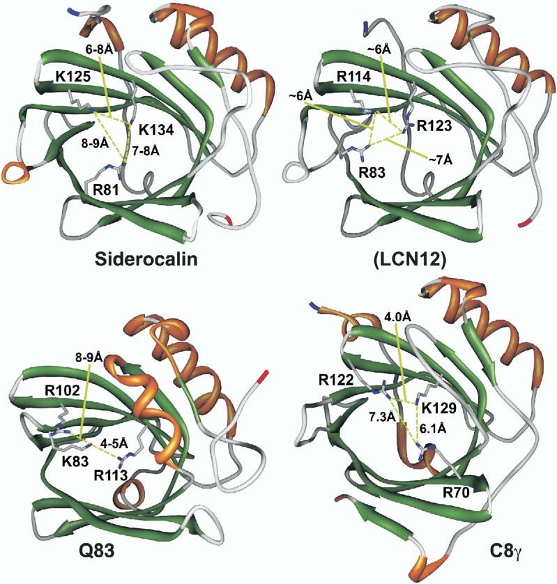
Structure
32

Figure 2. Stereoviews of Siderocalin Ligands Positioned in the Siderocalin Calyx
(A) Ent, (B), CMB-S, and (C), NCA. Ligands are shown in a licorice stick representation, colored by atom-type (C, gray; N, blue; O, red; Fe,
dark red). The molecular surface of the protein (as displayed by SwissPDBViewer [Guex and Peitsch, 1997]) is shown, colored by electrostatic
potential (positive, blue; negative, red). Surfaces corresponding to Trp79, Arg81, Lys125, and Lys134 are labeled. In these views, the three
pockets of the trilobate siderocalin calyx are 1, at the top; 2, at the lower right; and 3, at the lower left. The deepest portion of the calyx is
pocket 2.
(D and E) Stereoviews of superpositions (based only on protein atoms) of all of the different models of Ent and TRENCAM-3,2-HOPO (intact
and partial structures) and the six total CMB-S/T models. The arrowhead indicates the position of the R4 methyl group, the only difference
observed between the CMB-S and -T ligands in the complex crystal structures.
(F) Stereoview of the superposition of key ligand contacting side chains from the 15 available views of siderocalin from the crystal structures,
colored by the ligand present in the complex (NCA or none, blue; phenolate-type siderophore, red; and CMBs, orange). The position of the
iron atom varies by over 1.1 Å among the phenolate-type siderophore calyces and by no more than 0.46 Å among the CMB calyces. All
molecular figures were generated with SwissPDBViewer (Guex and Peitsch, 1997) and rendered using the MegaPOV extensions (megapov.
inetart.net) to POV-Ray (www.povray.org) except where noted.

aminations of the preliminary electron density maps,                        TRENCAM-3,2-HOPO apparently also interacts with
phased by molecular replacement (dmin = 2.5 or 2.1 Å,                       siderocalin identically to FeEnt (Figures 2A and 2D).
respectively), showed that the density corresponding to                     (The quality of the density did not permit resolving the
the ligands was as poor as in the initial FeEnt complex                     preferred position, if any, of the asymmetrical HOPO
(data not shown). The conclusion is that while ligand                       group of TRENCAM-3,2-HOPO in the siderocalin calyx.)
degradation may contribute to poor ligand density, it                       The conservation of ligand orientation in the calyx con-
does not solely account for the effect. The TRENCAM                         firms the observation that the “backbone” of the sid-
complex was therefore not refined, as the interaction                       erophore, which is completely different between the na-
with siderocalin appeared identical to FeEnt, though                        tive and synthetic molecules, does not contribute
the TRENCAM-3,2-HOPO complex structure was fully                            significant protein contacts affecting binding (Goetz et
refined (Table 1), as this compound represents a dis-                       al., 2002). This point is also supported by the high-affin-
tinct siderophore type, again modeling the ligand using                     ity binding of MECAM, yet another synthetic Ent ana-
multiple substructures (dihydroxybenzamide). However,                       log, with a third, distinct backbone structure (Figure 1).
within the limitation of the poor quality of the density,                      Though the current complex crystallization condi-
Siderocalin Also Binds Carboxymycobactins
33

Figure 3. Binding of CMB-S to Siderocalin
(A and B) Stereoviews ([A], from the side; [B], from above, looking down into the calyx) of CMB-S bound to siderocalin. The protein is
represented by a superposition of the Cα backbones of all fifteen independent views of siderocalin (from a total of six crystal structures,
three reported here and three previously reported [Goetz et al., 2000, 2002]), colored by secondary structure (α, orange; β, green; and
coil, gray). N termini are colored blue; C termini are colored red; the Cα of Asn65, the location of the single N-linked oligosaccharide, is
colored yellow.
(C) Stereoview of the simulated annealing composite-omit 2Fobs − Fcalc electron density map contoured around the CMB-S ligand (molecule
C; C, gray; N, blue; and O, red) at 1σ (amber) and 8σ (dark red).

tions are at low pH (4.5), rather than neutrality, there is              phores are known to undergo a sequential shift in bind-
essentially no effect on the overall structure of the pro-               ing mode in solution, from phenolate-mode binding to
tein (Figures 3A and 3B). Catecholate-type sidero-                       salicylate-mode binding, concurrent with acidification,
Structure
34

Table 1. Crystallographic Statistics

Siderophore                            TRENCAM-3,2-HOPO           TRENCAM                    CMB-S                      CMB-T
Data Collection
Space group                            P41212                     P41212                     P41212                     P41212
Lattice constants (Å)                  a = b = 114.4, c = 119.0   a = b = 115.1, c = 119.5   a = b = 114.2, c = 119.3   a = b = 114.8, c = 119.1
Resolution range (Å)                   20.0–2.10 (2.14–2.10)a     20.0–2.50 (2.54–2.50)      20.0–2.10 (2.14–2.10)      20.0–2.20 (2.24–2.20)
Observations                           456,603                    204,054                    381,305                    418,554
Unique reflections                     45,047                     27,376                     46,438                     40,016
Completeness (%)                       96.6 (93.0)                96.4 (98.4)                99.9 (100.0)               97.6 (99.4)
I/σ(I)                                 11.1 (1.5)                 10.6 (2.8)                 16.0 (2.9)                 14.6 (2.6)
Rsym (%)                               4.7 (39.9)                 5.7 (41.3)                 5.5 (34.5)                 6.8 (37.5)
Fe peak heightsb (A, B, C; σ)          9, 4, 6                    11, 7, 14                  11, 12, 17                 17, 12, 18
Refinement Statistics
Rwork (%)                              22.5                       —                          22.0                       22.0
Rfreec (%)                             25.7                       —                          25.1                       25.3
Number of atoms
  Protein                              4,127                      —                          4,154                      4,113
  Siderophore                          58                         —                          162                        159
  Waters                               116                        —                          95                         105
  Estimated coordinate errord (Å)      0.24                       —                          0.20                       0.23
Geometry
  Rmsd bonds (Å)                       0.024                      —                          0.027                      0.024
  Rmsd angles (°)                      2.2                        —                          2.2                        2.2
  Rmsd dihedrals (°)                   27.5                       —                          27.3                       27.2
  Rmsd impropers (°)                   1.4                        —                          1.6                        1.4
  Average B factor (Å2)                38.3                       —                          38.3                       37.1
  Protein monomer B factors            33.3, 50.2, 28.8                                      33.9, 51.9, 27.2           31.9, 52.8, 26.1
    (A, B, C; Å2)                                                 —
  Siderophore B factors                72.1, 85.6, 61.3           —                          47.5, 59.2, 39.1           38.2, 59.0, 36.5
    (A, B, C; Å2)
  Water B factors (Å2)                 36.0                       —                          33.1                       32.2
Ramachandrane
  Most favored (%)                     90.2                       —                          90.7                       90.5
  Additional allowed (%)               8.4                        —                          7.9                        8.0
  Generously allowed (%)               0.0                        —                          0.0                        0.5f
  Disallowed (%)                       1.3f                       —                          1.4f                       1.1f
a
  Numbers in parentheses correspond to the highest resolution shell.
b
  Peak heights of electron density features associated with ligand iron atoms in simulated annealing composite-omit Fourier syntheses.
c
  Calculated on 10% of the data (Kleywegt and Brunger, 1996) and matched between data sets.
d
  Crossvalidated σA error as calculated in CNS (Brunger et al., 1998).
e
  Calculated with PROCHECK (Laskowski et al., 1992).
f
  The Ramachandran outliers are the same two residues (Y115 and C175) in all three molecules in all of the refined structures, despite careful
rebuilding, arguing that these residues are not poorly modeled but truly adopt unfavorable conformations.

correlating with reduced iron affinity (Cohen and Ray-                       2002]; therefore, any iron present in the calyx is part of
mond, 2000). The transition occurs one phenolate                             a ferric siderophore complex.) What variation is seen in
group at a time as pH decreases. FeEnt starts to shift                       iron peak heights correlates well with ligand B factors,
at around a pH of 4.5. While poor, the electron density                      across or within individual asymmetric units, rather
is good enough to argue that both TRENCAM and                                than reflecting lower iron occupancies in the TRECAM
TRENCAM-3,2-HOPO remain in phenolate mode in the                             or TRENCAM-3,2-HOPO complexes versus the CMB
complex, suggesting that complexation with sidero-                           complexes. Therefore, the poor quality of the ligand
calin stabilizes phenolate-mode iron binding. The ob-                        electron density is likely mostly a consequence of un-
served iron location, which is well determined in all of                     usually pronounced ligand mobility. This conclusion is
the structures, also constrains the siderophore to a po-                     compatible with the very tight KD in that the siderocalin
sition in the calyx sterically incompatible with salicyl-                    recognition mechanism depends on electrostatic and
ate-mode binding.                                                            cation-π interactions with few hydrogen bonds and only
   In order to confirm that the ligands are fully, or near                   limited van der Waals contacts (Figure 4B). Indeed, the
fully, occupied, simulated-annealing composite-omit                          cationic groups are not even in van der Waals contact
maps were calculated in CNS (Brunger et al., 1998),                          with any atoms of the ligands. Superposition of all the
strictly excluding all ligand atoms. The peak heights of                     full FeEnt and DHBA/DHBS partial models of FeEnt and
the electron density features corresponding to the iron                      FeTRENCAM-3,2-HOPO from the two structures (Figure
atoms (Table 1) are high enough to eliminate low ligand                      2D) does reveal that the most constrained parts of the
occupancy as contributing to poor ligand density.                            ligands are the catecholate moieties in pocket 1 of the
(Siderocalin shows no appreciable affinity for iron in any                   trilobate calyx. This pocket, defined by the two lysine
form in the absence of a siderophore [Goetz et al.,                          side chains (125 and 134) contributing cation-π bonds,
Siderocalin Also Binds Carboxymycobactins
35
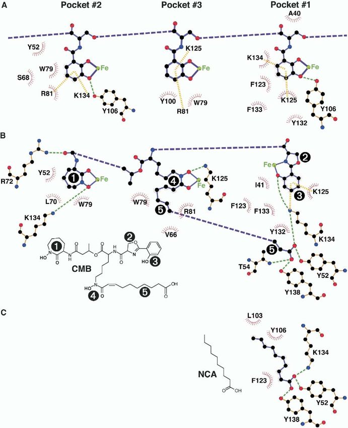
Figure 4. Exploded LIGPLOT Schematics of the Interactions between Siderocalin and Ent, CMB-S/T, and NCA, Broken Down by Calyx Pocket
(A) Ent, (B) CMB-S/T, and (C) NCA. The pictured interactions represent the union of all the protein/ligand interactions observed across all the
complexes in the asymmetric unit. Ligand covalent bonds are colored purple; protein covalent bonds are colored orange. Hydrogen bonds
are indicated with dashed green lines; cation-π interactions are indicated by dashed yellow bars. Atoms are colored by type (C, black; O, red;
N, blue; and Fe, green). Van der Waals contacts are indicated by red sunbursts. Chemical structures for CMB (the predominant variant
observed in the CMB-S crystal structures) and NCA are also shown; CMB substituent groups (1, the cyclic hydroxamate; 2, the oxazoline; 3,
the phenolate; 4, the linear hydroxamate; and 5, the fatty acid tail) are numbered on the chemical structure and the corresponding parts of
the LIGPLOT diagram for clarity.
is thus revealed as likely the key pocket for binding phores was qualitatively tested for binding by surface
phenolate-type siderophores. plasmon resonance (SPR), copurification and, when
possible, confirmed by cocrystallization and the pres-
Siderocalins Bind a Variety of Phenolate ence of a fully or near-fully occupied iron peak (Table
and CMB Siderophores 1). While the SPR assay remains a quick, simple and
In order to address whether siderocalin could bind fer- powerful method to qualitatively test for ligand binding,
ric siderophores other than FeEnt, a panel of 18 addi- fitting the kinetic data to model binding curves remains
tional, representative, natural and synthetic sidero- challenging due to the complexity of the multiphasicStructure
36
association and dissociation phases (data not shown). moved as much as 2.5 Å up, out of the calyx, from their
The slow component/s of the dissociation phase also positions in the Ent complexes. The linear hydroxamate
prevent the system from achieving equilibrium within a and the linking elements (“backbone”) of CMB make
reasonable time, precluding the estimation of KD from the fewest siderocalin contacts, again analogous to the
equilibrium responses. Siderophores negative for sidero- Ent complex.
calin binding in the SPR assay also fail to bind when Binding either CMB-S or CMB-T results in essentially
soaked in excess with the protein at millimolar concen- identical crystal structures (Figure 2E). Even though
trations; resultant concentrated protein solutions are heterogeneous CMB mixtures, isolated directly from
unchanged in color after washing (data not shown). culture supernatants (Lane et al., 1995; Ratledge and
These results show that, as expected, siderocalin binds Ewing, 1996), were used in cocrystallizations, the
to a family of phenolate/catecholate siderophores chemi- electron density is clear enough, and the resolution
cally related to Ent but not to yeast and bacterial sid- high enough, to show that a single molecular species
erophores based on hydroxamate or α-hydroxycarbox- predominates in the calyx in both complex structures,
ylate iron binding groups. However, quite unexpectedly, corresponding to an aliphatic linker in the fatty acid tail
siderocalin also binds the heterogeneous, mixed-type, eight methylene groups long. Deletion of even one
soluble carboxymycobactin siderophores of Mycobac- methylene group from ligand models worsens the re-
terium smegmatis (CMB-S) and M. tuberculosis (CMB-T) finement statistics. The only difference between the
(Gobin et al., 1995; Lane et al., 1998, 1995; Wong et al., CMB-S and -T ligand structures is the absence of the
1996). These compounds are quite distinct chemically methyl group at the R4 position on the CMB-T ligands.
from FeEnt and its relatives, with iron binding moieties The structures have been refined with a double bond in
contributed by 2-hydroxyphenyloxazoline, a seven- the fatty acid tail (between C47 and C48 in the coordi-
cyclic hydroxamate and a linear hydroxamate trailing nate file) as suggested by the electron density and con-
off into a fatty acid tail (Figure 1). CMBs vary species sistent with Rfree values monitored during rebuilding,
to species and within a single species in the length of though this assignment is by no means certain at this
the fatty acid tail and by the identity of several substitu- resolution. The CMB-T structure clearly seen in the
ent groups (Figure 1). As predicted from the pattern of complex structures is also one methylene group longer
sequence conservation in the calyx, murine siderocalin in the fatty acid tail than previously reported for com-
displays an equivalent binding specificity for the panel pounds isolated from M. tuberculosis cultures (Gobin
of 18 siderophores (data not shown). et al., 1995). It has so far not been possible to purify
CMB variants to the homogeneity necessary to test in-
CMBs More Completely Fill the Siderocalin Calyx, dividual isoforms for binding.
Binding in a Single Orientation
In order to elucidate the details of the interaction be- Ligand-Sensitive Calyx Structural Elements
tween siderocalin and CMB ligands, complexes be- The only structural elements in the siderocalin calyx
tween siderocalin and either FeCMB-S or -T were crys- that display any significant conformational variation in
tallized and analyzed crystallographically at 2.1 or response to the presence or absence of any of the three
2.2 Å resolution, respectively (Table 1; Figures 2–4). The types of ligands (NCA, phenolate-type siderophores, or
FeCMB ligands occupy a volume in the calyx overlap- CMBs) are the side chains of two residues: Trp79 and
ping that of FeEnt, with the 2-hydroxyphenyloxazoline Arg81 (Figure 2F). The side chain of Trp79 is either dis-
group sitting in pocket 1, the cyclic hydroxamate group ordered (modeled as an alanine) or adopts one of three
sitting in the upper part of pocket 2 and the linear hy- rotamers fairly consistently, progressively moving out
droxamate sitting in pocket 3 (Figure 2B). The fatty acid of the way in response to the increasing size of the
tail curls under the rest of the siderophore, crossing various ligands. In the CMB structures, the most calyx-
from pocket 3 into pocket 1, positioning the carboxyl- filling ligands, Trp79 is oriented parallel to the calyx
ate group into the bottom of pocket 2. Interestingly, the wall, with the distal nitrogen of the imidazole moiety
CMB carboxylate essentially superimposes on the car- pointing outward. Given the difficulty in confidently
boxylate group of an adventitious ligand, tentatively modeling arginine side chain conformations, even at
identified as n-capric acid (NCA), in the original crystal this resolution, the pattern is less clear, though, again,
structure of siderocalin expressed recombinantly in a the side chain of Arg81 adopts various rotamers. The
baculovirus system (Goetz et al., 2000) (Figures 2C and Arg81 side chain can either intercalate between cate-
3C). Lysines 125 and 134 participate in cation-π bonds cholate rings in the FeEnt structures, providing cation-π
to the CMB hydroxybenzoyl moiety completely analo- interactions, or move away from the ligand, substituted
gous to those in Ent, with the hydroxybenzoyl of CMB by the side chain of Trp79 providing alternate herring-
superimposing almost identically onto the DHBA moi- bone ring stacking interactions to the ligand. In the
ety of Ent in pocket 1. However, largely due to the orien- CMB structures, the Arg81 side chains also move to
tation of the fatty acid tail, CMBs fill the calyx more positions along the calyx wall to accommodate the
completely, generating more extensive hydrogen bond larger ligands. All the other calyx residue side chain
and van der Waals networks (Figure 4). The result is conformations are markedly well conserved among the
that the CMB ligands sit fully resolved in the calyx in a 15 views of the siderocalin structure, including the ly-
single, well-defined orientation (Figures 2E, 3B, and sines (125 and 134) donating cation-π bonds to all the
4C). The most constrained portion of the CMB structure different siderophore ligands. The structure of the or-
is again the hydroxybenzoyl group sitting in pocket 1. dered solvent shell is quite variable across all of the
The iron atoms in the CMB complex structures have structures, consistent with the variable crystallizationSiderocalin Also Binds Carboxymycobactins
37
conditions and crystal contacts, both over the surface ligand electron density features is the result of marked
of the protein and within the calyx. Siderophore binding ligand flexibility, reflecting fundamental aspects of sidero-
displaces most of the calyx waters observed in the calin-mediated recognition of phenolate/catecholate-type
empty structures and there are no uniformly conserved siderophores, and is not an artifact of previous crystallo-
water-mediated protein/ligand interactions. graphic analyses. Siderocalin has evolved a recognition
mechanism that generates tight binding while tolerating
Discussion such flexibility, in the absence of interactions to the
most variable element of this class of microbial sidero-
The Siderocalin Fold Is Imperturbable phores, the backbone linker, thus permitting siderocalin
Pair-wise superposition rmsds (on all common Cαs) to degenerately interact with a broad range of pheno-
across the 15 siderocalin models (Goetz et al., 2000, late/catecholate siderophores. Because the siderophore
2002) vary from 0.54 to 0.67 Å; these numbers improve phenolate moiety determines bound ligand orientation,
measurably if the most N-terminal two or three resi- and because the most constrained portion of the ligand
dues, the most variable siderocalin structural element sits in pocket 1, we conclude that it is the highly polar-
(Figures 3A and 3B), are excluded. These structures ized ferric hydroxybenzoyl group that is the key deter-
span two different expression systems, four different minant for siderocalin binding and that pocket 1 is the
crystallization conditions (from high to low salt and key binding pocket. Reliance on cation-π bonds to
neutral to low pH), six different packing environments, highly polarized hydroxybenzoyl iron ligands, either in
the presence or absence of three different ligand context of catecholate, 2-hydroxyphenyloxazoline or,
classes, and even the presence or absence of N-linked potentially, HOPO groups, complements the degener-
oligosaccharide (at Asn65). After the N terminus, the ate siderocalin recognition mechanism, broadening po-
next greatest flexibility is seen in the loops rimming the tential responses. These conclusions are confirmed by
calyx, where even greater variation is typically seen in the analysis of siderocalin/CMB complexes, where the
other lipocalins (Flower, 1996). The secondary structure 2-hydroxyphenyl group superimposes on the pocket 1
elements, and their relative arrangement, are fully con- catecholate groups of the phenolate/catecholate-type
served. This behavior is consistent with observations siderophores, even though the rest of the structures of
that extreme conditions are required to denature sid- these siderophores are essentially completely different.
erocalin and release bound siderophores (Goetz et al., Variable solvent structures are unlikely to contribute to
2002). However, these results directly contradict con- degenerate recognition in that there are few water-
clusions based on the solution NMR structure of sid- mediated interactions. The CMB complex structures do
erocalin (Coles et al., 1999), where structural mobility, show that many of the complex calyx features, sub-
particularly the orientation of the α helix to the β barrel, pockets irrelevant for the binding of phenolate/cate-
was much more marked. We conclude that the sidero- cholate-type enterobacterial siderophores, allow for the
calin fold is very stable and well defined. Structural sta- binding of whole different classes of siderophores,
bility extends to the calyx, where the only flexibility of broadening, in this case, potential siderocalin-medi-
any significance is seen in the side chains of two resi- ated antibacterial defenses to mycobacteria, which are
dues, Trp79 and Arg81. While these two side chains significant human pathogens. However, antimycobac-
do move to accommodate the presence or absence of terial siderocalin responses may be limited by the high
different ligands, the degree of movement is nowhere selectivity of siderocalin for a single CMB species, in
near the level of conformational flexibility typically as- terms of fatty acid tail length, in both the CMB-S and
sociated with “induced-fit” recognition mechanisms. -T complex structures, even though CMB mixtures
For instance, this level of binding site plasticity is con- were used in the cocrystallizations. While siderocalin
siderably less than the remodeling observed in the li- may be able to tolerate binding of CMB variants ± one
gand binding site of the insect ecdysone receptor, or perhaps two methylene groups in the fatty acid moi-
which binds disparate ecdysteroid hormones and non- eties, though likely with significant concurrent reduc-
steroidal synthetic agonists (Billas et al., 2003). How- tions in affinity, it seems unlikely that siderocalin could
ever, such side chain movements observed in the accommodate the extremes of the reported CMB-S/T
siderocalin calyx could account for the multiphasic as- spectrum (Figure 1), at least while retaining the overall
sociation kinetics seen in the SPR experiments, where ligand orientation seen in the current cocrystal struc-
ligand approach, possibly dominated by the electro- tures. The variation seen in CMB structures may, in-
static component of the interaction, is fast, and estab- deed, reflect mycobacterial responses to siderocalin-
lishing the optimal calyx conformation, possibly opti- mediated defenses, evidenced by the obvious success
mizing the cation-π network, is slow. of mycobacteria as human pathogens. The efficacy of
siderocalin in thwarting mycobacterial infections in vivo
Siderocalin Tightly Binds Phenolate/Catecholate remains to be tested.
Siderophores without Securely Constraining
Their Orientation, Enabling Degeneracy Are There Other Siderophore Binding Lipocalins?
By comparing the complex structures of nonhydrolyz- While broadly and degenerately recognizing enterobac-
able phenolate/catecholate siderophores with the terial phenolate/catecholate siderophores and specifi-
FeEnt complex structure, optimizing crystallization cally interacting with at least a limited spectrum of my-
conditions to minimize crystallization time, and con- cobacterial CMBs, siderocalin fails to bind to many
firming ligand occupancy by careful analysis of iron bacterial siderophores and essentially all types of fun-
peak heights, we have shown that the poor quality of gal siderophores (Figure 1). It is likely that evading sid-Structure
38
Figure 5. Ribbon Representations of Sidero-
calin and Three Potential Siderophore Bind-
ing Lipocalins, Colored by Secondary
Structure
The side chains of characteristic triads of
positively charged residues are also shown,
with interresidue distances indicated. The
representations are based on the crystal
structures of siderocalin (1L6M.pdb) and C8γ
(1IW2.pdb), the NMR structure of Q83
(1JZU.pdb), and a simplistic homology model
of Lcn12 based on siderocalin using the
SWISS-Model server (Peitsch, 1996). At 86%
sequence identity, it is presumed that Ex-
FABP will be essentially identical in structure
to Q83 at this resolution. Molecular ribbons
were generated with Chimera (Huang et al.,
1996) (www.cgl.ucsf.edu/chimera). α, orange;
β, green; and coil, gray.
erocalin contributes to virulence, as the ability to syn- eling of Lcn12 using the SwissMODEL server (Peitsch,
thesize aerobactin, which does not bind to siderocalin, 1996) reveals a triad of positively charged side chains
is a virulence factor for enteric bacteria (Wooldridge in the Lcn12 calyx completely analogous to siderocalin
and Williams, 1993). However, it seems unusual that the (Figure 5). The next most related lipocalins are the
innate immune system would limit such powerful anti- highly homologous (86% identical to each other) fowl
siderophore responses to only those pathogens whose proteins Ex-FABP (Descalzi Cancedda et al., 2000)
siderophores bind to siderocalin, cleverly degenerate (from chicken, w20% identical to siderocalins) and
as it is. Since siderocalin is, to our knowledge, the first quail Q83 (Hartl et al., 2003) (20%–22% identical to sid-
nonbacterial siderophore binding protein characterized, erocalins). Ex-FABP is expressed during chicken
the search for other candidate siderophore binders embryo development in hypertrophic cartilage, muscle
must begin with neighboring lipocalins. Comparisons fibers, and granulocytes and is also a component of
show that sequence identities rapidly plummet as the egg whites. In chondrocyte and myoblast cultures, Ex-
alignments move from the siderocalins themselves. A FABP expression is induced by inflammatory agents
number of neighboring lipocalins are also readily elimi- and inhibited by anti-inflammatory agents. Q83 is a pro-
nated as having characterized functions or ligands that tein strongly induced in v-myc-transformed avian fibro-
preclude siderophore binding: the prostaglandin D2 blasts, though no specific candidate ligands or func-
synthases (Helliwell et al., 2004) (w30%–35% identical tions have been proposed. The NMR structure of Q83
to siderocalins), including distant homologs in Xenopus again shows a triad of positively-charged amino acids
and Bufo, and human HC, a lipocalin associated with in the calyx (conserved in Ex-FABP) very reminiscent,
IgA which binds a nonsiderophore chromophore (Åker- in arrangement and character, of the key siderophore
strom et al., 2000b) (16%–23% identical to sidero- binding residues of siderocalin (Arg81, Lys125, and
calins). However, given that the structural homology be- Lys134), and most uncharacteristic of lipocalins in
tween even distantly related lipocalins is high and that general. This arrangement is also echoed in the calyx
the hallmark features of the siderocalin calyx (a charac- of the more distantly related protein C8γ (Schreck et
teristic triad of positively charged side chains) are so al., 2000), a well-studied member of the complement
pronounced, it is possible to identify candidate sidero- cascade and the subject of high-resolution crystallo-
phore binding lipocalins (“siderocalins”). graphic analyses (Ortlund et al., 2002). However, no
Murine lipocalin 12 (Lcn12, 28%–29% identical to candidate ligands for C8γ have been proposed, nor has
siderocalins) has recently been identified as a compo- its precise role in the membrane attack complex been
nent of seminal fluid (Suzuki et al., 2005), but little else delineated. However, particularly for C8γ, the shape of
is known about its function. Simplistic homology mod- the calyx, at least in the absence of any hypotheticalSiderocalin Also Binds Carboxymycobactins
39
ligand-induced conformational changes, is incompati- buffered at pH 4.5 with 100 mM Na acetate. Crystals were cryo-
ble with the binding of any known siderocalin ligand in preserved in a mother liquor with 20% glycerol at −170°C. The
TRENCAM complex data set was collected on a Rigaku Raxis IV
a comparable geometry. Nevertheless, these observa-
area detector using CuKα radiation; the other three data sets were
tions do suggest that there may be other siderophore collected at beamlines 5.0.1 and 5.0.2 at the Advanced Light
binding lipocalins, currently unrecognized, perhaps Source (Lawrence Berkeley National Laboratory, Berkeley, CA). The
with nonoverlapping specificities, that complement the space group is P41212, with three monomers in the asymmetric
function of siderocalin in innate immunity. Subsequent unit, nearly isomorphous with crystals used in the previous struc-
binding and crystallographic analyses, however, fail ture determination (Goetz et al., 2002) (Table 1). Diffraction data
were processed with DENZO and SCALEPACK. Reflections re-
to convincingly demonstrate specific interactions be-
served for calculating Rfree (Kleywegt and Brunger, 1996) values
tween C8γ and any of the siderophores we have access were strictly matched between all three refined data sets. Model
to (data not shown and J.M. Sodetz, personal com- coordinates were refined with the CNS software package (Brunger
munication), showing that calyx elements beyond tri- et al., 1998) using standard protocols. Noncrystallographic symme-
ads of positively charged residues may be necessary try restraints were not used in the refinement, consistent with Rfree
for siderophore binding. However, a recent report calculations. Relevant statistics are presented in Table 1. Initial
model coordinates and target geometry for DHBA, used to model
(Fluckinger et al., 2004) shows that Lcn1 (tear lipocalin,
TRENCAM-3,2-HOPO, and CMB-S/T were derived, respectively,
von Ebner’s Gland protein), a lipocalin too distant in from the structure of enterobactin (Cambridge Structural Database
sequence space to have been identified in this analysis, entry JOSLOS) or the structures of carboxymycobactin P (Hough
is also functionally a “siderocalin,” broadly inhibiting and Rogers, 1974) and sebacic acid (Cambridge Structural Data-
the growth of bacteria and fungi through ferric sidero- base entry SEBAAC).
phore sequestration, though the nature of the recogni-
tion mechanism has yet to be elucidated.
Acknowledgments
Thus, proposals to use siderophores and siderophore
analogs as therapeutics, either as antibiotics (Budzikie- The authors wish to thank Kenneth N. Raymond (University of Cali-
wicz, 2001; Roosenberg et al., 2000) or, through the fornia, Berkeley) for much insightful input and discussion,
bound iron, as oxygen radical scavengers in various Sudeshna Seal for assistance with crystallization, Niels Borregaard
clinical settings (Horwitz et al., 1998), will still need to and coworkers (Rigshospitalet) for supplying MMP-9, Edward
be tempered by the possibility that endogenous sidero- Hough for supplying ferricarboxymycobatin P (from M. phlei) coor-
dinates, and James M. Sodetz and his colleagues at the University
phore binding specificities will limit, defeat, or con-
of South Carolina for providing unpublished results of their crystal-
found such approaches. lographic studies of siderophore binding to C8γ. This study was
supported by the National Institutes of Health (grants AI48675,
Experimental Procedures AI59432, and AI11744).
Protein Expression and Purification and Siderophore
Received: September 27, 2004
Binding Assays
Revised: October 25, 2004
Human siderocalin was expressed as a glutathione S-transferase
Accepted: October 28, 2004
fusion protein and purified as previously described (Bundgaard et
Published: January 11, 2005
al., 1994; Goetz et al., 2002). Murine siderocalin was expressed in
an identical procedure. Binding of siderophores to either human or
mouse siderocalin was assayed by SPR as previously described References
(Flo et al., 2004) using a Biacore System 3000 (Biacore AB) in HBS-P
buffer. Briefly, approximately 2000–5500 resonance units (RU) of
Åkerstrom, B., Flower, D.R., and Salier, J.-P. (2000a). Lipocalins:
protein were coupled to research-grade CM5 sensor chips using
unity in diversity. Biochim. Biophys. Acta 1482, 1–8.
amine coupling chemistry following the manufacturer’s recommended
procedure. Surface-specific activities were typically greater than Åkerstrom, B., Logdberg, L., Berggard, T., Osmark, P., and Lind-
75%; siderophore responses ranged from 5 to 180 RUs. Surfaces qvist, A. (2000b). Alpha(1)-microglobulin: a yellow-brown lipocalin.
were regenerated with 20% acetonitrile and 5 mM NaOH. A binding Biochim. Biophys. Acta 1482, 172–184.
call corresponds to an interaction with an estimated KD at less than Berman, H.M., Westbrook, J., Feng, Z., Gilliland, G., Bhat, T.N.,
100 nM; nonbinding siderophores interact with siderocalins with a Weissig, H., Shindyalov, I.N., and Bourne, P.E. (2000). The Protein
KDs estimated to be greater than 1 mM. Siderophores were ob- Data Bank. Nucleic Acids Res. 28, 235–242.
tained from Sigma-Aldrich, Biophore Research Products, or were Billas, I.M., Iwema, T., Garnier, J.M., Mitschler, A., Rochel, N., and
synthesized (Thulasiraman et al., 1998) or isolated (Lane et al., Moras, D. (2003). Structural adaptability in the ligand-binding
1995; Ratledge and Ewing, 1996) as previously described. SPR pocket of the ecdysone hormone receptor. Nature 426, 91–96.
measurements of FeEnt binding are confounded by its extreme so-
lution instability, which is not a problem for the other siderophores Brunger, A.T., Adams, P.D., Clore, G.M., DeLano, W.L., Gros, P.,
listed in Figure 1. All of the binding positive compounds also copur- Grosse-Kunstleve, R.W., Jiang, J.S., Kuszewski, J., Nilges, M.,
ify (after extensive washing) with siderocalin, yielding obviously Pannu, N.S., et al. (1998). Crystallography & NMR system: a new
colored solutions, and cocrystallize with human siderocalin, though software suite for macromolecular structure determination. Acta
the resultant crystals are not always diffraction quality. FeEnt bind- Crystallogr. D Biol. Crystallogr. 54, 905–921.
ing, the use of which is problematic in the SPR assay due to solu- Budzikiewicz, H. (2001). Siderophore-antibiotic conjugates used as
tion instability, has been previously demonstrated by alternate trojan horses against Pseudomonas aeruginosa. Curr. Top. Med.
means (Goetz et al., 2002). Chem. 1, 73–82.
Bundgaard, J.R., Sengelov, H., Borregaard, N., and Kjeldsen, L.
Crystallization and Crystallography (1994). Molecular cloning and expression of a cDNA encoding
Siderocalin was cocrystallized with four different siderophores NGAL: a lipocalin expressed in human neutrophils. Biochem. Bio-
(TRENCAM, TRENCAM-3,2-HOPO, CMB-S, and CMB-T) using con- phys. Res. Commun. 202, 1468–1475.
ditions that accelerated crystal growth time to 2 weeks or less from Cohen, S.M., and Raymond, K.N. (2000). Catecholate/salicylate
greater than 6 months (Goetz et al., 2002): vapor diffusion at 22°C heteropodands: demonstration of a catecholate to salicylate coor-
over reservoirs of 1.4–1.8 M ammonium sulfate, 50–100 mM NaCl, dination change. Inorg. Chem. 39, 3624–3631.Structure
40
Coles, M., Diercks, T., Muehlenweg, B., Bartsch, S., Zolzer, V., lipocalin 24p3, which is an essential molecule in IL-3 withdrawal-
Tschesche, H., and Kessler, H. (1999). The solution structure and induced apoptosis, is not involved in the G-CSF withdrawal-
dynamics of human neutrophil gelatinase-associated lipocalin. J. induced apoptosis. Eur. J. Haematol. 71, 412–417.
Mol. Biol. 289, 139–157. Kjeldsen, L., Cowland, J.B., and Borregaard, N. (2000). Human neu-
Descalzi Cancedda, F., Dozin, B., Zerega, B., Cermelli, S., and trophil gelatinase-associated lipocalin and homologous proteins in
Cancedda, R. (2000). Ex-FABP: a fatty acid binding lipocalin de- rat and mouse. Biochim. Biophys. Acta 1482, 272–283.
velopmentally regulated in chicken endochondral bone formation Kleywegt, G.J., and Brunger, A.T. (1996). Checking your imagina-
and myogenesis. Biochim. Biophys. Acta 1482, 127–135. tion: applications of the free R value. Structure 4, 897–904.
Devireddy, L.R., Teodoro, J.G., Richard, F.A., and Green, M.R. Lane, S.J., Marshall, P.S., Upton, R.J., Ratledge, C., and Ewing, M.
(2001). Induction of apoptosis by a secreted lipocalin that is tran- (1995). Novel extracellular mycobactins, the carboxymycobactins
scriptionally regulated by IL-3 deprivation. Science 293, 829–834. from Mycobacterium avium. Tetrahedron Lett. 36, 4129–4132.
Dougherty, D.A. (1996). Cation-π interactions in chemistry and biol- Lane, S.J., Marshall, P.S., Upton, R.J., and Ratledge, C. (1998). Iso-
ogy: a new view of benzene, Phe, Tyr, and Trp. Science 271, 163– lation and characterization of carboxymycobactins as the second
168. extracellular siderophores in Mycobacterium smegmatis. Biomet-
Ecker, D.J., Matzanke, B.F., and Raymond, K.N. (1986). Recognition als 11, 13–20.
and transport of ferric enterobactin in Escherichia coli. J. Bacteriol.
Laskowski, R.A., MacArthur, M.W., Hutchinson, E.G., and Thornton,
167, 666–673.
J.M. (1992). PROCHECK: a program to check the stereochemical
Ecker, D.J., Loomis, L.D., Cass, M.E., and Raymond, K.N. (1988). quality of protein structures. J. Appl. Crystallogr. 26, 283–291.
Substituted complexes of enterobactin and synthetic analogs as
Matzanke, B.F., Ecker, D.J., Yang, T.S., Huynh, B.H., Muller, G., and
probes of the ferric-enterobactin receptor in Escherichia coli. J.
Raymond, K.N. (1986). Escherichia coli iron enterobactin uptake
Am. Chem. Soc. 110, 2457–2464.
monitored by Mossbauer spectroscopy. J. Bacteriol. 167, 674–680.
Flo, T.H., Smith, K.D., Sato, S., Rodriguez, D.J., Holmes, M.A.,
Nizet, V., Ohtake, T., Lauth, X., Trowbridge, J., Rudisill, J.,
Strong, R.K., Akira, S., and Aderem, A. (2004). Lipocalin 2 mediates
Dorschner, R.A., Pestonjamasp, V., Piraino, J., Huttner, K., and
a novel innate immune response to bacterial infection by seques-
Gallo, R.L. (2001). Innate antimicrobial peptide protects the skin
trating iron. Nature 432, 917–921.
from invasive bacterial infection. Nature 414, 454–457.
Flower, D.R. (1996). The lipocalin protein family: structure and func-
Ortlund, E., Parker, C.L., Schreck, S.F., Ginell, S., Minor, W., Sodetz,
tion. Biochem. J. 318, 1–14.
J.M., and Lebioda, L. (2002). Crystal structure of human comple-
Flower, D.R., North, A.C., and Sansom, C.E. (2000). The lipocalin ment protein C8γ at 1.2 A resolution reveals a lipocalin fold and a
protein family: structural and sequence overview. Biochim. Bio- distinct ligand binding site. Biochemistry 41, 7030–7037.
phys. Acta 1482, 9–24.
Peitsch, M.C. (1996). ProMod and Swiss-Model: internet-based
Fluckinger, M., Haas, H., Merschak, P., Glasgow, B.J., and Redl, tools for automated comparative protein modelling. Biochem. Soc.
B. (2004). Human tear lipocalin exhibits antimicrobial activity by Trans. 24, 274–279.
scavenging microbial siderophores. Antimicrob. Agents Chem-
Ratledge, C., and Ewing, M. (1996). The occurrence of carboxymy-
other. 48, 3367–3372.
cobactin, the siderophore of pathogenic mycobacteria, as a se-
Gobin, J., Moore, C.H., Reeve, J.R., Jr., Wong, D.K., Gibson, B.W., cond extracellular siderophore in Mycobacterium smegmatis.
and Horwitz, M.A. (1995). Iron acquisition by Mycobacterium tuber- Microbiol. 142, 2207–2212.
culosis: isolation and characterization of a family of iron-binding
exochelins. Proc. Natl. Acad. Sci. USA 92, 5189–5193. Raymond, K.N., Müller, G., and Matzanke, B.F. (1984). Complex-
ation of iron by siderophores. A review of their solution and struc-
Goetz, D.H., Willie, S.T., Armen, R.S., Bratt, T., Borregaard, N., and tural chemistry and biological function. In Topics in Current Chem-
Strong, R.K. (2000). Ligand preference inferred from the structure istry, F.L. Boschke, ed. (Berlin: Springer-Verlag), pp. 50–102.
of neutrophil gelatinase associated lipocalin. Biochemistry 39,
1935–1941. Raymond, K.N., Dertz, E.A., and Kim, S.S. (2003). Enterobactin: an
archetype for microbial iron transport. Proc. Natl. Acad. Sci. USA
Goetz, D.H., Holmes, M.A., Borregaard, N., Bluhm, M.E., Raymond,
100, 3584–3588.
K.N., and Strong, R.K. (2002). The neutrophil Lipocalin NGAL is a
bacteriostatic agent that interferes with siderophore-mediated iron Roosenberg, J.M., 2nd, Lin, Y.M., Lu, Y., and Miller, M.J. (2000).
acquisition. Mol. Cell 10, 1033–1043. Studies and syntheses of siderophores, microbial iron chelators,
and analogs as potential drug delivery agents. Curr. Med. Chem. 7,
Guex, N., and Peitsch, M.C. (1997). SWISS-MODEL and the Swiss-
159–197.
PdbViewer: an environment for comparative protein modeling.
Electrophoresis 18, 2714–2723. Salzman, N.H., Ghosh, D., Huttner, K.M., Paterson, Y., and Bevins,
C.L. (2003). Protection against enteric salmonellosis in transgenic
Hartl, M., Matt, T., Schuler, W., Siemeister, G., Kontaxis, G., Kloiber,
mice expressing a human intestinal defensin. Nature 422, 522–526.
K., Konrat, R., and Bister, K. (2003). Cell transformation by the
v-myc oncogene abrogates c-Myc/Max-mediated suppression of a Schreck, S.F., Parker, C., Plumb, M.E., and Sodetz, J.M. (2000). Hu-
C/EBP β-dependent lipocalin gene. J. Mol. Biol. 333, 33–46. man complement protein C8γ. Biochim. Biophys. Acta 1482, 199–
208.
Helliwell, R.J., Adams, L.F., and Mitchell, M.D. (2004). Prostaglandin
synthases: recent developments and a novel hypothesis. Prosta- Stintzi, A., and Raymond, K.N. (2002). Siderophore chemistry. In
glandins Leukot. Essent. Fatty Acids 70, 101–113. Molecular and Cellular Iron Transport, D.M. Templeton, ed. (To-
ronto: Marcel Dekker, Inc.), pp. 273–320.
Horwitz, L.D., Sherman, N.A., Kong, Y., Pike, A.W., Gobin, J., Fen-
nessey, P.V., and Horwitz, M.A. (1998). Lipophilic siderophores of Suzuki, K., Lareyre, J.-J., Sánchez, D., Gutierrez, G., Araki, Y., Ma-
Mycobacterium tuberculosis prevent cardiac reperfusion injury. tusik, R.J., and Orgebin-Crist, M.C. Molecular evolution of a
Proc. Natl. Acad. Sci. USA 95, 5263–5268. multigene subfamily encoding epididymal lipocalins localized on
Hough, E., and Rogers, D. (1974). The crystal structure of ferrimy- the [A3] locus of mouse chromosome 2. Gene, in press.
cobactin P, a growth factor for the mycobacteria. Biochem. Bio- Thulasiraman, P., Newton, S.M., Xu, J., Raymond, K.N., Mai, C.,
phys. Res. Commun. 57, 73–77. Hall, A., Montague, M.A., and Klebba, P.E. (1998). Selectivity of fer-
Huang, C.C., Couch, G.S., Pettersen, E.F., and Ferrin, T.E. (1996). ric enterobactin binding and cooperativity of transport in gram-
Chimera: an extensible molecular modeling application con- negative bacteria. J. Bacteriol. 180, 6689–6696.
structed using standard components. Pacific Symposium on Bio- Tschesche, H., Zolzer, V., Triebel, S., and Bartsch, S. (2001). The
computing 1, 724. human neutrophil lipocalin supports the allosteric activation of ma-
Kamezaki, K., Shimoda, K., Numata, A., Aoki, K., Kato, K., Takase, trix metalloproteinases. Eur. J. Biochem. 268, 1918–1928.
K., Nakajima, H., Ihara, K., Haro, T., Ishikawa, F., et al. (2003). The Wallace, A.C., Laskowski, R.A., and Thornton, J.M. (1995). LIG-You can also read