Melanocortin System in Kidney Homeostasis and Disease: Novel Therapeutic Opportunities - Frontiers
←
→
Page content transcription
If your browser does not render page correctly, please read the page content below
REVIEW
                                                                                                                                              published: 24 February 2021
                                                                                                                                         doi: 10.3389/fphys.2021.651236

                                                Melanocortin System in Kidney
                                                Homeostasis and Disease: Novel
                                                Therapeutic Opportunities
                                                Mingyang Chang, Bohan Chen, James Shaffner, Lance D. Dworkin and Rujun Gong*
                                                Division of Nephrology, Department of Medicine, University of Toledo College of Medicine, Toledo, OH, United States

                                                Melanocortin peptides, melanocortin receptors, melanocortin receptor accessory
                                                proteins, and endogenous antagonists of melanocortin receptors are the key
                                                components constituting the melanocortin hormone system, one of the most complex
                                                and important hormonal systems in our body. A plethora of evidence suggests that
                                                melanocortins possess a protective activity in a variety of kidney diseases in both
                                                rodent models and human patients. In particular, the steroidogenic melanocortin
                                                peptide adrenocorticotropic hormone (ACTH), has been shown to exert a beneficial
                          Edited by:
                                                effect in a number of kidney diseases, possibly via a mechanism independent of
                         Zhanjun Jia,           its steroidogenic activity. In patients with steroid-resistant nephrotic glomerulopathy,
     Nanjing Medical University, China          ACTH monotherapy is still effective in inducing proteinuria remission. This has inspired
                      Reviewed by:              research on potential implications of the melanocortin system in glomerular diseases.
                             Ran You,
 Key Laboratory of Pediatrics, Nanjing          However, our understanding of the role of the melanocortinergic pathway in kidney
           Children’s Hospital, China           disease is very limited, and there are still huge unknowns to be explored. The most
                               Mi Bai,
    Nanjing Medical University, China
                                                controversial among these is the identification of effector cells in the kidney as well as
                  *Correspondence:
                                                the melanocortin receptors responsible for conveying the renoprotective action. This
                         Rujun Gong             review article introduces the melanocortin hormone system, summarizes the existing
            Rujun.Gong@UToledo.edu              evidence for the expression of melanocortin receptors in the kidney, and evaluates the
                   Specialty section:
                                                potential strategy of melanocortin therapy for kidney disease.
         This article was submitted to
                                                Keywords: melanocyte-stimulating hormone, adrenocorticotropic hormone, pro-opiomelanocortin, glomerulus,
       Renal and Epithelial Physiology,         kidney disease, melanocortin receptors
               a section of the journal
               Frontiers in Physiology
         Received: 08 January 2021              INTRODUCTION
        Accepted: 03 February 2021
        Published: 24 February 2021
                                                The melanocortin system is a neuroimmune endocrine hormone system that encompasses
                              Citation:         five melanocortin receptors (MC1R∼MC5R), four pro-opiomelanocortin (POMC)-derived
        Chang M, Chen B, Shaffner J,            melanocortin peptides (ACTH, α-MSH, β-MSH, and γ-MSH) (Figure 1), endogenous antagonists,
      Dworkin LD and Gong R (2021)
                                                i.e., agouti-signaling protein (ASP or ASIP) and agouti-related protein (AGRP), and melanocortin
       Melanocortin System in Kidney
     Homeostasis and Disease: Novel
                                                receptor accessory proteins (MRAP) (Table 1) (Gantz and Fong, 2003). In the early 20th century,
            Therapeutic Opportunities.          Atwell (1919) found that there might be some substances in the pituitary gland that darkens skin
            Front. Physiol. 12:651236.          color. They implanted pituitary extracts into frogs that became albino after pituitary removal, and
     doi: 10.3389/fphys.2021.651236             found that the frogs regained pigmentary responses (Atwell, 1919). The underlying mechanism has

Frontiers in Physiology | www.frontiersin.org                                         1                                        February 2021 | Volume 12 | Article 651236
Chang et al.                                                                                                                 Melanocortins and Kidney Diseases

                                                                                       therapy for a myriad of diseases, including nephrotic syndrome.
                                                                                       Mechanistically, the use of ACTH was primarily based on its
                                                                                       ability to increase the adrenal production of glucocorticoids. As
                                                                                       such, its use was later substituted by synthetic corticosteroids.
                                                                                       However, a growing body of evidence recently suggests that
                                                                                       ACTH monotherapy is able to effectively alleviate steroid-
                                                                                       resistant nephrotic syndrome, denoting that ACTH achieves
                                                                                       its renal protection via mechanisms beyond adrenocortical
                                                                                       steroidogenesis (Gong, 2011). Given that multiple kidney
                                                                                       cells express MCRs, it is posited that the kidney may be a
                                                                                       quintessential effector organ of the melanocortin hormone
                                                                                       system and that melanocortin peptides may directly target
                                                                                       diverse types of kidney cells to convey a renoprotective activity.

                                                                                       THE MELANOCORTIN HORMONE
                                                                                       SYSTEM

  FIGURE 1 | Melanocortin peptides (ACTH, α-MSH, β-MSH, and γ-MSH) are
                                                                                       Melanocortin Receptors
  all derived from the common precursor protein pro-opiomelanocortin (POMC).           The five melanocortin receptors (MCRs, MC1R∼MC5R) are
  The 3-dimensional structure modeling of human melanocortin peptides was              expressed in a diverse array of tissues and belong to the
  generated based on amino acid sequences by using the following two servers           guanine nucleotide-binding protein-coupled receptor (GPCR)
  for automated protein structure modeling                                             family. MCRs have different affinities to agonists and antagonists
  (http://protein.ict.ac.cn/FALCON/#focus;
  http://bioserv.rpbs.univ-paris-diderot.fr/services/PEP-FOLD/).
                                                                                       causing a vast array of physiological effects (Gong, 2014). As
                                                                                       opposed to their 7-transmembrane GPCR counterparts whose
                                                                                       primary function is the intracellular induction of cyclic adenosine
TABLE 1 | Constituent components of the melanocortin hormone system.                   monophosphate (cAMP), MCRs can also activate the inositol
                                                                                       triphosphate pathway (Konda et al., 1994) and the protein kinase
The melanocortin hormone system
                                                                                       C (PKC) pathway (Kapas et al., 1995), of which the downstream
Ligands            Melanocortin           Melanocortin         Melanocortin            functions have not yet been fully elucidated. In addition,
                    receptors             antagonists            receptor              some MCRs signal through guanine nucleotide-dependent and
                                                                accessory
                                                                                       independent mechanisms and their functional coupling to
                                                                 proteins
                                                                                       agonists at the cell surface is regulated by interacting accessory
ACTH,                 MC1R,                      Agouti,         MRAP1,                proteins, like MRAPs and β-arrestins (Rodrigues et al., 2015).
α-MSH,                MC2R,                     ASIP, and        MRAP2,                It is worth noting that MCRs have phylogenetic differences, as
β-MSH,                MC3R,                      AGRP,       mahogany protein,
                                                                                       reported by Logan et al. (2003) that there are six types of MCRs
and γ-MSH            MC4R, and                                and syndecan-3
                      MC5R                                                             (including two MC5R orthologs) in Zebrafish whereas only four
                                                                                       types of MCRs (lacking MC3R) in Fugu.
ASIP, agouti-signaling protein; AGRP, agouti-related protein; MRAP, melanocortin
receptor accessory protein.
                                                                                           MC1R is expressed abundantly in skin cells where it is a
                                                                                       key control point in determining skin and hair pigmentation
                                                                                       (Jackson et al., 2007). In support of this, loss-of-function or
been elusive until about 60 years later after the development                          null mutations in MC1R are associated with a switch from
of the amino acid sequences and solid-phase synthesis of                               eumelanin to phaeomelanin production, resulting in red hair
synthetic polypeptides. It was finally confirmed that melanocortin                     color, freckles, and fair skin (Fitzpatrick skin type 1) in humans
peptides were derived from a common precursor molecule, which                          and yellow coat color in mice. MC1R was originally named MSH-
earned its name POMC due to its opiate derivative β-endorphin                          R before Mountjoy et al. (1992) completed the cloning of the
(Eipper and Mains, 1980).                                                              human MSH receptor, and later the other four distinct MSH
   The melanocortin hormone system exerts a diverse array of                           receptors, using the cDNA library prepared from melanoma.
physiological functions, including pigmentation, adrenocortical                        MC1R is also widely expressed in other tissues including adrenal
steroidogenesis, energy homeostasis, natriuresis, erectile                             gland, kidney (Figure 2), lung, brain, lymph nodes, spleen, and
response, exocrine gland secretion, analgesia, inflammation,                           leukocytes where it plays a key role in regulating inflammatory
immunomodulation, and temperature control (Cone,                                       reaction and immune response (Cone et al., 1996; Lindskog et al.,
2006). A number of melanocortin peptides and small                                     2010). Indeed, as compared with wild-type mice, dextran sodium
molecule melanocortin mimetics that are able to potentiate                             sulfate or Citrobacter rodentium-induced colitis significantly was
or mitigate these functions are undergoing preclinical testing                         aggravated in MC1R null (MC1Re/e ) mice that have a frameshift
or clinical trial. Among these, ACTH has been used since 1952                          mutation between exon 4 and 5 and are lacking a functional
as Food and Drug Administration (FDA)-approved first line                              MC1R (Maaser et al., 2006). Moreover, atherosclerosis in the

Frontiers in Physiology | www.frontiersin.org                                      2                                  February 2021 | Volume 12 | Article 651236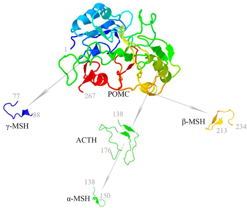
Chang et al.                                                                                                                     Melanocortins and Kidney Diseases

  FIGURE 2 | Distribution of the MCRs in kidney cells. MCRs are widely expressed in renal parenchymal cells. MCR, melanocortin receptor.

aortic sinus and in the whole aorta caused by apolipoprotein E                           MC4R is largely expressed in the central nervous system,
deficiency plus high-fat diet was exacerbated in recessive yellow                    where some MC4R expression overlaps with the localization of
(Mc1re/e) mice, associated with an enhanced arterial recruitment                     MC3R (Ghamari-Langroudi et al., 2018). Its function is believed
of Ly6Chigh monocytes (Rinne et al., 2018).                                          to be regulation of energy homeostasis and food intake (Huszar
   MC2R is predominantly expressed in adrenal cortices                               et al., 1997). Targeted deletion of MC4R gene is associated with
and adipose tissues. MC2R is unique that it only binds                               early-onset severe obesity, hyperphagia, and hyperinsulinemia.
to ACTH, which is produced by the pituitary gland and                                Even in cachexia models, MC4R knockout mice still have normal
circulated to the adrenal cortex where MC2R mediates                                 food intake, growth, and activity as compared with wild-type
steroidogenesis. In adipose tissues, the MC2R is known to                            mice. Furthermore, the study of MC4R knockout heterozygotes
mediate lipolysis (Cone et al., 1996). Mutations in human                            and homozygotes demonstrated that body weight homeostasis
MC2R are associated with familial glucocorticoid deficiency                          is more severely affected in homozygotes than in heterozygotes,
(FGD), which manifests with low levels of cortisol and                               consistent with a gene dosage effect (Cone, 2006).
high levels of ACTH due to the impairment in cortisol-                                   MC5R is highly expressed in exocrine glands and peripheral
mediated negative feedback to the hypothalamic release of                            tissues including, but not limited to, the adrenal gland, adipose
corticotropin-releasing factor and to the pituitary release of                       tissue, kidney, and leukocytes. Mice lacking MC5R were found
ACTH (Ramachandrappa et al., 2013).                                                  to have a severe defect in water repulsion and thermoregulation
   MC3R is expressed mainly in the brain, kidney, and other                          due to decreased production of sebaceous lipids. This finding
periphery tissues. It has the same affinity to ACTH as other                         may have implications for future research and treatment of skin
types of MCRs. While the exact pathobiological functions                             disorders, such as acne and dermatitis (Chen et al., 1997). Other
of MC3R still remain enigmatic, more and more evidence                               than the regulation of exocrine glands, MC5R may also play
suggests that MC3R may be involved in energy homeostasis                             an important role in farnesene-stimulated aggression in rodents,
(Ghamari-Langroudi et al., 2018). In support of this, MC3R                           as well as in anti-inflammation and immunomodulation (Gong,
knockout mice have increased fat mass, decreased lean mass,                          2014). Nevertheless, the exact function of MC5R in many tissues
hyperphagia, less activity, and mild late-onset obesity. Moreover,                   remains unclear.
experimental models of cachexia in the MC3R knockout mouse
are more susceptible to body weight loss. However, MC3R gene                         Agonists and Antagonists
polymorphism in humans has not yet been definitively associated                      The melanocortins include ACTH and the three melanocyte-
with obesity (Mencarelli et al., 2011).                                              stimulating hormones, namely α-MSH, β-MSH, and γ-MSH.

Frontiers in Physiology | www.frontiersin.org                                    3                                       February 2021 | Volume 12 | Article 651236
Chang et al.                                                                                                    Melanocortins and Kidney Diseases

Among these hormones, α-MSH is the most potent agonist for                syndecan-3 can, respectively, modulate the function of ASIP and
all MCRs except for MC2R, which is activated only by ACTH.                AGRP via promoting their competition with MSH for MCRs.
Furthermore, the β- and γ-MSH bind to MC3R with the same                  In addition, some endoplasmic reticulum-resident chaperones,
affinity as that of α-MSH (Kirwan et al., 2018). All melanocortin         like glucose-regulated protein (Yoon et al., 2018), have been
peptides, including MSH and ACTH, share a conserved core                  shown to promote trafficking and the intracellular signal of MCRs
tetrapeptide sequence His-Phe-Arg-Trp, which is essential to              (Rodrigues et al., 2015).The molecular mechanism underlying the
recognize and bind to MCRs. However, this sequence is not                 interaction between MCRs and the specific accessory proteins
sufficient for binding to MC2R. Another tetrapeptide motif Lys-           warrants further research (Gantz and Fong, 2003).
Lys-Arg-Arg is additionally required as the address sequence that            In an effort to reveal the functional association between
permits MC2R recognition and is unique to ACTH.                           the melanocortin system and other key pathways, a protein
    ASIP and AGRP are endogenous antagonists of MCRs, and                 interaction network diagram was constructed using Search Tool
play crucial roles, respectively, in the regulation of pigmentation       for the Retrieval of Interacting Genes/Proteins (STRING), a
and energy balance. ASIP is the human homolog of the mouse                biological database and web resource of known and predicted
agouti gene and encodes a 132 amino acid protein. ASIP is                 protein-protein interactions, based on the search of the
widely expressed in diverse human tissues, including the adipose          protein-protein interaction network database and functional
tissue, testis, ovary, heart, kidney, and liver, denoting a diverse       enrichment analyses. These analyses are capable of determining
range of functions (Voisey et al., 2003). In the rodents, ASIP,           the relationship of the melanocortin system with other proteins
encoded by the agouti gene, is primarily expressed in the skin,           located in a network hub. The protein POMC was located in
where it competes with MSH to bind with MC1R, resulting                   the most central area of the network, followed by corticotropin
in a decreased intracellular cAMP induction and thus affecting            releasing hormone (CRH), corticotropin releasing hormone
coat color. ASIP is able to bind to all five MCRs with the                receptor (CRHR), neuropeptide Y (NPY), Neuropeptide
highest affinity for MC1R and MC4R (Cortes et al., 2014). In              Y receptor (NPYR), neuropeptide S (NPS), agouti related
the hypothalamus, ectopic expression of agouti causes obesity             protein homolog (AGRP), mu-type opioid receptor (OPRM1),
due to its antagonism of MC4R. AGRP is found in the adrenal               corticotropin-releasing factor-binding protein (CRHBP), and
gland, kidney, lung, and plasma (Shen et al., 2002), but is mainly        MCRs (Figure 3).
expressed in the hypothalamus, where it acts as an antagonist
to MC3R and MC4R (Voisey et al., 2003). Interestingly, over-
expression of AGRP or administration of exogenous AGRP                    THE ROLE OF MELANOCORTIN SYSTEM
stimulates feeding, leading to obesity. However, unlike ASIP,             IN KIDNEY PATHOPHYSIOLOGY
there are no changes in pigmentation because AGRP doesn’t bind
to MC1R (Shen et al., 2002).                                              The key physiologic function of the kidney is to filtrate and
                                                                          excrete metabolic waste/toxins and to regulate fluid and
                                                                          electrolyte homeostasis and acid-base balance. The glomerular
Melanocortin Receptor Accessory                                           filtration barrier (GFB) is instrumental for plasma filtration
Proteins (MRAPs)                                                          and consists of a highly specialized blood filtration interface
The activity of MCRs is precisely regulated, not only by                  that exhibits a high permeability to small and midsized
their ligands (melanocortins or antagonists), but also by other           solutes in plasma but retains relative impermeability to
accessory proteins, named MRAP. MRAP is a single-pass                     macromolecules. The GFB is made up of 3 anatomical layers
transmembrane protein consisting of anti-parallel homodimers              known as the fenestrated capillary endothelium, glomerular
and plays an important role in the regulation of trafficking or           basement membrane, and podocytes, respectively. The
signaling of the 5 MCRs (Kim et al., 2014). MRAP exists in 2              podocyte, a terminally differentiated epithelial cell, plays a
isoforms, i.e., MRAP1 and MRAP2. MRAP1 is expressed in very               vital role in controlling the permselectivity of the GFB, and its
few tissues like adipocytes and the adrenal glands, where it is           dysfunction is centrally implicated in various glomerular diseases
essential for proper trafficking and signaling of the MC2R. In            (Haraldsson and Jeansson, 2009).
contrast, MRAP2 is widely expressed in a myriad of tissues in                 Adrenocorticotropic Hormone was approved in 1952 by the
addition to the adrenal glands. This distinct pattern of expression       U.S. FDA for the treatment of nephrotic syndrome. At that
may explain why mutations of MRAP1 account for 15–20% FGD                 time, ACTH was believed to act mainly via adrenocortical
(Rodrigues et al., 2015). MRAPs have no effect on the trafficking         steroidogenesis, and thus had been widely used in the treatment
of MC1R and MC3R but reduce surface expression of MC4R and                of a number of inflammatory or autoimmune disorders,
MC5R (Sebag and Hinkle, 2007, 2010; Chan et al., 2009). Indeed,           including rheumatoid arthritis (RA), gout, lupus, rheumatic
it appears that MRAP2 may act as a competitive inhibitor to               fever, psoriasis, and ulcerative colitis (Gallo-Payet, 2016).
MRAP1, but its exact role is still unclear. Mutations of MRAP2            However, due to its injectable route of administration and high
are associated with obesity but it has not been found to be linked        cost, ACTH was later replaced by synthetic glucocorticoids,
to FGD as MRAP1, suggesting that MRAP1 and MRAP2 are not                  which are much more affordable and provide the convenient oral
functionally interchangeable (Kim et al., 2014). Besides MRAPs,           route of administration. Recently, a growing body of evidence
a number of additional proteins also serve as potential accessory         indicates that ACTH is likely distinct from glucocorticoids in
proteins for MCRs. For instance, the mahogany protein and                 terms of clinical effectiveness as well as adverse effect profiles

Frontiers in Physiology | www.frontiersin.org                         4                                  February 2021 | Volume 12 | Article 651236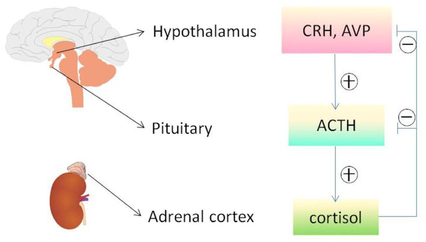
Chang et al. Melanocortins and Kidney Diseases
types, including vascular endothelial cells, glomerular mesangial
cells, glomerular podocytes and parietal epithelial cells, tubular
epithelial cells in different renal tubule segments, and renal
interstitial cells. Unfortunately, to date, it has been barely clarified
which MCR is expressed in what type of kidney cells. Recently,
based on reverse transcription-polymerase chain reaction (RT-
PCR) assay, Lindskog et al. (2010) demonstrated that MC1R
is the major MCR expressed in human and rat kidneys, more
specifically in podocytes, endothelial cells, mesangial cells, and
tubular epithelial cells. RT-PCR may be a useful molecular
biological technique, but it harbors potential pitfalls due to the
subjective nature in data analysis and reporting as well as the
technical limitations inherent in the assay like template quality
and operator variability (Bustin and Nolan, 2004). As such,
their initial results were likely unreliable, and not supported by
their subsequent studies, in which cAMP induction was barely
triggered in podocytes by selective MC1R agonists (Elvin et al.,
2016). In a later study, this group posited that MC1R expression
may be induced in podocytes upon stress or injury, despite very
low or no expression of MC1R under physiological conditions.
As such, they overexpressed human MC1R in cultured murine
podocytes (Elvin et al., 2016) and confirmed that activation of
MC1R by ACTH or by MC1R specific agonists was able to
FIGURE 3 | Protein-protein interactions network involving human
stimulate a cellular protective signaling cascade and protect the
melanocortin system and other key proteins as revealed by the STRING
(Search Tool for the Retrieval of Interacting Genes/Proteins), a biological
podocytes against injury. Nevertheless, somewhat contradictory
database and web resource of known and predicted protein–protein to this finding, a number of other studies demonstrated that
interactions. The associations between key proteins are indicated by the MC1R expression is predominant in renal tubules but very
connecting lines. The thickness of the connecting lines represents the weak in glomeruli (Siegrist et al., 1994; Gatti et al., 2006; Lee
strength of the associations. The associations do not necessarily suggest that
et al., 2008; Botte et al., 2014). To determine the expression
the proteins are physically binding each other, but may jointly contribute to a
shared function. The analysis was performed using STRING V11.0 (STRING profile of MC1R in diseased kidneys, the web-based gene
https://string-db.org/). CRH, corticotropin releasing hormone; NPY, expression database and analysis platform for transcriptomic
neuropeptide Y; NPS, neuropeptide S; AGRP, agouti related protein homolog; data of human kidney diseases (Nephroseq1 , version Nephroseq
OPRM1, mu-type opioid receptor; CRHBP, corticotropin-releasing v5) was employed. Shown in Figure 5, glomerular expression
factor-binding protein; MCR, melanocortin receptor.
of MC1R is comparable and not statistically different between
healthy living donors and renal patients with MN, FSGS or MCD,
based on post hoc analysis of gene expression microarray data
(Getting et al., 2002; Ko et al., 2004; Zaidi et al., 2010; Montero- derived from the Nephroseq Ju CKD Glom dataset (Ju et al.,
Melendez, 2015). For instance, one of the serious complications 2013), suggesting that glomerular expression of MC1R, if any, is
of glucocorticoid treatment is the risk of osteoporosis and not augmented upon glomerular injury.
osteonecrosis. In stark contrast, patients with ACTH-producing In addition to MC1R, multiple other MCRs have also been
adenomas typically do not develop osteonecrosis (Ko et al., reported to express in the kidney. Based on PCR amplification
2004) despite having very high levels of circulating glucocorticoid of human kidney-specific cDNA, Chhajlani and Wikberg (1996)
secondary to over-activation of the Pituitary-Adrenal hormone found strong expression of MC5R and weak expression of MC2R
axis (Figure 4). By using a rabbit model of steroid-induced in human kidney. Ni et al. (2006) found that MC3R, MC4R, and
avascular necrosis of the femoral head, Zaidi et al. (2010) MC5R were expressed in rat kidney using the same method.
demonstrated that ACTH therapy actually protects against It appears that these discrepant findings may reflect species
osteoporosis and osteonecrosis. In addition, Getting et al. (2002) differences, but may also be explained by potential pitfalls of
found in experimental models of gouty arthritis that ACTH the detection method, i.e., RT-PCR. Because all of the MCR
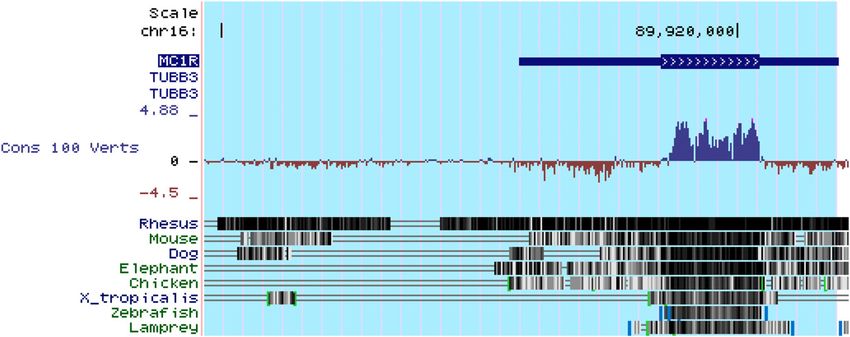
treatment has a potent anti-inflammatory effect without altering genes are intronless (Figure 6), genomic contamination may
circulating corticosterone levels and even in adrenalectomized confound the RT-PCR results. To address this issue, some studies
rats. When examining all of the data, it appears that ACTH prepared kidney mRNA in the presence of DNase and the quality
confers a better therapeutic efficacy but fewer side effects than was ascertained by the absence of amplicons of glyceraldehyde
glucocorticoids (Montero-Melendez, 2015), entailing that some 3-phosphate dehydrogenase (GAPDH) introns. It turned out that
steroidogenic-independent mechanisms may contribute to the rat kidneys mainly express MC1R and MC5R. However, MC1R
unique beneficial effect of ACTH. is mainly located to renal tubules but very weakly expressed
For a long time, MCRs have been known to express in the
kidney. The kidney is made up of a number of heterogeneous cell 1
www.nephroseq.org
Frontiers in Physiology | www.frontiersin.org 5 February 2021 | Volume 12 | Article 651236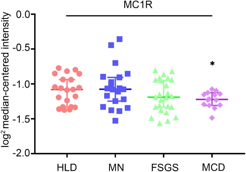
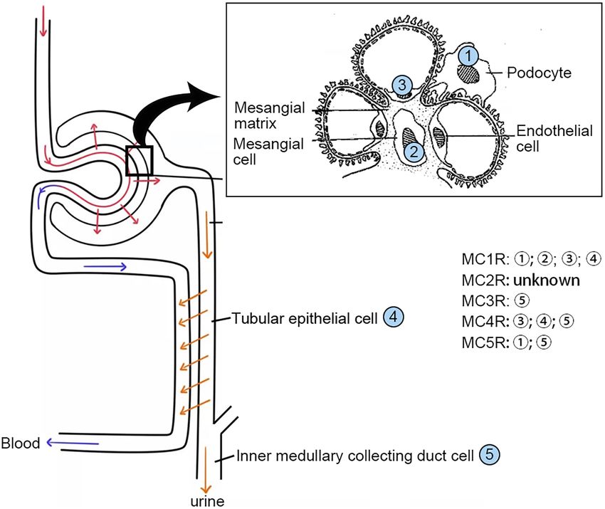
Chang et al.                                                                                                                          Melanocortins and Kidney Diseases

  FIGURE 4 | The hypothalamic-pituitary-adrenal axis. Cortisol released by the adrenal cortex in response to ACTH stimulation can negatively regulate the
  hypothalamus and pituitary, reduce the secretion of both corticotropin releasing hormone (CRH) and arginine vasopressin (AVP), and directly inhibit the pituitary
  production of ACTH and other melanocortins. The release of ACTH and other melanocortins is also affected by other factors via HPA axis, including stress and
  circadian rhythms. ACTH, adrenocorticotropic hormone; HPA, hypothalamic– pituitary–adrenal.

  FIGURE 5 | Expression levels of MC1R mRNA in glomeruli procured from healthy living donors and patients with diverse glomerular disease. Data were derived from
  www.Nephroseq.org on the basis of the Ju CKD Glom dataset. Glomerular expression of MC1R mRNA is not altered in glomerular diseases. Minimal Change
  Disease vs. Healthy Living Donor, ∗ P-value: 0.011. Focal Segmental Glomerulosclerosis vs. Healthy Living Donor, P-value: 0.111. Membranous
  Glomerulonephropathy vs. Healthy Living Donor, P-value: 0.697. HLD, healthy living donor; MN, membranous glomerulonephropathy; FSGS, focal segmental
  glomerulosclerosis; MCD, minimal change disease.

in glomeruli in rats (Si et al., 2013). Similar findings were also                      with loss-of-function or null mutations in MC1R. Furthermore,
made in murine kidneys (Qiao et al., 2020), suggesting that                             in complementary clinical studies, patients with steroid-resistant
MC1R is unlike a major mediator of the protective effect of                             nephrotic glomerulopathies, like idiopathic membranous
melanocortins on glomeruli. In consistency, the glomerular                              nephropathy or focal segmental glomerulosclerosis, responded
protective and anti-proteinuric effect of NDP-MSH in murine                             satisfactorily to ACTH monotherapy and ultimately achieved
models of podocytopathy elicited by LPS (Qiao et al., 2016) or                          clinical remission, despite the dominant-negative mutation
Adriamycin (Qiao et al., 2020) was completely retained in mice                          status of their MC1R gene as evidenced by the congenital

Frontiers in Physiology | www.frontiersin.org                                       6                                         February 2021 | Volume 12 | Article 651236
Chang et al.                                                                                                            Melanocortins and Kidney Diseases

  FIGURE 6 | All of the five types of melanocortin receptors are encoded by intronless genes, as exemplified here by the MC1R gene. MC1R is located at
  chr16:89,914,847-89,920,951. As shown above in genomic annotation of MC1R with UCSC Genome Browser (GRCh38/hg38), MC1R is highlighted and there is no
  intron in MC1R. Comparative Genomics indicated that MC1R is highly conserved among different species (http://genome.ucsc.edu/).

red hair color and by gene sequencing (Qiao et al., 2016),                     the pathogenic mechanisms and to aid in the research and
suggesting that a steroidogenic-independent non-MC1R-                          development of prevention and treatment measures (Pippin
mediated melanocortinergic signaling contributes to the                        et al., 2009). α-MSH, a melanocortin with the highest affinity
beneficial effect of melanocortin therapy in glomerular disease.               for all MCRs (except for MC2R), has been reproducibly shown
The exact identity of the MCR that mediates the glomerular                     to effectively protect the kidney in pre-clinical models of AKI
protection warrants in-depth investigations. Since most                        elicited by renal ischemia/reperfusion or by nephrotoxic drugs
glomerular diseases involve both glomerular cell autonomous                    (Kolgazi et al., 2007; Doi et al., 2008).
injury and systemic immunopathogenic mechanisms, the                               Chiao et al., were the first to demonstrate that α-MSH
melanocortinergic signaling driven by this MCR may have renal                  is able to inhibit inflammation and protect against AKI
and systemic effects.                                                          (Lipton et al., 1994; Chiao et al., 1997). They found that
   One of the critical physiological functions of the kidney is                α-MSH significantly reduced plasma blood urea nitrogen (BUN),
to maintain electrolyte homeostasis. This process is known to                  creatinine levels, and improved mortality in rats and mice
be heavily regulated by the melanocortin peptides, in particular               with renal ischemia/reperfusion injury elicited by renal artery
γ-MSH. γ-MSH has a high affinity for MC3R and thereby                          clamping for 30 or 40 min. This was consistent with histologic
regulates a variety of physiological activities, including energy              evidence that α-MSH treatment significantly inhibited necrosis,
homeostasis, food intake, hemodynamics, natriuresis and blood                  sloughing, and obstruction of the proximal straight tubules,
pressure regulation. MC3R is expressed in the brain and also                   which were severely damaged in the ischemic kidney (Chiao et al.,
abundantly in the heart and renal distal tubules (Humphreys,                   1997). Further study also found effective renal protection from
2004). γ-MSH promotes urinary salt excretion via activating                    α-MSH in the isolated kidney where neutrophils were completely
MC3R in distal renal tubules. In support of this, MC3R knockout                eliminated, thus suggesting a direct kidney protective effect of
or inhibition of γ-MSH production in mice caused salt-sensitive                α-MSH (Chiao et al., 1998). In agreement, another study by
hypertension (Kathpalia et al., 2011).                                         Jo et al. (2001b) suggested that α-MSH could directly mitigate
                                                                               tubular cell apoptosis and thus may play a role in mediating the
                                                                               beneficial effect in ischemic AKI. In addition, Deng et al., found
THE EFFECT OF MELANOCORTINS ON                                                 that α-MSH improved recovery of renal function in a model that
KIDNEY DISEASE                                                                 mimics kidney transplantation (Deng et al., 2001). Moreover, Li
                                                                               et al. (2006) showed that α-MSH can prevent deterioration of
Acute Kidney Disease (AKI)                                                     renal function in obstructive nephropathy.
Acute kidney injury (AKI) is a common and devastating                              In order to test the possible effect of α-MSH in an animal
complication in critically ill patients, characterized by an                   model more relevant to human AKI, Simmons et al. (2010)
abrupt deterioration of kidney function, and associated with                   employed the ischemia induced acute kidney injury in a porcine
high morbidity and mortality. AKI is a complex syndrome                        surgical model and found a robust renoprotective effect of AP214,
with a variety of different etiologies and pathophysiological                  an analog of α-MSH. Besides the ischemic AKI model, AP214
mechanisms. Most often, AKI is caused by ischemia or toxins                    had been tested in mice with cecal ligation and puncture (CLP),
and is referred to as acute tubular necrosis (ATN) pathologically              a model of sepsis-induced AKI. Intravenous injection of AP214
(Kohda et al., 1998). There has been little improvement in the                 improved blood pressure and heart rate in CLP mice, suggesting a
treatment of AKI despite hemodialysis and fluid replacement                    beneficial effect of AP214 on hemodynamics. In addition, AP214
therapy in the past 30 years. So the demand for novel                          inhibited inflammation, improved kidney function, and reduced
interventions of AKI is great. Animal models resembling                        mortality in sepsis-induced AKI, even when AP214 therapy was
human AKI has allowed scientists to effectively understand                     started 6h after injury (Doi et al., 2008).

Frontiers in Physiology | www.frontiersin.org                              7                                     February 2021 | Volume 12 | Article 651236
Chang et al. Melanocortins and Kidney Diseases
The efficacy of melanocortins was also tested in animal models A2 receptor (PLA2R), several studies examined the longitudinal
of nephrotoxic AKI. Miyaji et al., examined the effect of α-MSH response of the anti-PLA2R antibodies to ACTH treatment in
in murine models of mercuric chloride (HgCl2 ) induced AKI and iMN patients, and found that the titers of anti-PLA2R antibodies
found that α-MSH failed to reduce the level of serum creatinine decreased prior to proteinuria remission, entailing a potential
and tubular damage. They posited that α-MSH may be more role of ACTH in inducing immunological remission in iMN.
effective when the renal injury involves leukocyte-endothelial Because most patients had been resistant to corticosteroids or
interactions rather than direct tubular toxicity (Miyaji et al., the Ponticelli regimen, this immunosuppressive effect of ACTH
2002). In contrast, in the study by Kolgazi et al., α-MSH exhibited cannot be explained by the steroidogenic dependent pathway, but
a beneficial effect on gentamicin-induced nephrotoxicity and is more likely mediated directly through the MCRs expressed in
AKI, possibly via suppression of neutrophil infiltration and immune cells (Gong, 2014).
reactive oxygen metabolite induced lipid peroxidation (Kolgazi
et al., 2007). Although these pre-clinical studies are promising, MCD and FSGS
further research is still required to determine the usefulness of In experimental models of podocytopathy (LPS or Adriamycin-
α-MSH in clinical settings in patients. induced nephropathy), Qiao et al., found that NDP-MSH therapy
Although ACTH is also a potent agonist to all of the five protected against podocyte injury and glomerular damage and
MCRs, the effect of ACTH on AKI, unlike α-MSH, has been ameliorated proteinuria (Lindskog Jonsson et al., 2014). In
barely investigated. The only study by Si et al., examined the effect agreement in clinical settings, ACTH demonstrated great efficacy
of ACTH in two models of AKI, namely tumor necrosis factor- as a valuable alternative choice to treat FSGS and/or MCD
induced AKI in rats and CLP-induced AKI in mice (Si et al., for patients intolerant or resistant to conventional therapies,
2013). ACTH therapy demonstrated a remarkable protective including corticosteroids (Filippone et al., 2016). In support of
effect in both models. Of note, the beneficial effect of ACTH on this, ACTH therapy successfully induced complete or partial
AKI continued to increase even when the level of corticosteroid remission in 7 out of 24 FSGS patients (29%) who had previously
reached its peak, suggesting both steroidogenic-dependent and - failed other immunosuppressive regimens (Hogan et al., 2013).
independent (α-MSH-like) mechanisms may contribute. As such, Of these 7 patients, 5 were steroid resistant and 2 were steroid
ACTH is likely superior to α-MSH in treating AKI, owing to its dependent. There is little data on the effect of melanocortin
unique steroidogenic effect (Si et al., 2013). therapy in MCD, but two case series, respectively, involving 2 and
3 patients with MCD showed great remission rate after ACTH
Nephrotic Glomerulopathy therapy (Filippone et al., 2016; Madan et al., 2016).
A plethora of evidence supports the beneficial effect of
melanocortins in glomerulopathy (Table 2), including IgA Nephropathy
membranous nephropathy (MN), minimal change disease While IgA nephropathy is a common cause of chronic kidney
(MCD), focal segmental glomerular sclerosis (FSGS), and disease in some Asian countries, the published data regarding the
immunoglobulin A nephropathy (IgAN) (Gong, 2014). effectiveness of melanocortins in this population is little except
for several case series reports (Wyatt and Julian, 2013). Recently,
MN Prasad et al. (2018) found that the use of synthetic ACTH,
A number of melanocortins, including ACTH, α-MSH and the either as monotherapy or as a steroid-sparing agent, achieved
MC1R agonist MS05, have been tested in rats with Passive excellent outcomes in IgAN, and in most cases induced complete
Heymann nephritis (PHN), a classical model of human MN proteinuria remission. This is consistent with the experience of
(Pippin et al., 2009). Both α-MSH and MS05 significantly reduced using natural ACTH in patients with IgAN in the United States
proteinuria and improved glomerular injury. ACTH therapy had (Bomback et al., 2011, 2012; Prasad et al., 2018).
a tendency to reduce the level of proteinuria, but it did not
reach statistical significance, possibly due to insufficient dosing Diabetic Nephropathy (DN)
or lack of statistical power (Lindskog et al., 2010). Subsequently, Diabetic nephropathy is the leading cause of chronic kidney
in the clinical trial by Hladunewich et al., patients with biopsy disease (CKD) in Western societies, and one of the serious
proven idiopathic MN received ACTH gel monotherapy, and complications of diabetes mellitus with no definite therapy
60% (12 out of the 20 patients) achieved a complete or partial available yet (Webster et al., 2017). So far, the effect of
remission (Hladunewich et al., 2014). The synthetic ACTH was melanocortins on DN has been barely examined by pre-clinical
also tested in another study and showed a similar efficacy with a studies. In terms of clinical trials, due to the long-standing
55% complete and partial remission rate (11 out of 20 patients) controversy over the therapeutic benefit versus the risk of adverse
(van de Logt et al., 2015). A meta-analysis of the efficacy of effect of corticosteroids in diabetes associated kidney diseases,
ACTH in the treatment of glomerular diseases was carried out very few have been done to evaluate the effect of ACTH in
and revealed an 86% (25 out of 29 patients) remission rate for DN. Tumlin et al., performed the first randomized open-label
patients with iMN, who were converted to ACTH monotherapy pilot trial to test the effect of low-dose ACTH in DN (Jauregui
after having failed other immunosuppressive therapy including et al., 2009; Leeuwis et al., 2010; Tumlin et al., 2013). Their
glucocorticoids (Kittanamongkolchai et al., 2016). In view of results showed that ACTH treatment achieved complete or partial
the newly discovered pathogenic mechanism underlying iMN in remission in 57% (8 out of 14 patients) of the patients. No
men that involves the autoantibodies against anti-phospholipase significant difference in the efficacy of reducing proteinuria was
Frontiers in Physiology | www.frontiersin.org 8 February 2021 | Volume 12 | Article 651236Chang et al. Melanocortins and Kidney Diseases
TABLE 2 | The effect of melanocortins in kidney disease.
Kidney Diseases References Models/Diseases Effect of melanocortin treatment
AKI Chiao et al., 1997 Mice and rats with renal Inhibits inflammation (neutrophils, chemokines),
ischemia/reperfusion NO; attenuates necrosis, sloughing and
obstruction of the proximal straight tubules;
reduces BUN, Scr, and mortality
Chiao et al., 1998 ICAM-1 knock-out mice with Reduces BUN, renal cortex necrosis, and NO
renal ischemia/reperfusion;
isolated mouse kidneys
Jo et al., 2001b Rats with renal Improves tubular-cell apoptosis; reduces BUN,
ischemia/reperfusion Scr, tubular necrosis and tubular obstruction;
inhibits inflammation
Deng et al., 2001 Rat kidney transplantation Increases recovery of renal function
model
Li et al., 2006 Rats with bilateral ureteral Attenuates the downregulation of AQP2, AQP3,
obstruction Na-K-ATPase; reduces renal tubular cell
apoptosis; improves GFR
Simmons et al., 2010 Pigs with left nephrectomy and Reduces Scr; increases eGFR
ischemia of the right kidney
Doi et al., 2008 Mice with septic AKI induced Improves blood pressure and heart rate; inhibits
by cecal ligation and puncture inflammation; improves kidney function; and
reduces mortality
Miyaji et al., 2002 Mice with mercuric Fails to reduce the level of serum creatinine and
chloride-induced AKI tubular damage
Kolgazi et al., 2007 Rats with gentamicin-induced Reduces the severity of renal histological
AKI damage; fails to restore the impaired renal
function.
Si et al., 2013 Rats with TNF-elicited AKI; Improves survival and eGFR; reduces
Mice with septic AKI induced proteinuria, tubulointerstitial injury score,
by cecal ligation and puncture vacuolization area, dilation/sloughing and
tubular cell apoptosis
Nephrotic glomerulopathy MN Elvin et al., 2016 Rats with passive Heymann Restores serum albumin levels; improves
nephritis proteinuria, glomerular morphology, podocyte
injury; reduced oxidative stress, urine TBARS
Hladunewich et al., 2014 Patients with iMN Maintains renal function; reduces proteinuria;
60% (12/20) of patients achieve complete or
partial remission of proteinuria
van de Logt et al., 2015 Patients with iMN Reduces proteinuria; 55% (11/20) of patients
attain complete or partial remission of
proteinuria
Kittanamongkolchai et al., iMN patients resistant to other Reduces proteinuria; 86% (25/29) of patients
2016 immunosuppressants attain complete or partial remission of
proteinuria
MCD or FSGS Lindskog Jonsson et al., 2014 Mice with Adriamycin-induced No statistical significance in albuminuria, degree
podocytopathy of foot process effacement, disrupted
glomerular structures after melanocortin
therapy
Qiao et al., 2016, 2020 Mice with Adriamycin or NDP-MSH prominently improved proteinuria,
LPS-induced podocytopathy glomerular damage, podocyte ultrastructure via
an MC1R-independent mechanism
Filippone et al., 2016 FSGS or MCD patients All three patients with MCD achieved complete
resistant to conventional and partial remission; 40% (4/10) of FSGS
therapy patients attained complete and partial remission
Hogan et al., 2013 Patients with refractory FSGS 29% (7/24) of patient achieved complete or
partial remission, of which 5 had steroid
resistance
Madan et al., 2016 Patients with refractory FSGS 2/2 patients achieved complete or partial
remission
Wang et al., 2017 Pediatric patients with Ineffective at preventing disease relapses in
frequently relapsing or pediatric nephrotic syndrome (14/15 relapsed
steroid-dependent nephrotic on ACTH treatment)
syndrome
IgAN Prasad et al., 2018 Patients with refractory IgAN 1 patient achieved complete remission after
ACTH monotherapy; and ACTH was prescribed
as a steroid-sparing agent in combination with
cyclophosphamide for the other 2 patients
(Continued)
Frontiers in Physiology | www.frontiersin.org 9 February 2021 | Volume 12 | Article 651236Chang et al. Melanocortins and Kidney Diseases
TABLE 2 | Continued
Kidney Diseases References Models/Diseases Effect of melanocortin treatment
Bomback et al., 2012 Patients with refractory IgAN One patient achieved complete remission in
ACTH monotherapy
Bomback et al., 2011 Patients with refractory IgAN Two patients with steroid-resistant IgAN
demonstrated 50% reductions in proteinuria
DN Tumlin et al., 2013 Patients with DN 57% (8/14) patients achieved complete or partial
remission as a long-term effect.
Madan et al., 2016 Patients with DN 1 patient showed ≥ 30% proteinuria reduction; 2
had no response and 1 end up in early termination
LN Botte et al., 2014 Murine lupus-like models Reduces glomerular IgG deposits and reduces
lupus activity
Bomback et al., 2012 Patients with LN Patients with LN (class V) showed no response to
ACTH treatment
Other glomerular Berg and Arnadottir, 2004 Patients 6 MsPGN, 1 MCGN, 1 hereditary nephritis
diseases achieved complete or partial remission
Bomback et al., 2011 Patients 1 monoclonal DPGN patient failed to respond to
ACTH therapy
Interstitial nephritis and kidney fibrosis Jo et al., 2001a cultured human renal tubular α-MSH treatment significantly reduced
cells CsA-induced cellular apoptosis
Lee et al., 2004 Rats with CsA nephrotoxicity Improves renal cell apoptosis, inflammation and
tubulointerstitial fibrosis
AKI, acute kidney disease; NO, nitric oxide; ICAM, intercellular adhesion molecule; AQP, aquaporin; CLP, cecal ligation and puncture; TBARS, thiobarbituric acid-
reactive substances; MsPGN, mesangioproliferative glomerulonephritis; MCGN, mesangiocapillary glomerulonephritis; DPGN, diffuse proliferative glomerulonephritis;
CsA, cyclosporine A; TNF, tumor necrosis factor; MN, membranous nephropathy; MCD, minimal change disease; FSGS, focal segmental glomerular sclerosis; LPS,
lipopolysaccharides; NDP-MSH, [Nle4, DPhe7]-α-melanocyte-stimulating hormone; IgAN, immunoglobulin A nephropathy; DN, diabetic nephropathy; LN, lupus nephritis.
detected between the daily 16 IU dose group and the 32 IU nephritis, though pre-clinical evidence is still lacking (Berg and
dose group (Tumlin et al., 2013). Another study done by Madan Arnadottir, 2004; Bomback et al., 2011).
et al. (2016) involved only 4 DN patients, of which one patient
showed ≥ 30% proteinuria reduction, and the rest either had no Progressive CKD
response or ended the study early due to side effects. Collectively, Regardless of the original etiology, the final common pathway
clinical evidence suggests that melanocortin therapy might be for the progression of CKD is kidney fibrosis, characterized
useful in the treatment of DN, but large scale clinical trials are by glomerulosclerosis, tubular atrophy, inflammation, and
warranted to test the exact efficacy. interstitial fibrosis (Webster et al., 2017). The effect of
melanocortins on progressive CKD has been explored in
Lupus Nephritis (LN) pre-clinical studies by using in vivo or in vitro models of
Systemic lupus erythematosus (SLE) is an autoimmune disorder renal interstitial injury and fibrosis, including the model of
that affects many organ systems with one of the most common Cyclosporine A (CsA) nephropathy, which recapitulates key
and serious complications being LN. Several case reports features of renal tubular atrophy and interstitial fibrosis elicited
suggested a possible benefit of ACTH therapy in patients with by the use of CsA in human patients with high fidelity
LN (Bomback et al., 2011; Li et al., 2015). On the contrary, (Shihab et al., 1999). Jo et al. (2001a) found that α-MSH
in a murine lupus-like model induced by pristane, NDP-MSH treatment significantly attenuated CsA-induced apoptosis in
treatment did not reduce proteinuria or albuminuria, but it did cultured human tubular cells. In vivo in the rat model of CsA
improve histological markers of renal injury like glomerular IgG nephrotoxicity, Lee et al. (2004) demonstrated that α-MSH
deposition and did reduce lupus activity, marked by reduction can mitigate the CsA-induced tubulointerstitial fibrosis as well
in hypergammaglobulinemia, anti-nuclear antibodies, and anti- as tubular cell apoptosis. The results of these two studies
neutrophil cytoplasmic plasma antibodies (Botte et al., 2014). may pave the way to expand the clinical indications of
The mechanism responsible for this discrepancy was not fully melanocortin therapy.
understood. But a retrospective study to evaluate the role of
ACTH in SLE treatment suggested ACTH as an invaluable
alternative to corticosteroids in the treatment of SLE. ACTH SIDE EFFECTS
appears to be safe and well-tolerated after 6 months of treatment,
with a significant reduction in lupus activity (Li et al., 2015). All data to date has shown that the adverse effects of
melanocortins, in particular ACTH, are mild, tolerable,
Other Glomerular Disease and reversible, though it was commonly mentioned that
Based on some case series reports, ACTH treatment seems ACTH therapy may cause corticosteroid-like side effects. In
to be effective in a number of other glomerular diseases, contrast, patients treated with glucocorticoids experienced more
including mesangioproliferative glomerulonephritis (MsPGN), debilitating side effects including infection, hypertension, glucose
mesangiocapillary glomerulonephritis (MCGN), and hereditary intolerance, obesity, sleep disorders and others (Gong, 2014).
Frontiers in Physiology | www.frontiersin.org 10 February 2021 | Volume 12 | Article 651236Chang et al. Melanocortins and Kidney Diseases
Of note, most melanocortins are peptides or peptide derivatives ACTH protects the kidney through at least two mechanisms: (1)
and thus are biological macromolecules, which is inevitably stimulating the production of corticosteroids; and (2) activating
antigenic and may trigger immune reactions. Indeed, the use MCRs expressed by diverse kidney parenchymal cells. The latter
of animal-derived natural ACTH to treat glomerular disease one still needs continued research, considering the complexity
has been associated with de novo formation of neutralizing of the types of kidney cells, and the cross-interaction between
antibodies in some sensitive patients, followed by an acquired MCRs. However, with the advances in developing more specific
resistance to ACTH therapy (Wang et al., 2017; Shrivastava synthetic melanocortins and with the application of transgenic
et al., 2020). As such, there is a pressing need to develop animals with genetic ablation of specific components of the
small molecule MCR agonists or melanocortin analogs with melanocortin system, it is believed that much progress will be
less immunogenicity for improving the therapeutic efficacy in made in the near future regarding the role of melanocortinergic
patients with kidney diseases. pathways in kidney pathobiology.
CONCLUSION
AUTHOR CONTRIBUTIONS
The melanocortin hormone system is a complex and
incompletely understood neuroimmunoendocrine circuitry RG devised the conceptual ideas. MC performed the research.
in the mammalian body. The physiological interaction of LD contributed to discussion. MC and RG wrote the manuscript.
its constituent components increases the complexity of this BC and JS contributed to revision of the manuscript. All authors
hormone system. In recent years, new understandings about the approved the final version of the manuscript.
mechanisms of action of melanocortins promoted tremendous
exploration in this field. There has been a lot of evidence showing
that melanocortins confer renoprotective effects in animal FUNDING
models and in humans. Much of the compelling clinical evidence
is obtained from the use of ACTH in patients with steroid- This work was supported in part by the U.S. National Institutes
resistant glomerular disease. As a typical melanocortin peptide, of Health grant DK114006.
REFERENCES ischemia in mice and rats. J. Clin. Invest. 99, 1165–1172. doi: 10.1172/jci11
9272
Atwell, W. J. (1919). On the nature of the pigmentation changes following Chiao, H., Kohda, Y., McLeroy, P., Craig, L., Linas, S., and Star, R. A. (1998).
hypophysectomy in the frog larva. Science 49, 48–50. doi: 10.1126/science.49. Alpha-melanocyte-stimulating hormone inhibits renal injury in the absence of
1254.48 neutrophils. Kidney Int. 54, 765–774.
Berg, A. L., and Arnadottir, M. (2004). ACTH-induced improvement in the Cone, R. D. (2006). Studies on the physiological functions of the melanocortin
nephrotic syndrome in patients with a variety of diagnoses. Nephrol. Dial. system. Endocr. Rev. 27, 736–749. doi: 10.1210/er.2006-0034
Transplant. 19, 1305–1307. doi: 10.1093/ndt/gfh110 Cone, R. D., Lu, D., Koppula, S., Vage, D. I., Klungland, H., Boston, B., et al. (1996).
Bomback, A. S., Canetta, P. A., Beck, L. H. Jr., Ayalon, R., Radhakrishnan, J., The melanocortin receptors: agonists, antagonists, and the hormonal control of
and Appel, G. B. (2012). Treatment of resistant glomerular diseases with pigmentation. Recent Prog. Horm. Res. 51, 287–317; discussion 318.
adrenocorticotropic hormone gel: a prospective trial. Am. J. Nephrol. 36, 58–67. Cortes, R., Navarro, S., Agulleiro, M. J., Guillot, R., García-Herranz, V., Sánchez,
doi: 10.1159/000339287 E., et al. (2014). Evolution of the melanocortin system. Gen. Comp. Endocrinol.
Bomback, A. S., Tumlin, J. A., Baranski, J., Bourdeau, J. E., Besarab, A., Appel, 209, 3–10.
A. S., et al. (2011). Treatment of nephrotic syndrome with adrenocorticotropic Deng, J., Kohda, Y., Chiao, H., Wang, Y., Hu, X., Hewitt, S. M., et al. (2001).
hormone (ACTH) gel. Drug Des. Devel. Ther. 5, 147–153. doi: 10.2147/dddt. Interleukin-10 inhibits ischemic and cisplatin-induced acute renal injury.
s17521 Kidney Int. 60, 2118–2128. doi: 10.1046/j.1523-1755.2001.00043.x
Botte, D. A., Noronha, I. L., Malheiros, D. M., Peixoto, T. V., and de Mello, S. B. Doi, K., Hu, X., Yuen, P. S., Kim, S. M., Leelahavanichkul, A., Yasuda, H.,
(2014). Alpha-melanocyte stimulating hormone ameliorates disease activity in et al. (2008). AP214, an analogue of alpha-melanocyte-stimulating hormone,
an induced murine lupus-like model. Clin. Exp. Immunol. 177, 381–390. doi: ameliorates sepsis-induced acute kidney injury and mortality. Kidney Int. 73,
10.1111/cei.12336 1266–1274. doi: 10.1038/ki.2008.97
Bustin, S. A., and Nolan, T. (2004). Pitfalls of quantitative real-time reverse- Eipper, B. A., and Mains, R. E. (1980). Structure and biosynthesis of pro-
transcription polymerase chain reaction. J. Biomol. Tech. 15, 155–166. adrenocorticotropin/endorphin and related peptides. Endocr. Rev. 1, 1–27. doi:
Chan, L. F., Webb, T. R., Chung, T. T., Meimaridou, E., Cooray, S. N., Guasti, 10.1210/edrv-1-1-1
L., et al. (2009). MRAP and MRAP2 are bidirectional regulators of the Elvin, J., Buvall, L., Lindskog Jonsson, A., Granqvist, A., Lassén, E., Bergwall,
melanocortin receptor family. Proc. Natl. Acad. Sci. U.S.A. 106, 6146–6151. L., et al. (2016). Melanocortin 1 receptor agonist protects podocytes through
doi: 10.1073/pnas.0809918106 catalase and RhoA activation. Am. J. Physiol. Renal. Physiol. 310, F846–F856.
Chen, W., Kelly, M. A., Opitz-Araya, X., Thomas, R. E., Low, M. J., and Cone, Filippone, E. J., Dopson, S. J., Rivers, D. M., Jafari, G., Monk, R. D., Udani, S. M.,
R. D. (1997). Exocrine gland dysfunction in MC5-R-deficient mice: evidence et al. (2016). Adrenocorticotropic hormone analog use for podocytopathies. Int.
for coordinated regulation of exocrine gland function by melanocortin peptides. Med. Case Rep. J. 9, 125–133. doi: 10.2147/imcrj.s104899
Cell 91, 789–798. doi: 10.1016/s0092-8674(00)80467-5 Gallo-Payet, N. (2016). 60 Years of pomc: adrenal and extra-adrenal functions of
Chhajlani, V., and Wikberg, J. E. (1996). Molecular cloning and expression of the acth. J. Mol. Endocrinol. 56, T135–T156.
human melanocyte stimulating hormone receptor cDNA (FEBS 11553). FEBS Gantz, I., and Fong, T. M. (2003). The melanocortin system. Am. J. Physiol.
Lett. 390:238. Endocrinol. Metab. 284, E468–E474.
Chiao, H., Kohda, Y., McLeroy, P., Craig, L., Housini, I., and Star, R. A. (1997). Gatti, S., Colombo, G., Turcatti, F., Lonati, C., Sordi, A., Bonino, F., et al.
Alpha-melanocyte-stimulating hormone protects against renal injury after (2006). Reduced expression of the melanocortin-1 receptor in human liver
Frontiers in Physiology | www.frontiersin.org 11 February 2021 | Volume 12 | Article 651236Chang et al. Melanocortins and Kidney Diseases
during brain death. Neuroimmunomodulation 13, 51–55. doi: 10.1159/00009 and head osteonecrosis in Cushing’s disease. J. Formos Med. Assoc. 103,
4513 234–238.
Getting, S. J., Christian, H. C., Flower, R. J., and Perretti, M. (2002). Kohda, Y., Chiao, H., and Star, R. A. (1998). alpha-Melanocyte-stimulating
Activation of melanocortin type 3 receptor as a molecular mechanism for hormone and acute renal failure. Curr. Opin. Nephrol. Hypertens. 7, 413–417.
adrenocorticotropic hormone efficacy in gouty arthritis. Arthritis Rheum. 46, doi: 10.1097/00041552-199807000-00011
2765–2775. doi: 10.1002/art.10526 Kolgazi, M., Arbak, S., and Alican, I. (2007). The effect of alpha-melanocyte
Ghamari-Langroudi, M., Cakir, I., Lippert, R. N., Ellacott, K. L. J., Sweeney, P., stimulating hormone on gentamicin-induced acute nephrotoxicity in rats.
Litt, M. J., et al. (2018). Regulation of energy rheostasis by the melanocortin-3 J. Appl. Toxicol. 27, 183–188. doi: 10.1002/jat.1191
receptor. Sci. Adv. 4:eaat0866. doi: 10.1126/sciadv.aat0866 Konda, Y., Gantz, I., DelValle, J., Shimoto, Y., Miwa, H., and Yamada, T.
Gong, R. (2011). The renaissance of corticotropin therapy in proteinuric (1994). Interaction of dual intracellular signaling pathways activated by the
nephropathies. Nat. Rev. Nephrol. 8, 122–128. doi: 10.1038/nrneph.2011.190 melanocortin-3 receptor. J. Biol. Chem. 269, 13162–13166. doi: 10.1016/s0021-
Gong, R. (2014). Leveraging melanocortin pathways to treat glomerular diseases. 9258(17)36813-8
Adv. Chronic Kidney Dis. 21, 134–151. doi: 10.1053/j.ackd.2013.09.004 Lee, S. Y., Jo, S. K., Cho, W. Y., Kim, H. K., and Won, N. H. (2004). The
Haraldsson, B., and Jeansson, M. (2009). Glomerular filtration barrier. Curr. Opin. effect of -melanocyte stimulating hormone on renal tubular cell apoptosis and
Nephrol. Hypertens. 18, 331–335. tubulointerstitial fibrosis in cyclosporine A nephrotoxicity. Transplantation 78,
Hladunewich, M. A., Cattran, D., Beck, L. H., Odutayo, A., Sethi, S., Ayalon, 1756–1764. doi: 10.1097/01.tp.0000144332.44435.ab
R., et al. (2014). A pilot study to determine the dose and effectiveness of Lee, Y. S., Park, J. J., and Chung, K. Y. (2008). Change of melanocortin receptor
adrenocorticotrophic hormone (H.P. Acthar(R) Gel) in nephrotic syndrome expression in rat kidney ischemia-reperfusion injury. Transplant. Proc. 40,
due to idiopathic membranous nephropathy. Nephrol. Dial. Transplant. 29, 2142–2144. doi: 10.1016/j.transproceed.2008.07.101
1570–1577. doi: 10.1093/ndt/gfu069 Leeuwis, J. W., Nguyen, T. Q., Dendooven, A., Kok, R. J., and Goldschmeding,
Hogan, J., Bomback, A. S., Mehta, K., Appel, G. B., Canetta, P. A., Rao, M. K., et al. R. (2010). Targeting podocyte-associated diseases. Adv. Drug Deliv. Rev. 62,
(2013). Treatment of idiopathic FSGS with adrenocorticotropic hormone gel. 1325–1336. doi: 10.1016/j.addr.2010.08.012
Clin. J. Am. Soc. Nephrol. 8, 2072–2081. doi: 10.2215/cjn.02840313 Li, C., Shi, Y., Wang, W., Sardeli, C., Kwon, T. H., Thomsen, K., et al. (2006). alpha-
Humphreys, M. H. (2004). Gamma-MSH, sodium metabolism, and salt-sensitive MSH prevents impairment in renal function and dysregulation of AQPs and
hypertension. Am. J. Physiol. Regul. Integr. Comp. Physiol. 286, R417–R430. Na-K-ATPase in rats with bilateral ureteral obstruction. Am. J. Physiol. Renal.
Huszar, D., Lynch, C. A., Fairchild-Huntress, V., Dunmore, J. H., Fang, Q., Physiol. 290, F384–F396.
Berkemeier, L. R., et al. (1997). Targeted disruption of the melanocortin-4 Li, X., Golubovsky, J., Hui-Yuen, J., Lomeo, R., Shah, U., Olech, E., et al.
receptor results in obesity in mice. Cell 88, 131–141. doi: 10.1016/s0092- (2015). Adrenocorticotropic hormone gel in the treatment of systemic lupus
8674(00)81865-6 erythematosus: a retrospective study of patients. F1000Res 4:1103. doi: 10.
Jackson, I. J., Budd, P. S., Keighren, M., and McKie, L. (2007). Humanized MC1R 12688/f1000research.7192.1
transgenic mice reveal human specific receptor function. Hum. Mol. Genet. 16, Lindskog, A., Ebefors, K., Johansson, M. E., Stefánsson, B., Granqvist, A.,
2341–2348. doi: 10.1093/hmg/ddm191 Arnadottir, M., et al. (2010). Melanocortin 1 receptor agonists reduce
Jauregui, A., Mintz, D. H., Mundel, P., and Fornoni, A. (2009). Role of altered proteinuria. J. Am. Soc. Nephrol. 21, 1290–1298. doi: 10.1681/asn.2009101025
insulin signaling pathways in the pathogenesis of podocyte malfunction and Lindskog Jonsson, A., Granqvist, A., Elvin, J., Johansson, M. E., Haraldsson, B., and
microalbuminuria. Curr. Opin. Nephrol. Hypertens. 18, 539–545. doi: 10.1097/ Nystrom, J. (2014). Effects of melanocortin 1 receptor agonists in experimental
mnh.0b013e32832f7002 nephropathies. PLoS One 9:e87816. doi: 10.1371/journal.pone.0087816
Jo, S. K., Lee, S. Y., Han, S. Y., Cha, D. R., Cho, W. Y., Kim, H. K., et al. (2001a). Lipton, J. M., Ceriani, G., Macaluso, A., McCoy, D., Carnes, K., Biltz, J., et al. (1994).
Alpha-Melanocyte stimulating hormone (MSH) decreases cyclosporine a Antiinflammatory effects of the neuropeptide alpha-MSH in acute, chronic, and
induced apoptosis in cultured human proximal tubular cells. J. Korean Med. systemic inflammation. Ann. N. Y. Acad. Sci. 741, 137–148. doi: 10.1111/j.1749-
Sci. 16, 603–609. doi: 10.3346/jkms.2001.16.5.603 6632.1994.tb39654.x
Jo, S. K., Yun, S. Y., Chang, K. H., Kim, H. K., Cha, D. R., Cho, W. Y., et al. (2001b). Logan, D. W., Bryson-Richardson, R. J., Pagan, K. E., Taylor, M. S., Currie, P. D.,
alpha-MSH decreases apoptosis in ischaemic acute renal failure in rats: possible and Jackson, I. J. (2003). The structure and evolution of the melanocortin and
mechanism of this beneficial effect. Nephrol. Dial. Transplant. 16, 1583–1591. MCH receptors in fish and mammals. Genomics 81, 184–191. doi: 10.1016/
doi: 10.1093/ndt/16.8.1583 s0888-7543(02)00037-x
Ju, W., Greene, C. S., Eichinger, F., Bitzer, M., Nair, V., Hodgin, J. B., et al. (2013). Maaser, C., Kannengiesser, K., Specht, C., Luger, T. A., Lügering, A., Brzoska, T.,
Defining cell-type specificity at the transcriptional level in human disease. et al. (2006). Crucial role of the melanocortin receptor MC1R in experimental
Genome Res. 23, 1862–1873. doi: 10.1101/gr.155697.113 colitis. Gut 55, 1415–1422.
Kapas, S., Purbrick, A., and Hinson, J. P. (1995). Role of tyrosine kinase and protein Madan, A., Mijovic-Das, S., Stankovic, A., Teehan, G., Milward, A. S., and Khastgir,
kinase C in the steroidogenic actions of angiotensin II, alpha-melanocyte- A. (2016). Acthar gel in the treatment of nephrotic syndrome: a multicenter
stimulating hormone and corticotropin in the rat adrenal cortex. Biochem. J. retrospective case series. BMC Nephrol. 17:37. doi: 10.1186/s12882-016-0241-7
305(Pt 2), 433–438. doi: 10.1042/bj3050433 Mencarelli, M., Dubern, B., Alili, R., Maestrini, S., Benajiba, L., Tagliaferri, M.,
Kathpalia, P. P., Charlton, C., Rajagopal, M., and Pao, A. C. (2011). The natriuretic et al. (2011). Rare melanocortin-3 receptor mutations with in vitro functional
mechanism of Gamma-Melanocyte-Stimulating Hormone. Peptides 32, 1068– consequences are associated with human obesity. Hum. Mol. Genet. 20, 392–
1072. doi: 10.1016/j.peptides.2011.02.006 399. doi: 10.1093/hmg/ddq472
Kim, J. D., Leyva, S., and Diano, S. (2014). Hormonal regulation of the Miyaji, T., Hu, X., and Star, R. A. (2002). alpha-Melanocyte-simulating hormone
hypothalamic melanocortin system. Front. physiol. 5:480. doi: 10.3389/fphys. and interleukin-10 do not protect the kidney against mercuric chloride-induced
2014.00480 injury. Am. J. Physiol. Renal. Physiol. 282, F795–F801.
Kirwan, P., Kay, R. G., Brouwers, B., Herranz-Pérez, V., Jura, M., Larraufie, P., Montero-Melendez, T. (2015). ACTH: the forgotten therapy. Semin. Immunol. 27,
et al. (2018). Quantitative mass spectrometry for human melanocortin peptides 216–226. doi: 10.1016/j.smim.2015.02.003
in vitro and in vivo suggests prominent roles for beta-MSH and desacetyl alpha- Mountjoy, K. G., Robbins, L. S., Mortrud, M. T., and Cone, R. D. (1992). The
MSH in energy homeostasis. Mol. Metab. 17, 82–97. doi: 10.1016/j.molmet. cloning of a family of genes that encode the melanocortin receptors. Science
2018.08.006 257, 1248–1251. doi: 10.1126/science.1325670
Kittanamongkolchai, W., Cheungpasitporn, W., and Zand, L. (2016). Efficacy and Ni, X. P., Bhargava, A., Pearce, D., and Humphreys, M. H. (2006). Modulation
safety of adrenocorticotropic hormone treatment in glomerular diseases: a by dietary sodium intake of melanocortin 3 receptor mRNA and protein
systematic review and meta-analysis. Clin. Kidney J. 9, 387–396. doi: 10.1093/ abundance in the rat kidney. Am. J. Physiol. Regul. Integr. Comp. Physiol. 290,
ckj/sfw045 R560–R567.
Ko, J. Y., Chen, S. H., Chen, C. E., Chen, S. H., and Eng, H. L. Pippin, J. W., Brinkkoetter, P. T., Cormack-Aboud, F. C., Durvasula, R. V.,
(2004). Femoral head preservation in non-united femoral neck fracture Kowalewska, J., Hauser, P. V., et al. (2009). Inducible rodent models
Frontiers in Physiology | www.frontiersin.org 12 February 2021 | Volume 12 | Article 651236You can also read