Regulation of gliomagenesis and stemness through acid sensor ASIC1a
←
→
Page content transcription
If your browser does not render page correctly, please read the page content below
INTERNATIONAL JOURNAL OF ONCOLOGY 59: 82, 2021

                        Regulation of gliomagenesis and stemness
                              through acid sensor ASIC1a
                              PENDELTON KING1, JINGWEI WAN1,2, ALYSSA AIHUI GUO3,
                                SHANCHUN GUO4, YUGANG JIANG2 and MINGLI LIU1

    1
   Department of Microbiology, Biochemistry and Immunology, Morehouse School of Medicine, Atlanta, GA 30310, USA;
2
 Department of Neurosurgery, The Second Xiangya Hospital, Central South University, Changsha, Hunan 410008, P.R. China;
       3
        Department of Biomedical Sciences, School of Medicine Greenville, University of South Carolina, Greenville,
                 SC 29605; 4Department of Chemistry, Xavier University, New Orleans, LA 70125, USA

                                         Received May 7, 2021; Accepted July 28, 2021

                                                   DOI: 10.3892/ijo.2021.5262

Abstract. Glioblastoma multiforme (GBM) is the most preva‑         that ASIC1a expression was negatively associated with glioma
lent and aggressive type of adult gliomas. Despite intensive       grading. Functional studies revealed that the downregulation
therapy including surgery, radiation, and chemotherapy,            of ASIC1a promoted glioma cell proliferation and invasion,
invariable tumor recurrence occurs, which suggests that glio‑      while upregulation of ASIC1a inhibited their proliferation
blastoma stem cells (GSCs) render these tumors persistent.         and invasion. Furthermore, ASIC1a suppressed growth and
Recently, the induction of GSC differentiation has emerged as      proliferation of glioma cells through G1/S arrest and apoptosis
an alternative method to treat GBM, and most of the current        induction. Mechanistically, ASIC1a negatively modulated
studies aim to convert GSCs to neurons by a combination of         glioma stemness via inhibition of the Notch signaling pathway
transcriptional factors. As the tumor microenvironment is          and GSC markers CD133 and aldehyde dehydrogenase 1.
typically acidic due to increased glycolysis and consequently      ASIC1a is a tumor suppressor in gliomagenesis and stemness
leads to an increased production of lactic acid in tumor cells,    and may serve as a promising prognostic biomarker and target
in the present study, the role of acid‑sensing ion channel 1a      for GBM patients.
(ASIC1a), an acid sensor, was explored as a tumor suppressor
in gliomagenesis and stemness. The bioinformatics data from        Introduction
The Cancer Genome Atlas revealed that ASIC1 expression
levels in GBM tumor tissues were lower than those in normal        Glioma are aggressive lethal solid brain tumors derived from
brain, and glioma patients with high ASIC1 expression had          astrocytes and oligodendrocytes present in the central nervous
longer survival than those with low ASIC1 expression. Our          system (CNS). The most prevalent and aggressive type of adult
immunohistochemistry data from tissue microarray revealed          gliomas is the grade IV astrocytomas, which are also known as
                                                                   glioblastoma multiforme (GBM) (1). In the present study, the
                                                                   role of acid‑sensing ion channel 1a (ASIC1a) in gliomagenesis
                                                                   and stemness was explored as the tumor microenvironment
Correspondence to: Dr Shanchun Guo, Department of Chemistry,       is typically acidic due to increased glycolysis in tumor cells.
Xavier University, 1 Drexel Drive, New Orleans, LA 70125, USA      In general, due to an oxygen poor environment, tumor cells
E‑mail: sguo@xula.edu                                              switch to aerobic glycolysis to generate considerable amounts
Dr Mingli Liu, Department of Microbiology, Biochemistry and        of energy to support their rapid growth and progression. This
Immunology, Morehouse School of Medicine, Hugh Gloster Building,   results in the continuous generation of metabolic acids. While
Room 325, 720 Westeview Drive SW, Atlanta, GA 30310, USA           acidity is harmful to normal cells, long‑time coevolution of
E‑mail: mliu@msm.edu                                               tumor cells with the host has enabled them to be more adapt‑
                                                                   able to acidic microenvironments (2,3). Accumulating evidence
Abbreviations: ASIC1a, acid‑sensing ion channel 1a; ASICs,         has indicated that the acidity of the tumor microenvironment is
acid‑sensing ion channels; BAAA, BODIPY‑aminoacetaldehyde;         associated with stemness phenotype, poor prognosis of tumor
CNS, central nervous system; DEAB, diethylaminobenzaldehyde;
                                                                   patients, and stimulation of a chemo‑ and radio‑therapy resis‑
GBM, glioblastoma multiforme; GSCs, glioblastoma stem cells;
                                                                   tant phenotype (4). Ion channels are transmembrane proteins
IHC, immunohistochemistry; MTT, 3‑(4,5‑dimethylthiazol‑2‑yl)‑
2,5‑diphenyltetrazolium bromide; OS, overall survival              involved in regulating various physiological and pathological
                                                                   functions across biological membranes. The precise role of ion
Key words: ASIC1a, tumor suppressor, Notch, GSCs, CD133,           channels during regulation of cell survival and death is far from
aldehyde dehydrogenase 1                                           being understood, as ion channels may cause cell proliferation,
                                                                   cancer development, and metastasis in some cell types, but
                                                                   they may support regulated cell death in other cell types. The
                                                                   acid‑sensing ion channels (ASICs) are extracellular pH sensors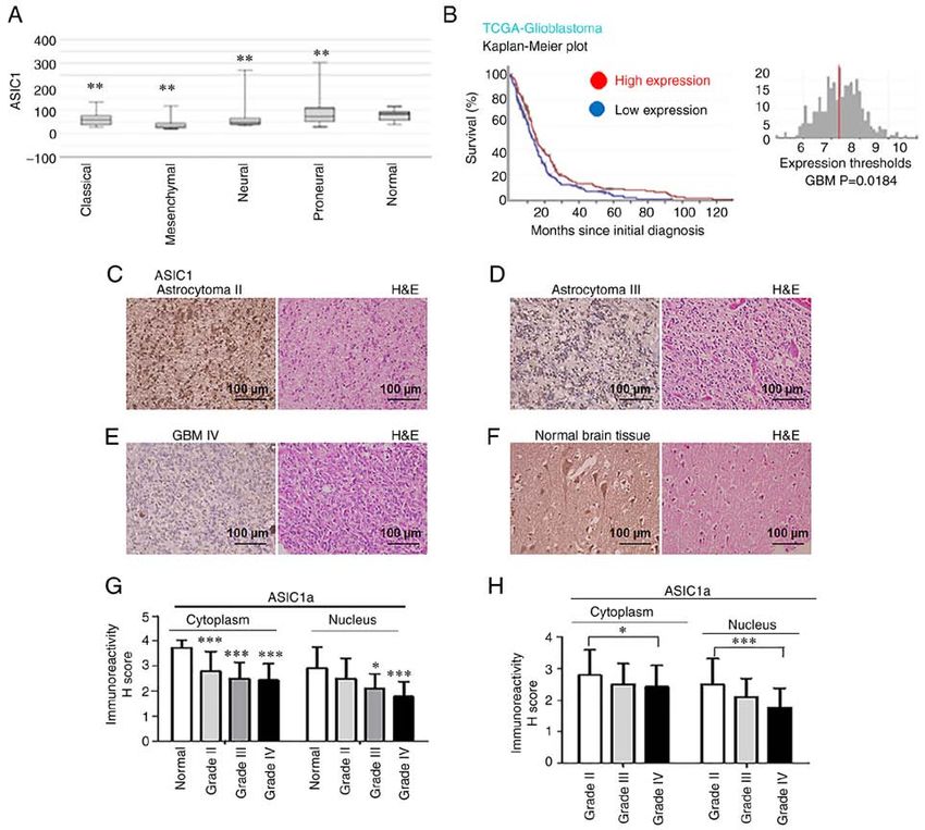
2 KING et al: REGULATION OF GLIOMAGENESIS THROUGH ACID SENSOR ASIC1a
that are acid responsive and can be transiently activated by (https://www.biomax.us/). Biopsy features included age, sex,
extracellular acidosis to be cation permeable (5). ASIC1 has organ or anatomic site involved, grading, and pathological
been reported to contribute to tumorigenesis in breast, pros‑ diagnosis (H&E‑stained sections). Slides from BioCoreUSA
tate, and pancreatic cancers. The conclusion regarding the role (product no. GL1001b) contained 75 cases of glioma: grade II,
of ASIC1 in glioma is inconsistent among different groups. n=51 (astrocytoma, n=47; oligodendroglioma, n=2; oligoas‑
Previous studies revealed that the knockdown of ASIC1 inhib‑ trocytoma, n=2); grade III, n=12 (anaplastic astrocytoma);
ited glioblastoma cell migration (6‑10). However, previous grade IV, n=12 (glioblastoma), and 10 cases of normal brain
available microarray data from The Cancer Genome Atlas tissues. Slides from Biomax (product no. GL803c) contained
(TCGA) revealed that glioma patients with high ASIC1 expres‑ 68 cases of glioma: grade II, n=27 (astrocytoma, n=14; oligoas‑
sion had increased survival compared with those with low trocytoma, n=13), grade III, n=4 (astrocytoma); grade IV, n=37
ASIC1 expression, which indicates that the preserved suscep‑ (glioblastoma, n=31; pleomorphic glioblastoma, n=6), and
tibility to extracellular pH may impair tumor growth (11). In 5 cases of normal brain tissues.
addition, Tian et al recently revealed that glioblastoma stem
cells (GSCs), which mainly account for the failure of current Immunohistochemistry (IHC). IHC staining was performed
treatment against malignant glioma, express functional on 5‑µm thick microarray slides. The slides were fixed using
ASIC1 and ASIC3 channels (11). Glioblastoma is driven by 4% paraformaldehyde for 30 min at room temperature and
stem cell‑like cells and is characterized by a block of cellular blocked by 10% normal horse serum at room temperature
differentiation. However, the mechanisms that accompany for 20 min. The immunohistochemical staining for ASIC1a
differentiation remain poorly understood. Any mechanisms was performed using the rabbit monoclonal anti‑ASIC1a anti‑
identified in GSCs with regard to astrocytes, oligodendrocytes, body, which is specific for ASIC1a, and a streptavidin‑biotin
and neuron differentiation will potentially lead to new strategies unlabeled immunoperoxidase technique (ABC‑Elite; Vector
to treat glioblastoma (12,13). The scope that GSCs permanently Laboratories, Inc.) with diaminobenzidine (DAB) as a chro‑
develop into a non‑proliferative and terminally differentiated mogen for ASIC1a. The sections were pretreated in citrate
state highlights the significance of differentiation therapy. In buffer of pH 6 for 10 min at 100˚C, and incubated with
the present study, it was revealed that ASIC1a functions as a primary antibody ASIC1a diluted at 1:100 at 4˚C overnight.
tumor suppressor in glioma stemness and tumorigenesis, which The secondary antibody was diluted at 1:200 and incubation
may provide therapeutic applications for GBM patients by was conducted at room temperature for 60 min. Mayer's hema‑
directing GSCs toward differentiation. toxylin was used for nuclear counterstaining for 2 min. The
slides were then visualized under a light microscope.
Materials and methods
HSORE determination. The staining intensity of cells in TMA
Antibodies and reagents. The following primary antibodies was evaluated as negative or positive in three different bright
were used in the present study: Rabbit monoclonal anti‑ASIC1a fields (≥100 cells/field). The semi‑quantitative HSCORE
antibody (cat. no. 35‑156465) was purchased from American was calculated for ASIC1a using the following equation:
Research Products, Inc. Rabbit polyclonal anti‑Notch4 antibody HSCORE=Σpi (i + 1), where ‘i’ is the intensity with a value of
(cat. no. 07‑189) and anti‑β‑actin antibody (product no. A3854) 0, 1, 2, or 3 (negative, weak, moderate or strong, respectively)
were purchased from Sigma‑Aldrich; Merck KGaA. Rabbit and ‘pi’ is the percentage of stained cells for each intensity (14).
polyclonal anti‑Notch3 antibody (product code ab60087) was Immunohistochemically stained slides were blindly reviewed
purchased from Abcam. Mouse monoclonal anti‑Notch1 anti‑ and scored by two independent investigators.
body (product no. N6786) was purchased from Sigma‑Aldrich;
Merck KGaA. Rabbit monoclonal anti‑Notch2 antibody Plasmids. The generation of short hairpin ASIC1a (shASIC1a),
(product no. 5732) and rabbit monoclonal anti‑survivin anti‑ the plasmid pEGFP‑ASIC1a, and corresponding controls
body (clone 71G4B7; product no. 2808) were purchased from were previously described (15). Briefly, shASIC1a and
Cell Signaling Technology, Inc. Rabbit polyclonal anti‑CD133 control shRNA were purchased from SuperArray Bioscience
antibody (cat no. NB120‑16518) was purchased from Novus Corporation. Each vector contained shRNA under the control
Biologicals, LLC. Rabbit polyclonal anti‑aldehyde dehydroge‑ of U1 promoter and green fluorescent protein (GFP) gene
nase 1 antibody (ALDH1; cat no. GTX123973) was purchased for enrichment of transiently transfected cells. In detail,
from GeneTex, Inc. Mouse monoclonal anti‑p21 antibody (cat SureSilencing shRNA plasmid for human ACCN2 (ASIC1a,
no. sc‑817), mouse monoclonal anti‑Fas antibody (sc‑8009), amiloride‑sensitive cation channel 2, neuronal) was designed
and mouse monoclonal anti‑cyclin D1 antibody (sc‑8396) to specifically knockdown the expression of ASIC1a gene
were purchased from Santa Cruz Biotechnology, Inc. All by RNA interference under transient transfection condi‑
secondary antibodies (goat anti‑rabbit, peroxidase‑conjugated, tions after performing appropriate enrichment. The vector
cat. no. AP132P; and goat anti‑mouse antibody, peroxi‑ contained shRNA under the control of U1 promoter and
dase‑conjugated, cat. no. AP124P) used for western blotting GFP gene for enrichment of transiently transfected cells.
were purchased from Calbiochem; Merck KGaA. Psalmotoxin The RefSeq accession number (NM_020039) refers to the
(PcTx1) was obtained from Tocris (cat. no. 5042). representative sequence used to design the enclosed shRNA.
The insert sequence is: GCCA AGA AGT TCA ACA AATCT.
Tissue microarray (TMA). Glioma tissue arrays from Chinese The sequence of normal control (NC) is GGAATCTCATTC
patients were purchased from BioCoreUSA Corporation GATG CATAC. The plasmid overexpressing ASIC1a named
(https://biocoreusa.com/default.aspx) and US Biomax, Inc. pEGFP‑ASIC1a was constructed as previously described (15).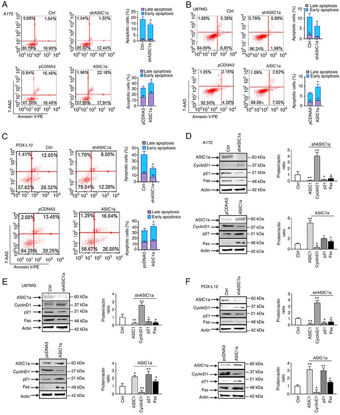
INTERNATIONAL JOURNAL OF ONCOLOGY 59: 82, 2021                                             3

Briefly, the rat ASIC1a cDNA (NM_024153) was fused with             for the indicated time‑points. Another set of glioma cells was
a GFP at the c‑terminal and inserted into pcDNA3. The rat           treated with PcTx1 and the corresponding control. A total of
ASIC1a cDNA (NM_024153) tagged with epitope FLAG                    10 µl of 3‑(4,5‑dimethylthiazol‑2‑yl)‑2,5‑diphenyltetrazolium
(YKDDDDK) at the C terminus was constructed in plasmid              bromide (MTT) reagent (the ratio of MTT reagent to the
pCDNA3.                                                             medium was 1:10; Sigma‑Aldrich; Merck KGaA) was added
                                                                    into each well and incubated in the dark at 37˚C for 2‑4 h.
Cell culture. Human glioblastoma cell lines: A172 (RRID:            Isopropanol was used to dissolve the formazan. The absor‑
CVCL_0131) and U87MG (HTB‑14), a glioblastoma of                    bance was measured at 570 nm using 690 nm as the reference
unknown origin (RRID: CVCL_0022) were obtained from                 and the absorbances were obtained using a CytoFluor™ 2300
American Type Culture Collection (ATCC). All cells were             plate reader.
cultured in Dulbecco's modified Eagle's medium (DMEM)
supplemented with 10% fetal bovine serum (FBS; both                 Cell migration and invasion assays. The migration and inva‑
from Thermo Fisher Scientific, Inc.), 50 U/ml penicillin            sion potential were assessed as previously described (16,17).
and 50 µg/ml streptomycin at 37˚C. All cell lines had been          Briefly, cell culture chambers with 8‑µm pore size polycar‑
authenticated using short tandem repeat profiling within            bonate membrane filters (Corning, Inc.) were used for cell
the last three years. Patient‑derived xenoline (PDX) lines:         invasion assays with the filters precoated with Matrigel at 37˚C
Primary tumor tissue cubes stored at liquid nitrogen [provided      for 3 h (50 µl; 1.25 mg/ml). Each of the glioma cell lines/PDX
by Dr Yancey G. Gillespie at the University of Alabama at           were transfected with or without shASIC1 or ASIC1‑GFP for
Birmingham (UAB), Birmingham, USA] were implanted                   48 h, and harvested and seeded with 5x10⁵ cells in 200 µl of
subcutaneously into the flanks of male or female 6‑8 week‑old       DMEM supplemented with 1% FBS in the upper chambers.
nude mice under anesthesia (ketamine/xylazine 90/6 mg/kg            The bottom chambers were filled with 500 µl DMEM supple‑
BW). A total of 4 athymic nude mice that were 6‑8‑weeks old         mented with 10% FBS. Following another 24 h of incubation
and with an average weight of 25 g, obtained from Charles           at 37˚C, Matrigel and cells on the upper surface of the filter
River Laboratories, Inc., were maintained on 12‑h light/dark        were wiped off thoroughly with Q‑tips. Cells attached on the
cycle with access to food and water ad libitum. The mice were       lower surface of the membrane filters were fixed with 4% para‑
housed at temperature of 18‑23˚C with 40‑60% humidity.              formaldehyde/PBS for 10 min and stained with 0.5% crystal
Mice with tumors exceeding 1,000 mm 3 were euthanized               violet/methanol for 10 min at room temperature. The cells
(by cervical dislocation) and the tumors were removed for           were then counted using light microscopy with a magnifica‑
further study. The time interval between implantation and the       tion of x10 in 3‑4 random fields. Cell numbers under different
end of the experiment ranged from 3‑4 weeks. Briefly, cryo‑         treatments were normalized to the appropriate controls. Assays
preserved tumor tissues were thawed at 37˚C and washed with         were performed in triplicate samples and performed in three
phosphate‑buffered saline (PBS) before subcutaneous implan‑         independent experiments.
tation. To prepare single‑cell suspension of viable tumor cells,
the xenograft tumor tissues were harvested and minced with          Western blotting. Cells were lysed with lysis buffer (50 mM
scalpel blades followed by passing through cell strainers. The      HEPES, 150 mM NaCl, 1.5 mM MgCl2, 1 mM EGTA, 10%
cells were then cultured in DMEM/F‑12 media plus 10% FBS,           glycerol, 1% Nonidet P‑40, 100 mM NaF, 10 mM sodium
50 U/ml penicillin and 50 µg/ml streptomycin for future use.        pyrophosphate, 0.2 mM sodium orthovanadate, 1 mM phenyl‑
All experiments were performed with mycoplasma‑free cells.          methylsulfonyl fluoride, 10 µg/ml aprotinin and 10 µg/ml
The study was carried out in strict accordance with the recom‑      leupeptin). The protein concentration was determined by BCA
mendations in the Guide for the Care and Use of Laboratory          assay. Samples (50 µg) were separated using 4‑15% SDS
Animal of the National Institutes of Health. The protocol was       PAGE, and separated proteins were transferred to nitrocel‑
approved (approval no. 20‑14) by the Institutional Animal           lulose membranes and identified by immunoblotting. The
Care and Usage Committee (IACUC) of Morehouse School of             membranes were blocked using 5% milk for 1 h at room
Medicine (Atlanta, USA).                                            temperature. Primary antibodies, incubated at 4˚C overnight,
                                                                    were obtained from commercial sources and were diluted at a
Transfection of shRNA and DNA constructs. When the glio‑            ratio of 1:1,000 according to the manufacturer's instructions.
blastoma cells reached ~50‑75% confluency in 35‑mm dishes,          Subsequently, the membranes were incubated with secondary
5 µg of ASIC1a shRNA, or pEGFP‑ASIC1a, FLAG‑ASIC1a                  peroxidase‑conjugated antibodies (at a dilution of 1:2,000)
or corresponding controls were transfected into glioma cells        at room temperature for 1 h. The blots were developed with
using the Lipofectamine RNAiMAX or Lipofectamine 3000               Supersignal Pico or Femto substrate (Pierce; Thermo Fisher
transfection reagent (Invitrogen; Thermo Fisher Scientific,         Scientific, Inc.). Densitometric analysis of the bands was
Inc.) at room temperature for 30 min, according to the manu‑        performed with ImageQuant software version 6.1 (Bio‑Rad
facturer's instructions. The transiently transfected glioma cells   Laboratories, Inc.).
expressing each specific construct were maintained in DMEM
containing 10% FBS for further growth for 72 h.                     Flow cytometry. For cell cycle analysis and the apoptosis
                                                                    assay, a total of 1x10⁶ cells were harvested, fixed in ice‑cold
MTT assay. All glioma cells were seeded at 2.5x10⁴ cells in         70% ethanol at 4˚C for 20 min, and resuspended in PBS
100 µl of medium per well into 96‑well plates and transfected       for 1 min at room temperature. Following room tempera‑
with specific shRNA or DNA constructs or controls using             ture centrifugation at 450 x g for 5 min with the brake
Lipofectamine RNAiMAX or Lipofectamine 3000 reagents                on low, the cells were resuspended in 200 µl Guava Cell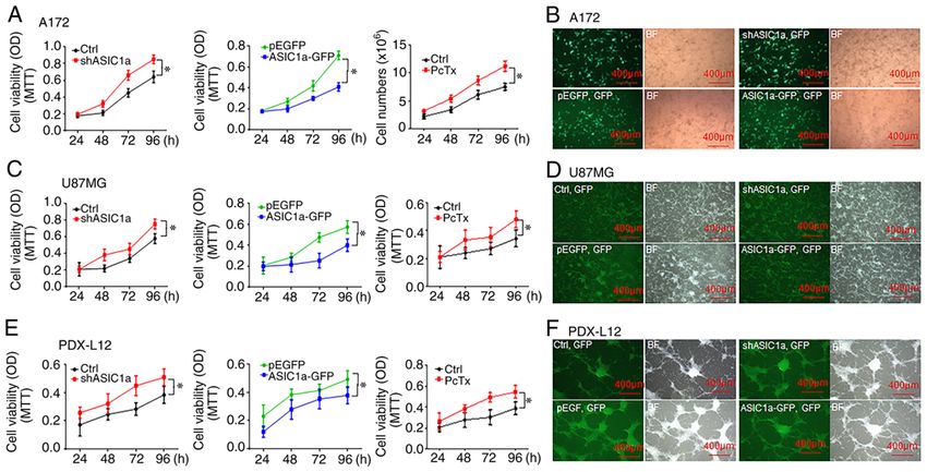
4                          KING et al: REGULATION OF GLIOMAGENESIS THROUGH ACID SENSOR ASIC1a

Cycle Reagent (part no. 4500‑0220; Luminex Corporation;           Results
containing propidium iodide) and incubated at room
temperature for 30 min while shielded from the light. All         Expression of ASIC1a is associated with improved survival
samples were transferred to 96‑well microplate plates with a      in GBM patients and reduced ASIC1a protein expression
round bottom and acquired on a Guava easyCyte 8HT Base            is associated with grade in glioma patients. To determine
System (Luminex Corporation). The percentage of cells in          whether ASIC1 gene expression is related to patient survival,
G0/G1, S, and G2/M phases was determined from the DNA             the TCGA database was analyzed. ASIC1 mRNA expression
content using guavaSoft 3.1.1 (Luminex Corporation). The          levels in glioma tissue were lower than those in normal brain,
apoptotic glioma cells were detected by flow cytometry            irrespectively of GBM subtypes (classical, mesenchymal,
using Annexin V‑PE and 7‑AAD. The staining procedure              neural, and proneural) as detected by Affymetrix HT HG
was conducted with a Guava Nexin Reagent kit (part                U133A (P
INTERNATIONAL JOURNAL OF ONCOLOGY 59: 82, 2021                                                                  5

Figure 1. Expression of ASIC1 is associated with improved survival in glioblastoma patients, and reduced ASIC1a protein expression is associated with
grade progression in glioma patients. (A) ASIC1 mRNA expression levels in brain tissues of different molecular subtypes of glioma patients were analyzed
by Affymetrix HT HG U133ATCGA data. (B) Kaplan‑Meier survival curve for the 5‑year OS rate of glioma patients. The cut‑off was set at the median.
(C‑F) ASIC1a protein was expressed in (C) grade II gliomas, (D) grade III gliomas, (E) grade IV GBM and (F) normal brain tissue. Images were captured at a
magnification of x40. (G) Quantification of IHC results in grade IV GBM, grade III, and II gliomas compared with that of normal brain tissue. (H) Quantification
of IHC results compared among different grades (IV GBM, grade III, and II gliomas). *P
6                               KING et al: REGULATION OF GLIOMAGENESIS THROUGH ACID SENSOR ASIC1a

Figure 2. Downregulation of ASIC1a promotes glioma cell proliferation, while overexpression of ASIC1a inhibits its growth. (A) A172 cells were transfected
with shASIC1a (left), treated with ASIC1a selective inhibitor PcTx1 (right), and transfected with ASIC1a‑GFP (middle) for 24 to 96 h followed by an MTT
assay. (B) Transfection efficiency of A172 cells was revealed by the expression of GFP marker. (C) U87MG cells were transfected with shASIC1a (left), treated
with ASIC1a selective inhibitor PcTx1 (right), and transfected with ASIC1a‑GFP (middle) for 24 to 96 h followed by an MTT assay. (D) Transfection efficiency
of U87MG cells was revealed by the expression of GFP marker. (E) PDX‑L12 cells were transfected with shASIC1a (left), treated with ASIC1a selective
inhibitor PcTx1 (right), and transfected with ASIC1a‑GFP (middle) for 24 to 96 h followed by an MTT assay. (F) Transfection efficiency of PDX‑L12 cells was
revealed by the expression of GFP marker. *P
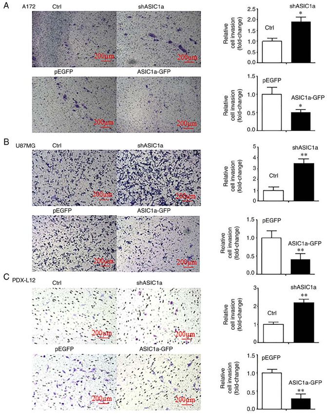
INTERNATIONAL JOURNAL OF ONCOLOGY 59: 82, 2021                                                             7

Figure 3. Downregulation of ASIC1a increases glioma cell invasion, while overexpression of ASIC1a decreases its invasion. (A) Cell invasion assays were
conducted to examine the effects of ASIC1a on the invasion after transfecting A172 cells with shASIC1a targeting ASIC1a mRNA (upper panel) or transfected
with the plasmid overexpression ASIC1a (ASIC1a‑GFP, lower panel). Images were captured at a magnification of x10. (B) Cell invasion assays were conducted
to examine the effects of ASIC1a on invasion after transfecting U87MG cells with shASIC1a (upper panel) or transfected with ASIC1a‑GFP (lower panel).
(C) Cell invasion assays were conducted to examine the effects of ASIC1a on invasion after transfecting PDX‑L12 cells with shASIC1a (upper panel) or
transfected with ASIC1a‑GFP (lower panel). *P
8                              KING et al: REGULATION OF GLIOMAGENESIS THROUGH ACID SENSOR ASIC1a

Figure 4. ASIC1a suppresses the growth and proliferation of glioma cells through G1/S arrest. Cell cycle progression was analyzed by flow cytometry.
Representative histograms of cell cycle progression and bar graphs revealed the mean percentage of cells in G0/G1, S and G2/M phases of (A) A172,
(B) U87MG, and (C) PDX‑L12 cells by shASIC1a treatment. Overexpression of ASIC1a induced G0/G1 cell cycle accumulation of (D) A172, (E) U87MG,
and (F) PDX‑L12 cells. The bar graphs indicate the mean ± SD of three independent experiments. All data represent a representative experiment from three
independent experiments. *P
INTERNATIONAL JOURNAL OF ONCOLOGY 59: 82, 2021                                                               9

Figure 5. ASIC1a suppresses the growth and proliferation of glioma cells through the induction of apoptosis. (A‑C) Apoptosis was analyzed by flow cytometric
analysis of Annexin V‑PE staining after shASIC1 treatment in (A, upper panel) glioma cells A172, (B, upper panel) U87MG and (C, upper panel) PDX‑L12
cells. Apoptosis was also examined by overexpression of ASIC1a in (A, lower panel) A172, (B, lower panel) U87MG and (C, lower panel) PDX‑L12 cells.
(D‑F) The cell cycle regulatory proteins cyclin D1 and p21, as well as apoptotic‑related protein Fas were examined when the ASIC1 gene was silenced by
shASIC1a in (D, upper panel) A172, (E, upper panel) U87MG, and (F, upper panel) PDX‑L12 cells. Cyclin D1, p21, and Fas were also detected when the ASIC1a
gene was overexpressed in (D, lower panel) A172, (E, lower panel) U87MG and (F, lower panel) PDX‑L12 cells. The bar graphs indicate the mean ± SD of three
independent experiments. All data represent a representative experiment from three independent experiments. *P
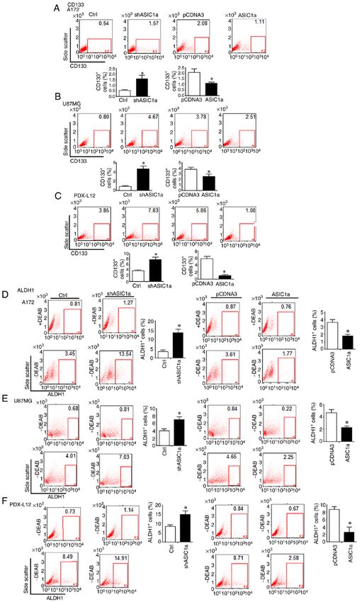
10                               KING et al: REGULATION OF GLIOMAGENESIS THROUGH ACID SENSOR ASIC1a

Figure 6. GSC markers CD133 and ALDH1 are negatively associated with ASIC1a in glioblastoma multiforme. (A‑C) Flow cytometric analysis to assess
CD133 expression in (A, left two panels) A172, (B, left two panels) U87MG, and (C, left two panels) PDX‑L12 glioma cells by ASIC1a silencing. CD133
expression was determined when (A, right two panels) A172, (B, right two panels), U87MG, and (C, right two panels) PDX‑L12 cells overexpressed ASIC1a.
(D‑F) ALDH1 enzymatic activities were determined by the ALDEFLUOR assay which was performed in ASIC1a‑knockdown (D, left five panels) A172,
(E, left five panels), U87MG and (F, left five panels) PDX‑L12 cells. ALDH1 enzymatic activities were also determined in (D, right five panels) A172, (E, right
five panels) U87MG and (F, right five panels) PDX‑L12 cells overexpressing ASIC1a. Bars on the right of each image represent the mean ± SD after normal‑
ization to control. All results are representative of three separate experiments. *P
INTERNATIONAL JOURNAL OF ONCOLOGY 59: 82, 2021                                               11

ASIC1a expression levels were elevated by introducing             Discussion
ASIC1a‑FLAG into glioma cells, the number of CD133 +
cells revealed a decrease in A172 (Fig. 6A, right two             In the present study, two glioma cell lines, A172 and U87MG
panels), U87MG (Fig. 6B, right two panels) and PDX‑L12            and one PDX line were utilized based on their different molec‑
cells (Fig. 6C, right two panels). To further verify our          ular characteristics. A172 and U87MG both have wild‑type
findings, the effects of PcTx1 on the expression of CD133         TP53, PTEN mutations, and CDKN2A (p14ARF/p16 INK4a)
on PDX‑L12 glioma cells were examined. As anticipated,            deletion (25). However, U87MG cells have another CDKN2C
PcTx1 effectively increased expression of CD133 compared          (p18INK4c) mutation, express high levels of VEGF as compared
with that of the control (Fig. S2). To address the question of    with A172 expressing high levels of bFGF (26). PDX‑L12 cells,
whether ASIC1a would affect ALDH1, another GSC marker,            a PDX with neural subtype, have wild‑type genes of EGFR,
the ALDEFLUOR assay was performed on an identical                 PTEN, CDKN2A, NF‑κB, and amplified genes of CDK4/MDM
model of glioma cells aforementioned, in which ASIC1a             and CSNK2A with a deleted TP53. Major findings from the
was either underexpressed or overexpressed by shASIC1‑            present study include: i) Expression of ASIC1 was associated
or ASIC1‑FLAG‑related constructs. ASIC1a silencing                with improved survival in glioblastoma patients and reduced
increased the number of ALDH1‑positive cells from 3.45            ASIC1a protein expression was associated with grade progres‑
to 13.54% in A172 (Fig. 6D, left five panels), from 4.01 to       sion in glioma patients; ii) downregulation of ASIC1a increased
7.03% in U87MG (Fig. 6E, left five panels), and from 8.49 to      glioma cell proliferation and invasion, while upregulation of
14.91% in PDX‑L12 cells (Fig. 6F, left five panels). In accor‑    ASIC1a decreased their proliferation and invasion; iii) ASIC1a
dance with the relationship of ASIC1a and CD133, here, our        suppressed the growth and proliferation of glioma cells through
results produced further evidence that ASIC1a knockdown           G1/S arrest and induced apoptosis; and iv) ASIC1a causatively
resulted in an increased GSC population as defined by the         induced the inactivation of Notch, reduced expression of GSC
ALDH1+ population. Similarly, overexpression of ASIC1a            markers CD133 and ALDH1, and played an important role in
decreased the ALDH1+ cells in all of the three glioma             glioma stemness.
cells examined. In detail, compared with corresponding                A total of 4 ASIC genes (ASIC1, ASIC2, ASIC3 and ASIC4)
controls, the ALDH1+ cells decreased from 3.61 to 1.77%           and splice variants for ASIC1 (ASIC1a, ASIC1b, and ASIC1b2)
in A172‑transfected ASIC1a‑FLAG cells (Fig. 6D, right             and ASIC2 (ASIC2a and ASIC2b) have been identified and
five panels), from 4.65 to 2.25% in U87MG‑transfected             found to be expressed in a variety of cell types (27,28). The
ASIC1a‑FLAG cells (Fig. 6E, right five panels), and from          functional ASICs are trimeric assemblies with each subunit
8.71 to 2.58% in PDX‑L12‑transfected ASIC1a‑FLAG cells            consisting of two transmembrane domains (2). ASICs are
(Fig. 6F, right five panels).                                     voltage‑independent ion channels and have the highest expres‑
    It was previously reported that ASIC1 promotes differen‑      sion in the brain, mainly in the central nervous system (28,29);
tiation of neuroblastoma by negatively regulating the Notch       in addition, they are also expressed in the retina (27,30,31), lung
signaling pathway (14). The Notch signaling pathways plays        epithelia (31), bone and cartilage (32), pituitary gland (33), and
critical roles in the maintenance and differentiation of neural   testis (34). As extracellular acidosis is typically concomitant
stem cells (NSC) (23) and can maintain GSCs in an undif‑          with brain injury, ASICs, the main neuronal H+ receptor in
ferentiated state (24). It was therefore determined whether       neurons, play an important role in neuronal injury under various
ASIC1a is a critical regulator of Notch1 gene expression during   injurious conditions in the brain (35). In ischemia‑induced
gliomagenesis. Upon ASIC1a downregulation by shASIC1a,            brain injury, multiple endogenous factors (lactate, speri‑
in A172 cells, the active form of Notch, intracellular domains    mine, and dynorphins) potentiate ASIC1a channel‑mediated
of Notch1 (Notch1/NICD), Notch2/NICD, Notch3/NICD,                ischemic injury. Multiple sclerosis (MS) is a demyelinating
Notch4/NICD, along with the Notch target survivin expression      disease in CNS and is associated with prolonged inflamma‑
were increased. The GSC markers CD133 and ALDH1 were              tion and acidification. A recent clinical study conducted by
increased as well (Fig. 7A). U87MG (Fig. 7B) and PDX‑L12          Arun et al revealed that amiloride, an ASIC1 inhibitor, has
cells (Fig. 7C) exhibited similar patterns based on ASIC1a        a promising neuroprotective effect by reducing brain atrophy
silencing, with enhanced expression of Notch active forms of      of MS patients, axonal damage, and myelin loss (36). Brain
Notch1/NICD, Notch2/NICD, Notch3/NICD, Notch4/NICD,               pH is reduced in traumatic brain injury (TBI) patients due to
Notch target survivin, and GSC markers CD133 and ALDH1            massive disruption of metabolism; and in TBI patients whose
in response to ASIC1a knockdown. To further detect the asso‑      ASIC1a expression was increased in the brain, amiloride or
ciation between ASIC1a and Notch receptors, A172, U87MG           PcTx1 (ASIC1 selective inhibitor) attenuated the severity of
and PDX‑L12 cells were transfected with ASIC1‑FLAG to             brain injury (37). ASIC‑mediated responses result in loss of
overexpress ASIC1a protein along with the control vector          dopaminergic neurons in Parkinson's disease (PD). In patients
pCDNA3. The anticipated results were observed in that             with PD, ASIC1 inhibition with amiloride or PcTx1 alleviates
ASIC1a‑overexpressing A172 (Fig. 7D), U87MG (Fig. 7E)             the reduction of immunoreactivity of tyrosine hydroxylase and
and PDX‑L12 (Fig. 7F) cells exhibited decreased Notch             dopamine transporter, and consequently prevents the loss of
active forms of Notch1/NICD, Notch2/NICD, Notch3/NICD,            dopaminergic neurons, and therefore impedes PD progres‑
Notch4/NICD, Notch target survivin and GSC makers CD133           sion (38).
and ALDH1. These results indicated that ASIC1a causatively            Adaptions to the highly acidic microenvironment are crucial
induced the inactivation of Notch, reduced the expression of      steps in the development of invasive cancer (3). As a result,
GSC markers CD133 and ALDH1, and played a critical role in        proton (H+) concentration increases within the lumen and causes
glioma stemness.                                                  the interior of the lumen to become highly acidic. Cancer cells
12 KING et al: REGULATION OF GLIOMAGENESIS THROUGH ACID SENSOR ASIC1a Figure 7. Notch signaling is negatively associated with ASIC1a in glioblastoma multiforme. (A‑C) The glioma cells were transfected with shASIC1a and control followed by assaying protein expression of ASIC1a, active form of Notch1, Notch2, Notch3, Notch4, and Notch target survivin, as well as GSC markers CD133 and ALDH1 by western blotting in (A) A172, (B) U87MG, and (C) PDX‑L12 cells. (D‑F) Subsequently, the glioma cells (D) A172, (E) U87MG and (F) PDX‑L12 were transfected with ASIC1a‑FLAG followed by assaying protein expression of ASIC1a, active form of Notch1, Notch2, Notch3, Notch4 and Notch target survivin, as well as GSC markers CD133 and ALDH1 by western blotting. *P
INTERNATIONAL JOURNAL OF ONCOLOGY 59: 82, 2021                                               13

express RNA for numerous different subunits of the ASIC and        actual disease more closely than the established glioma cell
ENaC families (6). Unlike ENaC, ASICs are proton‑gated             lines, which may not resemble the original tumor, by adapting
cation‑selective channels most permeable to Na+ ions (29,47,48).   to the environment and acquiring mutations. In the future, the
ASIC1a and heteromeric ASIC1a/2b channels are permeable            conclusion from this study, especially from PDX, should be
to Ca2+ and can cause an accumulation of intracellular Ca2+ in     tested in immunocompromised animals.
neurons (49,50). The studies on the role of ASIC1 in glioma‑
genesis are controversial. Sun et al reported that ASIC1 and       Acknowledgements
CaMKII form a functional complex at the plasma membrane
in GBM cells, which promotes GBM migration. However, their         We are grateful to Dr Yancey G. Gillespie at the University
results were only based on experimentation in the U251‑MG          of Alabama at Birmingham (UAB) (Birmingham, USA) for
cell line, which may represent a selection bias (51). Previous     providing us with the PDX lines.
studies performed in 2003 and 2009, that support the mito‑
genic role of ASIC1 reported that silencing of ASIC1 inhibits      Funding
glioblastoma cell migration (6,7). The mechanical studies from
this group demonstrated that ASIC interacted with several          The present study was supported by the SC3 grant from NIH
biochemical molecules such as integrin‑β and α‑actinin (9),        NIGMS GM121230 (ML) and partly supported by the Morehouse
ENaC subunits (8), Hsc70 (52) or cleaved by serine protease        School of Medicine Tx Pilot grant (ML). The funding agencies
matriptase (53) to accomplish its functions. The apparent limi‑    had no role in the design, collection, analysis, and interpretation
tation of their studies is lack of prognostic information drawn    of the study's data and in writing the manuscript.
from big data bioinformatics, which is critical to identify the
difference between tumor suppressor genes and oncogenes.           Availability of data and materials
    The study supporting a tumor suppressor role of ASIC1
originated from previous research from Tian et al. In rat C6       The datasets used and/or analyzed during the current study are
glioma cells, functional activation of ASIC1 induced a short       available from the corresponding author on reasonable request.
depolarization or a transient calcium influx even with persis‑
tent acidic stimulation. Notably, GSC expresses functional         Authors' contributions
ASIC1a and ASIC3. Microarray data from their study revealed
that the expression of ASIC1 and ASIC3 was associated with         SG and ML designed the study protocol. PK, JW, AAG, and
improved survival of glioma patients, which indicated that         YJ performed experiments based on glioma cell cultures
the preserved susceptibility to extracellular pH may impair        and evaluated the data with the help of ML. ML performed
tumor growth (11). In 2017, our group first revealed that          the biostatistical evaluation of the data. AAG and ML wrote
ASIC1 induces neuroblastoma differentiation (14). Later,           the manuscript with contributions and final approval by all
Zhang et al revealed that both human‑induced pluripotent stem      authors. SG and YJ contributed to the critical reading and
cell‑(hiPSC)‑derived neural progenitor cell (hiPSC‑NPC)            revision of the manuscript. All authors read and approved the
and hiPSC‑NPC‑derived neurons express abundant ASIC1               final manuscript.
mRNA (54). These findings provided an indication of the
important relationship between stem cells and ASIC1. As            Ethics approval and consent to participate
acidic stress maintains (55) or promotes (40) glioblastoma
stem cell‑like phenotype, the acid‑sensor ASIC1a regulation        The study was carried out in strict accordance with the recom‑
of GSC markers was therefore evaluated. In the present study,      mendations in the Guide for the Care and Use of Laboratory
different glioma cell lines and PDX were utilized to reveal        Animals of the National Institutes of Health. The protocol
that ASIC1a serves as a tumor suppressor in glioma develop‑        was approved (approval no. 20‑14) by the Institutional Animal
ment and progression, which is consistent with the research of     Care and Usage Committee (IACUC) of Morehouse School of
Tian et al (11). It was also revealed that ASIC1a expression is    Medicine (Atlanta, USA).
inversely associated with glioma grade progression by using
human glioma tumor tissues. The role for ASIC1a as a tumor         Patient consent for publication
suppressor is further strengthened by the bioinformatic data
from TCGA, which demonstrated that GBM patients with               Not applicable.
high expression ASIC1 have improved OS, indicating that
ASIC1 is a promising prognostic biomarker for GBM patients.        Competing interests
The antitumor function of ASIC1 was also supported by our
previous work (14), which revealed that ASIC1a promotes            The authors declare that they have no competing interests.
neurite growth and differentiation by negatively regulating
Notch signaling. In summary, our data strongly indicated that      References
ASIC1a functions as a tumor suppressor in glioma stemness
                                                                    1. Louis DN, Perry A, Reifenberger G, von Deimling A, Figarella-
and tumorigenesis. Stimulation of ASIC1 activity may inhibit           Branger D, Cavenee WK, Ohgaki H, Wiestler OD, Kleihues P
GSC self‑renewal and glioma progression.                               and Ellison DW: The 2016 World Health Organization classifica‑
    All the major findings from the present study were drawn           tion of tumors of the central nervous system: A summary. Acta
                                                                       Neuropathol 131: 803‑820, 2016.
from in vitro cell cultures from established glioma cell lines      2. Damaghi M, Wojtkowiak JW and Gillies RJ: pH sensing and
or glioma PDX. PDX has its advantages by recapitulating the            regulation in cancer. Front Physiol 4: 370, 2013.
14 KING et al: REGULATION OF GLIOMAGENESIS THROUGH ACID SENSOR ASIC1a
3. Lee WY, Huang SC, Hsu KF, Tzeng CC and Shen WL: Roles for 23. Hitoshi S, Alexson T, Tropepe V, Donoviel D, Elia AJ, Nye JS,
hypoxia‑regulated genes during cervical carcinogenesis: Somatic Conlon RA, Mak TW, Bernstein A and van der Kooy D: Notch
evolution during the hypoxia‑glycolysis‑acidosis sequence. pathway molecules are essential for the maintenance, but not
Gynecol Oncol 108: 377‑384, 2008. the generation, of mammalian neural stem cells. Genes Dev 16:
4. Ward G, Meehan J, Gray ME, Murray AF, Argyle DJ, Kunkler IH 846‑858, 2002.
and Langdon SP: The impact of tumor pH on cancer progression: 24. Purow BW, Haque RM, Noel MW, Su Q, Burdick MJ, Lee J,
Strategies for clinical intervention. Explor Target Antitumor Sundaresan T, Pastorino S, Park JK, Mikolaenko I, et al:
Ther 1: 71‑100, 2020. Expression of notch‑1 and its ligands, delta‑like‑1 and jagged‑1,
5. Xiong ZG, Chu XP and Simon RP: Acid sensing ion chan‑ is critical for glioma cell survival and proliferation. Cancer
nels‑novel therapeutic targets for ischemic brain injury. Front Res 65: 2353‑2363, 2005.
Biosci 12: 1376‑1386, 2007. 25. Ishii N, Maier D, Merlo A, Tada M, Sawamura Y, Diserens AC and
6. Berdiev BK, Xia J, McLean LA, Markert JM, Gillespie GY, Van Meir EG: Frequent co‑alterations of TP53, p16/CDKN2A,
Mapstone TB, Naren AP, Jovov B, Bubien JK, Ji HL, et al: p14ARF, PTEN tumor suppressor genes in human glioma cell
Acid‑sensing ion channels in malignant gliomas. J Biol Chem 278: lines. Brain Pathol 9: 469‑479, 1999.
15023‑15034, 2003. 26. Ke LD, Shi YX, Im SA, Chen X and Yung WK: The relevance
7. Kapoor N, Bartoszewski R, Qadri YJ, Bebok Z, Bubien JK, of cell proliferation, vascular endothelial growth factor, and
Fuller CM and Benos DJ: Knockdown of ASIC1 and epithelial basic fibroblast growth factor production to angiogenesis and
sodium channel subunits inhibits glioblastoma whole cell current tumorigenicity in human glioma cell lines. Clin Cancer Res 6:
and cell migration. J Biol Chem 284: 24526‑24541, 2009. 2562‑2572, 2000.
8. Kapoor N, Lee W, Clark E, Bartoszewski R, McNicholas CM, 27. Ettaiche M, Guy N, Hofman P, Lazdunski M and Waldmann R:
Latham CB, Bebok Z, Parpura V, Fuller CM, Palmer CA and Acid‑sensing ion channel 2 is important for retinal func‑
Benos DJ: Interaction of ASIC1 and ENaC subunits in human tion and protects against light‑induced retinal degeneration.
glioma cells and rat astrocytes. Am J Physiol Cell Physiol 300: J Neurosci 24: 1005‑1012, 2004.
C1246‑C1259, 2011. 28. Krishtal O: The ASICs: Signaling molecules? Modulators?
9. Rooj AK, Liu Z, McNicholas CM and Fuller CM: Physical and Trends Neurosci 26: 477‑483, 2003.
functional interactions between a glioma cation channel and 29. Wemmie JA, Price MP and Welsh MJ: Acid‑sensing ion chan‑
integrin‑β1 require α‑actinin. Am J Physiol Cell Physiol 309: nels: Advances, questions and therapeutic opportunities. Trends
C308‑C319, 2015. Neurosci 29: 578‑586, 2006.
10. Rooj AK, McNicholas CM, Bartoszewski R, Bebok Z, Benos DJ 30. Brockway LM, Zhou ZH, Bubien JK, Jovov B, Benos DJ and
and Fuller CM: Glioma‑specific cation conductance regu‑ Keyser KT: Rabbit retinal neurons and glia express a variety of
lates migration and cell cycle progression. J Biol Chem 287: ENaC/DEG subunits. Am J Physiol Cell Physiol 283: C126‑C134,
4053‑4065, 2012. 2002.
11. Tian Y, Bresenitz P, Reska A, El Moussaoui L, Beier CP and 31. Lingueglia E: Acid‑sensing ion channels in sensory perception.
Gründer S: Glioblastoma cancer stem cell lines express func‑ J Biol Chem 282: 17325‑17329, 2007.
tional acid sensing ion channels ASIC1a and ASIC3. Sci Rep 7: 32. Jahr H, van Driel M, van Osch GJ, Weinans H and van
13674, 2017. Leeuwen JP: Identification of acid‑sensing ion channels in bone.
12. Carén H, Stricker SH, Bulstrode H, Gagrica S, Johnstone E, Biochem Biophys Res Commun 337: 349‑354, 2005.
Bartlett TE, Feber A, Wilson G, Teschendorff AE, Bertone P, et al: 33. Grunder S, Geissler HS, Bässler EL and Ruppersberg JP: A
Glioblastoma stem cells respond to differentiation cues but fail to new member of acid‑sensing ion channels from pituitary gland.
undergo commitment and terminal cell‑cycle arrest. Stem Cell Neuroreport 11: 1607‑1611, 2000.
Reports 5: 829‑842, 2015. 34. Ishibashi K and Marumo F: Molecular cloning of a DEG/ENaC
13. Park NI, Guilhamon P, Desai K, McAdam RF, Langille E, sodium channel cDNA from human testis. Biochem Biophys Res
O'Connor M, Lan X, Whetstone H, Coutinho FJ, Vanner RJ, et al: Commun 245: 589‑593, 1998.
35. Huang Y, Jiang N, Li J, Ji YH, Xiong ZG and Zha XM: Two
ASCL1 reorganizes chromatin to direct neuronal fate and aspects of ASIC function: Synaptic plasticity and neuronal
suppress tumorigenicity of glioblastoma stem cells. Cell Stem injury. Neuropharmacology 94: 42‑48, 2015.
Cell 21: 209‑224.e7, 2017. 36. Arun T, Tomassini V, Sbardella E, de Ruiter MB, Matthews L,
14. Lopes C, Madureira TV, Gonçalves JF and Rocha E: Disruption Leite MI, Gelineau‑Morel R, Cavey A, Vergo S, Craner M, et al:
of classical estrogenic targets in brown trout primary hepatocytes Targeting ASIC1 in primary progressive multiple sclerosis:
by the model androgens testosterone and dihydrotestosterone. Evidence of neuroprotection with amiloride. Brain 136: 106‑115,
Aquat Toxicol 227: 105586, 2020. 2013.
15. Liu M, Inoue K, Leng T, Zhou A, Guo S and Xiong ZG: ASIC1 37. Yin T, Lindley TE, Albert GW, Ahmed R, Schmeiser PB,
promotes differentiation of neuroblastoma by negatively regu‑ Grady MS, Howard MA and Welsh MJ: Loss of acid sensing ion
lating Notch signaling pathway. Oncotarget 8: 8283‑8293, 2017. channel‑1a and bicarbonate administration attenuate the severity
16. Larco DO, Semsarzadeh NN, Cho‑Clark M, Mani SK and Wu TJ: of traumatic brain injury. PLoS One 8: e72379, 2013.
β‑Arrestin 2 is a mediator of GnRH‑(1‑5) signaling in immortal‑ 38. Chu XP and Xiong ZG: Physiological and pathological functions
ized GnRH neurons. Endocrinology 154: 4726‑4736, 2013. of acid‑sensing ion channels in the central nervous system. Curr
17. Liu M, Inoue K, Leng T, Guo S and Xiong ZG: TRPM7 channels Drug Targets 13: 263‑271, 2012.
regulate glioma stem cell through STAT3 and Notch signaling 39. Pilon‑Thomas S, Kodumudi KN, El‑Kenawi AE, Russell S,
pathways. Cell Signal 26: 2773‑2781, 2014. Weber AM, Luddy K, Damaghi M, Wojtkowiak JW, Mulé JJ,
18. Salinas M, Rash LD, Baron A, Lambeau G, Escoubas P and Ibrahim‑Hashim A and Gillies RJ: Neutralization of tumor
Lazdunski M: The receptor site of the spider toxin PcTx1 on the acidity improves antitumor responses to immunotherapy. Cancer
proton‑gated cation channel ASIC1a. J Physiol 570: 339‑354, Res 76: 1381‑1390, 2016.
2006. 40. Hjelmeland AB, Wu Q, Heddleston JM, Choudhary GS,
19. Sherwood TW, Lee KG, Gormley MG and Askwith CC: MacSwords J, Lathia JD, McLendon R, Lindner D, Sloan A and
Heteromeric acid‑sensing ion channels (ASICs) composed Rich JN: Acidic stress promotes a glioma stem cell phenotype.
of ASIC2b and ASIC1a display novel channel properties and Cell Death Differ 18: 829‑840, 2011.
contribute to acidosis‑induced neuronal death. J Neurosci 31: 41. Arcangeli A, Pillozzi S and Becchetti A: Targeting ion channels
9723‑9734, 2011. in leukemias: A new challenge for treatment. Curr Med Chem 19:
20. Wan J, Guo AA, King P, Guo S, Saafir T, Jiang Y and Liu M: 683‑696, 2012.
TRPM7 induces tumorigenesis and stemness through notch 42. Lehen'kyi V, Shapovalov G, Skryma R and Prevarskaya N: Ion
activation in glioma. Front Pharmacol 11: 590723, 2020. channnels and transporters in cancer. 5. Ion channels in control
21. Omoruyi SI, Ekpo OE, Semenya DM, Jardine A and Prince S: of cancer and cell apoptosis. Am J Physiol Cell Physiol 301:
Exploitation of a novel phenothiazine derivative for its C1281‑C1289, 2011.
anti‑cancer activities in malignant glioblastoma. Apoptosis 25: 43. Li M and Xiong ZG: Ion channels as targets for cancer therapy.
261‑274, 2020. Int J Physiol Pathophysiol Pharmacol 3: 156‑166, 2011.
22. Hiyama H, Iavarone A, LaBaer J and Reeves SA: Regulated 44. Bubien JK, Keeton DA, Fuller CM, Gillespie GY, Reddy AT,
ectopic expression of cyclin D1 induces transcriptional activation Mapstone TB and Benos DJ: Malignant human gliomas express
of the cdk inhibitor p21 gene without altering cell cycle progres‑ an amiloride‑sensitive Na+ conductance. Am J Physiol 276:
sion. Oncogene 14: 2533‑2542, 1997. C1405‑C1410, 1999.INTERNATIONAL JOURNAL OF ONCOLOGY 59: 82, 2021 15
45. Olsen ML, Schade S, Lyons SA, Amaral MD and Sontheimer H: 52. Vila‑ Ca r r iles W H, Kovacs GG, Jovov B, Zhou ZH,
Expression of voltage‑gated chloride channels in human glioma Pahwa AK, Colby G, Esimai O, Gillespie GY, Mapstone TB,
cells. J Neurosci 23: 5572‑5582, 2003. Markert JM, et al: Surface expression of ASIC2 inhibits the
46. Kellenberger S and Schild L: International union of basic and amiloride‑sensitive current and migration of glioma cells. J Biol
clinical pharmacology. XCI. Structure, function, and phar‑ Chem 281: 19220‑19232, 2006.
macology of acid‑sensing ion channels and the epithelial Na+ 53. Clark EB, Jovov B, Rooj AK, Fuller CM and Benos DJ:
channel. Pharmacol Rev 67: 1‑35, 2015. Proteolytic cleavage of human acid‑sensing ion channel 1 by the
47. Wemmie JA, Taugher RJ and Kreple CJ: Acid‑sensing ion chan‑ serine protease matriptase. J Biol Chem 285: 27130‑27143, 2010.
nels in pain and disease. Nat Rev Neurosci 14: 461‑471, 2013. 54. Zhang XH, Šarić T, Mehrjardi NZ, Hamad S and Morad M:
48. Jasti J, Furukawa H, Gonzales EB and Gouaux E: Structure Acid‑sensitive ion channels are expressed in human induced
of acid‑sensing ion channel 1 at 1.9 a resolution and low pH. pluripotent stem cell‑derived cardiomyocytes. Stem Cells
Nature 449: 316‑323, 2007. Dev 28: 920‑932, 2019.
49. Sherwood TW and Askwith CC: Endogenous arginine‑phenylal‑ 55. Haley EM, Tilson SG, Triantafillu UL, Magrath JW and Kim Y:
anine‑amide‑related peptides alter steady‑state desensitization of Acidic pH with coordinated reduction of basic fibroblast growth
ASIC1a. J Biol Chem 283: 1818‑1830, 2008. factor maintains the glioblastoma stem cell‑like phenotype
50. Sherwood TW, Frey EN and Askwith CC: Structure and activity in vitro. J Biosci Bioeng 123: 634‑641, 2017.
of the acid‑sensing ion channels. Am J Physiol Cell Physiol 303:
C699‑C710, 2012.
51. Sun X, Zhao D, Li YL, Sun Y, Lei XH, Zhang JN, Wu MM, This work is licensed under a Creative Commons
Li RY, Zhao ZF, Zhang ZR and Jiang CL: Regulation of ASIC1 Attribution-NonCommercial-NoDerivatives 4.0
by Ca2+/calmodulin‑dependent protein kinase II in human glio‑ International (CC BY-NC-ND 4.0) License.
blastoma multiforme. Oncol Rep 30: 2852‑2858, 2013.You can also read