TPPU treatment of burned mice dampens inflammation and generation of bioactive DHET which impairs neutrophil function
←
→
Page content transcription
If your browser does not render page correctly, please read the page content below
www.nature.com/scientificreports
OPEN TPPU treatment of burned
mice dampens inflammation
and generation of bioactive DHET
which impairs neutrophil function
Christian B. Bergmann1, Bruce D. Hammock2, Debin Wan2, Falk Gogolla3, Holly Goetzman1,
Charles C. Caldwell1 & Dorothy M. Supp4,5*
Oxylipins modulate the behavior of immune cells in inflammation. Soluble epoxide hydrolase (sEH)
converts anti-inflammatory epoxyeicosatrienoic acid (EET) to dihydroxyeicosatrienoic acid (DHET).
An sEH-inhibitor, TPPU, has been demonstrated to ameliorate lipopolysaccharide (LPS)- and sepsis-
induced inflammation via EETs. The immunomodulatory role of DHET is not well characterized.
We hypothesized that TPPU dampens inflammation and that sEH-derived DHET alters neutrophil
functionality in burn induced inflammation. Outbred mice were treated with vehicle, TPPU or 14,15-
DHET and immediately subjected to either sham or dorsal scald 28% total body surface area burn
injury. After 6 and 24 h, interleukin 6 (IL-6) serum levels and neutrophil activation were analyzed. For
in vitro analyses, bone marrow derived neutrophil functionality and mRNA expression were examined.
In vivo, 14,15-DHET and IL-6 serum concentrations were decreased after burn injury with TPPU
administration. In vitro, 14,15-DHET impaired neutrophil chemotaxis, acidification, CXCR1/CXCR2
expression and reactive oxygen species (ROS) production, the latter independent from p38MAPK
and PI3K signaling. We conclude that TPPU administration decreases DHET post-burn. Furthermore,
DHET downregulates key neutrophil immune functions and mRNA expression. Altogether, these data
reveal that TPPU not only increases anti-inflammatory and inflammation resolving EET levels, but also
prevents potential impairment of neutrophils by DHET in trauma.
The concept that oxylipins can modulate the behavior of immune cells in inflammatory diseases has evolved
over the last decades1–3. Recently, clinical observational studies in inflammatory diseases and conditions such
as sepsis, pneumonia, or surgical trauma assess oxylipin levels with the aim to establish health predictions and
alter outcomes based on oxylipin dynamics4–6.
In the largely anti-inflammatory branch of the arachidonic acid cascade, epoxy-fatty acids (EpFA) are among
the dominant and best studied oxylipins. Four epoxyeicosatrienoic acid (EET) regioisomers are produced from
arachidonic acid by cytochrome P-450 (CYP) enzymes by epoxidation of the double bonds2. Beside the role
of EETs to regulate vascular t one2,7, they were found to exert potent anti-inflammatory f unctions2,3,8. EETs are
converted to dihydroxyeicosatrienoic acids (DHETs) largely by soluble epoxide hydrolase (sEH)2. However, in
some tissues and with some EpFA the microsomal epoxide hydrolase (mEH) plays a significant role9. Several
molecules, e.g. N-[1-(1-oxopropyl)-4-piperidinyl]-N’-[4-(trifluoromethoxy)phenyl)-urea (TPPU), are known to
inhibit sEH10. In vivo experimental models revealed sEH-inhibitors dampen inflammation. Administering sEH
reduced the release of pro-inflammatory cytokines in murine endotoxemia10,11, increased survival in a mouse
sepsis model12, and reduced neuronal death in an intracerebral hemorrhage model13. Some studies attributed
these effects to the anti-inflammatory properties of E ETs11,12, which are increased upon inhibition of sEH. What
none of these studies examined is whether the effect could be attributed to the eradication of potential immu-
nomodulation by DHET.
1
Division of Research, Department of Surgery, College of Medicine, University of Cincinnati, Cincinnati, OH,
USA. 2Department of Entomology, University of California, Davis, CA, USA. 3Institute of Bioinformatics, Medical
University of Innsbruck, Innsbruck, Austria. 4Division of Plastic, Reconstructive and Hand Surgery/Burn Surgery,
Department of Surgery, University of Cincinnati College of Medicine, Cincinnati, OH, USA. 5Scientific Staff,
Shriners Children’s Ohio, Dayton, OH, USA. *email: Dorothy.Supp@UC.edu
Scientific Reports | (2021) 11:16555 | https://doi.org/10.1038/s41598-021-96014-2 1
Vol.:(0123456789)www.nature.com/scientificreports/
Figure 1. Acutely after burn injury TPPU decreases serum DHET levels and serum IL-6 levels, which prevents
DHET induced decrease of neutrophil activation. TPPU increases the serum 14,15-EpETrE levels and decreases
serum 14,15-DHET levels after burn injury. TPPU is an inhibitor of soluble epoxide hydrolase (sEH), which
converts 14,15-EpETrE into 14,15-DHET. 10 mg/kg body weight TPPU in polyethylene glycol 400 (PEG) or
PEG as control were administered intraperitoneally directly post-burn or sham intervention in male CD1 IGS
mice (n = 3–5/group). Injury was inflicted by a third degree burn of 28% of total body surface on the back and
blood was harvested after 6 or 24 h. Serum levels of TPPU, 14,15 EpETrE and 14,15-DHET were assessed using
mass spectroscopy (A–F). Serum levels of IL-6 were analyzed using Cytometric Bead Array after 6 h (n = 15–20/
group) (G). Skin from the burn wound border was harvested and dissected after 24 h to measure the activation
of neutrophils after burn injury and intraperitoneal injection of 15 µg/kg body weight 14,15-DHET (n = 12–15/
group) or phosphate buffered saline (PBS). Activation was quantified by the total MFI of CD11b expression (H).
Data are expressed as means ± SEM. *p < 0.05.
Burn trauma models are well suited to examine potential immunomodulatory effects of oxylipins. Burn
injury results in a robust systemic activation of both innate and adaptive i mmunity14. Murine burn injury acutely
increases relative and absolute numbers of n eutrophils15, induces acute activation and increase in numbers of
Regulatory T cells15,16 and impairs CD4 and CD8 T cell f unction17. Neutrophils are a key cellular component
umbers18.
for the resolution of the injury as they are first recruited to the site of injury in quantitatively high n
Neutrophils’ key functions are the ability to migrate to the site of injury and subsequently clear cell debris and
pathogens from injured tissue, by phagocytizing and dissolving them internally by producing reactive oxygen
species18–20. Recent studies showed that different neutrophil subgroups, distinguished by the expression of inter-
cellular adhesion molecule 1 (ICAM-1, also known as CD54) and CD62L (L-Selectin), differ in their maturation
and potency to exert these functions21–23.
Previous studies examining modulation of inflammation by oxylipins focused on the effect on
macrophages12,24, therefore the regulation of neutrophil functionality is not yet well characterized. Based upon
this limited literature, we tested whether TPPU would alter systemic inflammation after burn injury and hypoth-
esized that sEH-derived DHETs alter immune function in neutrophils that play key roles in burn induced
inflammation.
Results
TPPU treatment decreases serum DHET levels and serum IL‑6 levels following burn injury,
preventing DHET induced neutrophil depression. To evaluate the effect of the sEH-inhibitor TPPU
on inflammation in burn injury, we conducted a 28% total body surface area (TBSA) burn injury model and
evaluated the effects of TPPU administration. Previous studies revealed a beneficial effect of sEH-inhibitors in
multiple inflammatory diseases and injuries12,25–27. The enzyme sEH metabolizes EETs to DHETs, and TPPU
inhibits this process. We hypothesized that the administration of TPPU would systemically increase 14,15-EpE-
TrE levels and decrease 14,15-DHET levels post-burn. We also hypothesized that a beneficial clinical effect can
be seen reflected by the decrease of systemic IL-6 levels and that the product 14,15-DHET impairs neutrophil
functionality. After 6 and 24 h post-burn injury, we saw a TPPU-dependent systemic increase of 14,15-EpE-
TrE levels and decrease 14,15-DHET (Fig. 1A–F). All oxylipin levels are provided in the supplemental material
(Suppl. Table 1 and 2). Moreover, systemic IL-6 levels were significantly decreased 6 h after burn injury and
TPPU administration (Fig. 1G). When 14,15-DHET was administered after burn injury in a pilot study, we
did not detect increased systemic IL-6 levels (Suppl. Figure 1A). However, the same intervention significantly
decreased neutrophil activation, reflected by their total CD11b expression, 24 h post injury (Fig. 1H). Addition-
Scientific Reports | (2021) 11:16555 | https://doi.org/10.1038/s41598-021-96014-2 2
Vol:.(1234567890)www.nature.com/scientificreports/
ally, we discovered in vitro, that the CD62L-/ICAM-1- and CD62L-/ICAM-1 + phenotypes express the highest
CD11b levels (Suppl. Figure 1B) and that 14,15-DHET decreases CD11b also in vitro after at least 4 h up until
24 h (Suppl. Figure 1C). The 14,15-DHET concentrations used for incubation did not affect viability of the
neutrophils (Suppl. Figure 2). The results suggest that TPPU effectively inhibits sEH function resulting in ben-
eficial prevention of excessive inflammation, as indicated by IL-6 levels, and reducing 14,15-DHET levels and
neutrophil activation.
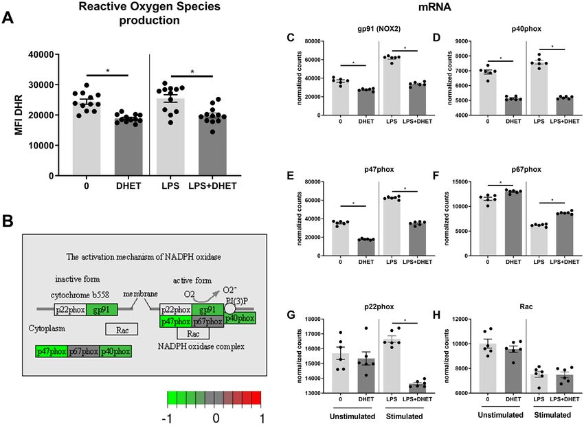
DHET functionally impairs neutrophil Reactive Oxygen Species (ROS) production by tran-
scriptionally impairing the NADPH oxidase complex. To further assess if 14,15-DHET also blunts
other neutrophil functions, we examined its effect on other key neutrophil functions for pathogen killing28. We
hypothesized that neutrophil ROS production would be functionally impaired by 14,15-DHET and that this
would be maintained by transcriptionally reducing the function of the nicotinamide adenine dinucleotide phos-
phate (NADPH) oxidase complex. In vitro incubation of unstimulated, as well as lipopolysaccharide (LPS)-stim-
ulated bone marrow with 14,15-DHET did decrease neutrophil ROS production (Fig. 2A). We then analyzed
expression of genes encoding the enzymes involved in NADPH oxidase (NOX) complex activation (illustrated in
Fig. 2B, comparing unstimulated neutrophils with or without incubating them with DHET). On a transcriptional
level, mRNA levels for three enzymes in this pathway, including gp91 (NOX2), p40phox and p47phox decreased
in neutrophils treated with 14,15-DHET (Fig. 2C–E) under unstimulated conditions, whereas under LPS stimu-
lation the same molecules plus p22phox (G) were found decreased. Under unstimulated conditions two other
genes involved in this pathway, p22phox and Rac, were slightly but not significantly reduced (Fig. 2G,H). We
concluded that 14,15-DHET hampers neutrophil ROS production likely in a transcriptional manner.
DHET decreases Reactive Oxygen Species (ROS) production in mature neutrophils in a p38
and PI3K independent manner. After revealing the impairment of neutrophil ROS production, we exam-
ined which neutrophil subtypes are mainly responsible for ROS production. Moreover, we assessed whether
14,15-DHET impairs ROS production transcriptionally via p38MAPK or PI3K, both of which are known to
modulate neutrophil ROS production29,30. We hypothesized that 14,15-DHET would also functionally alter the
signaling pathways of p38MAP kinase and PI3K, and the impairment of ROS production can be reversed when
these pathways are blocked. We found that neutrophil ROS production is mainly maintained by the mature
CD62L + /ICAM-1 + and CD62L-/ICAM-1 + phenotype (Fig. 3A). At the gene expression level, incubation with
14,15-DHET increased p38MAP kinase and PI3K mRNA transcription (Fig. 3B,C). Interestingly, the functional
inhibition of PI3K did not modulate neutrophil ROS production overall or in any of the subtypes (0 vs LY)
(Fig. 3E,G,I,K,M). Blockage of p38MAPK showed a trend to increase ROS production in CD62L + ICAM-1- and
CD62L + ICAM-1 + neutrophils, however not statistically significantly (0 vs SB) (Fig. 3F,J), but did decrease ROS
production in the CD62L-ICAM-1- and CD62L-ICAM-1 + (0 vs SB) populations significantly (Fig. 3H,L). To
assess the effect of 14,15-DHET on p38MAPK/PI3K-mediated ROS production we hypothesized that blockage
of p38MAPK or PI3K might restore ROS production in 14,15-DHET treated neutrophils. However, the addi-
tion of the specific blockers did not change in ROS production in the 14,15-DHET treated cells (Fig. 3D-M).
The gating strategy is displayed in Suppl. Figure 3. These results suggest that the impairment of neutrophil ROS
production by 14,15-DHET is independent from p38MAP kinase and PI3K signaling pathways.
DHET affects neutrophil functionality as it impairs the acidification of cell compartments, but
does not change the phagocytic capacity, except for the CD62L‑/ICAM‑1‑subtype. Beside ROS
production, the ability to phagocytize and digest pathogens via phagosomes and lysosomes is key for pathogen
clearance28. We therefore hypothesized that 14,15-DHET might impair acidification of these cell compartments
and the ability to phagocytize cells. We examined which neutrophil subtypes are capable of acidification and
phagocytosis, and were able to attribute these functions mainly to CD62L-/ICAM-1- and CD62L-/ICAM-1 + phe-
notypes (Fig. 4A,E). Moreover, the acidification of cell compartments such as phagosomes and lysosomes were
significantly diminished in all neutrophils and the aforementioned phenotypes after 14,15-DHET incubation
(Fig. 4B–D). The phagocytic capacities in the same cells, however, were unchanged (Fig. 4F,H), or in the case of
the CD62L-/ICAM-1-subtype even slightly increased (Fig. 4G). The gating strategy is displayed in Suppl. Fig-
ure 3. Mechanistically, the acidification of cell compartments is less important for neutrophils compared with
macrophages19,31. However, neutrophil bacterial containment was found to be associated with acidification of the
phagolysosome32. Therefore, the data suggest that 14,15-DHET impairs neutrophil cell compartment acidifica-
tion while mostly not changing neutrophil phagocytosis.
DHET impairs neutrophil migration in vitro in a CXCR1 and CXCR2 dependent manner, and
reduces CXCR1 and CXCR2 expression in vivo. Lastly, the migratory behavior of neutrophils was stud-
ied. Neutrophils have to be recruited to the inflamed tissue to be able to clear pathogens and debris28. A potent
chemokine attracting neutrophils to the inflamed tissue is KC (also called CXCL1), which binds to the recep-
tors CXCR1 and C XCR233,34. We hypothesized that neutrophil migration would be decreased by 14,15-DHET.
The receptors and their ligands are illustrated in Fig. 5A. We observed that neutrophil migration is signifi-
cantly impaired upon treatment with 14,15-DEHT in vitro (Fig. 5B). We observed that 14,15-DHET impairs the
expression of CXCR1 (Fig. 5C) but not CXCR2 (Fig. 5D) on a transcriptional level. CXCR1 is mainly expressed
on CD62L-/ICAM-1 + neutrophils (Suppl. Figure 4A) and CXCR2 in the CD62L + /ICAM-1- and the CD62L + /
ICAM-1 + phenotypes (Suppl. Figure 4B). To determine whether these receptors are reduced in vitro we exam-
ined their expression in stimulated and non-stimulated bone marrow-derived cells (Fig. 5E,F). CXCR1 was
reduced by 14,15-DHET in only stimulated cells, whereas CXCR2 was reduced by 14,15-DHET in both non-
Scientific Reports | (2021) 11:16555 | https://doi.org/10.1038/s41598-021-96014-2 3
Vol.:(0123456789)www.nature.com/scientificreports/
Figure 2. DHET functionally impairs neutrophil Reactive Oxygen Species (ROS) production in stimulated
and unstimulated condition by transcriptionally impairing the NADPH oxidase complex. DHET functionally
impairs neutrophil ROS production by reducing the mRNA production of key elements of the NADPH oxidase
complex. Bone marrow from female C57Bl/6 mice was harvested (n = 18/ group) and incubated with 14,15-
DHET followed by dihydrorhodamine (DHR) with or without lipopolysaccharide (LPS). Afterwards cells were
harvested and labelled using flow cytometry. Neutrophils were identified as Ly6G positive cells. DHR passively
diffuses into the cell and becomes oxidized by ROS to highly fluorescent rhodamine 123, an indicator for ROS
production. The MFI of rhodamine 123 was assessed using flow cytometry (A). Neutrophils from bone marrow
of female C57Bl/6 mice were isolated (n = 6/group). They then were incubated with 5 μM DHET or 100 ng of
LPS for 3 h. mRNA sequencing was conducted. mRNA levels coding for gp91 (NOX2), p40phox, p47phox,
p67phox, p22phox, Rac, were assessed (B–H). Data shown in the activation mechanism of NADPH oxidase
(B) shows the changes of untreated neutrophils versus DHET treated neutrophils, without the addition of
LPS. Data are expressed as means ± SEM. *p < 0.05. (A, C–H). Data are expressed in gray as p adjusted > 0.01.
Green: log fold change > -1 (downregulated in DHET). Red: log fold change > 1. (upregulated in DHET) (B).
The figure contains graphical elements of the Kyoto Encyclopedia of Genes and Genomes (KEGG) Pathways
from the Kanehisa Laboratories. Written permission for usage in this publication was obtained from Kanehisa
Laboratories on March 17, 2021.
stimulated and stimulated cells. We then examined the expression in an in vivo burn injury model after 6 h and
found that both CXCR1 and CXCR2 trended towards decreased expression (Fig. 5G,H). We concluded that
14,15-DHET impairs neutrophil chemotaxis most likely in a CXCR1 and CXCR2 dependent manner.
Discussion
Several sEH-inhibitors have been found to potently reduce inflammation in various inflammatory models, but
not trauma. For example, TPPU was proven to dampen inflammation in e ndotoxemia11, sepsis12 and intracer-
ebral hemorrhage13. However, the effect on all innate immune cells has not been sufficiently studied. We sought
to elucidate its effects in a trauma model and examine the mechanisms preventing excessive inflammation.
Burn injury leads to an acute and robust upregulation of inflammation, mirrored by the upregulation of the
pro-inflammatory cytokine IL-6 whose serum levels increase within hours and positively correlate with the size
of the burned skin area35. Our rationale was to acutely assess IL-6 serum levels after sEH inhibition to evaluate
dampening effects on inflammation in trauma. Consistent with previous reports10,12 we observed a robust increase
Scientific Reports | (2021) 11:16555 | https://doi.org/10.1038/s41598-021-96014-2 4
Vol:.(1234567890)www.nature.com/scientificreports/
Figure 3. DHET decreases ROS production in mature neutrophils in an p38 and PI3K independent manner.
ROS production is most potently exerted by mature (CD62L-/ + ICAM-1 +) neutrophil subtypes whereas DHET
functionally decreases it. DHET increases mRNA transcription of p38 MAPK and PI3K significantly, but their
blockage does not change ROS production impaired by DHET. Bone marrow from female C57Bl/6 mice was
harvested (n = 18/group) and incubated with SB239063 (p38 MAP kinase inhibitor) or LY294002 (PI3Kα/δ/β
inhibitor) before adding 14,15-DHET followed by dihydrorhodamine (DHR). The cells were then harvested,
labeled, and analyzed using flow cytometry. Neutrophils were identified as Ly6G positive cells and subtypes
identified using CD62L and ICAM-1. The MFI of the oxidized DHR was used as indicator for ROS production
via flow cytometry (A, D–M). mRNA sequencing was conducted on isolated neutrophil cells of female C57Bl/6
mice (n = 6/group) after being incubated with 5 μM DHET for 3 h. The mRNA levels of p38MAPK and PI3K
were evaluated (B, C). Data are expressed as means ± SEM. *p < 0.05.
of EETs and a decrease of DHETs with TPPU administration (Fig. 1A–F). The enzymatic pathway is nicely dis-
played in the work of Spector and Norris2. EETs were shown to exert potent anti-inflammatory properties2,12,36,
e.g. by inhibiting the activation of the nuclear factor kappa B (NFκB)37. It is of note that genes coding for epoxide
hydrolase are expressed in the skin38, however to our knowledge the local effects in burn injured skin tissue has
not been studied. Systemically, we discovered that TPPU treatment decreased IL-6 levels, indicating that TPPU
effectively ameliorates inflammation. This dampening effect on the release of pro-inflammatory cytokines can
also be seen in s epsis12. We found the administration of DHET post burn not leading to changes in systemic IL-6
levels (Suppl. Figure 1A). This might suggest that the increase of EETs and not decrease in DHETs after TPPU
administration in burn might be responsible for the decrease in IL-6. However, we suggest that given the wide
distribution of systemic IL-6 levels seen in burn injury (Fig. 1G), a higher sample number would be needed to
provide a clear answer to the question if DHETs significantly affect systemic IL-6 levels.
Previous sepsis and endotoxemia models only studied the anti-inflammatory effects of sEH-inhibitors and
14,15-EET11,12. In contrast, DHETs are less studied which led us to examine if they are responsible for detrimental
effects driving inflammation. We found TPPU to decrease systemic 14,15-DHET levels, leading us to investigate
whether 14,15-DHET directly effects immune function. We focused on the effect on neutrophils, as they are the
first immune cells to be recruited to the injury site and quantitatively and functionally key to clear the wound
of debris and potential pathogens18. Although our data revealed systemic 14,15-DHET levels ranging in single
to low double-digit nmol/l levels, we estimate concentrations in murine skin might range from single to double
digit μM concentrations when calculated based on data provided in previous m urine39 studies. Significantly
higher concentrations were also shown for liver and heart in vivo in chicken embryos40 and intracellularly in
human non-cancerous and cancerous breast t issue41. As 14,15-DHET was revealed to effect cell function and
gene expression at concentrations of 3 to 10 μmol/l2 we selected the dose of 5 μM 14,15-DHET as appropriate to
estimate the effect on neutrophils mainly in the wounded skin tissue. A mechanistic examination of the 14,15-
DHET effects revealed ROS production was impaired transcriptionally but was independent from p38MAPK and
PI3K pathways. The acidification of cell compartments such as phagosomes was decreased, and the phagocytic
capacities unchanged except for an increased in the CD62L-/ICAM-1-neutrophil subtype. Chemotaxis in vitro
was impaired by the transcriptional reduction of CXCR1 but not CXCR2. Both were shown to be decreased
by 14,15-DHET in vitro and trended towards decreased expression in vivo. We therefore conclude that the
administration of sEH-inhibitors benefits the host in trauma-related inflammation by reducing the amount of
bioactive DHET, which impairs neutrophilic key functions and consequently might lead to an impaired clear-
ance of pathogens and debris.
CD11b serves as a marker for neutrophil a ctivation42. Interestingly neutrophil activation was decreased after
24 h in the wound border (Fig. 1H). Past studies showed that increased CD11b expression can be a sign of exces-
sive neutrophil a ctivation43 which potentially fuels excessive inflammation. One might therefore argue that DHET
decreasing CD11b expression shows it is dampening excessive inflammation. We believe that this is beneficial
only for later time points, as in the acute phase of inflammation neutrophil activation is needed to maintain a
Scientific Reports | (2021) 11:16555 | https://doi.org/10.1038/s41598-021-96014-2 5
Vol.:(0123456789)www.nature.com/scientificreports/
Figure 4. DHET affects neutrophil functionality as it impairs the acidification of cell compartments, but does
not change the phagocytic capacity, except for the CD62L-/ICAM-1-subtype. The acidification in neutrophil
internal cell compartments such as phagosomes and lysosomes is highest in the CD62L-/ICAM-1- and mature
CD62L-/ICAM-1 + neutrophil subtypes and is significantly impaired in these subtypes by DHET. The phagocytic
capacity of neutrophils was unchanged by DHET, except for the CD62L-/ICAM-1-subtype, in which it was
increased. A neutrophil acidification assay was performed using pHrodo dye which fluoresces in the acidic
environment of the internal cell compartments such as phagosomes and lysosomes. Bone marrow from female
C57Bl/6 mice was used (n = 12). First, cells were incubated with 14,15-DHET, followed by pHrodo opsonized
particles. Subsequently they were fixed and labelled for flow cytometry. Neutrophils were identified as Ly6G
positive cells. CD62L and ICAM-1 were used to differentiate subtypes and the percentage of pHrodo positive
cells was assessed (A–D). The phagocytic capacity of neutrophils was evaluated using opsonized Escherichia
coli particles labelled with a fluorescent dye. Bone marrow from female C57Bl/6 mice was used (n = 8). The
cells were incubated with 14,15-DHET and E. coli particles and consecutively labeled. The percentage of E. coli
positive cells was measured to assess the phagocytic capacity of neutrophils and their subtypes (E–H). Data are
expressed as means ± SEM. *p < 0.05.
proper immune response, otherwise leaving the host susceptible to infection44,45. Moreover, the analysis of neu-
trophil phenotypes using CD62L and ICAM-1 as markers for m aturation18,21–23 revealed that a significant share of
CD11b expression of the whole neutrophil population was attributed to the mature CD62L-/ICAM-1 + phenotype
(Suppl. Figure 1B). In previous studies, we and others depicted that the CD62L-/ICAM-1 + phenotype can be
considered mature and shows increased functionality compared to other phenotypes (Fig. 3A)18,23. Therefore, we
interpret the observed effect of neutrophils being compromised in their activation as an impairment of proper
immune activation and not a prevention of excessive inflammation.
To further harden this assumption, we examined another key neutrophil function, ROS production. ROS
produced by neutrophils is released to clear pathogens and cell debris46 and to dissolve phagocyted pathogens
and cell debris in the phagosomes20. 14,15-DHET impaired ROS production (Fig. 2A). Moreover, we could
Scientific Reports | (2021) 11:16555 | https://doi.org/10.1038/s41598-021-96014-2 6
Vol:.(1234567890)www.nature.com/scientificreports/
Figure 5. DHET impairs neutrophil migration in vitro in a CXCR1 and CXCR2 dependent way, and reduces
their expression in an in vivo burn model. DHET impairs neutrophil expression of CXCR1 and 2 in vitro and
in vivo, to which the strong chemoattractant chemokine (C-X-C motif) ligand 1 (CXCL1, or keratinocyte-
derived chemokine: KC) binds. The migratory capability is therefore shown to be impaired. The chemokine
CXCL1 (KC) is a strong chemoattractant for neutrophils and binds to CXCR1 and 2 (A). 14,15-DHET impairs
Keratinocyte-derived Cytokine (KC, CXCL1) driven neutrophil migration in vitro after 3 h. We performed
a neutrophil chemotaxis assay with bone marrow cells from C57Bl/6 mice (n = 12/ group). After harvesting
bone marrow, cells were incubated on top of a transwell plate in 14,15-DHET and 100 ng of CXCL1 (KC) was
added. After 3 h all cells were harvested and labelled using flow cytometry. Neutrophils were identified as Ly6G
positive cells. We divided the number of neutrophils from the bottom well by the number of all neutrophils to
assess the percentage of migrated neutrophils (B). To assess whether DHET reduces CXCR1 and 2 expression
under stimulated or non-stimulated conditions we performed mRNA sequencing on isolated neutrophils
of female C57Bl/6 mice after being incubated with 14,15-DHET with or without LPS (n = 6/group) (C,D).
Consecutively, the expression of CXCR1 and 2 was measured in vitro. Bone marrow was incubated overnight
with or without 14,15-DHET and with and without LPS. The cells were then labelled and total expression of
CXCR1 and 2 measured via flow cytometry (E,F). Lastly, a burn injury model was conducted, and male CD1
IGS mice were injected with 15 µg/kg body weight 14,15-DHET or PBS as control. After 6 h the mice were
euthanized and the skin surrounding the wound borders was cut out and dissociated. The cell suspension was
then labelled and analyzed to assess neutrophil CXCR1 and 2 expression as total MFI via flow cytometry (G,H).
Data are expressed as means ± SEM. *p < 0.05. The figure contains graphical elements of the KEGG Pathways
from the Kanehisa Laboratories. Written permission for usage in this publication was obtained from Kanehisa
Laboratories on March 17, 2021.
show that the impairment of the NADPH oxidase complex (Fig. 2B), which generates ROS20, is transcriptionally
downregulated by 14,15-DHET under unstimulated and LSP-stimulated conditions (Fig. 2B–H).
As described above, we assessed the contribution of neutrophil subtypes to ROS production. We confirm
previous results23 by showing that the mature CD62L-/ICAM-1 + and CD62L + /ICAM-1 + phenotypes contrib-
ute the most to ROS production (Fig. 3A). To examine what drives ROS production impairment we examined
p38MAPK and PI3K expression, which were increased by 14,15-DHET (Fig. 3D,E). Both are reported to regu-
late ROS production in neutrophils in a stimulatory m anner29,30 and both were shown to be downregulated
by TPPU in LPS-activated macrophages12. We hypothesized that p38MAPK and PI3K expression would be
impaired by DHET, thereby mediating the impairment of ROS production on a transcriptional and functional
level. To our surprise, p38MAPK and PI3K mRNA expression were increased by 14,15-DHET (Fig. 3D,E). We
then examined the post-translational, functional expression of the two enzymes by blocking p38MAPK and
PI3K pathways using specific inhibitors. The blockage of PI3K did not decrease neutrophil ROS production (0
vs LY) (Fig. 3E,G,I,K,M). In contrast p38MAPK blockage did, although not significantly, increase ROS produc-
tion in CD62L + ICAM-1- and CD62L + ICAM-1 + cells (0 vs SB) (Fig. 3F,J). It did decrease ROS production in
the CD62L-ICAM-1- and CD62L-ICAM-1 + cells (0 vs SB) (Fig. 3H,L). Thus we presume that the stimulatory
effect of p38MAPK on ROS production, which has been described47, might only apply to certain neutrophil
subtypes. We therefore suggested that DHET treatment might restore functionality in all neutrophils or at least
certain subtypes. However, no change in ROS production could be achieved by blocking p38MAPK and PI3K
in the 14,15-DHET treated neutrophils (Fig. 3D–M). We subsequently conclude that, contradicting our initial
hypothesis, the impairment of ROS production by 14,15-DHET is independent from p38MAPK and PI3K,
both on transcriptional as well as post-transcriptional levels. This is important as it leads to the suggestion that
DHETs do not modulate the high affinity leukotriene B4 receptor BLT1, of which DHET is a potential ligand48
Scientific Reports | (2021) 11:16555 | https://doi.org/10.1038/s41598-021-96014-2 7
Vol.:(0123456789)www.nature.com/scientificreports/
and which is suggested to activate PI3K d ownstream49. However, the exact mechanisms will need to be examined
in future studies. Moreover, multiple immune cell functions were described in the literature that are affected by
the p38MAPK and PI3K pathways50,51. We suggest future studies to examine if the transcriptional increase of
both enzymes affect neutrophil functionality in ways not studied in this manuscript.
Crucial for pathogen and debris clearance is the neutrophils’ ability to phagocytize and dissolve particles46.
The neutrophil subtypes CD62L-/ICAM-1 + and mature CD62L-/ICAM-1 + consume and acidify particles
more potently than other phenotypes (Fig. 4A,E). 14,15-DHET reduces the acidification of cell compartments
(Fig. 4B–D). Neutrophils dissolve particles in their phagosomes mainly by ROS release into the phagosome19,20, in
contrast to monocytes, which acidify their phagosomes by H + production utilizing the vacuolar (v)-ATPase19,31.
Therefore, the pH in neutrophil phagosomes is neutral or weakly acidic, whereas monocyte phagosomes reach
very low pH values19. It is of note that ROS release into neutrophil phagosomes is suggested to modulate pH-
sensitive measurements19, potentially by ROS-dependent quenching of p Hrodo52, so the pHrodo positivity
observed in these experiments might be due to this effect. However, recent studies revealed that neutrophil
bacterial containment is associated with acidification of the phagosome, measured with pHrodo-technique and
suggested to be ROS independent32. Consequently, their ability to digest pathogens is decreased. 14,15-DHET
did not show an effect on phagocytosis (Fig. 4F,H) except in CD62L-/ICAM-1-neutrophils were the phagocytic
functionality was increased (Fig. 4G). This is of note as the CD62L-/ICAM-1-neutrophils were the main phe-
notype discovered in the burn wounds (Suppl. Figure 5B). However, we assume an overall impairment of their
ability to clear pathogens and debris, as acidification and ROS release into phagolysosomes were shown to be
important for pathogen clearing52,53. Clarification will require future investigations using infection models to
compare the ability to clear pathogens quantified in colony forming units (CFUs). This is beyond the scope of
this study as we were using a sterile burn model.
As shown above, neutrophil functionality at the site of inflammation, which is necessary for proper wound
resolution, is impaired by DHET. An important prerequisite of neutrophils to exert these functions is the ability
to migrate into the inflamed tissue. KC is a key chemoattractant for neutrophils released by inflamed tissue and
is bound by CXCR1 and CXCR2 (Fig. 5A)33,34. CXCR1 and CXCR2 are relevant for neutrophil migration33,34,
therefore several drugs are currently being tested clinically to impair recruitment to dampen inflammation34.
More specifically CXCR2 seems to be crucial for neutrophil migration54 whereas CXCR1 modulation does
not necessarily affect c hemotaxis55. We showed that 14,15-DHET does impair KC-induced chemotaxis in vitro
(Fig. 5B) and subsequently showed the depression of CXCR1 and 2 expression in vitro (Fig. 5E,F) and a trend
towards decreased expression in vivo (Fig. 5G,H) with CXCR1 but not CXCR2 being decreased transcriptionally
(Fig. 5C,D). We did not observe decreased neutrophil recruitment to the wound borders (Suppl. Figure 5A). A
potential explanation might be increased systemic KC levels, driven by DHET, as other investigators showed a
dose-dependent decrease of systemic KC when mice were treated with sEH-inhibitor56. Interestingly, in contrast
to neutrophils, monocyte MCP-1 driven chemotaxis is improved by DHET57, leading to the assumption that
DHET might have different chemotactic effects on different innate leukocytes and the effect may depend on the
chemotactic stimulus. Exceeding their role in migration, numerous studies revealed that CXCR1 exerts pro-
inflammatory functions on neutrophil i nflammation55,58,59, e.g. impaired CXCR1 expression decreases neutrophil
ROS production55,59. Therefore, CXCR1 and CXCR2 depression observed here might contribute to impairing
neutrophil functionality aside from chemotaxis. In trauma patients, neutrophil CXCR2 expression was found
to be decreased and the response of neutrophil CXCR1 and CXCR2 activation desensitized, increasing the risk
for pneumonia60. Inhibition of sEH reducing systemic DHET levels might prevent this.
In summary, we were able to demonstrate that the sEH-inhibitor TPPU reduces inflammation in a murine
burn injury model and reduces systemic levels of bioactive 14,15-DHET. We found 14,15-DHET to functionally
impair neutrophil activation, ROS production, acidification, and the expression of CXCR1 and CXCR2, the latter
potentially hampering migration. In addition, we were able to reveal that the impairment of ROS production
and CXCR1 depression was compromised on a transcriptional level. These results allow three conclusions. First,
we further mechanistically revealed the beneficial effect of sEH inhibitors by showing that they not only lead
to increased anti-inflammatory EET levels but also reduces systemic levels of DHET, which has the capacity to
impair innate immunity. Second, this provides a rationale to measure DHET levels in inflammation or cancer to
evaluate potential neutrophil impairment. Third, these data provide the rationale to examine potential therapeutic
modulation of systemic DHET levels to restore or dampen innate immune cell activation.
Material and methods
Animal models. Male C57Bl/6 and male outbred CD1 IGS mice were purchased from Charles River (Wilm-
ington, MA, USA). C57Bl/6 mice were used for in vitro analyses and outbred CD1 IGS mice for the in vivo
trauma model. All animal experiments were performed under protocols approved by the Institutional Animal
Care and Use Committee (IACUC) of the University of Cincinnati (IACUC protocol no: 08-09-19-01) in accord-
ance with all institutional and federal guidelines, and reporting in the manuscript follows the recommendations
in the ARRIVE guidelines61. At the end of each experiment, euthanasia was performed using carbon dioxide
overdose followed by cervical dislocation, a method consistent with commonly accepted norms of veterinary
best practice and approved by the University of Cincinnati IACUC.
Burn injury model and application of vehicle, TPPU and DHET. A burn injury model was applied
escribed62. Briefly, burn injury was inflicted via scald under 4.5% inhaled isoflurane in oxygen for
as previously d
anesthesia. The mice were shaved on the back and placed in a plastic cylinder with a cut-out exposing an area of
their back equivalent to 28% of their TBSA calculated using the Meeh f ormula63,64. The exposed back was held in
a 90 °C water bath for 9 s, leading to a third degree (full-thickness) burn injury. The mice were resuscitated with
Scientific Reports | (2021) 11:16555 | https://doi.org/10.1038/s41598-021-96014-2 8
Vol:.(1234567890)www.nature.com/scientificreports/
1 ml of 0.9% normal saline injected intraperitoneally (i.p.) and placed on a 42 °C heating pad for three hours.
Sham-treated mice underwent the same procedure except for exposure to the 90 °C water bath. Directly prior to
burn injury (less than 60 s), mice were injected i.p. with 10 mg/kg body weight TPPU (Cayman Chemical, Ann
Arbor, MI, USA) diluted in 100% polyethylene glycol (PEG) with an average molecular weight of 400 Da (Sigma-
Aldrich, St. Louis, MO, USA), or with PEG vehicle for controls. In subsequent experiments, mice were injected
i.p. with 15 µg/kg body weight 14,15-DHET (Cayman Chemical) diluted in phosphate buffered saline (PBS);
untreated mice served as controls. Mice were euthanized after 6 and 24 h and samples were collected for analysis.
Mass spectrometry. Following euthanasia at 6 or 24 h post-burn, whole blood was collected and centri-
fuged to gain blood plasma. An antioxidant solution was added (2 µl of 0.2% triphenylphosphine/0.2% butylated
hydroxytoluene/0.1% ethylenediaminetetraacetic acid per 100 µl of plasma) and samples were frozen at −80 °C
until analyzed. Lipid profiles were determined using solid phase extraction (SPE) followed by liquid chromatog-
raphy-electrospray ionization/multi-stage mass spectrometry (LC–MS/MS)65.
Serum cytokine levels. To assess serum cytokine levels, whole blood collected after euthanasia at 6 and
24 h was centrifuged to obtain serum. The serum was then analyzed to determine IL-6 levels using the BD Cyto-
metric Bead Array (CBA) Mouse IL-6 Flex Set (BD Biosciences, San Jose, CA, USA) according to the manufac-
turer’s instructions.
Isolation of neutrophils from burn wound borders. Neutrophil surface expression of CD11b, CD62L,
ICAM-1, CXCR1 and CXCR2 from tissue resident cells in the burn wound borders was measured by cutting
out the burn wound borders after euthanizing the mice after 6 and 24 h. The skin was cut with a 1 mm distance
left and right of the visible transition between the third degree burn wound and healthy, non-burned skin, and
placed in cell culture media, consisting of RPMI to which gentamycin, Minimum Essential Medium (MEM),
sodium pyruvate, Penicillin–Streptomycin-Glutamine, L-Glutamine 200 mM and Cytiva HyClone Fetal Bovine
Serum (Thermo Fisher Scientific) were added. The skin was then dissociated into a cell suspension using a gen-
tle MACS Octo Dissociator with Heaters and the Multi Tissue Dissociation Kit 1 (both from Miltenyi Biotec,
Bergisch Gladbach, Germany) following the manufacturer’s customized protocol for mouse whole skin (3 h
incubation). The cell suspension was then washed and labelled for flow cytometry analysis as described below.
Transcriptional analysis of neutrophils. For all in vitro experiments and gene expression analyses, bone
marrow was harvested by flushing the tibia and femur of both legs. For the transcriptional analysis, neutro-
phils were then isolated. Magnetic bead sorting employing the autoMACS Pro Separator (Miltenyi Biotech)
was conducted according to the manufacturer’s instructions, using Anti-Ly6G MicroBeads Ultra Pure mouse
(Miltenyi Biotech). After isolation 2 × 10⁶ neutrophils were plated in the cell culture media described above. The
cells were incubated at 37 °C for 3 h with either no stimulus, 100 ng lipopolysaccharide (LPS) (Escherichia coli
0111:B4, Sigma-Aldrich), 5 μM 14,15-DHET (Cayman Chemical), or the combination of the two. After incuba-
tion, mRNA was prepared using Qiagen RNeasy Mini Kit columns (Qiagen, Inc., Germantown, MD) and mRNA
samples were shipped on dry ice to a commercial provider, GENEWIZ NGS bioinformatics solutions (South
Plainfield, NJ, USA) for standard mRNA sequencing analysis (Standard RNA-Seq Data Analysis Package).
Labeling and characterization of neutrophils using flow cytometry. For flow cytometry analysis,
cells were washed and incubated with Fc-receptor blockage using CD16/CD32 (Mouse BD Fc Block) (clone
2.4G2 (RUO), BD Pharmingen) and 5% rat serum (Invitrogen, Carlsbad, CA, USA) for 10 min prior to labeling.
Subsequently, the cells were incubated with labelling antibodies for 20 min, washed and analyzed on the Attune
NxT Acoustic Focusing Cytometer (Thermo Fisher Scientific). The following fluorescent-labeled antibodies
were used for cell labeling: Ly6G (clone: 1A-8), ICAM-1 (CD54) (clone: 3E2), CD62L (L-Selectin) (clone: MEL-
14), CXCR1 (clone: U45-632), all from BD Biosciences and CD11b (clone: M1/70) and CXCR2 (clone: SA044-
G4), from BioLegend (San Diego, CA, USA). Neutrophil subsets were characterized using CD62L (L-Selectin)
and ICAM-1 (CD54). Neutrophils were considered immature when expressing a CD62L + /ICAM-1(CD54)-
phenotype and mature when expressing CD62L + /ICAM-1(CD54) + or CD62L-/ICAM-1(CD54) + 21,22.
Viability assay. To assess the viability of cells, cells were labelled with Annexin V (BD Biosciences) and
propidium iodide (PI) (Fluka Chemie GmbH, Buchs, Switzerland). Bone marrow was incubated with either 0,
2, 5, or 10 μM 14,15-DHET for 24 h. Cells were then washed and resuspended in Annexin V Buffer (BD Bio-
sciences), counted and diluted into 100 μl buffer containing 1 × 10⁵ cells. Each of these samples was incubated
with 5 μl Annexin V antibody and 1 μl 100 μg/ml PI for 15 min. Afterwards, they were washed, placed on ice,
labeled, and analyzed via flow cytometry as described above. Neutrophils were considered viable when found
Annexin V and PI negative.
Reactive oxygen species (ROS) assay and blocking of p38MAPK and PI3K. Bone marrow was
incubated at 37 °C for 15 min with either no inhibitors (untreated controls), 10 μM SB239063 (p38 MAP kinase
inhibitor), or 10 μM LY294002 (PI3Kα/δ/β inhibitor) (both from Cayman Chemical). Subsequently, 5 μM 14,15-
DHET (Cayman Chemical) was added and incubated for 15 min followed by 1 mM dihydrorhodamine (DHR)
for another 15 min. The cells were then harvested, labeled, and analyzed using flow cytometry. The median
fluorescent intensity (MFI) of the oxidized DHR indicates ROS production.
Scientific Reports | (2021) 11:16555 | https://doi.org/10.1038/s41598-021-96014-2 9
Vol.:(0123456789)www.nature.com/scientificreports/
Acidification (pHrodo) assay. Opsonized E. coli particles labelled with pH-sensitive dyes (pHrodo
Green Escherichia coli BioParticles; Thermo Fisher) were processed according to the manufacturer’s instructions
and incubated with bone marrow at 37 °C, 5% CO2 for one hour, during which the neutrophils phagocyted these
particles. After one hour phagocytosis was stopped by placing cells on ice and fixing them with 1% paraformal-
dehyde (PFA). For the rest of the procedure, cells were kept on ice and subsequently labelled for flow cytometry
analysis as described above. The internal cell compartments such as phagosomes and lysosomes become acidi-
fied, which correlates with the increased intensity of the light signal of the pH-sensitive dyes, reflected in its MFI
in flow cytometry. Cells that did acidify the phagocyted particles were identified as pHrodo positive cells.
Phagocytosis. E. coli particles (Escherichia coli BioParticles Opsonizing Reagent; Thermo Fisher Scientific)
were opsonized, washed, and incubated at 37 °C for 15 min with 1 × 10⁶ cells, according to the manufacturer’s
instructions. Afterwards, cells were fixed by adding 500 μl 1% PFA for 5 min at 37 °C, washed, and labelled for
flow cytometry analysis as described above. The phagocytic uptake of particles of E. coli was then detected using
flow cytometry.
Chemotaxis. Harvested bone marrow cells were seeded on a transwell plate (Thermo Fisher Scientific) of
3 μm pore size and incubated with 5 μM 14,15-DHET (Cayman Chemical) at 37 °C for 30 min before add-
ing 100 ng of chemokine (C-X-C motif) ligand 1 (CXCL1, also known as keratinocyte-derived chemokine
(KC)). After 3 h all cells were harvested and labeled for flow cytometry analysis. The number of neutrophils that
migrated to the bottom well was divided by the number of all neutrophils to assess the percentage of migrated
neutrophils.
LPS stimulation of neutrophils in cell culture. The femur and tibia were flushed to harvest bone mar-
row. Two million cells of the suspension were plated per well, stimulated with 100 ng LPS (Escherichia coli
0111:B4, Sigma-Aldrich) and incubated at 37 °C at 5% CO2 for 24 h, before being labeled and analyzed via flow
cytometry.
Statistical analyses. The statistical analysis was performed with GraphPad Prism 9.0 (GraphPad Soft-
ware, La Jolla, CA; graphpad.com). Outliers were identified and removed using the ROUT method (Q = 1%). All
groups were tested for normality with the Shapiro–Wilk and the D`Agostino and Pearson normality test. If the
groups were normally distributed, a two tailed Student’s t test comparison of two groups or one-way ANOVA
with Tukey post-hoc analysis for comparisons of more than two groups was applied. A one tailed Student’s t test
was used to compare CXCR1 and CXCR2 in vivo expression of neutrophils after assessing a decrease in vitro.
If groups were not normally distributed, a Mann–Whitney test to compare two groups or Kruskal–Wallis test
with Dunn`s multiple-comparison test analysis for comparisons of more than two groups was applied. For all
in vitro experiments that included LPS-stimulation, we only compared the differences after DHET-treatment
within the stimulated and non-stimulated cohorts. The results are visualized in bars with the mean ± standard
error of the mean (SEM). A p-value of ≤ 0.05 was considered statistically significant. For the evaluation of the
mRNA sequencing data an additional differential expression analysis was performed using DEseq2 method66.
Significant results (Benjamini–Hochberg adjusted P value < 0.1) of KEGG pathway graphs were rendered using
Pathview67,68.
Data availability
All data described are contained within the manuscript. Additional data not discussed here are available from
the corresponding author upon reasonable request.
Received: 18 May 2021; Accepted: 3 August 2021
References
1. Yang, T. et al. The role of 14,15-dihydroxyeicosatrienoic acid levels in inflammation and its relationship to lipoproteins. Lipids
Health Dis. 12, 151 (2013).
2. Spector, A. A. & Norris, A. W. Action of epoxyeicosatrienoic acids on cellular function. Am. J. Physiol. Cell Physiol. 292, C996-1012
(2007).
3. Morisseau, C. & Hammock, B. D. Epoxide hydrolases: mechanisms, inhibitor designs, and biological roles. Annu. Rev. Pharmacol.
Toxicol. 45, 311–333 (2005).
4. Antcliffe, D. B. et al. Profiling inflammatory markers in patients with pneumonia on intensive care. Sci. Rep. 8, 14736 (2018).
5. Hamaguchi, M. et al. A case series of the dynamics of lipid mediators in patients with sepsis. Acute Med. Surg. 6, 413–418 (2019).
6. Wolfer, A. M. et al. Longitudinal analysis of serum oxylipin profile as a novel descriptor of the inflammatory response to surgery.
J. Transl. Med. 15, 83 (2017).
7. Hercule, H. C. et al. Interaction between P450 eicosanoids and nitric oxide in the control of arterial tone in mice. Arterioscler.
Thromb. Vasc. Biol. 29, 54–60 (2009).
8. Thomson, S. J., Askari, A. & Bishop-Bailey, D. Anti-inflammatory effects of epoxyeicosatrienoic acids. Int. J. Vasc. Med. 2012,
605101 (2012).
9. Edin, M. L. et al. Epoxide hydrolase 1 (EPHX1) hydrolyzes epoxyeicosanoids and impairs cardiac recovery after ischemia. J. Biol.
Chem. 293, 3281–3292 (2018).
10. Liu, J. Y. et al. Substituted phenyl groups improve the pharmacokinetic profile and anti-inflammatory effect of urea-based soluble
epoxide hydrolase inhibitors in murine models. Eur. J. Pharm. Sci. 48, 619–627 (2013).
11. Zhou, Y. et al. Soluble epoxide hydrolase inhibitor attenuates lipopolysaccharide-induced acute lung injury and improves survival
in mice. Shock 47, 638–645 (2017).
Scientific Reports | (2021) 11:16555 | https://doi.org/10.1038/s41598-021-96014-2 10
Vol:.(1234567890)www.nature.com/scientificreports/
12. Chen, Z. et al. sEH inhibitor Tppu ameliorates cecal ligation and puncture-induced sepsis by regulating macrophage functions.
Shock 53, 761–771 (2020).
13. Wu, C. H. et al. Genetic deletion or pharmacological inhibition of soluble epoxide hydrolase reduces brain damage and attenuates
neuroinflammation after intracerebral hemorrhage. J. Neuroinflam. 14, 230 (2017).
14. Jeschke, M. G. et al. Burn injury. Nat. Rev. Dis. Primers 6, 11 (2020).
15. Mulder, P. P. G. et al. Persistent systemic inflammation in patients with severe burn injury is accompanied by influx of immature
neutrophils and shifts in T cell subsets and cytokine profiles. Front. Immunol. 11, 621222 (2020).
16. Hanschen, M., Tajima, G., O’Leary, F., Ikeda, K. & Lederer, J. A. Injury induces early activation of T-cell receptor signaling pathways
in CD4+ regulatory T cells. Shock 35, 252–257 (2011).
17. Beckmann, N. et al. Scald Injury-induced T cell dysfunction can be mitigated by Gr1(+) cell depletion and blockage of CD47/
CD172a signaling. Front. Immunol. 11, 876 (2020).
18. Mortaz, E., Alipoor, S. D., Adcock, I. M., Mumby, S. & Koenderman, L. Update on neutrophil function in severe inflammation.
Front. Immunol. 9, 2171 (2018).
19. Nordenfelt, P. & Tapper, H. Phagosome dynamics during phagocytosis by neutrophils. J. Leukoc. Biol. 90, 271–284 (2011).
20. Roos, D., van Bruggen, R. & Meischl, C. Oxidative killing of microbes by neutrophils. Microbes Infect. 5, 1307–1315 (2003).
21. Woodfin, A. et al. ICAM-1-expressing neutrophils exhibit enhanced effector functions in murine models of endotoxemia. Blood
127, 898–907 (2016).
22. Ivetic, A., Hoskins Green, H. L. & Hart, S. J. L-selectin: A major regulator of leukocyte adhesion, migration and signaling. Front.
Immunol. 10, 1068 (2019).
23. Sengupta, S., Caldwell, C. C. & Nomellini, V. distinct neutrophil populations in the spleen during PICS. Front. Immunol. 11, 804
(2020).
24. Dong, L. et al. Soluble epoxide hydrolase inhibitor suppresses the expression of triggering receptor expressed on myeloid cells-1
by inhibiting NF-kB activation in murine macrophage. Inflammation 40, 13–20 (2017).
25. Chen, X. et al. Soluble epoxide hydrolase inhibition provides multi-target therapeutic effects in rats after spinal cord injury. Mol.
Neurobiol. 53, 1565–1578 (2016).
26. Kim, J., Imig, J. D., Yang, J., Hammock, B. D. & Padanilam, B. J. Inhibition of soluble epoxide hydrolase prevents renal interstitial
fibrosis and inflammation. Am. J. Physiol. Renal Physiol. 307, F971-980 (2014).
27. Tao, W., Li, P. S., Yang, L. Q. & Ma, Y. B. Effects of a soluble epoxide hydrolase inhibitor on lipopolysaccharide-induced acute lung
injury in mice. PLoS ONE 11, e0160359 (2016).
28. Mayadas, T. N., Cullere, X. & Lowell, C. A. The multifaceted functions of neutrophils. Annu. Rev. Pathol. 9, 181–218 (2014).
29. Kulkarni, S. et al. PI3Kbeta plays a critical role in neutrophil activation by immune complexes. Sci. Signal. 4, ra23 (2011).
30. Qian, F. et al. A non-redundant role for MKP5 in limiting ROS production and preventing LPS-induced vascular injury. EMBO
J. 28, 2896–2907 (2009).
31. Brisseau, G. F. et al. Interleukin-1 increases vacuolar-type H+-ATPase activity in murine peritoneal macrophages. J. Biol. Chem.
271, 2005–2011 (1996).
32. Leliefeld, P. H. C. et al. Differential antibacterial control by neutrophil subsets. Blood Adv. 2, 1344–1355 (2018).
33. Vieira, S. M. et al. A crucial role for TNF-alpha in mediating neutrophil influx induced by endogenously generated or exogenous
chemokines, KC/CXCL1 and LIX/CXCL5. Br. J. Pharmacol. 158, 779–789 (2009).
34. de Oliveira, S., Rosowski, E. E. & Huttenlocher, A. Neutrophil migration in infection and wound repair: Going forward in reverse.
Nat. Rev. Immunol. 16, 378–391 (2016).
35. Agay, D. et al. Interleukin-6, TNF-alpha and interleukin-1 beta levels in blood and tissue in severely burned rats. Eur. Cytokine
Netw. 19, 1–7 (2008).
36. Samokhvalov, V. et al. PPARgamma signaling is required for mediating EETs protective effects in neonatal cardiomyocytes exposed
to LPS. Front. Pharmacol. 5, 242 (2014).
37. Node, K. et al. Anti-inflammatory properties of cytochrome P450 epoxygenase-derived eicosanoids. Science 285, 1276–1279 (1999).
38. Hoopes, S. L. et al. Generation and characterization of epoxide hydrolase 3 (EPHX3)-deficient mice. PLoS ONE 12, e0175348
(2017).
39. Zhang, Q. et al. Anti-versus pro-inflammatory metabololipidome upon cupping treatment. Cell Physiol. Biochem. 45, 1377–1389
(2018).
40. Diani-Moore, S., Ma, Y., Gross, S. S. & Rifkind, A. B. Increases in levels of epoxyeicosatrienoic and dihydroxyeicosatrienoic acids
(EETs and DHETs) in liver and heart in vivo by 2,3,7,8-tetrachlorodibenzo-p-dioxin (TCDD) and in hepatic EET:DHET ratios by
cotreatment with TCDD and the soluble epoxide hydrolase inhibitor AUDA. Drug Metab. Dispos. 42, 294–300 (2014).
41. Luo, J. et al. 14, 15-EET induces breast cancer cell EMT and cisplatin resistance by up-regulating integrin alphavbeta3 and activat-
ing FAK/PI3K/AKT signaling. J. Exp. Clin. Cancer Res. 37, 23 (2018).
42. Zhou, X. et al. LPS activation of Toll-like receptor 4 signals CD11b/CD18 expression in neutrophils. Am. J. Physiol. Lung Cell Mol.
Physiol. 288, L655-662 (2005).
43. Orr, Y. et al. Conformational activation of CD11b without shedding of L-selectin on circulating human neutrophils. J. Leukoc.
Biol. 82, 1115–1125 (2007).
44. Solomkin, J. S. Neutrophil disorders in burn injury: Complement, cytokines, and organ injury. J. Trauma 30, S80-85 (1990).
45. Beckmann, N. et al. Burn injury impairs neutrophil chemotaxis through increased ceramide. Shock 56(1), 125–132 (2020).
46. Nathan, C. Neutrophils and immunity: Challenges and opportunities. Nat. Rev. Immunol. 6, 173–182 (2006).
47. Belambri, S. A. et al. NADPH oxidase activation in neutrophils: Role of the phosphorylation of its subunits. Eur. J. Clin. Investig.
48(Suppl 2), e12951 (2018).
48. Behm, D. J., Ogbonna, A., Wu, C., Burns-Kurtis, C. L. & Douglas, S. A. Epoxyeicosatrienoic acids function as selective, endogenous
antagonists of native thromboxane receptors: Identification of a novel mechanism of vasodilation. J. Pharmacol. Exp. Ther. 328,
231–239 (2009).
49. Jeon, W. K. et al. The proinflammatory LTB4/BLT1 signal axis confers resistance to TGF-beta1-induced growth inhibition by
targeting Smad3 linker region. Oncotarget 6, 41650–41666 (2015).
50. Canovas, B. & Nebreda, A. R. Diversity and versatility of p38 kinase signalling in health and disease. Nat. Rev. Mol. Cell Biol. 22,
346–366 (2021).
51. Hawkins, P. T. & Stephens, L. R. PI3K signalling in inflammation. Biochim. Biophys. Acta 1851, 882–897 (2015).
52. Rybicka, J. M., Balce, D. R., Chaudhuri, S., Allan, E. R. & Yates, R. M. Phagosomal proteolysis in dendritic cells is modulated by
NADPH oxidase in a pH-independent manner. EMBO J. 31, 932–944 (2012).
53. Karavolos, M. H., Horsburgh, M. J., Ingham, E. & Foster, S. J. Role and regulation of the superoxide dismutases of Staphylococcus
aureus. Microbiology (Reading) 149, 2749–2758 (2003).
54. Ness, T. L., Hogaboam, C. M., Strieter, R. M. & Kunkel, S. L. Immunomodulatory role of CXCR2 during experimental septic
peritonitis. J. Immunol. 171, 3775–3784 (2003).
55. Carevic, M. et al. CXCR1 regulates pulmonary anti-pseudomonas host defense. J. Innate Immun. 8, 362–373 (2016).
56. Podolin, P. L. et al. In vitro and in vivo characterization of a novel soluble epoxide hydrolase inhibitor. Prostaglandins Other Lipid
Mediat. 104–105, 25–31 (2013).
Scientific Reports | (2021) 11:16555 | https://doi.org/10.1038/s41598-021-96014-2 11
Vol.:(0123456789)You can also read