A Novel Venom-Derived Peptide for Brachytherapy of Glioblastoma: Preclinical Studies in Mice - MDPI
←
→
Page content transcription
If your browser does not render page correctly, please read the page content below
molecules
Article
A Novel Venom-Derived Peptide for Brachytherapy
of Glioblastoma: Preclinical Studies in Mice
Steve Swenson 1,2 , Radu O. Minea 2 , Cao Duc Tuan 3 , Thu-Zan Thein 2 , Thomas C. Chen 2
and Francis S. Markland 1, *
 1   Department of Biochemistry and Molecular Medicine, University of Southern California, Los Angeles,
     CA 90089, USA; sswenson@usc.edu
 2   Department of Neurological Surgery, University of Southern California, Los Angeles, CA 90089, USA;
     minea@usc.edu (R.O.M.); tthein@usc.edu (T.-Z.T.); tchen68670@gmail.com (T.C.C.)
 3   Faculty of Pharmacy, Haiphong University of Medicine and Pharmacy, Haiphong, Vietnam;
     caoductuan11@gmail.com
 *   Correspondence: markland@usc.edu

 Academic Editor: Jean-Marc Sabatier

 Received: 1 October 2018; Accepted: 3 November 2018; Published: 8 November 2018

 Abstract: We developed a bacterial expression system to produce a recombinant disintegrin, vicrostatin
 (VCN), whose structure is based on a natural disintegrin isolated from southern copperhead snake
 venom. Our goal is to develop VCN for potential clinical translation as an anti-cancer agent. VCN is a
 peptide of 69 amino acids with a single tyrosine residue. We have employed VCN as integrin-targeted
 radionuclide therapy (brachytherapy) for treatment of glioblastoma (GBM, glioma). GBM is a deadly
 brain cancer that doesn’t discriminate between sexes and knows no age limit. We established that
 the tyrosine residue in VCN can be radioiodinated with full retention of bioactivity. 131 I-VCN was
 utilized for integrin-targeted radionuclide therapy using mouse models of glioma. The combination of
 radioiodinated VCN plus temozolomide (a DNA alkylating agent) significantly prolonged survival of
 glioma-bearing mice. We also obtained similar results using an immunocompetent mouse model and a
 murine glioma cell line. In summary, as demonstrated in studies reported here we have shown that
 VCN as targeted radionuclide therapy for GBM has significant translational potential for therapy of
 this deadly disease.

 Keywords: disintegrin; glioblastoma; integrin; brachytherapy; radioiodine; vicrostatin; cancer therapy

1. Introduction
     Glioblastoma multiforme (GBM) affects males and females and knows no age barrier; it accounts
for more than 50% of all human primary brain tumors. Despite advances in surgery, chemotherapy,
and radiation therapy GBM remains a highly invasive cancer, and the average life span is only 12 to
15 months from time of diagnosis. Increasing incidence of GBM is one of the prime factors driving
the development of new therapeutic strategies. GBM is considered the most aggressive form of
brain cancer; it grows rapidly and spreads into the normal brain. The National Cancer Institute
(NCI) estimates that 23,880 adults will be diagnosed with brain and other nervous system forms of
cancer in 2018. The NCI also estimates that in 2018, 16,830 of these diagnoses will result in death [1].
The mainstay of treatment for GBM is surgery, followed by radiation and chemotherapy. The main
objective of surgery is to remove as much of the tumor as possible without injuring the surrounding
normal brain tissue needed for neurological function [2]. Surgical debulking can have multiple positive
effects on patient survival including relieving the tumor mass effect, decreasing intracranial pressure,
reducing associated brain edema, and lowering steroid dependence, all of which can lead to increased
overall patient survival and a higher quality of life [2]. However, GBM is surrounded by a zone

Molecules 2018, 23, 2918; doi:10.3390/molecules23112918                     www.mdpi.com/journal/molecules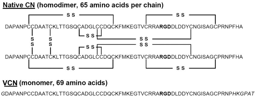
Molecules 2018, 23, 2918                                                                              2 of 13

of migrating, infiltrating tumor cells that invade surrounding tissues, which uniformly makes the
complete removal of the tumor very challenging.
      Temozolomide is a leading molecule for the treatment of newly diagnosed GBM [3]. The gold
standard of therapy for newly diagnosed patients with primary GBM was first introduced more
than a decade ago and is called the Stupp protocol [4]. This consists of a regimen of conformal
radiotherapy (dosed fractionated at about 2 Gy/day for a total cumulative dose of 60 Gy) administered
concurrently with temozolomide dosed at 75 mg/m2 for 42 consecutive days, followed by multiple
cycles of adjuvant temozolomide administered for 5 days on and 23 days off while dose is escalated
up to 150 mg/m2 . Although it represented an important therapeutic advancement at the time of its
implementation, the survival gains brought by the Stupp protocol are rather modest because resistance
to temozolomide emerges rapidly in GBM and recurrence is typical for these patients. In addition
to temozolomide there are only limited therapies available for the treatment of recurrent GBM with
very limited survival gains. For recurrent GBM surgical management is a significantly more difficult
problem and standard procedures are not fully developed at present. However, the current pipeline
of experimental therapeutics for recurrent GBM is strong and includes biologics, small molecules,
devices, innovative surgical strategies, and immunotherapy including several approaches in phase I
and phase II clinical trials. A large group of immunotherapeutic approaches are currently undergoing
clinical study for safety and efficacy in the treatment of newly diagnosed or recurrent high-grade
gliomas. Further, combination therapies using immunotherapy and other forms of therapy are being
explored [5]. Some of these new forms of immunotherapy have shown promise, but a significant
time delay will be involved for further testing and clinical trials before their incorporation into GBM
standard therapy [6]. Gaining better understanding of the disease will improve what can be achieved
through an increase in understanding of molecular mechanisms and the development of clinical trials
leading to more promising and effective therapeutic options. In aggregate, recurrent GBM poses a
difficult challenge, as the desire to provide significant lesion removal needs to be balanced by the need
to preserve neurological function and to retain a good quality of life [7].
      As indicated, due to its highly infiltrative nature, GBM is a complicated disease and thus
its treatment is a challenge, forcing researchers into investigating novel diagnostic strategies and
unique chemotherapeutic approaches. The tumor microenvironment plays a large role in cancer
progression and its pharmacological manipulation can repress progression in the recurrence setting.
Central to the ability of GBM cells to invade the surrounding brain distant from the tumor is a class of
glycoproteins called integrins. Integrins are heterodimeric cell surface adhesion receptors (containing α
and β chains) operating at the interface between the extracellular matrix and cytoskeletal apparatus [8–10].
Integrins link cytoskeletal proteins to the extracellular matrix and are involved in bi-directional signaling
to alter cellular functions. Among these interactions are the adhesion of both endothelial and glioma cells
to extracellular matrix proteins and other interactions that are integral to tumor growth, metastasis and
angiogenesis. Integrins allow cells to mechanically sense their environment [11] by integrating multiple
signaling pathways initiated by extracellular cues with the cellular locomotor apparatus. Integrins exhibit
structural diversity and undergo conformational changes that are central to the regulation of their receptor
function. They exist in three major conformational states: an inactive or low affinity state, a primed
or activated high affinity state, and a ligand bound or occupied state [8]. Activated integrins are not
usually present in quiescent tissues, but some, such as the RGD-dependent ones which recognize the
Arg-Gly-Asp (RGD) sequence present in key extracellular matrix proteins [12], play an important role in
neoplastic processes including glioma progression [13], invasion (via invadosome formation) [14,15],
angiogenesis [16,17] and dissemination. This subject was effectively reviewed in [18].
      Disintegrins represent evolution’s integrin targeting “invention”. Disintegrins are a class
of disulfide-rich peptides, originally isolated from snake venom [19–21]. These peptides hold
a significant translational potential with desirable pharmacological attributes based on their
high-affinity/high-specificity interaction with tumor integrins. The integrin-binding activity of
disintegrins depends on the appropriate pairing of several cysteine residues responsible for the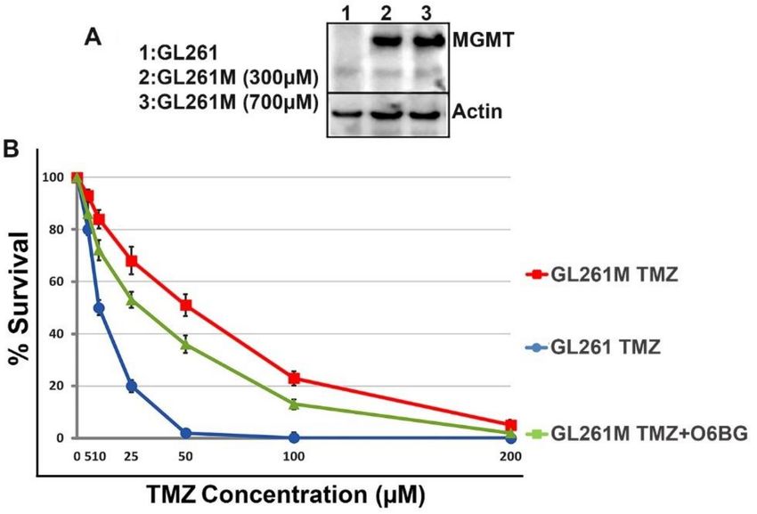
Molecules 2018, 23, 2918                                                                                3 of 13

disintegrin fold, a mobile 11-amino acid loop protruding from the polypeptide core displaying a
tripeptide motif, RGD in many disintegrins [22,23]. It is important to emphasize that disintegrins
bind only to the activated conformations of integrins on motile cells such as cancer cells and angiogenic
endothelial cells [8,21], making them attractive vehicles to specifically target cancerous tissues. We and
others demonstrated that disintegrins are well tolerated and can be infused without toxicity or
detrimental effect on blood pressure, body temperature, or other physiological parameters [24–26].
Subsequently, our laboratory designed a sequence-engineered RGD-disintegrin, vicrostatin (VCN),
that can be reliably produced in large quantity (~200 mg/L purified VCN from bacterial cell lysate) in a
robust recombinant bacterial system [27,28]. Recombinant VCN retains the binding properties of the
natural disintegrin from which it originated, the homodimer contortrostatin (CN) isolated from southern
copperhead snake venom, while showing improved binding affinity, compared to native CN, for an
important integrin in angiogenesis, integrin α5β1.
     The characteristic expression of very high densities of active RGD-dependent integrins by GBM
(expressed both by its florid neovasculature and by the tumor cells themselves) creates a unique
opportunity for precision delivery of minute doses of beta-emitting radionuclides (such as 131 I) into
the tumor microenvironment via highly targeted polypeptides such as VCN. In the present report we
describe our findings with the use of radioiodinated-VCN as a form of integrin-targeted radionuclide
therapy (brachytherapy) in combination with temozolomide in murine models of human GBM.

2. Results

2.1. Structures of VCN and CN
     The comparative amino acid sequences of VCN and its predecessor CN are shown (Figure 1).
The extension at the COOH-terminus of VCN is clearly visible and was purposefully engineered into the
VCN structure [28] to improve binding affinity to integrin α5β1, which it accomplished by about 13-fold
as compared to the affinity of CN for this integrin. Further, this alteration at the COOH-terminus had
very minimal effect on the affinities of VCN for integrins αvβ3 and αvβ5. Additionally, the tyrosine is
shown at residue 51 in VCN, which is the site of radioiodination of the peptide.

      Figure 1. Sequence alignment of native contortrostatin (CN) and recombinant vicrostatin (VCN).
      The recombinant disintegrin carries a short C-terminal amino acid sequence grafted from another
      snake venom disintegrin (echistatin) which improves VCN’s affinity toward α5β1 integrin and also
      causes the recombinant disintegrin to fold as a monomer. A unique tyrosine residue is available for
      radioiodination in the C-terminal half of VCN (residue 51). The RGD sequence is bold type.

2.2. Evaluation of Tumor Specific Brain Uptake of Radioiodinated VCN
     To identify the ability to iodinate and produce an effective iodination stoichiometry we examined
several methods of iodination. Ultimately, using either radioinert or radioactive iodine, we used
the chloramine-T procedure [29] for VCN iodination. We confirmed retention of biological activity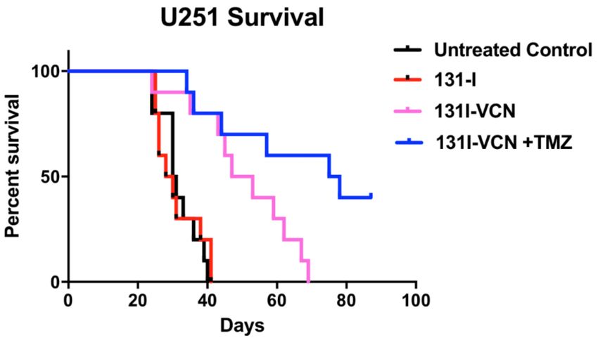
Molecules 2018, 23, 2918                                                                            4 of 13

of iodinated VCN by its ability to inhibit platelet aggregation with identical activity to that on
non-iodinated VCN. Additionally, the labeled material was analyzed by mass spectrometry and
we observed a ~70:30 mixture of mono-iodinated to di-iodinated material in the reaction which yielded
VCN with full activity. Allowing the reaction to yield more of the di-iodinated material resulted in
reduced biological activity, whereas attempting to produce only mono-iodinated material resulted in a
high level of un-iodinated species which defeats the ability to deliver radioactivity to the tumor.

2.3. Confirmation of Iodinated VCN Binding to Glioma Cells and Tumors
       All glioma cells used in the study were analyzed by FACS for integrin expression levels
(i.e., αv and α5 integrin subclasses) and found to overexpress active species of these integrin members
(data not shown). VCN labeled with radioinert iodine (i.e., I-VCN) was further labeled with Cy5-NHS to
assess the ability of Cy5-I-VCN to bind to glioma cells. This FACS analysis revealed avid binding by
Cy5-I-VCN to all glioma cell lines used in our studies. To confirm receptor specificity, the binding of
Cy5-I-VCN was inhibited when the cells were preincubated with 100-fold molar excess of cilengitide,
a methylated cyclic RGDfV peptide targeted to αvβ3 and αvβ5 integrins [30] (developed by Merck
KGaA, Darmstadt, Germany), before the addition of fluorescent disintegrin (Figure 2).

      Figure 2. Iodinated VCN avidly binds to glioma cell lines. A FACS analysis was conducted with
      Cy5-labeled iodinated VCN (Cy5-I-VCN). TMZ-sensitive human glioma cell lines (U87 and U251)
      or TMZ-resistant mouse glioma cells (GL261M) were incubated with Cy5-I-VCN in the presence or
      absence of molar excess (100-fold) of a small cyclic RGD peptide competitor (cilengitide).

     Furthermore, using 125 I-VCN, we evaluated the ability of the radioiodinated disintegrin to be
delivered intravenously (IV) and be retained in the brain and or blood vessels supplying the brain after
18 hours of circulation time. We found that a significant portion of the IV delivered material is found in
the brain and associated vasculature in U87 glioma-bearing animals while non-tumor bearing animals
display minimal uptake in the brain (Table 1). In the control group no counts above background were
observed of the 100 µCi (2.2 × 10⁸ counts per minute, cpm) injected, while in the tumor bearing animals
an average of 1.4% (3.1 × 10⁶ cpm) of the injected counts were concentrated in the brain.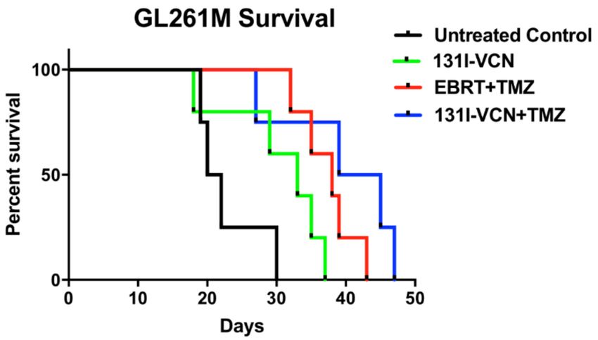
Molecules 2018, 23, 2918                                                                                           5 of 13

                                      Table 1. Brain specific uptake of 125 I-VCN.

                              Group                 % of Injected Counts in the Brain
                              Control       No counts detected above background (200 cpm)
                           Tumor Bearing              1.4% of the injected counts

2.4. Determination of Efficacy of 131 I-VCN in Limiting Tumor Progression in Xenograft Models of GBM
     We evaluated the efficacy 131 I-VCN in prolonging the progression-free survival of study animals
in two distinct xenograft models of glioma that are devoid of O-6-methylguanine methyltransferase
(MGMT) expression (i.e., U87 and U251) and therefore sensitive to TMZ. The U87 mice in the control
groups, began dying on day 20 and all mice in the control and 131 I alone groups were dead by day 25.
The mice treated with 131 I-VCN displayed no adverse effects of treatment and started to die on day 34
with all mice dead by day 44. The median survival for the controls, untreated and 131 I alone, were 22
and 21 days, respectively. While for 131 I-VCN, a median survival of 35 days was observed (Figure 3).

      Figure 3. Survival data following treatment with 131 I-VCN in the U87 xenograft model (5 mice per group).
      Treatment with 131 I-VCN extends survival of treated animals by more than 13 days (60% extension).
      Animals treated with 131 I-VCN maintained generally a good appearance and stable weight until a few
      days before succumbing to the disease. There is a significant therapeutic effect of 131 I-VCN (p value 0.0339)
      as compared to the control.

      Next, we examined the efficacy of 131 I-VCN in the U251 glioma model when delivered with the
DNA alkylating agent, temozolomide (TMZ). TMZ functions by alkylating/methylating DNA nucleotide
bases with >90% of lesions occurring at many different positions in all bases (i.e., the non O6-lesions)
and a minority (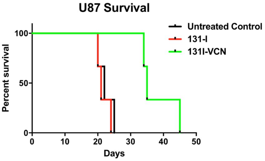
Molecules 2018, 23, 2918                                                                                    6 of 13

      Figure 4. Survival data following treatment with 131 I-VCN with or without temozolomide (TMZ) in
      the TMZ-sensitive U251 xenograft model. Treatment of tumor bearing animals (10 mice per group)
      with the combination of 131 I-VCN plus TMZ significantly enhances their survival over the animals
      treated with 131 I-VCN alone with a median survival in the combination group greater than 75 days.
      Furthermore, several mice from the combination group appeared tumor free at the conclusion of the
      experiment. Both the 131 I-VCN (p-value 0.0022) and 131 I-VCN + TMZ (p-value 0.0003) groups show a
      significant therapeutic advantage over the control while 131 I shows no significant therapeutic effect.

2.5. Comparison of Efficacy of 131 I-VCN in a Syngeneic Model of TMZ-Resistance Following a Schedule of
Administration Designed to Mimic the Stupp Protocol
       In this study, we compared the efficacy of 131 I-VCN plus TMZ against the current standard of care
{i.e., external beam radiation therapy (EBRT) plus TMZ} for glioma. We first derived a TMZ-resistant
population of murine glioma cells after transfecting the GL261 cells with a plasmid construct carrying
MGMT placed under the control of a medium-strength mouse promoter. To select highly resistant TMZ
cells, the transfected cells were further incubated with increasing concentrations of TMZ (up to 700 µM).
The cells derived after this procedure (i.e., GL261M) were further analyzed by Western blotting for MGMT
expression and confirmed that the GL261M cells are indeed producing MGMT. Furthermore, we validated
the functionality of this artificially expressed MGMT enzyme in a colony forming assay (CFA) by
seeding wildtype GL261 and GL261M at very low densities (50 cells/cm2 ) and incubating them in the
presence of a range of TMZ concentrations with or without O6-benzylguanine (O6BG), a potent MGMT
inhibitor (Figure 5).

      Figure 5. GL261M cells express functional mouse MGMT. (Panel A) Lysates were prepared from
      MGMT-transfected cells (GL261M) selected in the presence of increasing concentrations of TMZ and
      analyzed by Western blotting for MGMT expression. (Panel B) The functional activity of MGMT was
      further validated in a colony survival assay which shows that GL261M cells are resistant to TMZ but
      are re-sensitized to the drug in the presence of 10 µM of O6-benzylguanine (O6BG).
Molecules 2018, 23, 2918 7 of 13
Once the TMZ-resistant model was validated in vitro, we implanted syngeneic immunocompetent
mice with GL261M cells and allowed tumors to grow and become well established for 12 days after
implantation. These tumor-bearing mice were further randomized into four groups and treatment was
initiated 12 days post tumor implantation. The untreated control and 131 I-VCN groups began to show
severe signs of tumor growth (lethargy, weight loss and poor body condition score) by day 15 and
began to die on day 19 post implantation with all animals being sacrificed by day 30 in the control group
(median survival 21 days) and day 37 for the 131 I-VCN (median survival 33 days). The combination
of EBRT with TMZ, which was designed to mimic the Stupp protocol [4], demonstrated much better
survival compared to the controls (i.e., the first animal succumbing to the tumor at day 32), and with a
mean survival of 38 days. The combination of 131 I-VCN with TMZ, overall, performed the best with
the first animal succumbing on day 27, but with a median survival of 42 days for this group (Figure 6).
Figure 6. Survival data following a Stupp protocol-like schedule in a TMZ-resistant GL261M syngeneic
model. Animals (5 mice per group) in this model were treated with either external beam radiotherapy
(EBRT) dosed at 2 Gy/day plus TMZ dosed at 25 mg/kg/day both administered for five consecutive
days or with 2 doses of 100 µCi 131 I-VCN (administered 3 days apart) either alone or in combination
with TMZ dosed at 25 mg/kg/day for five consecutive days. In this glioma model of TMZ-resistance,
the 131 I-VCN/TMZ treatment combination shows better efficacy compared to the standard of care
EBRT/TMZ combination. There is a significant therapeutic effect between EBRT + TMZ (p-value 0.0047)
and 131 I-VCN + TMZ (p-value 0.0328) when compared to the control.
3. Discussion
Intratumor implantation of radioactive materials has been historically used to deliver a strong
dose of radiation to a small, defined area of glioma tissue, while limiting the amount of radiation
damage to nearby normal tissue. For instance, 125 I seeds have been used for glioma and there is
recent interest in the use of new forms of brachytherapy for glioma [31]. Two recent reviews detail
findings [32,33] of brachytherapy in glioma patients. It was noted that there was clear survival
advantage with postoperative radiation therapy at doses of 50 to 60 Gy, but dose escalation at doses
higher than this resulted in increased toxicity without any survival benefit. For relapsed glioma,
brachytherapy is also effective, but is associated with significant toxicity [32]. Here, we tested the
systemic administration of small amounts (dosed at 100 µCi) of beta-emitting radionuclides delivered
into the tumor microenvironment of GBM via a novel recombinant disintegrin called vicrostatin (VCN).
VCN was purposely designed with a carboxy-terminal extension, which was expected to enhance
affinity for integrin α5β1, and this was confirmed as binding affinity of VCN for α5β1 was found
to be ~13-fold higher than that of the native snake venom disintegrin, contortrostatin, VCN was
modelled after. Mass spectrometry shows that VCN is predominantly a monomer with a MW of
7146 Da, a size which makes this molecule an ideal candidate for efficient penetration of the tumor
microenvironment for payload delivery. Moreover, the high density of integrin targets expressed by
the glioma neovasculature obviates the need for VCN to penetrate the blood-brain-barrier in order to
deliver its 131 I payload. According to the brachytherapy principle and provided that VCN sticks to the
neovasculature, the beta particles emitted by 131 I will have an effective energy release radius of aboutMolecules 2018, 23, 2918 8 of 13
2 mm into the tumor tissue. Regarding the ability of VCN to effectively ligate integrins, unlike cyclic
RGD peptides and peptidomimetics, additional structural elements of VCN enable the disintegrin to
engage the receptor in an efficient and unique manner which leads to rapid integrin internalization.
Unlike cilengitide, cyclo(L-Arg-Gly-L-Asp-D-Phe-N-methyl-L-Val), a cyclic RGD peptide that was
extensively tested in the clinic in GBM [34], the additional residues flanking the RGD-loop in VCN
enable more extensive contacts with the integrin receptor. Also, NMR and crystallographic studies
have revealed that the C-terminal tail in disintegrins folds in close proximity to the RGD-containing
disintegrin loop, linking these two structural elements together in an extended conformational epitope,
suggesting that these two functional regions are engaged in extensive interactions with the target
integrin receptor [35–37]. This finding was further supported by our work on the rational design of
VCN, accounting for a 13-fold improved affinity for α5β1, by incorporating changes to the amino acid
sequence at the COOH-terminus of VCN [28]. By using structural and functional regions in addition
to the RGD motif, a sequence that serves as the sole basis for the design of cyclic RGD peptides
and RGD mimetics, disintegrins exhibit novel antitumor activities [27,28] compared to small cyclic
RGD peptides and peptidomimetics. Due to its robustness and reproducibility, we believe that our
recombinant production method will be easily translatable for scale up and cGMP production of VCN.
Furthermore, toxicity studies in rodents using dose escalation of VCN (250 mg/kg to 500 mg/kg),
a dose many times higher than that used in animal model studies of glioma, demonstrated complete
lack of toxicity.
In the present study, we show that radioiodinated VCN can be used for precision delivery of
therapeutic doses of 131 I into the microenvironment of glioma. In an earlier biodistribution study,
we demonstrated that radioiodinated VCN specifically binds to tumor tissue but not to the normal brain
after intravenous administration [13]. Moreover, in two distinct animal models of glioma we show
that radioiodinated VCN can significantly prolong the survival of treated animals. Specifically, in the
TMZ-sensitive U251 xenograft model we show that 131 I-VCN (100 µCi, intravenous) administered in
combination with TMZ (25 mg/kg, oral gavage) more than doubles the survival of treated animals
compared to the untreated controls. This result encouraged us to test the same therapeutic strategy in
a syngeneic model of TMZ-resistant glioma. To establish this model, we transfected GL261 murine
glioma cells with a plasmid that carries the murine MGMT gene under a mouse medium-strength
promoter (i.e., the murine phosphoglycerate kinase 1 promoter). The transfected cells were further
incubated in increased concentrations of TMZ (up to 700 µM) to select for highly TMZ-resistant clones
which were further implanted in syngeneic hosts. In this model, we compared head-to-head the
efficacy of 131 I-VCN in combination with TMZ versus external beam radiotherapy in combination
with TMZ. Although we expected all O6-methylguanine lesions to be directly reversed in the presence
of murine MGMT, we reasoned that TMZ could still synergize with radiotherapy in this model via
non-O6-methylguanine lesions, which in fact represent more than 90% of the DNA lesions inflicted
by TMZ. Indeed, the combination of 131 I-VCN plus TMZ proved superior to 131 I-VCN alone which
clearly suggest the possibility of a synergistic effect between TMZ and 131 I-VCN in this setting.
Moreover, two doses of 131 I-VCN (100 µCi given twice) proved to be superior to five consecutive
doses of whole-brain external beam radiotherapy (2 Gy given five times). These preliminary data
suggest that, if dosed according to rigorous dosimetry studies, 131 I-VCN may have the potential
to significantly synergize with TMZ in the clinical setting of TMZ resistance (i.e., typically newly
diagnosed patients with GBM tumors with unmethylated MGMT promoters). This could represent a
therapeutic alternative to the current Stupp protocol [4] which only provides very limited benefit to
this subset of patients.
We would like to make one additional point regarding the potential off-target effects of 131 I-VCN
in a clinical scenario. When compared to 131 I sodium iodide which, even though it cannot discriminate
between normal and cancerous thyroid tissue, is used in clinical practice for radioablation of cancerous
thyroid cells, radioiodinated VCN is a much bigger molecule (i.e., a polypeptide of ~7 KDa). Due to its
size, it is highly unlikely that VCN will fit into the Na-I symporter expressed by thyroid cells. For thisMolecules 2018, 23, 2918 9 of 13
reason, we predict that 131 I-VCN will not accumulate in the thyroid glands of GBM patients treated with
this form of intravenous brachytherapy. Moreover, the normal thyroid tissue is not known to express the
integrin receptors expressed by the GBM tissue, which are ligated by VCN. However, because a potential
off-target/off-tumor accumulation of radioactive material remains a concern with any form of radioiodine
brachytherapy in clinical practice, we believe that any therapeutic protocol for 131 I-VCN in glioma patients
should include a pre-brachytherapy dosing step with potassium iodide. This preparatory step is intended
to temporarily shut down the thyroid glands of GBM patients while 131 I-VCN is present in the circulation.
In aggregate, our animal studies demonstrate that VCN can be successfully used as a novel
delivery mechanism for targeting radioactivity to glioma cells. Although results are still preliminary,
they serve as a proof-of-principle demonstration that radioiodinated VCN, which we tested in several
animal models of GBM, can serve as an effective therapeutic approach for this deadly form of
cancer. Further, the complexity of the cancer microenvironment dictates that for optimal efficacy
an anti-integrin therapeutic must target at least two members of the RGD-binding integrin class and
preferably more [38] given the ability of cancer cells to change their integrin repertoire in response
to drug treatment. Our experience with VCN, which binds to multiple RGD-dependent integrins,
suggests that it admirably meets this criterion while also showing minimal binding to normal tissue.
Therefore, its excellent stability and high affinity for tumor integrins uniquely positions VCN for
precision delivery of targeted radioactivity in GBM, a type of solid tumor where radiotherapy is an
integral part of standard of care. In future investigations we will conduct dosimetry studies with VCN
labeled with 124 I (a positron emitter), which will allow us to precisely calculate by quantitative PET
(positron emission tomography) imaging the therapeutic dose of 131 I-VCN for each animal based on
its tumor burden and integrin biology.
4. Materials and Methods
4.1. Cells and Reagents
The U87 and U251 human glioma cell lines were obtained from ATCC (Manassas, VA, USA) and
maintained according to the manufacturer’s protocol. The GL261 murine glioma cell line was a gift from
Dr. Alan Epstein (Keck School of Medicine, University of Southern California, Los Angeles, CA, USA).
The TMZ-resistant variant of GL261 cell (i.e., GL261M) were generated by transfecting the wildtype cells
with a plasmid construct carrying the mouse O6-methylguanine-DNA-methyltransferase (MGMT) gene
under a mouse phosphoglycerate kinase 1 (mPGK) promoter. The transfected GL261 cells were further
cultured in increasing concentrations of TMZ (up to 700 µM) to select the GL261M variant. VCN was
produced and purified in the Markland laboratory [27,28]. Female 5-week-old 20 g Balb/c nu/nu mice
were obtained from Simonsen Laboratories (Gilroy, CA, USA), and 5-week old C57 bl/6 mice were obtained
from Charles River Laboratories (Boston, MA, USA). Sodium iodine (131 I) was obtained from Perkin-Elmer
(Billerica, MA, USA). All other reagents were purchased from Sigma Chemical Co. (St. Louis, MO, USA).
External beam radiotherapy was administered in a X-RAD320 irradiator (Precision X-Ray, North Branford,
CT, USA). Temozolomide (TMZ) was obtained from TCI America (Portland, OR, USA).
4.2. Radioactive and Radioinert Iodination of VCN
VCN in solution was directly iodinated with radioiodine (131 I) or radioinert sodium iodide using a
modification of the chloramine T method [29]. Briefly, chloramine T was dissolved in PBS and added to a
buffered VCN solution (100–600 µg at 0.3–2.0 mg/mL in PBS) containing Na 131 I. Five successive aliquots
of 15 µg chloramine T (in 5 µL) were added at 5-min intervals to the reaction mixture (final volume
400 µL). Following the final chloramine T addition, excess 131 I was quenched through the addition
of sodium metabisulfite (100 µg/400 µL reaction). Unreacted iodine was removed from the 131 I-VCN
solution and the buffer exchanged by 2× repeated filtration through a centrifugal 4-kDa cutoff filter
(VWR, Atlanta, GA, USA). This allows iodine to pass through the membrane and be separated from the
iodinated VCN solution.Molecules 2018, 23, 2918 10 of 13
4.3. Evaluation of Stoichiometry of Iodine Addition
The stoichiometry of iodine addition to VCN was determined by use of mass spectrometry.
A LC-Electro-spray ionization mass spectrometry system was employed in the positive ion mode
(Agilent, Santa Clara, CA, USA). A control of non-iodinated VCN was used as a standard.
4.4. FACS Analysis of VCN Binding to Glioma Cell Lines
The cold iodinated VCN (I-VCN) was further conjugated with Cy5-NHS (Lumiprobe, Hunt Valley,
MD, USA) according to manufacturer’s protocol. For FACS analysis, 106 glioma cells (U87, U251,
and GL261M) in complete DMEM (Dulbecco’s Modified Eagle Medium) were incubated with or without
100-fold molar excess of a small cyclic RGD peptide called Cilengitide (Selleck Chemicals, Houston,
TX, USA) [30] for 20 min at 37 ◦ C, then pelleted and resuspended in ice-cold PBS. The cells were further
incubated for 60 min at 4 ◦ C with Cy5-I-VCN, then pelleted, washed and resuspended in ice-cold PBS.
The cells were further analyzed on a BD FACSAria II instrument (BD Biosciences, Franklin Lakes, NJ,
USA) equipped with a 642 nm red laser. Cells incubated with unlabeled disintegrin (i.e., negative control)
were used to calibrate the instrument.
4.5. Confirmation of Biological Activity of Iodinated VCN
The inhibition of ADP-induced platelet aggregation by iodinated-VCN (both I-VCN and
Cy5-I-VCN) was measured by determining the light absorption of human platelet-rich plasma (PRP)
in a specialized spectrophotometer (Chrono-log 490 optical aggregometer, Chrono-log, Havertown,
PA, USA) as previously described [39]. The iodinated disintegrins (I-VCN and Cy5-I-VCN) were tested
for activity against unlabeled VCN.
4.6. Functional Analysis of GL261M Cells to Confirm MGMT Expression and Activity
Lysates prepared from wildtype GL261 and TMZ-resistant GL261M cells were analyzed by
Western-blotting for MGMT expression levels. A rabbit polyclonal antibody that cross-reacts with the
mouse MGMT protein was purchased from Boster Biological Technology (Pleasanton, CA, USA) and used
in this Western-blot analysis. This antibody was raised against a synthetic peptide PVFQQESFTRQVLWK,
which corresponds to a sequence in the middle region of human MGMT which is different from the
related rat and mouse sequences by only one amino acid. Furthermore, to check the functionality of the
MGMT enzyme resulting from transfection of the MGMT gene into GL261 cells, we completed a colony
formation assay (CFA) with GL261M cells incubated in a range of TMZ concentrations in the presence or
absence of the MGMT inhibitor O6-benzylguanine (Millipore Sigma, Burlington, MA, USA).
4.7. Establishment of Orthotopic Xenograft Glioma Models
All animal protocols were approved by the University of Southern California, IACUC (20707-CR001,
29 March 2018), and animals maintained according to approved guidelines. To perform this orthotopic
xenograft model, tumors were implanted 3 mm deep in the midline of mice brains using stereotactic
injections. Following surgical exposure of the skull and drilling a small-bore hole with a dental drill,
U87 or U251 (2 × 10⁵ /2 µL) glioma cells were injected using a slow controlled injection followed by a
one-minute rest period with syringe in place to minimize leakage from the injection track. The syringe
was slowly removed, the hole covered with wax, and the surgical incision was then closed with 2 stitches
(3.0 silk). Following implantation (7–14 days) tumor take was evaluated by bioluminescent imaging.
This was only possible in the U87 and U251 models in which the injected tumor cells were labeled with
GFP and firefly luciferase. In these xenograft models, the tumors were imaged by intravenous injection
of luciferin followed by a 90 s wait period when the animal was placed in a Xenogen IVIS 200 imaging
instrument (Perkin Elmer, Waltham, MA, USA) before acquiring optical images. In our experience
with these xenograft models, >90% of mice show a bioluminescent signal at 7 days post-implantation.
Positive, tumor bearing, mice were then randomized into treatment and control groups.Molecules 2018, 23, 2918 11 of 13
4.8. Evaluation of Brain-Uptake of Radioiodinated-VCN
To determine tumor specific brain uptake and retention of iodinated VCN, mice were implanted
with human gliomas (U87 as above) and the tumors were allowed to grow for 14 days. At this point
mice with confirmed tumors and control non-tumor bearing mice were injected with a single dose of
125 I-VCN (100 µCi) administered intravenously. Radioactively labeled VCN was allowed to circulate
for 18 h at which time the animals were euthanized, and brains removed, rinsed and counted in a
2480 WIZARD2 gamma counter available in the Molecular Imaging Center at the Health Sciences
Campus, University of Southern California.
4.9. Establishment of a Syngeneic Model of Glioma
For these studies, immunocompetent syngeneic C57BL/6 mice were implanted orthotopically
with TMZ-resistant GL261M glioma cells. Following a protocol identical to the method used with
the orthotopic xenograft model, tumors were implanted, and 13 days post implantation treatment
was initiated. Due to immune rejection concerns, these tumor cells were not labeled with foreign
transgenes such as GFP and luciferase, thus tumor growth could not be confirmed by bioluminescent
imaging in this syngeneic model. However, based on survival curves that were previously generated
by us after implanting various numbers of GL261M cells in vivo, we expected tumor growth in 100%
of the mice implanted with 1 × 10⁵ GL261M cells.
4.10. Treatment of Animal Models
For the U87 glioma studies, the following groups were employed (five mice per group): (i) untreated
control, (ii) treatment with 131 I alone (administered IV as Na131 I) and (iii) 131 I-VCN. The dose of
radioactivity administered was 100 µCi either iodine alone or radioiodinated VCN (131 I-VCN). The mice
were treated with a single dose and monitored daily for changes in weight and physical status.
For the 131 I-VCN combination with TMZ chemotherapy U251 glioma positive mice were randomized
into 4 groups of 10 mice each. These groups were: (i) untreated control, (ii) 131 I alone, (administered
IV as Na131 I), (iii) 131 I-VCN alone, and (iv) 131 I-VCN plus TMZ. The radioactivity was delivered as
100µCi either sodium iodine alone or radioiodinated VCN (131 I-VCN) given in two doses separated by
three days (each with 100 µCi 131 I-VCN). The groups receiving TMZ had the agent delivered as a daily
25 mg/kg gavage over five consecutive days. The efficacy of 131 I-VCN plus TMZ was further compared
to external beam radiation therapy (EBRT) plus TMZ in a TMZ-resistant syngeneic model. In this model,
we evaluated the following groups (five mice per group): (i) untreated control, (ii) 131 I-VCN alone, (iii)
EBRT plus TMZ and (iv) 131 I-VCN plus TMZ. The radioactivity was delivered as 100 µCi either iodine
alone or 131 I-VCN given in two doses separated by three days (each with 100 µCi). In comparison,
the EBRT plus TMZ group followed a dosing regimen mimicking the Stupp protocol [4] with TMZ
delivered as a daily 25 mg/kg gavage concurrently with 2 Gy EBRT daily for five consecutive days.
External beam irradiation was performed at 250 kv and 16 mA with a dose of 200 cGy delivered over
47 s to the skull of each animal over five consecutive days. Except for the skull area, the rest of each
animal’s body was protected by a 3 mm-thick lead shield.
4.11. Statistical Significance
To determine statistical significance, the outcomes of the individual treatments were compared to
the untreated controls using the Wilcoxon–Mann–Whitney two-sample rank-sum test. The treatment
effect (difference between treatments) was quantified using the Hodges–Lehmann (HL) estimator,
which is consistent with the Wilcoxon test. p values were determined and values less than 0.05 were
considered significant.
Author Contributions: Conceptualization, S.S., R.O.M., T.C.C. and F.S.M.; Funding acquisition, T.C.C.;
Investigation, S.S. and R.O.M.; Methodology, S.S., R.O.M., C.D.T. and T.-Z.T.; Writing—original draft,
S.S., R.O.M. and F.S.M.; Writing—review & editing, R.O.M., S.S. and F.S.M.Molecules 2018, 23, 2918 12 of 13
Funding: These studies were funded in part by grant R41 CA165626 from the National Cancer Institute of the
National Institutes of Health.
Conflicts of Interest: S.S., R.O.M., T.C.C. and F.S.M. are co-founders of an unfunded start-up company,
Disintegrin Therapeutics, Inc. The funder had no role in the design of the study; in the collection, analyses, or
interpretation of data; in the writing of the manuscript, or in the decision to publish the results.
References
1. Cancer Stat Facts: Brain and Other Nervous System Cancer. Available online: https://seer.cancer.gov/statfa
cts/html/brain.html. (accessed on 5 September 2018).
2. McCutcheon, I.E.; Chernov, M.F. Rationale for Aggressive Resection and General Surgical Principles for
Intracranial Gliomas. Prog. Neurol. Surg. 2018, 30, 63–105. [PubMed]
3. Eagles, M.E.; Nassiri, F.; Badhiwala, J.H.; Suppiah, S.; Almenawer, S.A.; Zadeh, G.; Aldape, K.D. Dendritic
cell vaccines for high-grade gliomas. Ther. Clin. Risk Manag. 2018, 14, 1299–1313. [CrossRef] [PubMed]
4. Stupp, R.; Hegi, M.E.; Mason, W.P.; van den Bent, M.J.; Taphoorn, M.J.; Janzer, R.C.; Ludwin, S.K.; Allgeier, A.;
Fisher, B.; Belanger, K.; et al. Effects of radiotherapy with concomitant and adjuvant temozolomide versus
radiotherapy alone on survival in glioblastoma in a randomised phase III study: 5-year analysis of the
EORTC-NCIC trial. Lancet Oncol. 2009, 10, 459–466. [CrossRef]
5. Miyauchi, J.T.; Tsirka, S.E. Advances in immunotherapeutic research for glioma therapy. J. Neurol. 2018, 265, 741–756.
[CrossRef] [PubMed]
6. Felthun, J.; Reddy, R.; McDonald, K.L. How immunotherapies are targeting the glioblastoma immune environment.
J. Clin. Neurosci. 2018, 47, 20–27. [CrossRef] [PubMed]
7. Ferguson, S.D.; Momin, E.N.; Weinberg, J.S. Surgical Management of Recurrent Intracranial Gliomas.
Prog. Neurol. Surg. 2018, 30, 218–231. [PubMed]
8. Askari, J.A.; Buckley, P.A.; Mould, A.P.; Humphries, M.J. Linking integrin conformation to function. J. Cell
Sci. 2009, 122, 165–170. [CrossRef] [PubMed]
9. Hynes, R.O. Integrins: Versatility, modulation, and signaling in cell adhesion. Cell 1992, 69, 11–25. [CrossRef]
10. Pignatelli, M.; Cardillo, M.R.; Hanby, A.; Stamp, G.W. Integrins and their accessory adhesion molecules in
mammary carcinomas: Loss of polarization in poorly differentiated tumors. Hum. Pathol. 1992, 23, 1159–1166.
[CrossRef]
11. Cheresh, D.A. Structural and biologic properties of integrin-mediated cell adhesion. Clin. Lab Med. 1992, 12, 217–236.
[CrossRef]
12. Ruoslahti, E. Integrins. J. Clin. Investig. 1991, 87, 1–5. [CrossRef] [PubMed]
13. Pyrko, P.; Wang, W.; Markland, F.S.; Swenson, S.D.; Schmitmeier, S.; Schönthal, A.H.; Chen, T.C. The role
of contortrostatin, a snake venom disintegrin, in the inhibition of tumor progression and prolongation of
survival in a rodent glioma model. J. Neurosurg. 2005, 103, 526–537. [CrossRef] [PubMed]
14. Cordes, N.; Hansmeier, B.; Beinke, C.; Meineke, V.; van Beuningen, D. Irradiation differentially affects
substratum-dependent survival, adhesion, and invasion of glioblastoma cell lines. Br. J. Cancer 2003, 89, 2122–2132.
[CrossRef] [PubMed]
15. Schmitmeier, S.; Markland, F.S.; Ritter, M.R.; Sawcer, D.E.; Chen, T.C. Functional effect of contortrostatin,
a snake venom disintegrin, on human glioma cell invasion in vitro. Cell Commun. Adhes. 2003, 10, 1–16.
[CrossRef] [PubMed]
16. Tabatabai, G.; Tonn, J.C.; Stupp, R.; Weller, M. The role of integrins in glioma biology and anti-glioma therapies.
Curr. Pharm. Des. 2011, 17, 2402–2410. [CrossRef] [PubMed]
17. Kumar, C.C. Integrin alpha v beta 3 as a therapeutic target for blocking tumor-induced angiogenesis.
Curr. Drug Targets 2003, 4, 123–131. [CrossRef] [PubMed]
18. Chamberlain, M.C.; Cloughsey, T.; Reardon, D.A.; Wen, P.Y. A novel treatment for glioblastoma: Integrin inhibition.
Expert Rev. Neurother. 2012, 12, 421–435. [CrossRef] [PubMed]
19. Gould, R.J.; Polokoff, M.A.; Friedman, P.A.; Huang, T.F.; Holt, J.C.; Cook, J.J.; Niewiarowski, S. Disintegrins:
A family of integrin inhibitory proteins from viper venoms. Proc. Soc. Exp. Biol. Med. 1990, 195, 168–171.
[CrossRef] [PubMed]
20. Swenson, S.; Ramu, S.; Markland, F.S. Anti-angiogenesis and RGD-containing snake venom disintegrins.
Curr. Pharm. Des. 2007, 13, 2860–2871. [CrossRef] [PubMed]Molecules 2018, 23, 2918 13 of 13
21. McLane, M.A.; Joerger, T.; Mahmoud, A. Disintegrins in health and disease. Front. Biosci. 2008, 13, 6617–6637.
[CrossRef] [PubMed]
22. Saudek, V.; Atkinson, R.A.; Pelton, J.T. Three-dimensional structure of echistatin, the smallest active RGD protein.
Biochemistry 1991, 30, 7369–7372. [CrossRef] [PubMed]
23. Moiseeva, N.; Bau, R.; Swenson, S.D.; Markland, F.S.; Choe, J.Y.; Liu, Z.J.; Allaire, M. Structure of acostatin,
a dimeric disintegrin from Southern copperhead (Agkistrodon contortrix contortrix), at 1.7 A resolution.
Acta Crystallogr. D Biol. Crystallogr. 2008, 64, 466–470. [CrossRef] [PubMed]
24. Shebuski, R.J.; Ramjit, D.R.; Bencen, G.H.; Polokoff, M.A. Characterization and platelet inhibitory activity of
bitistatin, a potent arginine-glycine-aspartic acid-containing peptide from the venom of the viper Bitis arietans.
J. Biol. Chem. 1989, 264, 21550–21556. [PubMed]
25. Yasuda, T.; Gold, H.K.; Leinbach, R.C.; Yaoita, H.; Fallon, J.T.; Guerrero, L.; Napier, M.A.; Bunting, S.; Collen, D.
Kistrin, a polypeptide platelet GPIIb/IIIa receptor antagonist, enhances and sustains coronary arterial thrombolysis
with recombinant tissue-type plasminogen activator in a canine preparation. Circulation 1991, 83, 1038–1047.
[CrossRef] [PubMed]
26. Cousins, G.R.; Sudo, Y.; Friedrichs, G.R.; Markland, F.S.; Lucchesi, B.R. Contortrostatin Prevents Reocclusion
after Thrombolytic Therapy in a Canine Model of Carotid Artery Thrombosis. FASEB J. 1995, 9, A938.
27. Minea, R.; Helchowski, C.; Rubino, B.; Brodmann, K.; Swenson, S.; Markland, F., Jr. Development of a
chimeric recombinant disintegrin as a cost-effective anti-cancer agent with promising translational potential.
Toxicon 2012, 59, 472–486. [CrossRef] [PubMed]
28. Minea, R.O.; Helchowski, C.M.; Zidovetzki, S.J.; Costa, F.K.; Swenson, S.D.; Markland, F.S., Jr.
Vicrostatin—An anti-invasive multi-integrin targeting chimeric disintegrin with tumor anti-angiogenic
and pro-apoptotic activities. PLoS ONE 2010, 5, e10929. [CrossRef] [PubMed]
29. Greenwood, F.C.; Hunter, W.M.; Glover, J.S. The Preparation of I-131-Labelled Human Growth Hormone of
High Specific Radioactivity. Biochem. J. 1963, 89, 114–123. [CrossRef] [PubMed]
30. Lombardi, G.; Pambuku, A.; Bellu, L.; Farina, M.; Della Puppa, A.; Denaro, L.; Zagonel, V. Effectiveness
of antiangiogenic drugs in glioblastoma patients: A systematic review and meta-analysis of randomized
clinical trials. Crit. Rev. Oncol. Hematol. 2017, 111, 94–102. [CrossRef] [PubMed]
31. Nachbichler, S.B.; Kreth, F.W. Brachytherapy of Intracranial Gliomas. Prog. Neurol. Surg. 2018, 31, 72–86. [PubMed]
32. Barani, I.J.; Larson, D.A. Radiation therapy of glioblastoma. Cancer Treat. Res. 2015, 163, 49–73. [PubMed]
33. Barbarite, E.; Sick, J.T.; Berchmans, E.; Bregy, A.; Shah, A.H.; Elsayyad, N.; Komotar, R.J. The role of brachytherapy
in the treatment of glioblastoma multiforme. Neurosurg. Rev. 2017, 40, 195–211. [CrossRef] [PubMed]
34. Scaringi, C.; Minniti, G.; Caporello, P.; Enrici, R.M. Integrin inhibitor cilengitide for the treatment of
glioblastoma: A brief overview of current clinical results. Anticancer Res. 2012, 32, 4213–4223. [PubMed]
35. Brown, M.C.; Eble, J.A.; Calvete, J.J.; Marcinkiewicz, C. Structural requirements of KTS-disintegrins for
inhibition of alpha(1)beta(1) integrin. Biochem. J. 2009, 417, 95–101. [CrossRef] [PubMed]
36. Marcinkiewicz, C.; Vijay-Kumar, S.; McLane, M.A.; Niewiarowski, S. Significance of RGD loop and
C-terminal domain of echistatin for recognition of alphaIIb beta3 and alpha(v) beta3 integrins and expression
of ligand-induced binding site. Blood 1997, 90, 1565–1575. [PubMed]
37. Chang, Y.T.; Shiu, J.H.; Huang, C.H.; Chen, Y.C.; Chen, C.Y.; Chang, Y.S.; Chuang, W.J. Effects of the RGD loop and
C-terminus of rhodostomin on regulating integrin alphaIIbbeta3 recognition. PLoS ONE 2017, 12, e0175321.
38. Sheldrake, H.M.; Patterson, L.H. Strategies to inhibit tumor associated integrin receptors: Rationale for dual and
multi-antagonists. J. Med. Chem. 2014, 57, 6301–6315. [CrossRef] [PubMed]
39. Swenson, S.; Bush, L.R.; Markland, F.S. Chimeric derivative of fibrolase, a fibrinolytic enzyme from southern
copperhead venom, possesses inhibitory activity on platelet aggregation. Arch. Biochem. Biophys. 2000, 384, 227–237.
[CrossRef] [PubMed]
Sample Availability: Samples of the compound vicrostatin are available from the authors with consent from the
company Disintegrin Therapeutics, Inc.
© 2018 by the authors. Licensee MDPI, Basel, Switzerland. This article is an open access
article distributed under the terms and conditions of the Creative Commons Attribution
(CC BY) license (http://creativecommons.org/licenses/by/4.0/).You can also read