Original Article Dental pulp stem cells expressing SIRT1 improve new bone formation during distraction osteogenesis
←
→
Page content transcription
If your browser does not render page correctly, please read the page content below
Am J Transl Res 2019;11(2):832-843
www.ajtr.org /ISSN:1943-8141/AJTR0085200

Original Article
Dental pulp stem cells expressing SIRT1 improve
new bone formation during distraction osteogenesis
Donghui Song, Ping Xu, Shu Liu, Senbin Wu

Department of Stomatology, Affiliated Hospital of Nantong University, Nantong 226001, China
Received September 7, 2018; Accepted December 12, 2018; Epub February 15, 2019; Published February 28,
2019

Abstract: Distraction osteogenesis (DO) is one of the most promising reconstructive methods for repairing large
craniofacial defects or growth deficiencies through bone regeneration, but it is also a challenge because of an unde-
sirably long process and its complications, which limit its application in clinical practice. The transplantation of mes-
enchymal stem cells (MSCs) is regarded as an innovative approach to accelerate bone regeneration. Dental pulp
stem cells (DPSCs) have shown some advantages over other human adult MSCs, and DPSCs have been regarded
as one of the most promising cell sources used in the endogenous tissue engineering. Furthermore, using stem
cells modified by gene engineering in DO has been reported in previous studies. It has been shown that Sirtuin-1
(SIRT1) can directly regulate the differentiation of MSCs into osteoblasts. In this study, DPSCs expressing SIRT1
were prepared and their effects on the new bone formation were further investigated in rabbits with tibia. Rabbits
were injected with the adenovirus (Adv)-SIRT1-green fluorescent protein (GFP)-transfected DPSCs (overexpression
group, Group OE), Adv-GFP transfected DPSCs (negative control group, Group NC) or physiologic saline (control
group, Groups CON) into the distraction gap. The new bone tissues in the distraction gap were harvested 8 weeks
later, and subjected to by radiographic examination, micro-CT evaluation, and histological and mechanical testing.
The better bone formation, the highest bone mineral density (BMD) and the highest bone mineral content (BMC)
were observed in the OE group. In conclusion, SIRT1-modified DPSCs in DO was more effective to promote new bone
formation during DO, which provides evidence for further investigation about the role of of SIRT1 in the DO.

Keywords: Distraction osteogenesis, dental pulp stem cells, SIRT1, bone formation

Introduction                                                     is necessary to develop a safe and efficient way
                                                                 for the improvement of DO in clinical practice.
Distraction osteogenesis (DO) is an endoge-
nous process that promotes bone regeneration                     Dental pulp stem cells (DPSCs), a type of mes-
in both long bones and maxillofacial bones,                      enchymal stem cells (MSCs), are multipotent
and simultaneously, the soft tissues including                   cells which can differentiate into distinct spe-
muscles, nerves, skin and blood vessels are                      cialized cell types, such as osteocytes, chon-
also elongated [1-3]. DO is also an orthopae-                    drocytes, and adipocytes [8-10]. Furthermore,
dics surgery that provides a way to reconstruct                  it has been reported that DPSCs are more
the skeleton deformities in clinical practice,                   effective in proliferation and osteogenesis, and
but the mechanism of DO has still not yet                        have lower immunogenicity than mesenchymal
completely revealed. DO activates the endoge-                    stem cells (MSCs) [11]. DPSCs originate from
nous tissue regeneration and new bone forma-                     dental pulp tissues, and have become a kind of
tion in the distraction gap. It has been report-                 seed cells in regenerative medicine and tissue
ed that there are proliferation, differentiation,                engineering because of their easier collection
angiogenesis, ossification, and remodeling dur-                  and higher osteogenic capacity as compared to
ing DO in the enlarging gap [4-6]. These com-                    MSCs [11]. Recently, it has been demonstrated
plicated processes have the risk of causing                      that MSCs play a pivotal role during DO, which is
many uncontrolled complications (such as de-                     regulated by series of signals such as proteins,
layed union and nonunion enlarging gap), which                   drugs [12-14]. We have found that DPSCs have
limits its application in clinical application [7]. It           the osteogenic potential and can be used dur-
SIRT1 improves new bone formation

ing DO [15]. Some studies have been conduct-       Dental Department of the Affiliated Hospital of
ed to investigate the effects of growth factors,   Nantong University, which was approved by the
hormonal proteins, miRNAs and some other           Ethics Committee of the Affiliated Hospital of
biological factors on the DO [6, 16, 17], and      Nantong University (2016--077). All subjects
findings reveal these factors can potentially      had neither carious lesions nor any other oral
enhance the osteogenesis and shorten the           infection. The pulp tissues were separated from
consolidation period after MSCs transplanta-       the crown and root completely, and then incu-
tion. In addition, injection of gene-modified      bated with digestive solution (3 mg/mL type I
DPSCs into the distraction gap can accelerate      collagenase, 4 mg/mL dispase in 4 mL of phos-
the distraction rate in a rabbit model [18].       phate-buffered saline (PBS), 100 U/mL penicil-
                                                   lin, 100 μg/mL streptomycin) for 1 h at 37°C.
Sirtuin 1 (SIRT1), an nicotinamide adenine         Single-cell suspensions were obtained by pass-
dinucleotide (NAD) - dependent class III protein   ing the digested tissues through a 70-μm cell
deacetylase, has been reported to involve in       strainer (BD Falcon). Cell suspensions of the
the regulation of various cellular processes,      dental pulp were added into 25-cm2 culture
including bone homeostasis, gene expression,       dishes, followed by incubation in Dulbecco’s
and metabolic pathways [19, 20]. SIRT1 knock-      modified Eagle medium (DMEM) supplemented
out mice show a reduction in bone mass char-       with 10% fetal bovine serum (FBS), 100 U/mL
acterized by increased marrow adipogenesis         penicillin and 100 μg/mL streptomycin at
and decreased bone formation [21]. Moreover,       37°C in 5% CO2 atmosphere. The medium was
SIRT1 is able to control the differentiation of    refreshed once every 3 days. Cells were pas-
MSCs into osteoblasts by up-regulating the         saged at the ratio of 1:3 when the confluence
expression of maker genes of osteogenesis          reached 85% to 90%. Cells of the third passage
and inhabiting adipogenic differentiation [22].    were used in following experiments.
SIRT1 overexpression or activation enhances
new bone formation characterized by the            Construction of Adv-hSIRT1-GFP and Adv-GFP
increased quality and quantity of bone regen-
eration [23]. These findings were confirmed in     Recombinant adenoviruses encoding human
our previous study [24]. Thus, we hypothesized     SIRT1 with green fluorescent protein (Adv-
that the up-regulation of SIRT1 expression         hSIRT1-GFP) and GFP alone (Adv-GFP) were
might promote bone regeneration during DO.         constructed by direct cloning. The titers of the
                                                   recombinant adenoviral vectors were as fol-
In this study, SIRT1 gene-modified DPSCs were      lows: Adv-hSIRT1-GFP, 5 × 10⁹ plaque-forming
prepared and injected into the tibia distraction   units (PFUs)/mL and Adv-GFP, 2 × 10¹⁰ PFU/mL.
gap, aiming to investigate the role of SIRT1 in
                                                   Transfection with adenovirus (Adv)-hSIRT1 and
the new bone formation after DPSCs transplan-
                                                   osteogenic differentiation
tation during DO.

Materials and methods                              The recombinant adenoviruses were trans-
                                                   duced into 293 cells and purified using stan-
Animals                                            dard cesium chloride banding techniques.
                                                   DPSCs were prepared as described above and
Thirty-six mature (2.5-3.0 kg) male New Zea-       seeded in six-well culture plates (1.4 × 10⁵
land white rabbits were used in this study. The    cells/well) on the day before transfection.
study protocol was approved by the Animal          DPSCs were transfected overnight with Adv-
Care Committee of Nantong University. All rab-     hSIRT1-GFP or Adv-GFP (multiplicity of infec-
bits were anesthetized by intravenous injection    tions [MOIs], XXX) at 37°C in α-minimum essen-
of ketamine hydrochloride and xylazine at 20       tial medium (MEM) control medium α-MEM.
mg/kg and 5 mg/kg body weight, respectively.       GFP expression was observed using fluores-
                                                   cence microscopy (Leica, Wetzlar, Germany) to
Isolation, culture and treatment of DPSCs          determine the transfection efficiency of Adv-
                                                   hSIRT1-GFP and Adv-GFP. At 96 h after trans-
The extracted normal human impacted third          fection, cells were collected for RT-PCR and
molars completely were collected from patients     Western blotting to determine the expression
aged 18-28 years (n = 9) and the informed con-     of SIRT1. DPSCs were plated at a density of
sent was obtained from each patient at the         2 × 10⁴ cells/cm2 and cultured in proliferation

833                                                         Am J Transl Res 2019;11(2):832-843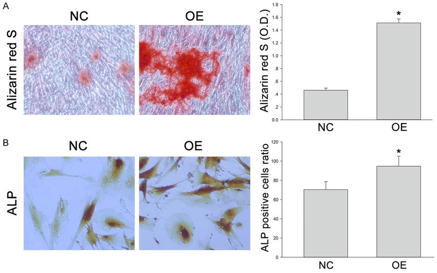
SIRT1 improves new bone formation

medium supplemented with 0.1 mM dexameth-           Surgical procedure, postoperative care, and
asone, 10 mM β-glycerophosphate (Sigma) and         distraction
50 mg/mL ascorbic acid (Sigma). The differen-
tiation of DPSCs was observed for 14 days. The      Each animal was placed in the dorsal position
degree of extracellular matrix calcification was    and a 2.0-cm vertical incision was made on the
estimated by Alizarin red S (Sigma) staining and    right hind leg to expose the tibia. Then, the peri-
Alkaline phosphatase (ALP, JianCheng, Nanjing,      osteum was carefully elevated circumferential-
China) staining. In brief, DPSCs were fixed with    ly. After a self-constructed external fixator had
4% paraformaldehyde (PFA) for 1 h and washed        been applied to the tibia with four self-taping
with phosphate-buffered saline (PBS). Then,         screws, a subperiosteal osteotomy was per-
DPSCs were incubated with 2% Alizarin red S         formed between the second and third screws
solution at 37°C for 2 h. Mineralization was        using a fine wire saw at a level immediately
quantified by extracting the stain using 100        below the area attached to the fibula. The peri-
mM cetylpyridinium chloride (Sigma-Aldrich) at      osteum, muscle, and skin were repositioned
room temperature for 2 h. The absorbance of         and closed with 3/0 silk sutures. The external
the extracted Alizarin red S stain was measured     fixator remained in place until necropsy. The
at 570 nm. DPSCs were subjected to ALP stain-       animals were kept in separate cages with food
ing using the ALP assay kit in accordance with      and water under the standardized environmen-
the manufacturer’s instructions.                    tal condition with 12-h light/dark cycle, and
                                                    allowed to move freely throughout the experi-
Determination of SIRT1 expression after trans-      mental period. Three days after osteotomy, the
fection                                             distraction was started at a rate of 2.0 mm per
                                                    24 h (twice per day and 1.0 mm per time) for 7
Total cellular RNA was isolated from cells and      days. Upon completion of the distraction, all
then reversely transcribed using conventional       animals were randomly divided into three
protocols. PCR amplification was performed          groups (n = 12): CON (control group or phos-
using the following primers: SIRT1: Forward         phate buffered saline group), NC (negative con-
5’-GCAACATCTTATGATTGGCACA-3’, reverse 5’-
                                                    trol or DPSCs infected with Adv-GFP), and OE
AAATACCATCCCTTGACCTGAA-3’, GAPDH: For-
                                                    (overexpression group or DPSCs infected with
ward 5’-TCCATGACAACTTTGGTATCG-3’, reverse
                                                    Adv-SIRT1-GFP). All rabbits were sacrificed at 8
5’-TGTAGCCAAATTCGTTGTCA-3’. All of the prim-
                                                    weeks after the completion of lengthening, and
er sequences were determined according to
                                                    the lengthened tibiae were harvested and pro-
the gene sequences from GenBank. Each sam-
                                                    cessed for further examinations.
ple was analyzed in triplicate and GAPDH was
used as a control. Cells were lysed in the buffer   Gene therapy
consisting of 50 mM Tris, 150 mM NaCl, 2%
sodium dodecyl sulfate (SDS) and a protease         DPSCs transfected with Adv-hSIRT1-GFP were
inhibitor mixture. After centrifugation at 12,000   harvested 2 days after transfection. They were
rpm for 12 min, protein concentrations were         washed with PBS and diluted in normal saline
determined using the Bradford assay (BioRad).       into 1 × 10⁷ cells/mL. On the last day of the
The resulting supernatant (50 mg of protein)        distraction period, 1 mL of DPSCs suspension
was subjected to SDS-polyacrylamide gel elec-       was injected directly into the distraction gap in
trophoresis (PAGE). The separated proteins          Group OE. Similarly, animals in Group NC
were transferred onto polyvinylidene fluoride       received an injection with 1 × 10⁷ DPSCs trans-
(PVDF) membranes at 350 mA for 2.5 h in a           fected with Adv-GFP, and animals in Group CON
blotting apparatus (Bio-RAD, CA, USA). Mem-         were injected with 1.0 mL of normal saline. The
branes were blocked with 5% nonfat milk, incu-      animals were held in rigid fixation until they
bated with primary antibodies (1:400) at 4°C        were sacrificed.
overnight and subsequently treated with anti-
rabbit horseradish peroxidase-conjugated sec-       Radiographic and dual-energy X-ray absorpti-
ondary antibodies (1:1000) for 2 h at room tem-     ometry (DXA) examinations
perature. Concomitantly, GAPDH served as a
reference. The following primary antibodies         The lengthened tibiae were harvested after
were used: GAPDH (anti-rabbit, Santa Cruz),         sacrifice, then lateral radiographs of the spe-
SIRT1 (anti-rabbit, Sigma).                         cimens were obtained, and the bone mineral

834                                                           Am J Transl Res 2019;11(2):832-843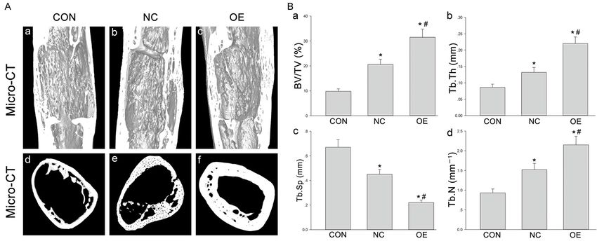
SIRT1 improves new bone formation

Figure 1. A. The green fluorescence of DPSCs after 3-day transfection under a fluorescence microscope. B. A large
amount of green fluorescence was observed under a microscope. C, D. Animals were divided into three groups: CON
(control group or phosphate buffered saline group), NC (negative control or DPSCs transfected with Adv-GFP) and
OE (overexpression group or DPSCs transfected with Adv-Runx2-GFP). C. The protein expression of SIRT1 in DPSCs
transfected by adenovirus vector containing human SIRT1 gene (Western blotting). GAPDH served as a control. The
optical density of SIRT1 was normalized to that of GAPDH at each time point. *P < 0.05. D. SIRT1 mRNA expres-
sion in DPSCs transfected by adenovirus vector containing human SIRT1 gene using (RT-PCR). GAPDH served as a
control. Quantification of RT-PCR products. The quantity of amplified product was analyzed by an image analyzer.
*P < 0.05.

density (BMD) was determined. Bone mineral                   Micro-CT evaluation
content (BMC) was detected by DXA (Lunar iDX-
ATM, GE Healthcare, USA). BMD and BMC of                     The samples were collected 5 mm outside the
the region of interest (ROI) were measured for               distracted regions, then placed in a custom jig
each distracted region. Both were also mea-                  with water, and scanned with a micro-CT 80
sured in the controlateral intact tibia as a nor-            scanner (Scanco Medical, Bassersdorf, Switzer-
mal control. All examinations were performed                 land) in an axial direction parallel to the long
three times by an operator blind to the experi-              axis of the tibiae. Nearly 1000 images with a
ment.                                                        resolution of 2048 × 2048 pixels and with an

835                                                                    Am J Transl Res 2019;11(2):832-843
SIRT1 improves new bone formation
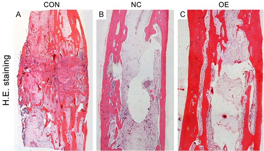
Figure 2. (A, B) DPSCs in Group NC and Group OE were cultured in osteogenic differentiation medium for 14 days,
and then stained with Alizarin red S (A) or ALP (B). Quantification of Alizarin red S positive deposits and the ratio of
ALP positive cells were described in right. *P < 0.05.
isotropic voxel size of 10 p.m were obtained. microtome (Leica, German) into 5-μm sections
The system was set to 70 kV, 114 mA, and an for hematoxylin eosin (H&E) staining.
integration time of 500, which allows accurate
analysis of the osteogenesis of the distracted Mechanical testing
regions. The volume of interest (VOI) was
selected as the distracted gap extending to a The samples were stored at -20°C until the day
total of 500 slices. The following micro-archi- of mechanical testing. The three-point bending
tecture parameters were assessed in VOI imag- strength was measured until failure between
es: bone volume to total volume ratio (BV/TV), the distraction regions with a support span of 5
trabecular thickness (Tb.Th), trabecular separa- mm, using an electronic universal material test
tion (Tb.Sp), and trabecular number (Tb.N). BV/ machine (Instron 5566, Instron, Norwood, MA,
TV indicates the portion of mineralized tissue, USA) with a 1-kN load cell under displacement
and Tb.Th, Tb.Sp, and Tb.N reflect the thick- control (5 mm/min). The load-displacement
ness, organization, and number of trabeculae. curve was recorded during the downward com-
After above examinations, six samples were pression and the ultimate load at failure (N;
randomly selected from each group and pro- maximum force that the specimen sustained)
cessed for histological examination. Remain- was calculated. The controlateral intact tibia
ing six samples were prepared for mechanical was also measured as a normal control.
testing.
Statistical analysis
Histological examination
All the data as expressed as mean ± standard
Samples were fixed in 4% paraformaldehyde for deviation (SDs). One-way analysis of variance
24 h, and then decalcified in 14.5% ethylenedi- (ANOVA) and Student-Newman Keuls (SNK)
aminetetraacetic acid buffer (Ph = 7.2) at room test were employed to compare the differen-
temperature for 6 weeks. The specimens were ces between groups using SPSS 19.0 (SPSS,
sectioned longitudinally along the long axial, Chicago, IL, USA). A value of P < 0.05 was con-
embedded in paraffin, and then cut with a sidered statistically significant.
836 Am J Transl Res 2019;11(2):832-843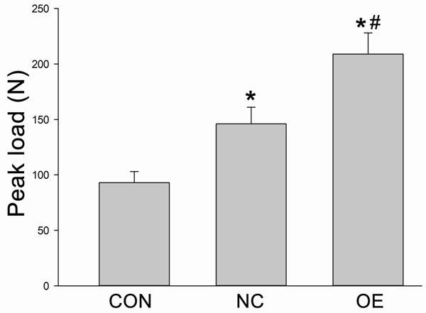
SIRT1 improves new bone formation
tion process was stabe and
the lengthened distraction
gaps maintained. At the
pre-designed time point,
the samples were harves-
ted for histological and
radiological examinations.
Results showed the newly
formed bone in Group OE
seemed to be more mature
than in Group NC and
Group CON.
Histological observation
Figure 3. (A-C) All samples from Group CON (A), Group NC (B) and Group OE (C) All samples in three groups
after 8-week consolidation were observed under a light microscope after H&E were observed under a
staining. The newly formed cortex in Group NC and Group OE was more con- light microscopy after H&E
tinuously than in Group CON. In Group NC, the newly formed trabeculae in the staining. In Group CON, the
distraction gap were thin, and partial trabeculae bridged discontinuously. More newly formed trabeculae
mature and regular trabecular bone were seen in Group OE.
were sparse, and focal de-
fects were seen in the dis-
Results traction gap (Figure 3A). In Group NC, the new-
ly formed trabeculae in the distraction gap
Evaluation of transfected cells were thin and partial trabeculae bridged dis-
continuously (Figure 3B). In Group OE, the
The expression of GFP in DPSCs was evaluated newly formed trabeculae in the distraction gap
by observation under a fluorescence micro- were thicker than in Group CON. More mature
scope. After 24-h transfection, the proportion and regular trabecular bone was observed in
of positive cells was approximately 100% Group OE (Figure 3C).
(Figure 1A). At the end of the distraction (at 7
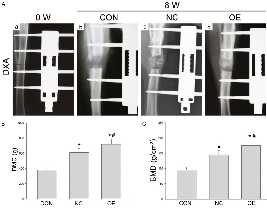
days), fibro-tissues had filled in the distracted Radiological findings, BMD and BMC
gap. A large amount of green fluorescence was
seen in Group OE and Group NC (Figure 1B), The distraction was displayed in the lateral
but in Group CON, little green fluorescence was radiograph (Figure 4A). At 0 (Figure 4Aa) and 8
observed. The expression of SIRT1 in Group OE weeks (Figure 4Ab-d), the focal defects were
was significantly higher than in Group NC and large, and tiny trabeculae could be found in the
Group CON (Figure 1C). The mRNA level, RT-PCR DO gaps of Group CON (Figure 4B). In the dis-
also showed the SIRT1 expression in Adv- traction gap of Group NC, the disordered tra-
SIRT1-GFP (Group OE) was much higher than beculae were observed, and the newly formed
in Adv-GFP group (Group NC) and control gro- trabeculae showed insufficiently robust (Figure
up (Group CON) (Figure 1D). More calcium 4C). The newly regenerated bone in Group OE
accumulation after Adv-SIRT1-GFP transfected displayed more mature and regular trabeculae
DPSCs injection was shown by Alizarin red S as compared to other groups (Figure 4Ad). The
staining (Figure 2A) (*P < 0.05 vs XXXXX). Simi- BMC and BMD of regenerate regions were
larly, more ALP positive cells were observed detected by DXA at the end of distraction. BMC
after injection of DPSCs transfected with Adv- and BMD in Group OE were significantly higher
SIRT1-GFP (90 - 93 ± 3.2%) than after inject- than in Group NC and Group CON (Figure 4B,
ion with DPSCs transfected with Adv2-GFP (73 4C) (*, #P < 0.05).
- 75 ± 2.4%) at 14 days (Figure 2B) (*P < 0.05).
Micro-CT
Clinical observation
High-resolution 3D images from micro-CT were
Generally, the experiment animals well tolerat- employed to display the microstructure of the
ed the distraction surgery. The whole distrac- regenerated gap in different groups. The corti-
837 Am J Transl Res 2019;11(2):832-843SIRT1 improves new bone formation Figure 4. (A) X-ray examination of new bone formation in three groups after 8-week consolidation (A: b-d). Lateral radiographs of the distracted tibia before the distraction (0 week) (a). The newly regenerated bone in Group OE showed more mature and regular trabeculae as compared to other two groups. (B, C) BMC (g) and BMD (g/cm2) of the regenerated bone in Group CON, Group NC and Group OE after 8-week consolidation as measured by DXA. *P < 0.05 vs Group CON; #P < 0.05 vs Group NC. cal bone in Group NC was thinner than in Group Mechanical testing OE, and the bone trabeculae in Group NC were incompact and immature (Figure 5A). Both lon- At the end of the distraction, the three-point gitudinal reconstruction and transversal sec- bending test were used to test the peak load of tion were built. The parameters of bone regen- the lengthened tibia. The loading of the sam- eration assessed by micro-CT were significantly ples in Group NC and Group OE had increased different among three groups. The samples in by 49% and 118% as compared to Group CON, Group CON had the lowest BV/TV (9.29% ± respectively (Figure 6) (*P < 0.05, respective- 0.103), Tb.Th (0.087 ± 0.011 mm), and Tb.N ly). The peak load was also significantly differ- (0.89 ± 0.08 mm) as well as the highest Tb.Sp ent between Group NC and Group OE (*P < (0.68 ± 0.12 mm), which were significantly dif- 0.05). ferent from those in other groups (Figure 5B) (*P < 0.05). The transfected DPSCs groups, Discussion both NC and OE, exhibited a significant effect in bone regeneration. Specially, the newly formed In this study, the SIRT1 was successfully intro- trabeculae in Group OE were the most mature duced into DPSCs via adenovirus vector, and in both morphology and texture, with the high- the increased calcium accumulation was ob- est BV/TV, Tb.Th and Tb.N as well as the lowest served in DPSCs transfected with SIRT1 as Tb.Sp as compared to other two groups (Figure compared to negative controls in vitro. These 5B) (*P < 0.05). DPSCs were transplanted into tibia DO of rab- 838 Am J Transl Res 2019;11(2):832-843
SIRT1 improves new bone formation

Figure 5. A. The 3D micro-CT images of the distal femur distraction gap in Group CON (a, d), Group NC (b, e) and Group OE (c, f); longitudinal reconstruction (upper)
and transversal section (lower) were shown. B. Parameters of the micro-structure of distraction gaps in all groups. *P < 0.05 vs Group CON; #P < 0.05 vs Group NC.

839                                                                                                                       Am J Transl Res 2019;11(2):832-843
SIRT1 improves new bone formation

                                                        DO is a tissue engineering technique which has
                                                        been widely applied in bone regeneration for
                                                        skeletal deformities and bone defects [30], and
                                                        it has been clinically used in some patients with
                                                        ankylosis of the temporomandibular joint (TMJ),
                                                        Robin Sequence (RS), obstructive sleep apnea-
                                                        hypopnea syndrome (OSAHS) and other defor-
                                                        mities. The elongation after DO may result in
                                                        more stable as compared to SSRO and the
                                                        inverted “L” osteotomy, because the process of
                                                        elongation is slow, and the attached soft tis-
                                                        sues can rebuild gradually. The therapeutic effi-
                                                        cacy can be evaluated effectively in terms of
Figure 6. The three-point bending test was used to      the appearance, improvements of maximum
test the peak load of the lengthened tibia. *P < 0.05   interincisal opening (MIO) and some other func-
vs Group CON; #P < 0.05 vs Group NC.
                                                        tions. However, some clinical trials and labora-
                                                        tory experiments indicate that the long treat-
bits in the following experiments, and the bone         ment period should be shorten and the risk for
regeneration was found to be accelerated sig-           fibrous nonunion under some unknown circum-
nificantly in both quantity and quality. These          stances should be controlled [31]. Otherwise,
results indicate that SIRT1 may facilitate the          the wide application of DO will be prevented.
osteogenic effect of DPSCs, and DPSCs trans-            Some special microenvironments in the dis-
fected with SIRT1 may have potential clinical           traction zone, including oxidative stress, in-
application value.                                      flammatory microenvironment, some proteins,
                                                        miRNAs and other kinds of molecules, may
MSCs are used as seedl cells in tissue engi-            influence this process [32-34]. To reduce the
neering which has been proven in many stud-             adverse effects and complications, a short
ies, and MSCs combined with scaffolds or                latency period and a rapid distraction rate are
modified by gene engineering have also been             needed. Some molecules have been found to
employed in some studies [5, 25]. It has been           significantly promote the osteogenesis of MSCs
shown that human MSCs (hMSCs) treated with              in vitro and mineralization in vivo during DO.
rat peripheral blood lymphocyte fail to induce          Therefore, it seems to be reasonable to supple-
significant lymphocyte proliferation because of         ment DPSCs modified with SIRT1 to promote
hMSCs’ low immunogenicity [14]. DPSCs are a             the bone regeneration into the distraction zone.
group of MSCs and have been identified as a
                                                        In our previous study, results showed SIRT1
source of multiple potentiality cells in vitro and
                                                        could promote the osteogenic differentiation of
in vivo. They can differentiate into various spe-       DPSCs in vitro in both osteogenic medium and
cific cell types and promote the regeneration of        inflammatory microenvironment [24]. SIRT1
some tissues, including bone, cartilage, and            has the ability to deacetylate many transcrip-
other tissues with high proliferative capability,       tion factors in the nucleus such as β-catenin,
low immune status preserved after differentia-          p53, NF-κB and STAT3, and then actives the
tion, and the use of these cells has no ethical         classical osteogenesis related pathways [35-
problems [26-28]. In this study, calcium accu-          38]. On the contrary, SIRT1 knockout mice
mulation in DPSCs transfected with SIRT1 was            exhibit reduced bone formation and increased
significantly higher rather than in control group       marrow adipogenesis [21]. Importantly, SIRT1
after osteogenic differentiation in vitro. Further-     can suppress inflammatory responses and
more, ALP is widely used to test the mineralized        senescence through inducing the deacetylation
matrices produced by osteoblast-like cells in           of some transcription factors and influence
early differentiation [29]. Our results indicated       some anti-inflammatory or anti-senescence
that SIRT1 increased the ALP activity signifi-          pathways, which promotes the differentiation
cantly. These demonstrate the SIRT1 plays a             of MSCs [10, 39-41]. In the present study,
key role in the calcium accumulation and may            SIRT1 accelerated the osteogenic differentia-
benefit the osteogenic differentiation of DPSCs.        tion of DPSCs in vitro. On the basis of findings

840                                                               Am J Transl Res 2019;11(2):832-843
SIRT1 improves new bone formation
from histological examination, radiological hances new bone formation in vivo. J Oral Max-
examination, micro-CT and mechanical testing, illofac Surg 2017; 75: 92-104.
SIRT1-transfected DPSCs significantly promot- [6] Xu J, Chen Y, Liu Y, Zhang J, Kang Q, Ho K, Chai
ed the bone regeneration after DO as com- Y and Li G. Effect of SDF-1/Cxcr4 signaling an-
tagonist AMD3100 on bone mineralization in
pared to empty vectors transfected DPSCs or
distraction osteogenesis. Calcif Tissue Int
the control group, indicating that SIRT1 may
2017; 100: 641-652.
enhance bone regeneration of DO in vivo. [7] Zhao K, Wang F, Huang W and Wu Y. Clinical
outcomes of vertical distraction osteogenesis
In summary, the SIRT1 modified DPSCs are suc- for dental implantation: a systematic review
cessfully established, and these DPSCs are and meta-analysis. Int J Oral Maxillofac Im-
more effective to enhance the osteogenic dif- plants 2018; 33: 549-564.
ferentiation in a tibia DO model of rabbits after [8] Mangano C, De Rosa A, Desiderio V, d’Aquino
injection into the distraction zone as compared R, Piattelli A, De Francesco F, Tirino V, Manga-
to control group. These findings suggest that no F and Papaccio G. The osteoblastic differ-
SIRT1-modified DPSCs can promote new bone entiation of dental pulp stem cells and bone
formation during DO and have the potential for formation on different titanium surface tex-
future clinical application. tures. Biomaterials 2010; 31: 3543-3551.
[9] Spath L, Rotilio V, Alessandrini M, Gambara G,
Acknowledgements De Angelis L, Mancini M, Mitsiadis TA, Vivarelli
E, Naro F, Filippini A and Papaccio G. Explant-
This work was supported by the “Top Six Types derived human dental pulp stem cells enhance
of Talents” Financial Assistance of Jiangsu differentiation and proliferation potentials. J
Cell Mol Med 2010; 14: 1635-1644.
Province Grant (No. 2013-WSW-048).
[10] Wang L, Chen K, Wan X, Wang F, Guo Z and Mo
Z. NLRP3 inflammasome activation in mesen-
Disclosure of conflict of interest
chymal stem cells inhibits osteogenic differen-
tiation and enhances adipogenic differentia-
None.
tion. Biochem Biophys Res Commun 2017;
484: 871-877.
Address correspondence to: Dr. Donghui Song, De-
[11] Ching HS, Luddin N, Rahman IA and Ponnuraj
partment of Stomatology, Affiliated Hospital of Nan-
KT. Expression of odontogenic and osteogenic
tong University, Nantong, China. Tel: +86-513-811- markers in dpscs and shed: a review. Curr
68412; Fax: +86-513-81168412; E-mail: sdh527@ Stem Cell Res Ther 2017; 12: 71-79.
163.com [12] Du Z, Wang L, Zhao Y, Cao J, Wang T, Liu P,
Zhang Y, Yang X, Cheng X, Liu B and Lei D.
References Sympathetic denervation-induced MSC mobili-
zation in distraction osteogenesis associates
[1] Lesensky J and Prince DE. Distraction osteo- with inhibition of MSC migration and osteo-
genesis reconstruction of large segmental genesis by norepinephrine/adrb3. PLoS One
bone defects after primary tumor resection: 2014; 9: e105976.
pitfalls and benefits. Eur J Orthop Surg Trau- [13] El Hadidi YN, El Kassaby M, El Fatah Ahmed
matol 2017; 27: 715-727. SA and Khamis NS. Effect of mesenchymal
[2] Lou S, Lv H, Li Z, Tang P and Wang Y. Effect of stem cell application on the distracted bone
low-intensity pulsed ultrasound on distraction microstructure: an experimental study. J Oral
osteogenesis: a systematic review and meta- Maxillofac Surg 2016; 74: 1463, e1461-1463,
analysis of randomized controlled trials. J Or- e1411.
thop Surg Res 2018; 13: 205. [14] Xu J, Wang B, Sun Y, Wu T, Liu Y, Zhang J, Lee
[3] Rossini G, Vinci B, Rizzo R, Pinho TM and WY, Pan X, Chai Y and Li G. Human fetal
Deregibus A. Mandibular distraction osteogen- mesenchymal stem cell secretome enhances
esis: a systematic review of stability and the bone consolidation in distraction osteogene-
effects on hard and soft tissues. Int J Oral Max- sis. Stem Cell Res Ther 2016; 7: 134.
illofac Surg 2016; 45: 1438-1444. [15] Feng G, Zhang J, Feng X, Wu S, Huang D, Hu J,
[4] Hvid I, Horn J, Huhnstock S and Steen H. The Zhu S and Song D. Runx2 modified dental pulp
biology of bone lengthening. J Child Orthop stem cells (DPSCs) enhance new bone forma-
2016; 10: 487-492. tion during rapid distraction osteogenesis
[5] Ma G, Zhao JL, Mao M, Chen J, Dong ZW and (DO). Differentiation 2016; 92: 195-203.
Liu YP. Scaffold-based delivery of bone marrow [16] Sun YX, Zhang JF, Xu J, Xu LL, Wu TY, Wang B,
mesenchymal stem cell sheet fragments en- Pan XH and Li G. MicroRNA-144-3p inhibits
841 Am J Transl Res 2019;11(2):832-843SIRT1 improves new bone formation
bone formation in distraction osteogenesis teogenic differentiation of mesenchymal stro-
through targeting Connexin 43. Oncotarget mal cells: a comparative analysis between hu-
2017; 8: 89913-89922. man subcutaneous adipose tissue and dental
[17] Wu B, Wang L, Yang X, Mao M, Ye C, Liu P, Yang pulp. Stem Cells Dev 2017; 26: 843-855.
Z, Yang X, Lei D and Zhang C. Norepinephrine [27] Paino F, La Noce M, Giuliani A, De Rosa A, Maz-
inhibits mesenchymal stem cell chemotaxis zoni S, Laino L, Amler E, Papaccio G, Desiderio
migration by increasing stromal cell-derived V and Tirino V. Human DPSCs fabricate vascu-
factor-1 secretion by vascular endothelial cells larized woven bone tissue: a new tool in bone
via NE/abrd3/JNK pathway. Exp Cell Res tissue engineering. Clin Sci (Lond) 2017; 131:
2016; 349: 214-220. 699-713.
[18] Zhang WB, Zheng LW, Chua DT and Cheung [28] Tabatabaei FS and Torshabi M. In vitro prolif-
LK. Treatment of irradiated mandibles with eration and osteogenic differentiation of endo-
mesenchymal stem cells transfected with metrial stem cells and dental pulp stem cells.
bone morphogenetic protein 2/7. J Oral Maxil- Cell Tissue Bank 2017; 18: 239-247.
lofac Surg 2012; 70: 1711-1716. [29] Cheng SL, Yang JW, Rifas L, Zhang SF and Avi-
[19] Rai E, Sharma S, Kaul S, Jain K, Matharoo K, oli LV. Differentiation of human bone marrow
Bhanwer AS and Bamezai RN. The interactive osteogenic stromal cells in vitro: induction of
effect of SIRT1 promoter region polymorphism the osteoblast phenotype by dexamethasone.
on type 2 diabetes susceptibility in the North Endocrinology 1994; 134: 277-286.
Indian population. PLoS One 2012; 7: e48621. [30] Savoldi F, Tsoi JKH, Paganelli C and Matinlinna
[20] Takeda-Watanabe A, Kitada M, Kanasaki K JP. Biomechanical behaviour of craniofacial su-
and Koya D. SIRT1 inactivation induces inflam- tures during distraction: an evaluation all over
mation through the dysregulation of autophagy the entire craniofacial skeleton. Dent Mater
in human THP-1 cells. Biochem Biophys Res 2017; 33: e290-e300.
Commun 2012; 427: 191-196. [31] Lai QG, Yuan KF, Xu X, Li DR, Li GJ, Wei FL, Yang
[21] Cohen-Kfir E, Artsi H, Levin A, Abramowitz E, ZJ, Luo SL, Tang XP and Li S. Transcription fac-
Bajayo A, Gurt I, Zhong L, D’Urso A, Toiber D, tor osterix modified bone marrow mesenchy-
Mostoslavsky R and Dresner-Pollak R. Sirt1 is mal stem cells enhance callus formation dur-
a regulator of bone mass and a repressor of ing distraction osteogenesis. Oral Surg Oral
Sost encoding for sclerostin, a bone formation Med Oral Pathol Oral Radiol Endod 2011; 111:
inhibitor. Endocrinology 2011; 152: 4514- 412-419.
4524. [32] Baj A, Trapella G, Lauritano D, Candotto V,
[22] Tseng PC, Hou SM, Chen RJ, Peng HW, Hsieh Mancini GE and Gianni AB. An overview on
CF, Kuo ML and Yen ML. Resveratrol promotes bone reconstruction of atrophic maxilla: suc-
osteogenesis of human mesenchymal stem cess parameters and critical issues. J Biol
cells by upregulating RUNX2 gene expression Regul Homeost Agents 2016; 30: 209-215.
via the SIRT1/FOXO3A axis. J Bone Miner Res [33] Santinoni CD, Oliveira HF, Batista VE, Lemos
2011; 26: 2552-2563. CA and Verri FR. Influence of low-level laser
[23] Simic P, Zainabadi K, Bell E, Sykes DB, Saez B, therapy on the healing of human bone maxil-
Lotinun S, Baron R, Scadden D, Schipani E and lofacial defects: a systematic review. J Photo-
Guarente L. SIRT1 regulates differentiation of chem Photobiol B 2017; 169: 83-89.
mesenchymal stem cells by deacetylating be- [34] Sun Y, Xu J, Xu L, Zhang J, Chan K, Pan X and
ta-catenin. EMBO Mol Med 2013; 5: 430-440. Li G. MiR-503 promotes bone formation in
[24] Feng G, Zheng K, Song D, Xu K, Huang D, distraction osteogenesis through suppressing
Zhang Y, Cao P, Shen S, Zhang J, Feng X and Smurf1 expression. Sci Rep 2017; 7: 409.
Zhang D. SIRT1 was involved in TNF-alpha-pro- [35] Jenwitheesuk A, Park S, Wongchitrat P, Tocha-
moted osteogenic differentiation of human DP- rus J, Mukda S, Shimokawa I and Govitrapong
SCs through Wnt/beta-catenin signal. In Vitro P. Comparing the effects of melatonin with ca-
Cell Dev Biol Anim 2016; 52: 1001-1011. loric restriction in the hippocampus of aging
[25] Sun Z, Tee BC, Kennedy KS, Kennedy PM, Kim mice: involvement of Sirtuin1 and the FOXOs
DG, Mallery SR and Fields HW. Scaffold-based pathway. Neurochem Res 2018; 43: 144-152.
delivery of autologous mesenchymal stem [36] Lee SC, Kim KH, Kim OH, Lee SK, Hong HE,
cells for mandibular distraction osteogenesis: Won SS, Jeon SJ, Choi BJ, Jeong W and Kim SJ.
preliminary studies in a porcine model. PLoS Determination of optimized oxygen partial
One 2013; 8: e74672. pressure to maximize the liver regenerative po-
[26] D’Alimonte I, Mastrangelo F, Giuliani P, Pierdo- tential of the secretome obtained from adi-
menico L, Marchisio M, Zuccarini M, Di Iorio P, pose-derived stem cells. Stem Cell Res Ther
Quaresima R, Caciagli F and Ciccarelli R. Os- 2017; 8: 181.
842 Am J Transl Res 2019;11(2):832-843SIRT1 improves new bone formation
[37] Thirupathi A and de Souza CT. Multi-regulatory [40] Fu Y, Wang Y, Du L, Xu C, Cao J, Fan T, Liu J, Su
network of ROS: the interconnection of ROS, X, Fan S, Liu Q and Fan F. Resveratrol inhibits
PGC-1 alpha, and AMPK-SIRT1 during exer- ionising irradiation-induced inflammation in
cise. J Physiol Biochem 2017; 73: 487-494. MSCs by activating SIRT1 and limiting NLRP-3
[38] Zainabadi K, Liu CJ and Guarente L. SIRT1 is a inflammasome activation. Int J Mol Sci 2013;
positive regulator of the master osteoblast 14: 14105-14118.
transcription factor, RUNX2. PLoS One 2017; [41] Yu Y, Liu Y, Zong C, Yu Q, Yang X, Liang L, Ye F,
12: e0178520. Nong L, Jia Y, Lu Y and Han Z. Mesenchymal
[39] Chen X, Li M, Yan J, Liu T, Pan G, Yang H, Pei M stem cells with Sirt1 overexpression suppress
and He F. Alcohol induces cellular senescence breast tumor growth via chemokine-depen-
and impairs osteogenic potential in bone mar- dent natural killer cells recruitment. Sci Rep
row-derived mesenchymal stem cells. Alcohol 2016; 6: 35998.
Alcohol 2017; 52: 289-297.
843 Am J Transl Res 2019;11(2):832-843You can also read