IP3R-Mediated Compensatory Mechanism for Calcium Handling in Human Induced Pluripotent Stem Cell-Derived Cardiomyocytes With Cardiac Ryanodine ...
←
→
Page content transcription
If your browser does not render page correctly, please read the page content below
ORIGINAL RESEARCH
                                                                                                                                           published: 12 August 2020
                                                                                                                                        doi: 10.3389/fcell.2020.00772

                                            IP3R-Mediated Compensatory
                                            Mechanism for Calcium Handling in
                                            Human Induced Pluripotent Stem
                                            Cell-Derived Cardiomyocytes With
                                            Cardiac Ryanodine Receptor
                                            Deficiency
                            Edited by:
                       Shinsuke Yuasa,      Xiaojing Luo 1† , Wener Li 1† , Karolina Künzel 1 , Sarah Henze 2 , Lukas Cyganek 2,3 ,
                  Keio University, Japan    Anna Strano 1 , Mareike S. Poetsch 1 , Mario Schubert 1 and Kaomei Guan 1,2*
                       Reviewed by:
                                            1
                        Keiko Uchida,        Institute of Pharmacology and Toxicology, Technische Universität Dresden, Dresden, Germany, 2 Clinic for Cardiology
                Keio University, Japan      and Pneumology, Universitätsmedizin Göttingen, Göttingen, Germany, 3 DZHK (German Center for Cardiovascular
                    Yoshinori Yoshida,      Research), Partner Site Göttingen, Göttingen, Germany
               Kyoto University, Japan
                      Masafumi Yano,
         Yamaguchi University, Japan
                                            In adult cardiomyocytes (CMs), the type 2 ryanodine receptor (RYR2) is an indispensable
               Mihaela Gherghiceanu,        Ca2+ release channel that ensures the integrity of excitation-contraction coupling, which
        Victor Babes National Institute     is fundamental for every heartbeat. However, the role and importance of RYR2 during
     of Pathology (INCDVB), Romania
                                            human embryonic cardiac development are still poorly understood. Here, we generated
                 *Correspondence:
                      Kaomei Guan           two human induced pluripotent stem cell (iPSC)-based RYR2 knockout (RYR2−/− )
         kaomei.guan@tu-dresden.de          lines using the CRISPR/Cas9 gene editing technology. We found that RYR2−/− -iPSCs
     † These   authors have contributed     could differentiate into CMs with the efficiency similar to control-iPSCs (Ctrl-iPSCs);
                    equally to this work
                                            however, the survival of iPSC-CMs was markedly affected by the lack of functional
                    Specialty section:      RYR2. While Ctrl-iPSC-CMs exhibited regular Ca2+ handling, we observed significantly
         This article was submitted to      reduced frequency and intense abnormalities of Ca2+ transients in RYR2−/− -iPSC-
                  Stem Cell Research,
                a section of the journal
                                            CMs. Ctrl-iPSC-CMs displayed sensitivity to extracellular Ca2+ ([Ca2+ ]o ) and caffeine
  Frontiers in Cell and Developmental       in a concentration-dependent manner, while RYR2−/− -iPSC-CMs showed inconsistent
                                Biology
                                            reactions to [Ca2+ ]o and were insensitive to caffeine, indicating there is no RYR2-
         Received: 13 January 2020
                                            mediated Ca2+ release from the sarcoplasmic reticulum (SR). Instead, compensatory
            Accepted: 22 July 2020
         Published: 12 August 2020          mechanism for calcium handling in RYR2−/− -iPSC-CMs is partially mediated by the
                              Citation:     inositol 1,4,5-trisphosphate receptor (IP3R). Similar to Ctrl-iPSC-CMs, SR Ca2+ refilling
      Luo X, Li W, Künzel K, Henze S,       in RYR2−/− -iPSC-CMs is mediated by SERCA. Additionally, RYR2−/− -iPSC-CMs
   Cyganek L, Strano A, Poetsch MS,
      Schubert M and Guan K (2020)
                                            showed a decreased beating rate and a reduced peak amplitude of L-type Ca2+ current.
        IP3R-Mediated Compensatory          These findings demonstrate that RYR2 is not required for CM lineage commitment but
     Mechanism for Calcium Handling
                                            is important for CM survival and contractile function. IP3R-mediated Ca2+ release is
  in Human Induced Pluripotent Stem
    Cell-Derived Cardiomyocytes With        one of the major compensatory mechanisms for Ca2+ cycling in human CMs with the
          Cardiac Ryanodine Receptor        RYR2 deficiency.
                            Deficiency.
           Front. Cell Dev. Biol. 8:772.    Keywords: ryanodine receptor 2, induced pluripotent stem cell-derived cardiomyocytes, CRISPR/Cas9, calcium
        doi: 10.3389/fcell.2020.00772       handling, inositol 1,4,5-trisphosphate receptor

Frontiers in Cell and Developmental Biology | www.frontiersin.org                 1                                               August 2020 | Volume 8 | Article 772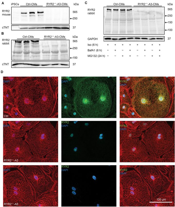
Luo et al.                                                                                     IP3R-Mediated Ca2+ Handling in RYR2−/− -iPSC-CMs

INTRODUCTION                                                              these cells into iPSC-CMs (Itzhaki et al., 2011; Lian et al.,
                                                                          2013; Cyganek et al., 2018). Previous studies have demonstrated
The type 2 ryanodine receptor (RYR2) represents the major                 that both RYR2 and IP3R are involved in calcium handling in
sarcoplasmic reticulum (SR) Ca2+ release channel in adult                 human ESC- and iPSC-derived CMs (Satin et al., 2008; Itzhaki
cardiomyocytes (CMs). It plays an essential role in excitation-           et al., 2011). However, our knowledge about how these two
contraction coupling, a process by which an electrical signal is          signaling pathways are cooperated during human embryonic
converted into a single contraction (Eisner et al., 2017). Upon           cardiac growth is still not sufficient.
spontaneous depolarization of membrane potential, voltage-                   In this study, we used iPSC technology in combination with
dependent L-type calcium channels (LTCC) are activated, which             the CRISPR/Cas9 gene editing technique to investigate the role
cause the influx of a small amount of external Ca2+ into the              of RYR2 in differentiation, development, and function of human
cytosol. The Ca2+ signal is then sensed and amplified by the              iPSC-CMs. We hypothesized that the generated RYR2−/− -iPSC-
Ca2+ -sensitive RYR2. The opening of RYR2 causes a substantial            CMs exhibit morphological and physiological abnormalities,
release of Ca2+ from the SR into the cytosol and thus elevates            providing insights into the function of RYR2 during human
intracellular Ca2+ levels. In the end, the binding of Ca2+ to             embryonic heart development.
troponin promotes sliding of thick and thin filaments, which
results in cardiac contraction (Eisner et al., 2017).
   A growing number of studies highlight the importance of                MATERIALS AND METHODS
RYR2 for the precise functionality of the adult heart. An inducible
cardiac-specific Ryr2 knockout (Ryr2−/− ) mouse model claimed             Culture and Maintenance of iPSCs
that RYR2 plays a non-redundant role in the control of heart              Human iPSC lines iWTD2.1 and iBM76.1 were used in this
rate and rhythmicity (Bround et al., 2012). An in-frame deletion          study as controls (Ctrl-iPSCs), which were generated from
of exon-3 in the Ryr2 gene in the mouse (Ex3-del+/− ) was                 dermal fibroblasts and mesenchymal stem cells from two healthy
associated with bradycardia and death. However, Ex3-del+/−                donors, respectively, using the STEMCCA lentivirus system, and
mice did not display some clinical phenotypes of patients with the        characterized as previously described (Streckfuss-Bomeke et al.,
RYR2 exon-3 deletion, including catecholaminergic polymorphic             2013; Cyganek et al., 2018). The study was approved by the
ventricular tachycardia (Liu et al., 2014).                               Ethics Committee of the University Medical Center Göttingen
   In the early stage of embryonic heart development in                   (approval number: 21/1/11), and carried out in accordance
mice, Ca2+ homeostasis is regulated not only by RYR2 but                  with the approved guidelines. Human iPSCs were cultured in
also by LTCC and inositol 1,4,5-trisphosphate receptor (IP3R)             chemically defined E8 medium (Thermo Fischer Scientific) on
(Takeshima et al., 1998; Rosemblit et al., 1999; Kapur and                Geltrex- (Thermo Fischer Scientific) coated cell culture plates
Banach, 2007; Sasse et al., 2007). Ryr2−/− mice possess the               at 37◦ C with 5% CO2 . The E8 medium was changed daily and
ability of repetitive Ca2+ signals and rhythmic contractions at           cells at ∼85% confluency were passaged using Versene (Thermo
embryonic day E9.5 (Takeshima et al., 1998), when LTCC and                Fischer Scientific).
IP3R may be responsible for the early Ca2+ cycling (Liu et al.,
2002; Kockskamper et al., 2008; Xie et al., 2018). However,               Generation of RYR2−/− -iPSC Lines Using
as the development of the embryo proceeds, the importance                 CRISPR/Cas9-Mediated Genome Editing
of Ca2+ cycling via SR increases progressively (Liu et al.,               In order to generate homozygous RYR2 knockout (RYR2−/− )
2002). Ryr2−/− mice died of cardiac arrest around embryonic               iPSC lines, two guide RNAs (gRNAs) were designed
day E10 with irregular arranged myocardium and trabeculae                 and inserted into the CRISPR/Cas9 plasmid (Sigma-
(Takeshima et al., 1998).                                                 Aldrich), respectively (Supplementary Figure 1A). While
   To date, studies on the expression and function of RYR2                gRNA1         (ACGAACTCTTCGTAGTCGAGGG)                   targets
during early embryonic cardiac development are limited                    exon 90 (g.741541-741563) of the RYR2 gene, gRNA2
to investigations using mouse Ryr2−/− models and mouse                    (CCTAGCCTGGTATATGACTATG) targets exon 99 (g.763747-
embryonic stem cell-derived CMs (mESC-CMs) (Takeshima                     763768). For transfection using the Amaxa Nucleofector II device
et al., 1998; Liu et al., 2002; Yang et al., 2002; Fu et al., 2006;       (Lonza), 2 × 10⁶ iPSCs were collected and re-suspended in a
Kapur and Banach, 2007; Sasse et al., 2007; Xie et al., 2018).            mixture of 82 µl Nucleofector solution and 18 µl supplement
The major challenge to study the origin and development of                1 (Nucleofector kit 1/2, Lonza) containing 4 µg plasmid. One
human embryonic cardiac myocytes is the limitation of a reliable          day after transfection, GFP+ cells were sorted and seeded on
human cell model, which can be used for long-term culture                 Geltrex-coated 96-well plates at a density of 1 × 10³ cells/well
experiments. The appearance of human induced pluripotent stem             for expansion. The genomic DNA from the colonies was
cell (iPSC) technology has shown its great potential to solve this        isolated and purified using the automated Maxwell 16 cell DNA
challenge (Takahashi et al., 2007; Yu et al., 2007). In the iPSC          purification kit (Promega) according to the manufacturer’s
approach, adult somatic cells can be efficiently reprogrammed             instruction. For genomic DNA sequencing, the DNA sequence
into pluripotent stem cells by ectopic expression of a set of             of RYR2 was initially amplified by PCR using the appropriate
transcription factors (Takahashi and Yamanaka, 2006). Based               primer set (Supplementary Table 1A). DNA sequencing of PCR
on this revolutionary finding, researchers have established the           products from CRISPR/Cas9-edited clones was performed by a
generation of human iPSCs and the efficient differentiation of            commercial sequencing facility (Seqlab, Göttingen).

Frontiers in Cell and Developmental Biology | www.frontiersin.org     2                                       August 2020 | Volume 8 | Article 772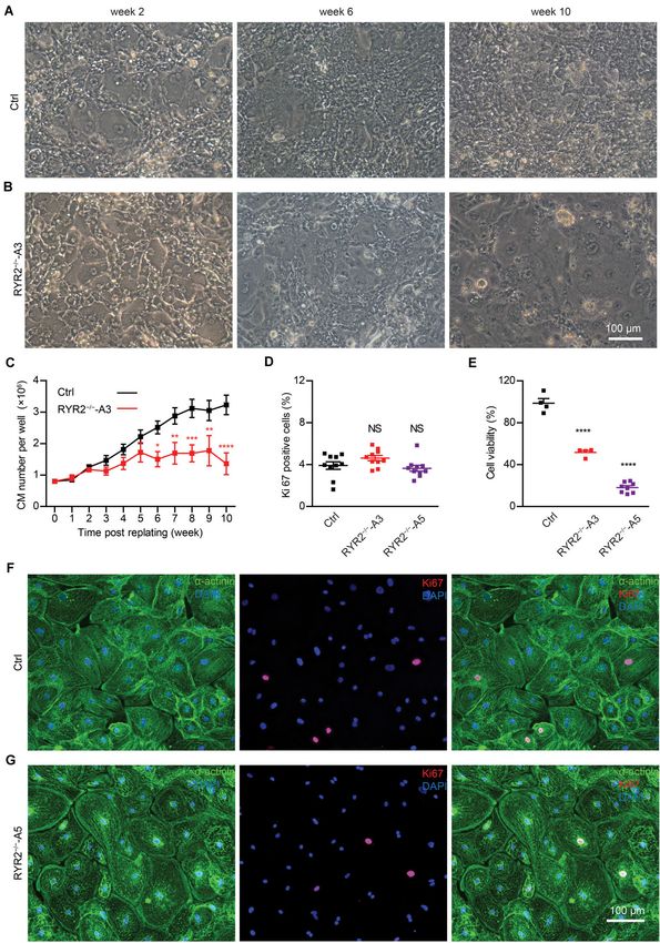
Luo et al.                                                                                       IP3R-Mediated Ca2+ Handling in RYR2−/− -iPSC-CMs

   To characterize the pluripotency of the CRISPR/Cas9-                    the cells, which were then incubated at 37◦ C for 2 h. Reaction was
edited RYR2−/− -iPSCs, reverse transcription-PCR analysis,                 stopped by the addition of 200 µl isopropanol supplemented with
immunofluorescence staining, and spontaneous differentiation               0.04 M HCl. Samples were incubated and shaken (300 rpm) for
in vitro were carried out using standard protocols as described            10 min at room temperature (RT). Absorbance of the formazan
earlier (Streckfuss-Bomeke et al., 2013). For detailed description,        at 570 and 630 nm was measured using a plate reader (Biotek
please see the Supplementary Material.                                     Synergy HTX). Cell viability was determined as absorbance (570–
                                                                           630 nm) and normalized to control group.
Directed Differentiation of iPSCs Into
Cardiomyocytes                                                             Immunofluorescence Staining of
Directed differentiation of Ctrl- and RYR2−/− -iPSCs into                  iPSC-CMs
CMs (Ctrl-iPSC-CMs, RYR2−/− -iPSC-CMs, respectively) was                   Ctrl- and RYR2−/− -iPSC-CMs grown on glass coverslips
induced by modulating WNT signaling as previously described                were fixed with 4% paraformaldehyde (PFA; Carl Roth),
(Lian et al., 2013; Cyganek et al., 2018). Briefly, when monolayer         blocked with 1% bovine serum albumin (BSA; Sigma-Aldrich),
cultures of iPSCs on 12-well plates reached 80–90% confluency,             and permeabilized with 0.1% Triton X-100 (Carl Roth).
differentiation was initiated by changing the E8 medium to                 Immunofluorescence staining was performed overnight using
cardio differentiation medium, which was composed of RPMI                  the following primary antibodies: anti-α-actinin (1:500; mouse
1640 with Glutamax and HEPES (Thermo Fischer Scientific),                  monoclonal, IgG1, Sigma-Aldrich), anti-Ki67 (1:400; rabbit
0.5 mg/ml human recombinant albumin (Sigma-Aldrich) and                    polyoclonal, abcam), anti-IP3R (1:100; rabbit polyclonal, Merck
0.2 mg/ml L-ascorbic acid 2-phosphate (Sigma-Aldrich). Cells               Millipore, used for all three subtypes of IP3R), and anti-RYR2
were first treated with 4 µM of the GSK3β inhibitor CHIR99021              (1:500; rabbit polyclonal, HPA020028; Sigma-Aldrich, used for
(Millipore) for 48 h and then with 5 µM of the WNT signaling               the full length of RYR2). Afterward, cells were washed three
inhibitor IWP2 (Millipore) for additional 48 h. Afterward, cells           times with PBS and incubated with the corresponding secondary
were cultured in cardio differentiation medium for another                 antibodies (1:1000; anti-rabbit Alexa fluor 546, Invitrogen; anti-
4 days. From day 8, cells were cultivated in cardio culture                rabbit Alexa fluor 488, Invitrogen; anti-mouse Alexa fluor
medium containing RPMI 1640 with Glutamax and HEPES,                       546, Invitrogen; anti-mouse Alexa fluor 488, Invitrogen) for
supplemented with 2% B27 (Thermo Fischer Scientific). At day               1 h at RT. Nuclei were co-stained with 4’,6-diamidino-2-
20, beating iPSC-CMs were detached from the plate by incubating            phenylindole (DAPI; 0.4 µg/ml; Sigma-Aldrich). Documentation
with 2 ml of 1 mg/ml collagenase B (Worthington Biochemical),              was performed using fluorescence microscopy (Carl Zeiss).
dissolved in cardio culture medium, for 1 h at 37◦ C. Floating
iPSC-CM sheet was gently transferred into a falcon tube and
                                                                           Western Blot
dissociated with 3 ml of 0.25% Trypsin/EDTA (Thermo Fischer
                                                                           Both Ctrl- and RYR2−/− -iPSC-CMs (90 days old) were scraped
Scientific) for 8 min at 37◦ C. Digestion was stopped by adding
                                                                           off from the culture plates and cell pellets were snap-frozen
the double volume of the cardio digestion medium (80% cardio
                                                                           into liquid nitrogen and stored at –80◦ C. For cell lysis,
culture medium, 20% FCS, and 2 µM Thiazovivin). Cells were
                                                                           frozen cell pellets were resuspended in lysis buffer containing
centrifuged at 200 g for 5 min, re-suspended in cardio digestion
                                                                           20 mM Tris/HCl (pH 7.4), 200 mM NaCl, 1 mM Na3 VO4 ,
medium, and replated into Geltrex-coated 6-well plates at a
                                                                           20 mM NaF, 1% IGEPAL CA-630 (Sigma-Aldrich), 1 mM
density of 800,000 cells/well. Afterward, iPSC-CMs were cultured
                                                                           dithiothreitol (Roth), PhosSTOP phosphatase inhibitor (Roche),
in cardio culture medium until 90 days.
                                                                           and cOmpleteTM protease inhibitor (Roche). After incubation for
                                                                           30 min on ice, lysates were centrifuged and protein concentration
Time-Dependent Proliferation Analysis                                      was determined using the Pierce BCA protein assay kit (Thermo
and Cell Viability Assay of iPSC-CMs                                       Fisher Scientific) according to the manufacturer’s instructions.
To investigate the proliferation of iPSC-CMs during long-term              A total amount of 40 µg protein lysate mixed with SDS
culture, both Ctrl- and RYR2−/− -iPSC-CMs were replated at a               loading buffer and DPBS in a volume of 20 µl was denatured
fixed density of 800,000 cells/well at day 20 post differentiation.        for 30 min at 37◦ C. Afterward, samples were run on a 6–
Cell number was determined weekly for 10 weeks until CMs                   15% polyacrylamide gel with a 5% stacking gel at 250 mA
reached the age of 90 days. CMs from two randomly selected wells           for around 1–2 h and then transferred to PVDF membranes
of one differentiation experiment were detached, dissociated, and          using the Wet/Tank blotting system. Unspecific binding sites
quantified by counting the cell numbers with a hemocytometer               on the membrane were blocked with 1% BSA in TBS with
and taking the average.                                                    0.1% Tween 20 (TBS-T) or with 5% non-fat dry milk in
   To assess the metabolic activity of iPSC-CMs, the MTT (3-               TBS-T for 1 h at RT. Membranes were incubated with
(4,5-dimethylthiazol-2-yl)-2,5-diphenyl tetrasodium bromide)               primary antibodies overnight at 4◦ C. The following primary
assay was performed according to the manufacturer’s instructions           antibodies were used: anti-RYR2 (1:500; rabbit polyclonal;
(MTT Kit CT02, Millipore). Briefly, iPSC-CMs at week 8 after               SAB4502707; Sigma-Aldrich, used for the detection of both the
replating were seeded in 48-well plates at a density of 60,000 cells       full-length and the truncated RYR2), anti-RYR2 (1:1000; mouse
per well, and cultured for another 2 weeks. Afterwards, 50 µg/ml           monoclonal, IgG1, MA3-916 C3-33; Thermo Fisher Scientific
MTT dissolved in 200 µl cardio culture medium was added onto               Pierce antibodies used for the detection of the full length

Frontiers in Cell and Developmental Biology | www.frontiersin.org      3                                        August 2020 | Volume 8 | Article 772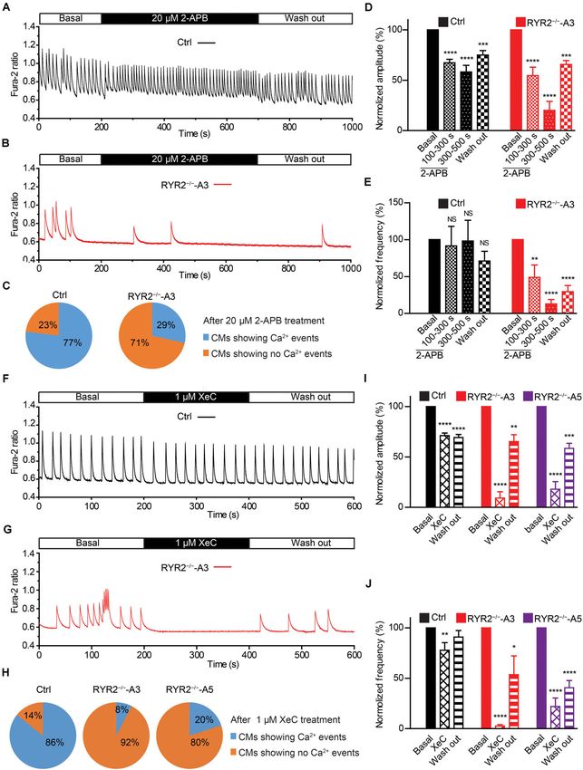
Luo et al.                                                                                     IP3R-Mediated Ca2+ Handling in RYR2−/− -iPSC-CMs

of RYR2), anti-IP3R (1:750; rabbit polyclonal), anti-SERCA2A             (Thermo Fisher Scientific) in Tyrode’s solution containing (in
(sarco/endoplasmic reticulum Ca2+ -ATPase type 2A; 1:1000;               mM): NaCl 140, KCl 5.4, CaCl2 1.8, MgCl2 1, HEPES 10,
mouse monoclonal, IgG2a, Thermo Fisher Scientific), anti-NCX1            and glucose 10 (pH adjusted to 7.3 with NaOH) for 30 min
(sodium-calcium exchanger type 1; 1:1000; mouse monoclonal,              at RT. Following incubation, the indicator-containing solution
IgG2b, Novus), anti-Cav 1.2 (voltage-dependent L-type calcium            was removed and cells were washed twice, and incubated for
channel alpha 1C subunit; 1:200; mouse monoclonal, IgG2b,                additional 10 min to allow de-esterification of the indicator. To
abcam), anti-α-actinin (1:1000; mouse monoclonal, IgG1), anti-           detect Ca2+ sparks, both Ctrl- and RYR2−/− -iPSC-CMs were
eukaryotic elongation factor 2 (EEF2; 1:50,000; rabbit polyclonal,       pre-treated with 100 nM isoprenaline for 10 min before starting
IgG, abcam), anti-cardiac troponin T (cTNT; 1:1000; rabbit               the recordings. Recordings were obtained using a LSM 710
polyclonal, IgG, abcam), and anti-GAPDH (glyceraldehyde 3-               confocal microscopy system in line scan mode (512 pixels, 45
phosphate dehydrogenase; 1:5000; rabbit polyclonal, Thermo               µm, 1057.7 Hz, 20,000 cycles). Cells were incubated in Tyrode’s
Fisher Scientific). The membranes were then incubated with               solution at RT and field stimulated at 0.25 Hz for at least 20 s to
HRP-conjugated goat anti-mouse or anti-rabbit secondary                  bring the cytosolic calcium concentration to a steady state. Fluo-
antibodies (1:10,000; Thermo Fisher Scientific) for 1 h at RT.           4 was excited at 488 nm and emitted fluorescence was captured
Afterwards, membranes were washed three times with TBS-T.                at 490–540 nm. Quantification of Ca2+ sparks was performed
Antigens of interest were detected by chemiluminescence (ECL;            using the SparkMaster plugin of ImageJ (NIH) and several key
GE Healthcare) and visualized using the ChemiDoc MP system.              parameters were determined: spark frequency (events per 100 µm
Quantification was performed by calculating the signal intensity         per second), amplitude (1F/F0 ), full duration at half maximum
with Image Lab software (Bio-Rad).                                       (FDHM), full width at half maximum (FWHM), and SR Ca2+
   To detect protein degradation, Ctrl-, A3 and A5 RYR2−/− -             leak per cell (spark frequency × amplitude × FDHM × FWHM).
iPSC-CMs (90 days old) were stimulated with 100 nM                       As fixed criteria, sparks with minimal amplitude of 0.2 1F/F0 ,
isoprenaline (Sigma-Aldrich) for 6 h and stepwise treated with           minimal width of 0.7 µm, and minimal duration of 7 ms were
the proteasome and calpain inhibitor MG132 (10 µM for                    selected for detailed analysis.
24 h; Sigma-Aldrich) and the autophagy inhibitor bafilomycin
A1 (BafA1; 100 nM for 6 h; Sigma-Aldrich) before the cell
pellets were collected.                                                  Calcium Transient Measurement of
                                                                         iPSC-CMs
Reverse Transcription-PCR Analysis                                       Both Ctrl- and RYR2−/− -iPSC-CMs around day 80 were
For gene expression analysis, Ctrl-iPSCs, CRISPR-edited A3 and           dissociated and replated on coverslips at a density of 200,000
A5 RYR2−/− -iPSCs as well as 90-day-old Ctrl- and RYR2−/− -              cells/well. Cells were allowed to recover for at least 10 days
iPSC-CMs were washed three times with PBS and cell pellets               post-replating. For measurement, cells were loaded with Fura-
were collected, snap-frozen into liquid nitrogen, and stored at –        2 (Thermo Fisher Scientific) at a final concentration of 5 µM
80◦ C. Isolation and purification of total RNA were performed            in cardio culture medium for 30 min at 37◦ C and washed twice
using the SV total RNA isolation system (Promega) in accordance          with the medium. Prior to measurement, cells were incubated for
to the manufacturer’s instructions. First strand cDNA synthesis          10 min to enable complete de-esterification of intracellular Fura-
was performed using MULV reverse transcriptase (Thermo                   2. Intracellular Ca2+ events were recorded using a 40 × objective
Fisher Scientific) and Oligo d(T)16 primer (Thermo Fisher                on an Olympus IX70 microscope fitted with an IonOptix system
Scientific). The expression level of SOX2, OCT4, NANOG,                  (Ionoptix, Milton, MA) at 35◦ C. Samples were excited at 340
LIN28, and FOXD3 were assessed in Ctrl- and CRISPR-edited                and 380 nm with a switching frequency of 200 Hz and the
iPSC lines by reverse transcription-PCR using the GoTaq DNA              emitted fluorescence was collected at 510 nm. The cytosolic
polymerase (Promega). In addition, the expression level of RYR2,         Ca2+ level was measured as the ratio of fluorescence at 340 and
IP3R1, IP3R2, CACNA1C, TNNT2, and ACTN2 were assessed                    380 nm (340/380 nm).
in Ctrl and RYR2−/− -iPSC-CMs. GAPDH was used as an                          Spontaneous whole-cell Ca2+ transients were recorded in
internal control. Primer sequences, annealing temperature, and           normal Tyrode’s solution containing (in mM): NaCl 138, KCl
cycles used for reverse transcription-PCR analyses are listed in         4, CaCl2 1.8, MgCl2 1, NaH2 PO4 0.33, HEPES 10, and glucose
Supplementary Table 1B.                                                  10 (pH adjusted to 7.3 with NaOH). To normalize the Ca2+
                                                                         transient frequency, Ctrl- and RYR2−/− -iPSC-CMs were field-
                                                                         stimulated using a MyoPacer (Ionoptix, Milton, MA) at a pacing
Calcium Spark Measurement of                                             frequency of 0.5 Hz (6 V, 10 ms). Monotonic transient analysis
iPSC-CMs                                                                 was performed using the LabChart Pro software (ADInstrument)
For calcium spark recordings, spontaneously beating Ctrl-                and the following parameters were determined: peak amplitude
and RYR2−/− -iPSC-CMs (around 80-day-old) were dissociated,              of Ca2+ transients (the Fura-2 ratio at systole subtracted by the
replated on Geltrex-coated coverslips at a density of 200,000            Fura-2 ratio at diastole), decay rate (tau), as well as duration and
cells per 6-well and then allowed to recover for at least 10             frequency of Ca2+ transients.
days in cardio culture medium. Before measurement, cells were                Calcium      sensitivity   analysis    was      performed     as
loaded with the fluorescent calcium indicator 5 µM fluo-4/AM             described previously (Jiang et al., 2007). Briefly, Ctrl- and
(Thermo Fisher Scientific) and 0.02% [w/v] pluronic F-127                RYR2−/− -iPSC-CMs loaded with Fura-2 were first exposed

Frontiers in Cell and Developmental Biology | www.frontiersin.org    4                                        August 2020 | Volume 8 | Article 772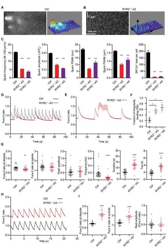
Luo et al.                                                                                       IP3R-Mediated Ca2+ Handling in RYR2−/− -iPSC-CMs

to 0 mM Ca2+ Tyrode’s solution until no spontaneous Ca2+                   Statistical Analysis
transient was detected and then continuously perfused with                 Results are presented as mean ± standard error of the mean
Tyrode’s solution containing Ca2+ of increasing concentrations             (SEM). Statistical analysis was performed using GraphPad Prism
(0, 0.1, 0.2, 0.3, 0.5, 1.0, and 2.0 mM). In the end, caffeine             5 software with student’s t-test, the one-way ANOVA with the
(10 mM; Sigma-Aldrich) was applied to confirm the activity of              Dunnett’s multiple comparison test or the two-way ANOVA with
RYR2.                                                                      Sidak’s correction for comparison of more groups and conditions.
   To examine the caffeine-induced Ca2+ release in iPSC-                   Results were considered statistically significant when the P-value
CMs, cells loaded with Fura-2 were washed with depolarization              was
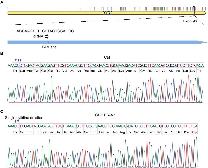
Luo et al.                                                                                               IP3R-Mediated Ca2+ Handling in RYR2−/− -iPSC-CMs

  FIGURE 1 | Generation of human homozygous RYR2 knockout iPSCs (RYR2−/− -iPSCs). (A) Illustration of the RYR2 locus and gRNA1 designed for
  CRISPR/Cas9-mediated gene editing in exon 90 of RYR2. (B,C) DNA sequencing of Ctrl- (B) and CRISPR/Cas9-edited A3 RYR2−/− -iPSC lines (C). The A3
  RYR2−/− -iPSC line displayed a single homozygous cytidine deletion in the RYR2 gene, which resulted in a premature termination codon (PTC).

These results indicate that loss of RYR2 does not alter the early                of Ctrl-iPSC-CMs quadrupled to 3.2 ± 0.3 million cells/well,
cardiac commitment of iPSCs and the differentiation efficiency                   and the number of RYR2−/− -iPSC-CMs only increased to
into CMs.                                                                        1.4 ± 0.3 million cells/well, P < 0.0001 (Figure 2C). To analyze
   Although RYR2−/− -iPSC-CMs could be cultivated for up to                      whether RYR2−/− -iPSC-CMs have a lower proliferation capacity,
3 months and remained a high percentage of cTNT-positive                         Ctrl- and RYR2−/− -iPSC-CMs at week 8 post replating were
cells similar to the Ctrl group (Ctrl: 98.4 ± 0.2%, n = 3; A3:                   double immunostained for sarcomeric α-actinin and Ki67, a
96.8 ± 2.4%, n = 3; A5: 96.7 ± 0.4%, n = 3; Supplementary                        marker of cell proliferation. The percentage of proliferating
Figure 2B), they grew differently with higher number of floating                 CMs was quantified as the number of Ki67-positive nuclei
cells and signs of cell death in comparison to Ctrl-iPSC-CMs                     divided by the total number of nuclei. The number of Ki67-
(Figures 2A,B and Supplementary Figures 2C,D) during long-                       positive cells was comparable in Ctrl- and RYR2−/− -iPSC-CMs
term culture. To analyze the survival and proliferation rate                     (Ctrl: 3.9 ± 0.4%; A3-RYR2−/− : 4.6 ± 0.3%; A5-RYR2−/− :
during long-term culture, both Ctrl- and RYR2−/− -iPSC-CMs                       3.6 ± 0.3%; Figures 2D,F,G), suggesting that the lower cell
were replated at a fixed density of 0.8 million cells/well on                    number in RYR2−/− -iPSC-CMs at week 10 is not due to a
day 20 post differentiation. No significant difference in cell                   change in the cell proliferation capacity. Next, we performed
numbers was observed between Ctrl- and A3 RYR2−/− -iPSC-                         the MTT assay to compare the cellular metabolic activity of
CMs during the first five weeks after replating. However, while                  Ctrl- and RYR2−/− -iPSC-CMs, which reflects the number of
Ctrl-iPSC-CMs revealed a steady increase in their number until                   viable cells presented. The results showed a significant reduction
week 8 and remained stable afterward, RYR2−/− -iPSC-CMs                          of cell viability in RYR2−/− -iPSC-CMs compared to Ctrl-
showed no increase in their number. At week 10, the number                       CMs (Figure 2E).

Frontiers in Cell and Developmental Biology | www.frontiersin.org            6                                            August 2020 | Volume 8 | Article 772
Luo et al.                                                                                                      IP3R-Mediated Ca2+ Handling in RYR2−/− -iPSC-CMs

  FIGURE 2 | Growth analysis of Ctrl- and RYR2−/− -iPSC-CMs during long-term culture. (A,B) Bright-field images of Ctrl-iPSC-CMs (A) and A3 RYR2−/− -iPSC-CMs
  (B) after replating at day 20 of differentiation. Ctrl-iPSC-CMs showed an increase in cell density from week 2 to week 10 post replating (A), while
  RYR2−/− -iPSC-CMs exhibited reduced cell density (B). Scale bar, 100 µm. (C) Quantification of the cardiomyocyte numbers. Ctrl- and A3 RYR2−/− -iPSC-CMs
  showed distinct growth curves during long-term cell culture. Ctrl-CMs from 2 different iPSC lines and 11 differentiation experiments (iBM76.1: n = 6 and iWTD2.1:
  n = 5) were analyzed. A3 RYR2−/− -CMs from six differentiation experiments were used. *P < 0.05, **P < 0.01, ***P < 0.001, and ****P < 0.0001, RYR2−/− vs. Ctrl
  by using the two-way ANOVA with the Sidak’s multiple comparison test. (D) Percentage of Ki67-positive CMs (n = 10 samples each for Ctrl, A3 and A5,
  n = 300–400 cells per sample counted). (E) Cell viability of Ctrl-, A3 and A5 RYR2−/− -iPSC-CMs (Ctrl: n = 4 from two differentiation experiments; A3 RYR2−/− :
  n = 4 from two differentiation experiments; A5 RYR2−/− : n = 8 from two differentiation experiments). ****P < 0.0001 by using the one-way ANOVA with the Dunnett’s
  multiple comparison test (D,E). (F,G) Representative immunostaining of Ctrl- and RYR2−/− -iPSC-CMs probing for Ki67 (red) and α-actinin (green). Cells were
  counterstained with DAPI (blue) to show the nucleus. Scale bar, 100 µm.

Frontiers in Cell and Developmental Biology | www.frontiersin.org                 7                                              August 2020 | Volume 8 | Article 772
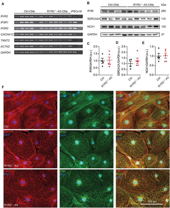
Luo et al.                                                                                   IP3R-Mediated Ca2+ Handling in RYR2−/− -iPSC-CMs

Loss of RYR2 Does Not Affect the                                        did not observed differences in the distribution of IP3R between
Expression of Ca2+ Signaling-Associated                                 Ctrl- and RYR2−/− -iPSC-CMs.
Genes                                                                   Loss of RYR2 Results in Significant
To assess whether the RYR2 expression is lost in RYR2−/− -
iPSC-CMs, different anti-RYR2 antibodies were applied for               Reduction of Spontaneous Release of
western blot analysis. Whereas the mouse monoclonal anti-               Ca2+ Sparks in RYR2−/− -iPSC-CMs
RYR2 antibody, produced by using canine cardiac Ryr2 as                 To determine functional consequences of the loss of RYR2,
the immunogen, was used to detect the full-length protein of            Ca2+ sparks were measured in Ctrl- and RYR2−/− -iPSC-
RYR2, the rabbit polyclonal anti-RYR2 antibody (SAB4502707)             CMs after pacing at 0.25 Hz, which were pre-treated with
recognizes both the full-length and the truncated proteins by           100 nM isoprenaline to activate the β-adrenergic signaling
binding the N-terminal region before the PTC. Neither the               and to promote the occurrence of Ca2+ sparks. In Ctrl-iPSC-
full length nor the truncated proteins of RYR2 were detectable          CMs, typical Ca2+ sparks occurred randomly throughout the
in A3 (Figures 3A,B and Supplementary Figure 3A) and A5                 cell (Figure 5A). In contrast, Ca2+ sparks appeared rarely in
RYR2−/− -iPSC-CMs (Supplementary Figure 3B), indicating the             RYR2−/− -iPSC-CMs (Figure 5B) and showed a significantly
loss of functional RYR2 in A3 and A5 RYR2−/− -iPSC-CMs.                 lower frequency with smaller amplitude (1F/F0 ) compared to
Immunofluorescence staining using anti-α-actinin and anti-              Ctrl-iPSC-CMs (Figure 5C). Additionally, RYR2−/− -iPSC-CMs
RYR2 antibodies further confirmed the absence of RYR2 in A3             showed significantly reduced FDHM and FWHM, indicating
and A5 RYR2−/− -iPSC-CMs (Figure 3D). However, the RYR2                 smaller sizes of the released Ca2+ sparks in RYR2−/− -iPSC-
mRNA levels were not altered in A3 and A5 RYR2−/− -iPSC-CMs             CMs compared to Ctrl-iPSC-CMs. This led to a 95.7 and
compared to Ctrl-iPSC-CMs (Figure 4A and Supplementary                  94.7% reduction of spontaneous SR Ca2+ leak in A3 and A5
Figure 3C). To investigate whether the absence of the truncated         RYR2−/− -iPSC-CMs, respectively, compared to Ctrl-iPSC-CMs
RYR2 in RYR2−/− -iPSC-CMs might be the result of protein                (Figure 5C). These data indicate that RYR2 plays a major role
degradation, isoprenaline-stimulated Ctrl- and RYR2−/− -iPSC-           in spontaneous Ca2+ leak from the SR at diastole. The remaining
CMs were treated with MG132 (10 µM for 24 h) to inhibit                 but smaller Ca2+ sparks detected in RYR2−/− -iPSC-CMs suggest
the activity of the proteasome and with bafilomycin A1 (BafA1,          the existence of some other Ca2+ regulatory channels, which may
100 nM for 6 h) to inhibit the fusion of autophagosomes with            mediate Ca2+ sparks independent of RYR2.
lysosomes. Interference of the two major protein degradation
pathways did not result in the detection of degraded RYR2               RYR2−/− -iPSC-CMs Show Abnormal
proteins in both A3 (Figure 3C) and A5 RYR2−/− -iPSC-CMs                Ca2+ Transients Compared to
(Supplementary Figure 3D), suggesting that nonsense-mediated            Ctrl-iPSC-CMs
mRNA decay leads to no translation of the truncated RYR2                To further investigate whether normal Ca2+ homeostasis was
protein in RYR2−/− -iPSC-CMs.                                           affected in RYR2−/− -iPSC-CMs, spontaneous Ca2+ transients
   Next, we evaluated the impact of loss of RYR2 on the                 were assessed. Although we detected spontaneous Ca2+
expression of other cardiac Ca2+ signaling-associated genes             transients in both Ctrl- and RYR2−/− -iPSC-CMs (Figure 5D),
in RYR2−/− -iPSC-CMs, such as genes encoding the cardiac                54.1 ± 6.1% of A3 and 65.7 ± 9.6% of A5 RYR2−/− -iPSC-CMs
IP3R types 1 and 2 (IP3R1, IP3R2) and the voltage-gated                 showed abnormalities in Ca2+ handling (Figures 5E,F). These
calcium channel subunit alpha-1C (CACNA1C). No tendency of              irregularities were mainly detectable as highly frequent Ca2+
increased or decreased expression of tested genes was observed in       release events at elevated diastolic Ca2+ levels (Figure 5E),
RYR2−/− -iPSC-CMs in comparison to their corresponding Ctrl-            which were not observed in Ctrl-iPSC-CMs. Furthermore,
iPSC-CMs (Figure 4A and Supplementary Figure 3C). However,              RYR2−/− -iPSC-CMs showed significantly lower diastolic Ca2+
as RYR2−/− -iPSC-CMs remained the ability to spontaneously              levels, but no significant differences in systolic Ca2+ levels
contract, we hypothesized that RYR2 may not be the only                 and peak amplitude of Ca2+ transients in comparison to Ctrl-
channel promoting intracellular calcium release. To verify              iPSC-CMs (Figure 5G and Supplementary Figure 3E). We
potential compensatory mechanisms in response to loss of                also observed a significant decrease in the frequency of Ca2+
RYR2, we studied the expression of several Ca2+ -regulation             transients and an increase in the duration of Ca2+ transients in
related proteins. Consistent with IP3R mRNA levels, western blot        RYR2−/− -iPSC-CMs compared to Ctrl-iPSC-CMs (Figure 5G
analysis revealed no differences in IP3R protein expression in          and Supplementary Figure 3E), indicating that loss of RYR2
RYR2−/− -iPSC-CMs compared to Ctrl-iPSC-CMs. In addition,               results in reduced but prolonged contraction-relaxation cycles.
we detected no significant changes in the protein expression of         Moreover, the time constant (tau) during the decay of Ca2+
SERCA2A and NCX1 between Ctrl- and RYR2−/− -iPSC-CMs                    transients was significantly increased in RYR2−/− -iPSC-CMs
(Figures 4B–E). To study the distribution of IP3R in iPSC-              compared to Ctrl-iPSC-CMs (Figure 5G and Supplementary
CMs, we conducted immunostaining by using antibodies against            Figure 3E). By applying a field stimulation, we normalized
IP3R and sarcomeric α-actinin (Figure 4F). IP3R was expressed           the Ca2+ cycling of Ctrl- and RYR2−/− -iPSC-CMs to 0.5 Hz.
throughout the cytosol (Figure 4F middle) and partially co-             Paced Ca2+ transients in A3 RYR2−/− -iPSC-CMs revealed
localized with sarcomeric α-actinin (Figure 4F right). The              a comparable peak amplitude (Figures 5H,I). However, the
perinuclear region displayed an intensive expression of IP3R. We        diastolic level of Ca2+ transients after pacing was much higher

Frontiers in Cell and Developmental Biology | www.frontiersin.org   8                                       August 2020 | Volume 8 | Article 772
Luo et al.                                                                                                     IP3R-Mediated Ca2+ Handling in RYR2−/− -iPSC-CMs

  FIGURE 3 | Expression of RYR2 in 3-month-old Ctrl- and RYR2−/− -iPSC-CMs. (A) Western blot analysis of the full-length RYR2 and cardiac troponin T (cTNT) in
  Ctrl- and A3 RYR2−/− -iPSC-CMs (Ctrl: n = 3 and A3 RYR2−/− : n = 3 independent differentiation experiments). (B) Western blot analysis showing protein expression
  of N-terminal RYR2 and cTNT in Ctrl- and A3 RYR2−/− -iPSC-CMs (Ctrl: n = 3 and A3 RYR2−/− : n = 3 independent differentiation experiments). (C) Representative
  western blot analysis of RYR2 in response to treatment with MG132 and/or Bafilomycin A (BafA1) in Ctrl- and RYR2−/− -iPSC-CMs (Ctrl: n = 3, A3 RYR2−/− : n = 3
  independent differentiation experiments). No degraded RYR2 proteins were detected in RYR2−/− -iPSC-CMs by inhibiting protein degradation with the proteasome
  and calpain inhibitor MG132 and the autophagy inhibitor BafA1. (D) Representative immunostaining of Ctrl- and RYR2−/− -iPSC-CMs using antibodies against RYR2
  (green) and α-actinin (red). The cells were counterstained with DAPI (blue) to show the nucleus. Scale bar, 100 µm.

Frontiers in Cell and Developmental Biology | www.frontiersin.org                9                                               August 2020 | Volume 8 | Article 772

Luo et al.                                                                                                   IP3R-Mediated Ca2+ Handling in RYR2−/− -iPSC-CMs

  FIGURE 4 | Expression of markers involved in Ca2+ signaling in RYR2−/− -iPSC-CMs compared to Ctrl-iPSC-CMs. (A) Reverse transcription-PCR analyses showing
  mRNA expression of transcripts for Ca2+ handling-related genes (RYR2, IP3R1, IP3R2, and CACNA1C), genes encoding sarcomeric proteins (TNNT2 and ACTN2),
  and the housekeeping gene GAPDH in Ctrl- and A3 RYR2−/− -iPSC-CMs (Ctrl: n = 6 and RYR2−/− -A3: n = 6 different differentiation experiments). M: DNA
  molecular-weight size marker. (B) Western blot showing expression of Ca2+ handling-associated proteins (IP3R, SERCA2A, and NCX1) in both Ctrl- and A3
  RYR2−/− -iPSC-CMs. (C–E) Scatter dot plot showing protein levels of IP3R (C), SERCA2A (D), and NCX1 (E) normalized to GAPDH between Ctrl- and A3
  RYR2−/− -iPSC-CMs (Ctrl: n = 4 and A3 RYR2−/− : n = 5 different differentiation experiments). (F) Representative immunostaining of Ctrl- and RYR2−/− -iPSC-CMs
  for IP3R (green) and α-actinin (red). Cells were counterstained with DAPI (blue). Scale bar, 100 µm.

Frontiers in Cell and Developmental Biology | www.frontiersin.org              10                                             August 2020 | Volume 8 | Article 772
Luo et al.                                                               IP3R-Mediated Ca2+ Handling in RYR2−/− -iPSC-CMs

  FIGURE 5 | Continued

Frontiers in Cell and Developmental Biology | www.frontiersin.org   11                  August 2020 | Volume 8 | Article 772
Luo et al. IP3R-Mediated Ca2+ Handling in RYR2−/− -iPSC-CMs
FIGURE 5 | RYR2−/− -iPSC-CMs showed defective Ca2+ cycling. (A,B) Ca2+ sparks in Ctrl- and A3 RYR2−/− -iPSC-CMs during β-adrenergic stimulation.
(A) Spontaneous Ca2+ spark images in Ctrl-iPSC-CMs (A; ncell = 358, nspark = 1521) and A3 RYR2−/− -iPSC-CMs (B; ncell = 21, nspark = 38) after treatment with
100 nM isoproterenol. Both Ctrl- and RYR2−/− -iPSC-CMs were paced at 0.25 Hz. (C) Bar graphs summarize the key parameters of Ca2+ sparks in mean ± SEM
values for Ctrl- and RYR2−/− -iPSC-CMs: spark frequency (100 µm−1 s−1 ), amplitude (1F/F0 ), FDHM (ms), FWHM (µm) and relative SR Ca2+ leak. For A5
RYR2−/− -iPSC-CMs, ncell = 12, nspark = 20 were analyzed. ***P < 0.001 by using the one-way ANOVA with the Dunnett’s multiple comparison test.
(D) Representative Ca2+ transients in spontaneously beating Ctrl- and A3 RYR2−/− -iPSC-CMs. (E) Representative abnormal Ca2+ transients in A3
RYR2−/− -iPSC-CMs. (F) Fractions of irregularly beating Ctrl- and A3- and A5 RYR2−/− -iPSC-CMs (Ctrl: CMs from six differentiation experiments; A3 RYR2−/− :
CMs from four differentiation experiments; A5 RYR2−/− : CMs from three differentiation experiments. n > 15 cells per experiment). *P < 0.05 by using the one-way
ANOVA with the Dunnett’s multiple comparison test. (G) Scatter dot plot showing the diastolic Ca2+ levels, systolic Ca2+ levels, peak amplitude, frequency and
duration of Ca2+ transients, as well as the time constant during the decay of Ca2+ transients (tau) in Ctrl- and A3 RYR2−/− -iPSC-CMs (Ctrl: n = 31 cells from five
differentiation experiments; A3 RYR2−/− : n = 32 cells from three differentiation experiments). (H) Representative Ca2+ transients in Ctrl- and A3
RYR2−/− -iPSC-CMs under a normalized field stimulation at a pacing frequency of 0.5 Hz. (I) Scatter dot plot showing the diastolic Ca2+ levels, systolic Ca2+ levels
and peak amplitude of paced Ca2+ transients in Ctrl- and A3 RYR2−/− -iPSC-CMs (Ctrl: n = 22 cells from three differentiation experiments; A3 RYR2−/− : n = 12
cells from two differentiation experiments). **P < 0.01; ****P < 0.0001 RYR2−/− vs. Ctrl by using the unpaired Student’s t-test.
in RYR2−/− -iPSC-CMs than in non-paced RYR2−/− -iPSC-CMs in Ctrl-iPSC-CMs (Figures 6A,B), which confirms the absence
as well as in Ctrl-iPSC-CMs (Figures 5H,I). Together with of functional RYR2.
the increased time constant of spontaneous Ca2+ transients Caffeine, a pharmacological agonist of RYR2 (Porta et al.,
in RYR2−/− -iPSC-CMs (Figure 5G and Supplementary 2011), is commonly used to monitor RYR2-mediated Ca2+
Figure 3E), these data suggest that the efficiency of Ca2+ removal release. To determine the impact of loss of RYR2 in iPSC-CMs,
from the cytosol either by reuptake into the SR or by pumping we measured cytosolic Ca2+ levels in Ctrl- and RYR2−/− -iPSC-
Ca2+ out of the cell is lower in RYR2−/− -iPSC-CMs than CMs before and after continuously supplementing caffeine with
in Ctrl-iPSC-CMs. increasing concentrations (from 0.025 to 5.0 mM). As shown in
Figure 6E, low concentration of caffeine (0.025 mM) resulted
in a reduction of cytosolic Ca2+ levels in Ctrl-iPSC-CMs, while
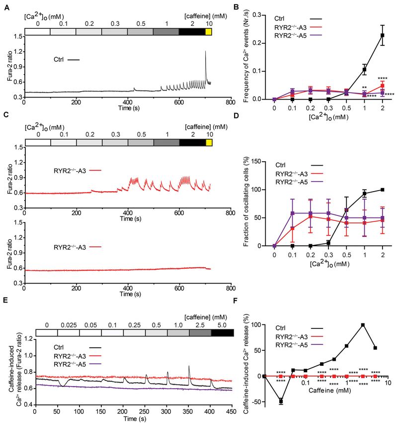
The Sensitivity of RYR2−/− -iPSC-CMs to the amplitudes of caffeine-induced Ca2+ release progressively
increased under the caffeine treatment at concentrations from
Extracellular Ca2+ and to Caffeine Is 0.05 to 2.5 mM and declined at the concentration of 5 mM
Changed (Figure 6F). We believe that the decrease in caffeine-induced
To evaluate the relationship between extracellular Ca2+ Ca2+ release at 5 mM caffeine might be the result of
([Ca2+ ]o ) levels and the occurrence of Ca2+ transients in depletion of internal Ca2+ stores in the SR due to previous
iPSC-CMs, we measured Ca2+ events under different [Ca2+ ]o caffeine supplementation. In contrast to Ctrl-iPSC-CMs, caffeine
concentrations using the fluorescent Ca2+ dye Fura-2 in treatment did not result in any change of cytosolic Ca2+
combination with Ca2+ imaging. To this end, spontaneously levels in both A3 and A5 RYR2−/− -iPSC-CMs (Figures 6E,F),
beating iPSC-CMs were first washed with Tyrode’s solution indicating that no functional RYR2 is present in RYR2−/− -iPSC-
containing no Ca2+ until no spontaneous Ca2+ transient was CMs and that Ca2+ transients observed in RYR2−/− -iPSC-CMs
detectable. Afterward, cytosolic Ca2+ changes were monitored (Figures 5D,E, 6B) are not caused by Ca2+ release via RYR2,
in response to continuous perfusion with increasing Ca2+ rather via other mechanisms.
concentrations (from 0 to 2.0 mM) and addition of 10 mM
caffeine at the end. To quantify the fraction of Ca2+ -oscillating IP3R-Mediated Ca2+ Release and
cells for each condition, the number of iPSC-CMs, which
displayed spontaneous Ca2+ transients at each [Ca2+ ]o
SERCA-Mediated SR Ca2+ Uptake Are
concentration was determined. As shown in Figure 6A, no Required for the Generation of Ca2+
Ca2+ transients in Ctrl-iPSC-CMs were detected with [Ca2+ ]o Transients in RYR2−/− -iPSC-CMs
lower than 0.5 mM. Raising [Ca2+ ]o concentrations stepwise Given that RYR2−/− -iPSC-CMs can spontaneously beat and
from 0.3 to 2.0 mM concomitantly increased the amplitude and release Ca2+ sparks and Ca2+ transients without RYR2 protein
frequency of Ca2+ transients in Ctrl-iPSC-CMs (Figures 6A,C), expression, we believe that these cells possess an alternative
as well as the fraction of Ca2+ -oscillating cells (Figure 6D). In Ca2+ regulatory mechanism to compensate for the missing
contrast, RYR2−/− -iPSC-CMs responded differently to increased RYR2. IP3R-involved Ca2+ signaling has been discovered to play
[Ca2+ ]o concentrations (Figures 6B–D). While 6 out of 16 A3 an important role during the process of cardiac development
RYR2−/− -iPSC-CMs did not display any Ca2+ transients from (Rosemblit et al., 1999; Poindexter et al., 2001). Therefore, we
0.1 to 2 mM [Ca2+ ]o , the remaining 10 cells revealed Ca2+ studied the effect of IP3R blockade on Ca2+ handling in iPSC-
transients already at 0.1 or 0.2 mM [Ca2+ ]o (Figure 6B). Similar CMs. To this end, spontaneous Ca2+ transients in Ctrl- and
results were observed in A5 RYR2−/− -iPSC-CMs; 5 out of 12 A3 RYR2−/− -iPSC-CMs were measured before and after the
cells showed no Ca2+ transients under all conditions while the application of 20 µM 2-APB. As shown in Figures 7A–C, 2-
remaining 7 cells exhibited Ca2+ transients already at 0.1 mM APB treatment for 500 s completely and reversibly blocked
[Ca2+ ]o (Figure 6D). Furthermore, both A3 and A5 RYR2−/− - all Ca2+ transients in 10 out of 14 tested A3 RYR2−/− -
iPSC-CMs showed no caffeine-induced Ca2+ release as observed iPSC-CMs (Figures 7B,C), while Ctrl-iPSC-CMs (10 out of
Frontiers in Cell and Developmental Biology | www.frontiersin.org 12 August 2020 | Volume 8 | Article 772Luo et al. IP3R-Mediated Ca2+ Handling in RYR2−/− -iPSC-CMs FIGURE 6 | Sensitivity of Ctrl- and RYR2−/− -iPSC-CMs to extracellular calcium and caffeine. (A,B) Cytosolic Ca2+ dynamics in Ctrl-iPSC-CMs (A) and A3 RYR2−/− -iPSC-CMs (B) perfused with increasing concentrations of extracellular Ca2+ ([Ca2+ ]o ). Cytosolic Ca2+ dynamics in A3 RYR2−/− -iPSC-CMs (B) were inconsistent from cell to cell. (C) Frequency of Ca2+ events detected in Ctrl- and RYR2−/− -iPSC-CMs under different [Ca2+ ]o . (D) Percentage of CMs displaying Ca2+ events under different [Ca2+ ]o concentrations among the three groups (Ctrl: n = 20 cells from four differentiation experiments; A3 RYR2−/− : n = 15 cells from three differentiation experiments; A5 RYR2−/− : n = 12 cells from two differentiation experiments). (E) Representative traces of caffeine-induced Ca2+ release in Ctrl- and RYR2−/− -iPSC-CMs. (F) Relationship between Ca2+ release and caffeine concentration in CMs from the two groups. The amplitude of each caffeine-induced Ca2+ release was normalized to the maximum peak for each experiment (Ctrl: n = 20 cells from four differentiation experiments; A3 RYR2−/− : n = 6 cells from two differentiation experiments; A5 RYR2−/− : n = 6 cells from two differentiation experiments). **P < 0.01, ****P < 0.0001 RYR2−/− vs. Ctrl by the two-way ANOVA with the Sidak’s multiple comparison test. 13) displayed intensive but slightly smaller Ca2+ transients 100∼300 s declined to 54.7 and 49.1%, respectively, whereas the (Figures 7A,C). Although 2-APB application decreased Ca2+ inhibitory effects increased as treatment lasted longer (300–500 transient amplitude in Ctrl-iPSC-CMs (Figure 7D), it did s): the amplitude and frequency decreased to 20.1 and 12.9%, not reduce the beating frequency of these cells (Figure 7E). respectively (Figures 7D,E). Previous studies reported that 2- On the contrary, in A3 RYR2−/− -iPSC-CMs, the amplitude APB is also a blocker of store-operated Ca2+ entry independently and frequency of Ca2+ transients during 2-APB treatment for of the function of IP3R (Iwasaki et al., 2001; Bootman et al., 2002). Frontiers in Cell and Developmental Biology | www.frontiersin.org 13 August 2020 | Volume 8 | Article 772
Luo et al.                                                                                                        IP3R-Mediated Ca2+ Handling in RYR2−/− -iPSC-CMs

  FIGURE 7 | Contributions of IP3R-mediated Ca2+ release to spontaneous Ca2+ transients in Ctrl- and RYR2−/− -iPSC-CMs. (A,B) Representative cytosolic Ca2+
  dynamics in Ctrl- (A) and RYR2−/− -iPSC-CMs (B) before and after the addition of IP3R antagonist 2-APB (20 µM), and after the washout of the drug. (C) Pie charts
  depict the percentage of cells maintaining Ca2+ transients after the treatment with 2-APB for 500 s. (D,E) Changes of Ca2+ transient amplitude (D) and frequency
  (E) after the treatment and the following removal of 2-APB, which were normalized to those from the same cell under the basal condition (Ctrl: n = 13 cells from three
  differentiation experiments; A3 RYR2−/− : n = 14 cells from three differentiation experiments). (F,G) Representative cytosolic Ca2+ dynamics in Ctrl- (F) and
  RYR2−/− -iPSC-CMs (G) before and after the addition of IP3R inhibitor Xestospongin C (XeC;1 µM), and after the washout of the drug. (H) Pie charts depict the
  percentage of cells maintaining Ca2+ transients after the treatment with XeC for 200 s. (I,J) Changes of Ca2+ transient amplitude (I) and frequency (J) after the
  treatment and following the washout of XeC, which were normalized to those from the same cell under the basal condition (Ctrl: n = 14 cells from three differentiation
  experiments; A3 RYR2−/− : n = 13 cells from three differentiation experiments; A5 RYR2−/− : n = 10 cells from two differentiation experiments). *P < 0.05;
  **P < 0.01; ***P < 0.001;****P < 0.0001 by using the one-way ANOVA with the Dunnett’s multiple comparison test.

Frontiers in Cell and Developmental Biology | www.frontiersin.org                  14                                                August 2020 | Volume 8 | Article 772
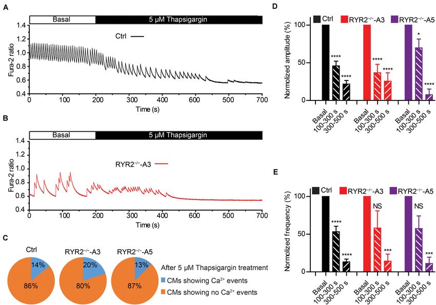
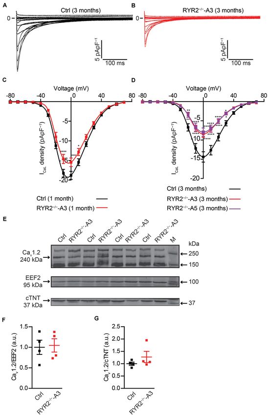
Luo et al.                                                                                                         IP3R-Mediated Ca2+ Handling in RYR2−/− -iPSC-CMs

  FIGURE 8 | Contributions of SERCA-mediated SR Ca2+ uptake to spontaneous Ca2+ transients in Ctrl- and RYR2−/− -iPSC-CMs. (A,B) Representative cytosolic
  Ca2+ dynamics in Ctrl- (A) and RYR2−/− -iPSC-CMs (B) before and after the addition of SERCA inhibitor thapsigargin (5 µM). (C) Pie charts depict the percentage
  of cells maintaining Ca2+ transients after the treatment with thapsigargin for 500 s. (D,E) Changes of Ca2+ transient amplitude (D) and frequency (E) after the
  treatment with thapsigargin, which were normalized to those from the same cell under the basal condition. (Ctrl: n = 14 cells from three differentiation experiments;
  A3 RYR2−/− : n = 10 cells from three differentiation experiments; A5 RYR2−/− : n = 8 cells from two differentiation experiments). *P < 0.05; ***P < 0.001;
  ****P < 0.0001 by using the one-way ANOVA with the Dunnett’s multiple comparison test.

Therefore, we used the IP3R inhibitor Xestospongin C (XeC) to                            time-dependent manner (Figures 8A–E). The amplitude and
confirm our hypothesis. As shown in Figures 7F–J, the Ca2+                               frequency of Ca2+ transients in Ctrl-iPSC-CMs declined to
transients in 12 out of 13 A3 and 8 out of 10 A5 RYR2−/− -iPSC-                          21.8 and 13.2%, respectively, during thapsigargin treatment for
CMs were completely and reversibly blocked after the addition                            300∼500 s, which was comparable with the changes found in
of 1 µM XeC (Figures 7G,H), while the majority of Ctrl-iPSC-                             RYR2−/− -iPSC-CMs (Figures 8D,E). These findings indicate
CMs (12 out of 14) still showed Ca2+ transients (Figures 7F,H).                          that the refilling of SR Ca2+ content by SERCA pumps is the
Additionally, RYR2−/− -iPSC-CMs showed a faster reaction to                              main mechanism in both Ctrl- and RYR2−/− -iPSC-CMs with no
XeC (1 µM) than 2-APB (20 µM), as 200 s of XeC (1 µM)                                    significant differences.
treatment was sufficient to block the Ca2+ transients in these
cells. These observations imply that an IP3-gated Ca2+ pool is
functional in iPSC-CMs, which is dominant in the modulation of                           RYR2−/− -iPSC-CMs Displayed Abnormal
Ca2+ handling in RYR2−/− -iPSC-CMs.                                                      Action Potentials
   We next studied the contribution of another important Ca2+                            It is well-known that LTCC are the main transporter for trans-
handling protein located on the SR membrane, namely SERCA.                               sarcolemmal Ca2+ influx responsible for the activation of RYR2.
To this end, we recorded spontaneous Ca2+ transients in Ctrl-                            To understand whether loss of RYR2 function affects LTCC,
and RYR2−/− -iPSC-CMs before and after the application of                                we analyzed ICaL in Ctrl- and RYR2−/− -iPSC-CMs. We found
5 µM thapsigargin, a specific SERCA inhibitor. It turned out                             that ICaL density was significantly lower in RYR2−/− -iPSC-CMs
that thapsigargin treatment for 500 s was sufficient to block                            (Figures 9A–D). Compared to 3-month-old Ctrl-iPSC-CMs,
all Ca2+ transients in most of Ctrl- (12 out of 14), A3 (8                               both A3 and A5 RYR2−/− -iPSC-CMs at the same age presented
out of 10) and A5 RYR2−/− -iPSC-CMs (7 out of 8) in a                                    a reduction of peak current density at 0 mV by half (Figure 9D).

Frontiers in Cell and Developmental Biology | www.frontiersin.org                   15                                               August 2020 | Volume 8 | Article 772
Luo et al.                                                                                                          IP3R-Mediated Ca2+ Handling in RYR2−/− -iPSC-CMs

  FIGURE 9 | Electrophysiological analysis of ICaL in Ctrl- and RYR2−/− -iPSC-CMs. (A,B) Representative ICaL traces of 3-month-old Ctrl- (A) and A3
  RYR2−/− -iPSC-CMs (B). (C) Average current-voltage (I–V) curves for peak ICaL in 1-month-old Ctrl- and RYR2−/− -iPSC-CMs (Ctrl: n = 12 cells from three
  differentiation experiments; RYR2−/− : n = 15 cells from two differentiation experiments). (D) Average current-voltage (I–V) curves for peak ICaL in 3-month-old Ctrl-
  and RYR2−/− -iPSC-CMs (Ctrl: n = 32 cells from four differentiation experiments; A3 RYR2−/− : n = 24 cells from three differentiation experiments; A5 RYR2−/− :
  n = 18 cells from two differentiation experiments). *P < 0.05; **P < 0.01; ***P < 0.001; ****P < 0.0001 RYR2−/− vs. Ctrl by the two-way ANOVA with Sidak’s
  multiple comparison test. (E–G) Western blot analysis of the voltage-dependent calcium channel alpha 1C subunit (Cav 1.2) in Ctrl- and A3 RYR2−/− -iPSC-CMs.
  Protein levels of Cav 1.2 normalized to EEF2 (F) and cTNT (G) in Ctrl- and A3 RYR2−/− -iPSC-CMs. Ctrl: n = 4 and A3 RYR2−/− : n = 4 independent differentiation
  experiments.

Frontiers in Cell and Developmental Biology | www.frontiersin.org                   16                                                 August 2020 | Volume 8 | Article 772
Luo et al.                                                                                      IP3R-Mediated Ca2+ Handling in RYR2−/− -iPSC-CMs

No significant difference in I-V curve of ICaL between A3 and             we performed the first in vitro study to investigate the effect of
A5 RYR2−/− -iPSC-CMs was detectable. In addition, a similar               homozygous knockout of RYR2 on the survival and function of
tendency was also found in 1-month-old RYR2−/− -iPSC-CMs                  human iPSC-CMs that are structurally and functionally similar
(Figure 9C). To investigate whether the loss of RYR2 has an               to human embryonic CMs (Kolanowski et al., 2017). Generated
effect on the protein expression of L-type Ca2+ channel Cav 1.2           human RYR2−/− -iPSCs retained stem cell morphology and
in iPSC-CMs, we performed western blot analysis and found no              pluripotency and were able to differentiate into spontaneously
differences between Ctrl- and RYR2−/− -iPSC-CMs (Figures 9E–              beating CMs, which is in line with the previous study using
G). These data suggest that the modulation of the LTCC (for               Ryr2−/− mESCs (Yang et al., 2002). We detected no differences
example, calmodulin binding, or phosphorylation) might be                 in the proliferation capacity of RYR2−/− -iPSC-CMs compared
altered in RYR2−/− -iPSC-CMs, leading to the inactivation of the          to Ctrl-iPSC-CMs. However, RYR2−/− -iPSC-CMs displayed a
channel or to a reduced open probability of the channel.                  tremendously low cell viability during long-term culture, which
    Calcium-induced calcium release is fundamental for                    resulted from increased cell death, indicating that RYR2 is critical
excitation-contraction coupling, in which RYR2 plays an                   for the survival of human embryonic CMs.
important role. To investigate if RYR2−/− -iPSC-CMs are still                 The most striking finding of the current study is that
able to generate cardiac action potentials (APs), we recorded             RYR2−/− -iPSC-CMs developed an alternative mechanism for
APs of Ctrl- and RYR2−/− -iPSC-CMs at single-cell level                   Ca2+ handling to compensate for the missing of RYR2. In
(Figures 10A,B) and analyzed main AP parameters, including                functional CMs, Ca2+ enters the cell through LTCC and activates
resting membrane potential (RMP), AP amplitude (APA),                     RYR2, which results in Ca2+ release from the SR into the
upstroke velocity (Vmax), and AP frequency (Figures 10C–F).               cytosol (Sham et al., 1995; Adachi-Akahane et al., 1996). It
While Ctrl-iPSC-CMs showed regular spontaneous APs with                   was reported that knockout of Ryr2 in mESC-derived CMs
typical ventricular-like AP morphology, APs in both A3 and A5             resulted in the absence of Ca2+ release from the SR and an
RYR2−/− -iPSC-CMs remained mainly abnormal, as indicated                  increase of ICaL (Fu et al., 2006). Inhibition of SERCA by
by more positive RMP (Figure 10C), smaller APA (Figure 10D),              thapsigargin hardly affected the dynamics of Ca2+ transients in
reduced Vmax (Figure 10E), as well as lower and irregular                 both Ryr2−/− mESC-CMs and embryonic Ryr2−/− CMs (Liu
beating frequency (Figures 10B,F).                                        et al., 2002; Guo and Yang, 2009). Ca2+ transients and CM
                                                                          contraction in the early embryonic stage of the mouse were
                                                                          attained by a compensatory Ca2+ influx into the cytosol via
DISCUSSION                                                                LTCC, accompanied by reduced Ca2+ release-induced Ca2+
                                                                          channel inactivation (Liu et al., 2002; Guo and Yang, 2009). In
In this study, we have generated two iPSC-based RYR2 knockout             contrast to these data, both 1- and 3-month-old RYR2−/− -iPSC-
lines using the CRISPR/Cas9 gene-editing technology to study              CMs in the current study displayed reduced ICaL , indicating
the physiological role of RYR2 in human iPSC-CMs. Our data                that trans-sarcolemmal Ca2+ influx via LTCC during each AP
reveal that (i) RYR2 is not essential for the early commitment            was lower in RYR2−/− -iPSC-CMs compared to Ctrl-iPSC-CMs.
of iPSCs into the cardiac lineage, while RYR2 is important for            Moreover, we noticed that treatment of iPSC-CMs with the
the viability and function of iPSC-CMs; (ii) RYR2-mediated                SERCA inhibitor thapsigargin decreased the amplitude of Ca2+
Ca2+ release is essential for the maintenance of normal Ca2+              transients in both Ctrl- and RYR2−/− -iPSC-CMs in a similar
transients, which directly modulates the beating rate of iPSC-            time-dependent manner, resulting in a complete inhibition
CMs; (iii) Ca2+ handling in iPSC-CMs is sensitive to [Ca2+ ]o ,           of Ca2+ transients over long time treatment. Therefore, we
which mainly depends on RYR2; (iv) in the absence of functional           assume that the differences observed between human iPSC-
RYR2, RYR2−/− -iPSC-CMs generate Ca2+ sparks and transients               CMs and mESC-CMs might be a result of different species.
via compensative IP3R-mediated pathway; and v) refilling of               Interestingly, a recent study demonstrated that mitochondrial
SR Ca2+ store by SERCA pumps is required for the Ca2+                     Ca2+ flux modulates the spontaneous electrical activity in
cycling in RYR2−/− -iPSC-CMs similar to Ctrl-iPSC-CMs. We                 Ryr2−/− mESC-CMs, which depends on IP3R-mediated SR Ca2+
conclude that SR Ca2+ release via RYR2 is critical for the                release (Xie et al., 2018).
survival and whole-cell Ca2+ handling of iPSC-CMs. When the                   IP3R shares structural and functional similarities to RYRs.
function of RYR2 is lost, IP3R-mediated Ca2+ release from the             IP3-operated releasable Ca2+ store has been demonstrated to
SR compensates the RYR2 function partially.                               be expressed and functional in human ESC- and iPSC-CMs
   So far, the importance of RYR2 in developing fetal heart               (Satin et al., 2008; Sedan et al., 2008; Itzhaki et al., 2011).
has mainly been investigated in animal models. As documented              In our study, although IP3R gene and protein expression
previously, the homozygous deletion of the mouse Ryr2 gene is             was not altered in RYR2−/− -iPSC-CMs, we discovered an
associated with embryonic cardiac arrest at around E10.5, even            increased sensitivity to the IP3R inhibitors 2-APB and XeC
the looped heart tubes were formed at E8.5 and spontaneous                in iPSC-CMs. Treatment of Ctrl-iPSC-CMs with 20 µM 2-
heartbeat was observed at E9.5 (Takeshima et al., 1998).                  APB or 1 µM XeC, respectively, showed minor or nearly no
Cardiomyocyte-specific deletion of Ryr2 in mice caused calpain-           effect on the Ca2+ transient frequency, while both inhibitors
10 dependent programmed cell death in vivo (Bround et al.,                completely and reversibly blocked all Ca2+ events in more than
2013). Moreover, homozygous deletion of exon-3 in mouse Ryr2              70% RYR2−/− -iPSC-CMs. These data demonstrate that the
gene led to embryonic lethality (Liu et al., 2014). In this study,        dependency of iPSC-CMs on IP3R-mediated Ca2+ release is

Frontiers in Cell and Developmental Biology | www.frontiersin.org    17                                        August 2020 | Volume 8 | Article 772
You can also read