NNOS regulates ciliated cell polarity, ciliary beat frequency, and directional flow in mouse trachea
←
→
Page content transcription
If your browser does not render page correctly, please read the page content below
Published Online: 2 March, 2021 | Supp Info: http://doi.org/10.26508/lsa.202000981
Downloaded from life-science-alliance.org on 15 July, 2021

Research Article

nNOS regulates ciliated cell polarity, ciliary beat
frequency, and directional flow in mouse trachea
Anatoly Mikhailik1,2, Tatyana V Michurina1,2 , Krikor Dikranian3 , Stephen Hearn4, Vladimir I Maxakov1,2, Saul S Siller5,
Ken-Ichi Takemaru5, Grigori Enikolopov1,2 , Natalia Peunova1,2

Clearance of the airway is dependent on directional mucus flow across                 et al, 2007; Francis et al, 2009; Vladar et al, 2012). Coordinated
the mucociliary epithelium, and deficient flow is implicated in a range                beating and efficient flow require proper polarization and orientation of
of human disorders. Efficient flow relies on proper polarization of the                the multiciliated cells in the trachea as well as a proper spacing pattern
multiciliated cells and sufficient ciliary beat frequency. We show that               and orientation of the cilia (Yoshimura et al, 2007; Hildebrandt et al, 2011;
NO, produced by nNOS in the multiciliated cells of the mouse trachea,                Werner et al, 2015). Mice are born with well polarized ciliated cells in the
controls both the planar polarity and the ciliary beat frequency and is              trachea, even though the refinement of cell polarity continues until up to
thereby necessary for the generation of the robust flow. The effect of                2 mo of age (Francis et al, 2009; Vladar et al, 2012). During embryonic
nNOS on the polarity of ciliated cells relies on its interactions with the           development, the polarity of ciliated cells is guided genetically, primarily
apical networks of actin and microtubules and involves RhoA acti-                    by core factors of the evolutionarily conserved planar cell polarity (PCP)
vation. The action of nNOS on the beat frequency is mediated by                      pathway. PCP genes provide ciliated cells with global positional infor-
guanylate cyclase; both NO donors and cGMP can augment fluid flow                      mation, thus ensuring that the cells’ orientation in the trachea is aligned
in the trachea and rescue the deficient flow in nNOS mutants. Our                      with the lung-to-larynx axis. Products of the PCP genes, including Dvl, Fz,
results link insufficient availability of NO in ciliated cells to defects in          Prickle, and Vangl, mark the proximal and distal sides of the cells and
flow and ciliary activity and may thereby explain the low levels of                   confer the initial positional bias (Wallingford, 2006, 2010; Wallingford &
exhaled NO in ciliopathies.                                                          Mitchell, 2011; Vladar et al, 2012). Furthermore, regulators of actin and
                                                                                     microtubule dynamics engage in cytoskeleton remodeling, which further
DOI 10.26508/lsa.202000981 | Received 11 January 2021 | Revised 8 February
2021 | Accepted 9 February 2021 | Published online 2 March 2021
                                                                                     drives cell polarization and determines the key features of planar
                                                                                     polarity of ciliated cells: the spacing pattern of cilia and their orientation
                                                                                     (rotational polarity) in the direction of the effective stroke.
Introduction                                                                             As the airway matures, along with ciliated cell polarization, the
                                                                                     ciliary beat frequency (CBF) increases, supported by the compressive
Airway clearance is crucial for the health of animals and humans                     and shear stress exerted by the airflow entering the airways with
and relies on the robust coordinated beating of cilia of the                         breathing (Button et al, 2007; Winters et al, 2007). Together, robust
mucociliary epithelium that lines the airway. Efficient performance                   ciliary beat and proper arrangement and polarity of cilia contribute
of cilia and the resultant propulsion of mucus serve as the airway’s                 to the overall efficiency of the mucus flow across the mucociliary
first line of defense: foreign particles and microorganisms are                       epithelium (Francis et al, 2009). No common factors that would
trapped in the mucus, transported toward the larynx, and expelled                    regulate both the beat frequency of cilia and their distribution and
from the airway. Defects in ciliary beating and mucus flow con-                       orientation in the tracheal ciliated cells have been reported so far.
tribute to numerous disorders, including bronchiectasis, cystic fi-                       Nitric oxide (NO) is a plausible candidate that may integrate
brosis, asthma, chronic obstructive pulmonary disease, and primary                   different modalities to modulate the activity of cilia in the ciliated
ciliary dyskinesia (PCD) (Zariwala et al, 2007; Hildebrandt et al, 2011;             epithelium. Neuronal isoform of NO synthase (nNOS/NOS1) has
Davis & Katsanis, 2012; Lobo et al, 2015; Werner et al, 2015; Bustamante-            been detected in tracheal ciliated cells, and chemical NO donors
Marin & Ostrowski, 2016; Horani et al, 2016; Knowles et al, 2016;                    added to tracheal ciliated cell preparations increase the CBF (Jain
Boucher, 2019; Wheway & Mitchison, 2019).                                            et al, 1993; Sisson et al, 2009; Jiao et al, 2011; Jackson et al, 2015).
    Efficient mucus flow is gradually established during the devel-                    Furthermore, an nNOS ortholog regulates cell polarization during
opment of the airway, with cilia beating with increased coordination                 axis elongation in Xenopus embryos, a process that relies on the
and frequency as the mucociliary epithelium matures (Mitchell                        PCP pathway (Peunova et al, 2007). In addition, in the cardiovascular

1
 Center for Developmental Genetics, Stony Brook University, Stony Brook, NY, USA 2Department of Anesthesiology, Renaissance School of Medicine at Stony Brook
University, Stony Brook, NY, USA 3Department of Neuroscience, Washington University, St. Louis, MO, USA 4Cold Spring Harbor Laboratory, Cold Spring Harbor, NY, USA
5
  Department of Pharmacological Sciences, Stony Brook University, Stony Brook, NY, USA

Correspondence: natalia.peunova@stonybrookmedicine.edu

© 2021 Mikhailik et al.                                                              https://doi.org/10.26508/lsa.202000981   vol 4 | no 5 | e202000981      1 of 24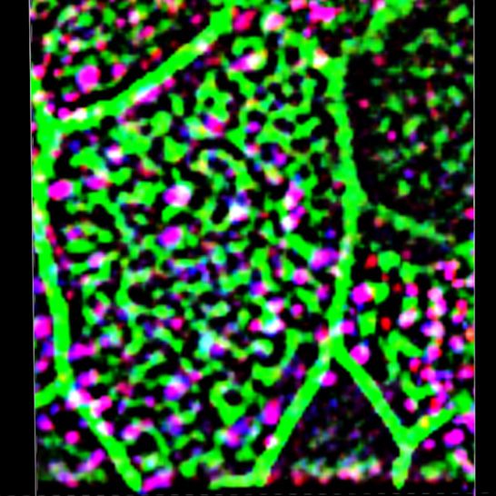
system, NO is produced by the endothelial isoform (eNOS/NOS3) in              protein (Fig 1B). Thus, immunocytochemical and genetic labeling
response to shear stress of the blood flow and rapidly remodels the            approaches demonstrated that nNOS, but not the other NOS isoforms,
cytoskeleton in smooth muscle cells, thus promoting vasorelaxation            is expressed in mouse tracheal multiciliated cells.
(Heiss et al, 2015; Shu et al, 2015); notably, nNOS isoform can similarly         We detected nNOS in ciliary axonemes (Fig 1C) and in the apical
respond to shear stress by producing NO to support basal blood flow            zones of ciliated cells (Fig 1D), where the basal bodies interact with
(Sato et al, 2008; Melikian et al, 2009; Hyndman et al, 2015; Zhang, 2016).   apical actin and microtubule cytoskeletons (Werner et al, 2011;
    Remarkably, low levels of exhaled NO strongly correlate with              Kunimoto et al, 2012; Vladar et al, 2012). We then applied structured
PCD, a human disorder associated with cilia dysfunction and                   illumination superresolution microscopy (SIM) to investigate the
manifested in affected patients as a constellation of syndromes,              details of interactions between nNOS, the basal body apparatus,
such as chronic sinusitis, bronchiectasis, male and female infer-             and the cytoskeleton of ciliated cells.
tility, reversal of left-right organ asymmetry, and hydrocephalus                 SIM revealed that the apical actin cortex of tracheal ciliated cells
(Walker et al 2012, 2013; Lobo et al, 2015; Werner et al, 2015; Horani        forms a ~2-μm deep grid with openings for the ciliary axonemes,
et al, 2016; Knowles et al, 2016). Notably, the link between insuffi-          with the basal bodies sandwiched between the apical actin grid at
cient NO production and poor cilia function is observed regardless            their upper limit and the network of cortical microtubules at their
of the specific genetic causes of the disorder. Even though the                lower limit (Fig 1E–K). The nNOS signal was associated with the basal
association between PCD and decreased production of NO is firmly               bodies and distributed along their entire height (Fig 1E). Within the
established and is routinely used as a diagnostic tool for triaging           apical aspect, the nNOS signal extended to the actin grid (Fig 1F, H,
PCD patients, the causal relationship between NO and PCD or the               and J), whereas in the basal aspect, it reached to the microtubule
cellular mechanisms underlying this association are unknown.                  network (Fig 1G, I, and K). The nNOS signal reached the basal foot, an
    Here, we investigate the roles of NO and nNOS in the functioning          appendage of the basal body that anchors microtubules, and it
of cilia in the mouse tracheal mucociliary epithelium and show that           partially overlapped with the signal of ODF2, an essential com-
nNOS has a versatile role in ciliated cells. First, it interacts with the     ponent of the basal foot (Fig1L and M). The zone of nNOS distri-
core factors of PCP to translate the global planar polarity of the            bution did not overlap with the distribution of rootletin, a marker of
tracheal tissue into the polarity of individual ciliated cells and            cilia rootlets (Fig 1N). Overall, the conspicuous distribution of nNOS
provides the cells with positional information in the tissue. In              with respect to the basal bodies and the components of the apical
addition, NO/nNOS is important for the polarization of both the               cytoskeleton, as well as its presence in the axonemes, prompted us
actin and microtubule apical networks, thus enabling correct                  to investigate the possible involvement of nNOS in the polarization
spacing and orientation of the basal bodies and the cilia. Finally, we        of tracheal ciliated cells and ciliary function.
show that NO/nNOS, through activation of soluble guanylate cy-
clase (sGC), is necessary for supporting the CBF. Together, our               Ciliated cells in nNOS knockout mouse trachea
results indicate a causal link between the cilia and flow dysfunction
and the insufficient availability of NO.                                       The spacing and orientation of the basal bodies are the crucial
                                                                              features of the polarity of ciliated cells in the trachea. To investigate
                                                                              the role of nNOS in ciliated cell polarity, we compared the tracheal
                                                                              mucociliary epithelium in wild-type and nNOS-deficient mice. We
Results                                                                       did not detect differences in the ratio of ciliated cells to other cell
                                                                              types in the epithelium, thus indicating that nNOS is not essential
nNOS in the mouse trachea                                                     for the determination of cell fate in the developing mucociliary
                                                                              epithelium. However, the morphology of ciliated cells, as analyzed
Prompted by the association of low levels of NO with PCD (Werner              by scanning electron microscopy, was notably different in the nNOS
et al, 2015; Knowles et al, 2016) and by the role of the nNOS ortholog in     KO than the wild-type animals (Fig 2A–F). In the trachea of wild-type
cell polarization and its interaction with the PCP pathway during             mice, the cilia were of consistent length, and their orientation was
Xenopus embryogenesis (Peunova et al, 2007), we investigated the              coordinated with adjacent cells, with a bias toward the direction of
role of nNOS in mouse tracheal multiciliated cells. Among the three           the presumptive mucus flow, in general alignment with the long (AP;
NOS isoforms tested, only the nNOS signal was present in the                  lung-to-larynx) axis of the trachea (Fig 2A and B). In the nNOS KO
mucociliary epithelium of the trachea (Fig 1), whereas the eNOS and           mouse trachea, a wide range of defects was observed (Fig 2C–F).
iNOS signals were associated with the tracheal vasculature (data not          Many ciliated cells were grossly misshapen, with the average apical
shown). We compared images of the tracheas of the wild-type and               area almost twice that in the wild type (110 ± 20 versus 62 ± 10 μm2)
nNOS-null mutant mice (nNOS knockouts; nNOS KO) (Packer et al,                (Fig 2C). In addition, numerous nNOS KO ciliated cells had cilia that
2003) after immunostaining with antibodies to nNOS; we detected the           were sparser and shorter than those in the wild type (3.8 ± 0.5 μm in
nNOS signal in the wild-type tracheal ciliated cells but not in cells of      nNOS KO versus 5.4 ± 0.6 μm in the wild type) and frequently bore
the mutant animals (Fig 1A). Additional evidence that nNOS is                 very short stub-like cilia (the length of axonemes was too short to
expressed in ciliated cells was obtained by genetically marking the           measure accurately) (Fig 2C–E).
nNOS-expressing cells: after crossing nNOS-CreER driver and Ai9                  The orientation of cilia in the nNOS KO trachea was poorly co-
reporter mouse lines and inducing recombination with tamoxifen, we            ordinated, although the degree of this defect varied: in the mild
found that ciliated cells in the trachea in the nNOS-CreER/Ai9 mice           phenotype, the orientation of the cilia within single cells showed
(Taniguchi et al, 2011) expressed nNOS promoter-driven fluorescent             coordination; however, poor alignment with the adjacent cells was

nNOS regulates tracheal ciliary cell activity   Mikhailik et al.              https://doi.org/10.26508/lsa.202000981   vol 4 | no 5 | e202000981   2 of 24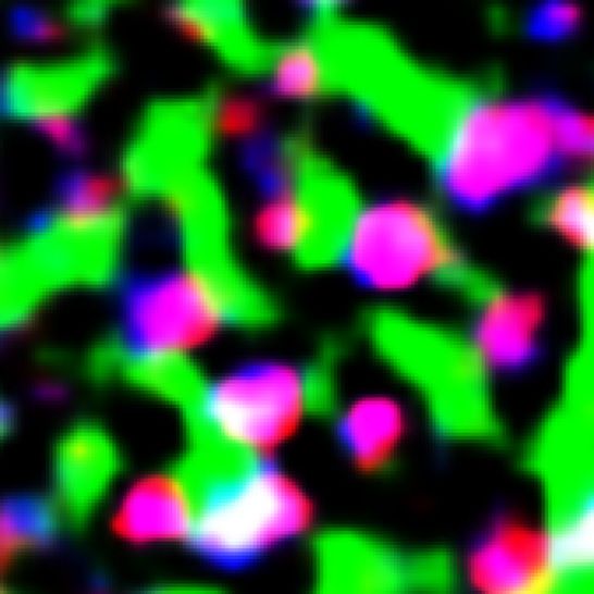
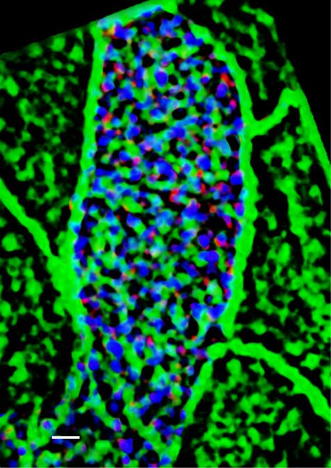
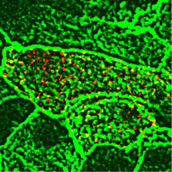
Figure 1. Localization of nNOS in the ciliated cells of trachea.
(A) Immunostaining for nNOS shows its expression in the ciliated cells of the mouse trachea and absence in the nNOS-null mutant trachea (inset): nNOS: red; acetylated
α-tubulin: blue (here and below the fluorophores for multiple labels are indicated on the figures by the corresponding colors). Scale bars are 5 μm in (A, B, C, D).
(B) Detection of nNOS expression in the tracheal ciliated cells of nNOS-CreER/Ai9 mice. dTomato expression, driven by nNOS promoter, is observed in the ciliated cells,
with the sample counterstained with phalloidin and acetylated α-tubulin. (C) nNOS is detected in the ciliary axonemes in the trachea, counterstained with phalloidin.
(D) nNOS is detected in the apical cortex of the ciliated cell of the trachea and in the ciliary axonemes, stained with antibodies to nNOS and acetylated α-tubulin.
(E) In cultured mouse tracheal ciliated cells nNOS is associated with basal bodies along their entire height. Z section from SIM in the inset. Scale bar is 2 μm. (F, G, H, I, J, K) In
the ciliated cells of the mouse trachea, nNOS is associated with the cortical apical actin grid and cortical microtubules. Scale bar is 2 μm. (F) The image is focused on the
apical-most area of the ciliated cells where nNOS is associated with the cortical actin grid; inset shows the entire cell (top view). (G) The same cell region as in (F), but
flipped over to show the interaction of nNOS with the cortical network of microtubules located below the actin grid; inset shows the entire cell as in (F). (H, I, J, K) Z sections
across the apical area of the ciliated cell of the tracheal explant, revealing the positions of the apical cortical actin grid and the microtubules (H); nNOS association with
the cortical microtubules (I); nNOS association with the cortical actin grid (J); and nNOS association with both actin and microtubule components of the cortical
cytoskeleton (K). (L) In the most basal aspects of the basal bodies nNOS is associated with microtubules and the basal feet (cultured mouse ciliated cells). Scale bars are 1
μm for (L, M, N). (M) nNOS is associated with basal bodies, labeled for γ–tubulin, and with basal feet, labeled for ODF2 (cultured mouse ciliated cells); higher
magnification in the inset. (N) nNOS is not detected in the cilia rootlets in cultured ciliated cells labeled for rootletin. Z section in the inset.

observed. In the severe phenotype, the ciliary orientation was poorly                         severe defects of the tracheal ciliated cells, with essentially no
coordinated even within single cells, and the cilia were often or-                            normal cells in sight; in a small fraction of animals (5–7%) mani-
ganized in a rosette-like pattern (Fig 2D). Besides the deformed cells,                       festation of the mutant phenotype was very weak.
there were also groups of cells present with apparently normal cilia                             The results of scanning electron microscopy were supported by
polarity. The penetrance of the mutants phenotype also differed                               confocal microscopy after immunocytochemical staining for cilia
between individual animals. In most cases, most of the ciliated cells                         and basal body markers, which similarly showed that in the nNOS
in a tracheal preparation would carry distinct and severe defects in                          KO trachea, the orientation of the cilia and their spacing pattern
polarity, with mildly or minimally affected cells observed in the same                        were disorganized, and the cilia were shorter, sparser, and not
preparation. However, a quarter of all animals showed particularly                            aligned with the direction of airway clearance (Fig 2G and H).

nNOS regulates tracheal ciliary cell activity     Mikhailik et al.                            https://doi.org/10.26508/lsa.202000981       vol 4 | no 5 | e202000981           3 of 24
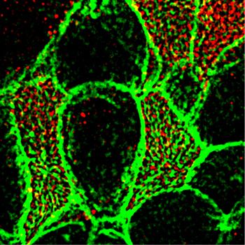
Figure 2. Distortion of the ciliated cell polarity in the trachea of nNOS-null (nNOS KO) mice.
(A) Scanning electron microscopy (SEM) of the ciliated cells in the wild-type mouse trachea. Scale bar is 5 μm in (A, C, D, E, G, H). (B) Circular plot of cilia orientation in
wild-type tracheal ciliated cells. Direction of the arrow represents the mean vector of the cilia orientation for a given cell, with the larynx position corresponding to the
top, and the arrow length represents the value of the mean vector (longer arrows indicating higher coordination of cilia orientation for that cell); r describes rotational
orientation of the cilia, n:number of evaluated cells. (C, D, E) SEM of the ciliated cells in nNOS KO mice. Note grossly misshapen cells with sparse cilia (C); cells with
rosette-like arrangement of cilia (D); and cells with short stub-like cilia (E). (F) Circular plot of cilia orientation in nNOS mutant tracheal ciliated cells, visualized by SEM. (G,
H) Immunocytochemical staining for basal bodies (γ-tubulin) and cilia (acetylated α-tubulin). (G, H) nNOS KO cells show defects in cilia orientation, chaotic spacing
pattern, and short and sparse cilia (G) as compared with cilia organization in the wild type (H). (I, J, K, L) Transmission electron microscopy. (I, K) Cilia in the wild-type (I)
and nNOS mutant (K) trachea, with arrows indicating basal feet orientation. (I, K) Scale bar is 0.5 μm in (I and K). (J, L) Circular plots, characterizing rotational polarity of
basal bodies shows coordination in the cilia orientation in the wild type (J), as compared with the nNOS mutant (L). (M, N, O, P) Transmission electron microscopy of a cross
sections of the trachea: there were no detectable defects in the docking of the basal bodies to the cell membranes (M, O); however, abnormal basal bodies associated
with abnormal short axonemes and not generated axonemes were observed in nNOS KO cells (P) in comparison to the wild type (N). Scale bar is 0.5 μm in (M, N, O, P).

nNOS regulates tracheal ciliary cell activity    Mikhailik et al.                            https://doi.org/10.26508/lsa.202000981        vol 4 | no 5 | e202000981          4 of 24

The orientation of the cilia in the direction of flow is established                    immediate vicinity of the apical membrane but not associated with
by rotation of the basal body, with the basal foot being correctly                        normal long axonemes (Fig 2N and P), indicating that nNOS may be
oriented. We applied transmission electron microscopy (TEM) to                            involved in the growth of cilia.
determine the orientations of the basal feet in horizontal sections                          Given the importance of the PCP pathway for multiciliated cell
of the trachea, and found that in the nNOS KO samples, the ori-                           polarization (Wallingford, 2006, 2010; Wallingford & Mitchell, 2011;
entation of the basal bodies within single ciliated cells and be-                         Vladar et al, 2012), we examined the changes evoked by NO on the
tween adjacent cells was less coordinated than that in the wild type                      distribution of the core PCP pathway factor Vangl1 (Kunimoto et al,
(Fig 2I–L). The fewer cilia observed at the surfaces of ciliated cells of                 2012; Vladar et al, 2012). We compared the patterns of Vangl1 in wild-
the mutants may potentially have been due to a failure of docking                         type and nNOS KO tracheas. In the wild-type animals, Vangl1
of the basal bodies to the apical membrane (e.g., basal bodies                            protein was found in the membranes and was localized asym-
stalled deeper in the cytoplasm). TEM of cross-sections of the                            metrically, as a crescent at the distal sides of ciliated cells (facing
mutants’ tracheas did not indicate basal bodies deep in the cy-                           the lungs); this stereotypic polarized pattern was common among
toplasm (Fig 2M and O). However, we detected ciliated cells with                          neighboring cells (Fig 3A, E, and I). In the nNOS KO trachea, a smaller
short abnormal cilia and with the basal bodies located in the                             fraction of Vangl1 protein was localized at the cell membranes,

Figure 3. Distortion of the planar cell polarity in the trachea of nNOS KO mice.
(A, E) Planar polarity of the ciliated cells in the wild-type tracheal cells, characterized by the asymmetric distribution of Vangl1, which is concentrated at the membranes
at the distal aspect of ciliated cells; membrane is marked by antibody to Na+/K+ ATPase. (B, F, I) The membrane enrichment of Vangl1 is distorted in the ciliated cells of the
tracheal explant of nNOS KO, as manifested by diminished association of the protein with the membranes and its excessive accumulation in the cytoplasm. (C, D, G, H, I)
SNAP treatment for 30 min improves the membrane association of the Vangl1 protein in the mutant, but not the wild-type tracheal explants. Bar in (I) shows mean ±
s.e.m. *P < 0.05, **P < 0.01. Scale bar is 5 μm.

nNOS regulates tracheal ciliary cell activity   Mikhailik et al.                         https://doi.org/10.26508/lsa.202000981      vol 4 | no 5 | e202000981        5 of 24
whereas a substantial fraction of Vangl1 accumulated as aggregates         nNOS KO trachea, the CBF was significantly lower (2.1 ± 0.75 Hz) (Fig
throughout the cytoplasm (Fig 3B, F, and I).                               4J), and the beating pattern showed profound uncoordination
   We next examined whether the treatment of trachea explants              between neighboring cells and often within single ciliated cells.
from wild-type and nNOS KO mice with the chemical NO donor                 Moreover, we observed numerous cells with very slowly moving and
S-nitroso-N-acetyl-penicillamine (SNAP) might evoke changes in the         even immotile cilia (5–20% in trachea samples from different
Vangl1 distribution in ciliated cells. Exposure of the wild-type trachea   mutant animals) (Video 2); however, we also observed rare areas
to 100 μM SNAP did not affect the polarity of the Vangl1 distribution or   with preserved coordination of the ciliary movement between
its localization to membranes (Fig 3C, G, and I). In contrast, SNAP        ciliated cells in the nNOS mutants.
treatment of nNOS KO trachea explants significantly rescued Vangl1              Similarly to the effects of the nNOS deletion, the addition of NOS
protein localization to the membranes, decreasing the fraction of          inhibitors to the wild-type tracheal preparations decreased the
cytoplasm-associated protein (Fig 3D, H, and I). These results indicate    flow velocity and CBF, to 63% and 52%, respectively, of the wild-type
that establishment of the ciliated cell polarity mediated by the core      values (Fig 5A–F).
factors of the PCP pathway is sensitive to NO signaling.

                                                                           NO donors restore directional flow and ciliary beating
Impaired fluid flow and ciliary beating in the nNOS KO trachea
                                                                           We next asked whether the lack of functional nNOS enzyme in nNOS
Having found that nNOS is important for the polarity of ciliated           KO trachea might be compensated by exogenous NO released by
cells, we examined the role of nNOS in the ability of cilia to generate    chemical NO donors. We found that after the addition of 10 μM 3-(2-
effective directional flow. To investigate the flow dynamics, we used        hydroxy-1-methyl-2-nitrosohydrazino)-N-methyl-1-propanamine
a live imaging assay to follow and quantify the movement of                (NOC7) to the tracheal preparations of nNOS KO mice, the velocity
fluorescent beads along the preparations of dissected tracheas,             and CBF significantly increased, 1.6-fold and 2.6-fold, respectively
which were longitudinally cut, flattened, and placed in a viewing           (Fig 4D and H–J). In the wild-type trachea explants, NOC7 also in-
chamber under a microscope. We analyzed the following param-               creased the flow velocity and CBF, to 128% and 127%, respectively
eters of the beads’ movement:                                              (Fig 4C, G, I, and J and Videos 1 and 2). Notably, in the nNOS KO
                                                                           preparations exposed to NOC7, we no longer observed areas with
  (i) flow velocity: the net displacement of the beads, that is, the        immotile cilia. However, we did not observe an obvious rescue of
      distance between the first and the last point of the path             cilia beating coordination throughout the tissue, commonly
      traveled by the bead per second;                                     detecting areas where cilia were moving with poor coordination
 (ii) overall trajectory traveled by a bead per second, and its de-        (albeit with higher CBF) after NOC7 treatment.
      viation from the net displacement;                                       We also found that a chemically distinct NO donor, SNAP, which
(iii) direction of the bead’s movement (vector of the net dis-             has a longer half-life than NOC7 (5 h versus 10 min at room tem-
       placement); and                                                     perature), elicited similar changes in the preparations of wild-type
(iv) coordination of vectors of the net displacement of individual         and mutant tracheas, thereby effectively rescuing the flow velocity
      beads as the deviation from the median of the vectors of all         and CBF in the nNOS KO (Fig S1A–H and Video 3).
      analyzed beads.                                                          Together, these results indicated that nNOS is critical for the
                                                                           efficiency of the CBF and for the velocity and directionality of the
    In the wild-type trachea, the fluorescent beads moved
                                                                           generated fluid flow; that exogenous NO donors can increase ve-
rapidly (46.8 ± 4.4 μm/s; mean ± sem), with an optimal trajectory
                                                                           locity and CBF in wild-type tracheal explants; and that NO donors
(i.e., the overall path traveled by a bead close to its net dis-
                                                                           are able to mitigate the defects in flow and CBF induced by the loss
placement) and well-coordinated directionality (with the bulk
                                                                           of nNOS.
of the beads’ movement vectors close to the median direction)
(Fig 4A, E, and I).
    A lack of nNOS impaired all analyzed flow parameters. In par-           cGMP mediates the action of nNOS in ciliated cells
ticular, in the trachea in nNOS mutant mice, the flow velocity was
diminished twofold to 22.6 ± 3.6 μm/s, and the beads moved in a            sGC is a key effector of NO in a range of biological settings: binding
poorly coordinated manner (Fig 4B, F, and I).                              of NO to the heme-containing catalytic center activates the enzyme
    CBF is a critical contributor to flow efficiency. Because chemical       and leads to a rapid increase in the cGMP level, which in turn
NO donors have been reported to stimulate the ciliary beat (Jain           activates cGMP-dependent kinase, with subsequent phosphoryla-
et al, 1993; Sisson et al, 2009; Jiao et al, 2011), we examined the        tion of target proteins. Therefore, to determine whether the ob-
contribution of nNOS to the CBF. In these experiments, cilia were          served effects of NO might be mediated by the cGMP signaling
visualized by live staining with fluorescein-conjugated wheat germ          pathway, we examined the effects of an sGC inhibitor and a cGMP
agglutinin. This method allowed us to follow changes in the ori-           analog on both the wild-type and mutant tracheas.
entation of cilia beating and beads movement in a live tissue in              Exposure of the wild-type trachea explants to the sGC inhibitor
parallel with measuring the CBF values.                                    1H-[1,2,4]-oxadiazolo-[4,3-alpha]-quinoxalin-1-one (ODQ) decreased
    In the trachea in wild-type animals, most cilia beat in unison with    the flow velocity sixfold and the CBF threefold (Fig 6A–F), thus re-
a consistent frequency (9.8 ± 0.84 Hz) and in a coordinated manner,        sembling the results observed with the nNOS deletion and NOS
generating an efficient directional flow (Fig 4J and Video 1). In the        inhibitors (Figs 4 and 5 and Video 4).

nNOS regulates tracheal ciliary cell activity   Mikhailik et al.           https://doi.org/10.26508/lsa.202000981   vol 4 | no 5 | e202000981   6 of 24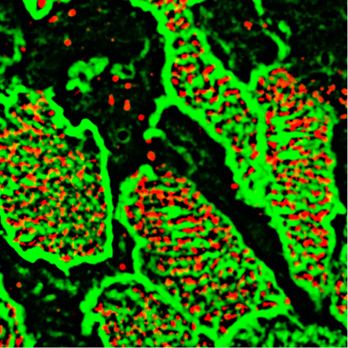
Figure 4. Nitric oxide is essential for polarized flow.
(A, B, C, D) Spot tracking of beads movement in the wild-type (A) and nNOS KO (B) trachea. Defective flow in the nNOS mutant (B) can be improved by the addition of NO
donor NOC7 (D). NOC 7 also increases the flow velocity in the wild type (C). Scale bar is 50 μm. (E, F, G, H) The trajectory path (colored), net displacement, and direction of
the beads in the flow (white arrow) probed in the wild-type (E) and nNOS KO (F) trachea specimens before (E, F) and after (G, H) addition of NOC7. (I, J) Effect of NOC7 on
ciliary beat frequency and flow velocity is summarized in charts (I, J). Scale bars are 50 μm. Corresponding Videos 1–3 are presented in Supplemental Material.

   We then asked whether a cell-permeant cGMP analog, 8-bromo-                            dynamics might be associated with the increase in CBF, the im-
guanosine 39,59-cyclic monophosphate (8-Br-cGMP), might repli-                            proved coordination of the ciliary beating, or both. To answer these
cate the effects of the NO donors in the wild-type and mutant                             questions, we modified the experimental setup to follow the be-
trachea. Addition of 8-Br-cGMP to the wild-type trachea explants                          havior of the same group of ciliated cells as they responded to a
increased the flow velocity 1.46-fold and the CBF—1.57-fold (Fig 7A, C,                    particular treatment. We placed the tracheal preparations in a
E, G, I, and J and Video 5). When added to the mutant trachea                             perfusion camera with the medium pumped through the camera at
explants, 8-Br-cGMP increased the flow velocity 1.9-fold and the CBF                       a low speed of 5 μm/s — conditions allowing the tissue to stay alive
threefold, thus bringing them close to the basal levels observed in                       for hours, with CBF maintained at consistent physiological levels,
the wild-type tracheas (Fig 7B, D, F, and H–J and Video 6).                               and without interfering with the velocity of cilia-generated flow
   Together, these results showed a strong response of both wild-                         (Winters et al, 2007). In addition, a T-valve switch, set next to the
type and nNOS-deficient tracheal ciliated cells to the sGC inhibitor                       chamber, allowed for rapid replacement of the medium with one
and the cGMP analog, thus pointing to the nNOS-NO-sGC-cGMP                                containing the 8-Br-cGMP and thus enabled continued recording
pathway as a critical effector of nNOS signaling in ciliated cells.                       the responses of the same group of ciliated cells to the treatment.
   At the same time, these experiments leave open the question of                            We first recorded ciliary beating while explants were perfused
whether the stimulating effect of the cGMP analog on the flow                              with regular medium, then switched to perfusion with medium

nNOS regulates tracheal ciliary cell activity   Mikhailik et al.                         https://doi.org/10.26508/lsa.202000981      vol 4 | no 5 | e202000981        7 of 24
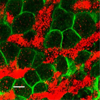
Figure 5. Inhibitors of NOS decrease flow velocity and ciliary beat frequency          Figure 6. Soluble guanylate cyclase (sGC) is a necessary factor of the nitric
(CBF).                                                                                oxide signaling pathway in controlling the fluid flow dynamics.
(A, B, C, D, E, F) Spot tracking of beads movement and CBF measurement in the wild-   (A, B, C, D, E, F) Spot tracking of beads movement and ciliary beat frequency
type trachea after exposure to NOS inhibitors. (A, B, C, D, E, F) Incubation of the   measurement in the wild-type trachea after exposure to sGC inhibitor. Treatment
preparations for 30 min with a mix of 1 mM L-NAME, 100 μM ETU and 100 μM S-           of the specimens of wild-type trachea explants with an inhibitor of sGC, [1H-
methyl-L-thiocitrulline hydrochloride (SMTC) resulted in a decrease in all fluid flow   [1,2,4]oxadiazolo-[4,3-a]quinoxalin-1-one], ODQ for 30 min decreases the flow
parameters (B, D) in comparison with non-treated control (A, C), demonstrating        velocity and the ciliary beat frequency (B and D, E, F) in comparison with control (A
inhibitory effects both on the flow velocity and CBF (charts in E, F).                 and C, E, F). Corresponding Video 4 is presented in Supplemental Material.

containing 8-Br-cGMP, focusing on the same area. We found that, as                        Importantly, the sequential recording setup allowed us to follow the
in the basic setup, addition of 8-Br-cGMP to the wild-type tracheal                   ciliary beating pattern and frequency in individual cells (Fig 8M–O),
preparations significantly increased the flow velocity and CBF, ~1.5-                   demonstrating that within seconds after addition, 8-Br-cGMP affected
fold for both parameters (Fig 8A–F).                                                  the CBF in most of the individual ciliated cells: increasing the CBF in cells
   We then used this sequential recording setup to analyze the                        with cilia that beat in apparent coordination with the neighboring cells;
nNOS KO tracheal preparations, focusing on the representative                         increasing the CBF in cells in which the beating was uncoordinated with
areas that showed various types of defects in ciliary function, as                    that of the neighbors; and converting immotile cilia into motile ones.
described earlier: (i) slower overall ciliary beating; (ii) cells with                However, we did not observe visible changes in the cilia beating di-
immotile cilia; (iii) cells lacking coordination with the neighboring                 rection: in cells in which the ciliary beating lacked coordination with that
cells in their beating direction; and (iv) cells with cilia still beating in          of the neighboring cells, cilia continued beating in an uncoordinated
apparent coordination with their close neighbors. As with the basic                   manner upon addition of 8-Br-cGMP, albeit with higher CBF (Video 7).
setup, addition of the cGMP analog increased the flow velocity and                         Remarkably, even in the presence of uncoordinated ciliary
CBF in nNOS-deficient cells (Fig 8G–L and Video 7).                                    beating, the flow dynamics parameters were improved for the

nNOS regulates tracheal ciliary cell activity   Mikhailik et al.                      https://doi.org/10.26508/lsa.202000981     vol 4 | no 5 | e202000981         8 of 24
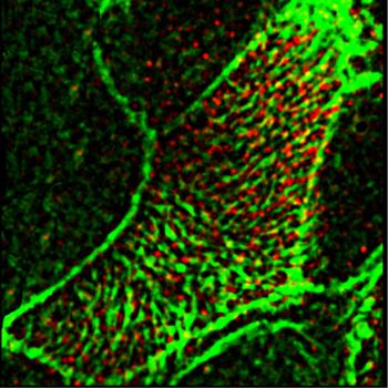
Figure 7. Nitric oxide positively controls ciliary beat frequency (CBF) and fluid flow through soluble guanylate cyclase-cGMP pathway.
(A, B, C, D, E, F, G, H, I, J) Treatment of the tracheal explants with cGMP analog 8BrcGMP for 30 min replicated the effect of nitric oxide donors, rescuing the CBF and flow
defects in the nNOS KO trachea. (A, B, D, E, I, J) Fluid flow and CBF in nNOS KO trachea, showing lower velocity, lower CBF, and poor directionality in comparison with the
wild type (A, B, D, E, I, J) are significantly improved by the treatment with 8-Br-cGMP. 8-Br-cGMP also augments CBF and flow velocity in the wild-type trachea explants.
Corresponding Videos 5 and 6 are presented in Supplemental Material.

entire recorded region. The rescuing effect of 8-Br-cGMP on the                          Loss of nNOS disrupts the rotational polarity of basal bodies
flow in nNOS KO mutants was commensurate with the severity of
the ciliary polarity and ciliary beating uncoordination defects: in                      Impaired coordination of the ciliary beating in the nNOS KO trachea
the regions of the trachea where the defects were moderate and                           implies the presence of defects in rotational polarity of the cilia. The
not more than half of all ciliated cells were beating without co-                        rotational polarity of ciliated cells is largely mediated by microtubules,
ordination with their neighbors, addition of 8-Br-cGMP improved                          which connect the basal feet into a joint network, thus enabling the
the flow velocity despite the presence of cells whose cilia beat                          concerted rotation of basal bodies in the direction of the flow (Werner
without coordination. However, in the regions with severe defects                        et al, 2011; Kunimoto et al, 2012; Vladar et al, 2012). Because nNOS was
(with most cells beating without coordination) treatment with 8-                         observed in association with microtubules and with the basal feet (Fig
Br-cGMP was not able to rescue the flow velocity despite the                              1L), we sought to determine whether nNOS might contribute to the
increased CBF. Together, these data suggested that the 8-Br-                             establishment of the cortical microtubule network.
cGMP-induced increase in the fluid flow velocity was mainly                                   To evaluate the input of nNOS to the rotational polarity of the
due to the increase in CBF.                                                              tracheal ciliated cells, we selected a pair of markers: Chibby1 (Cby1),

nNOS regulates tracheal ciliary cell activity   Mikhailik et al.                        https://doi.org/10.26508/lsa.202000981     vol 4 | no 5 | e202000981        9 of 24
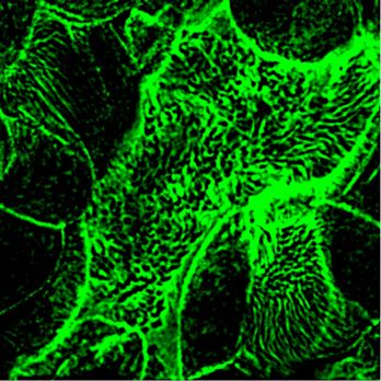
Figure 8. Increase of the ciliary beat frequency is the primary effect of 8-Br-cGMP treatment in rescuing fluid flow in nNOS KO tracheal explants.
(A, B, C, D, E, F, G, H, I, J, K, L, M, N, O) Focusing on the same area of the tracheal ciliated epithelium in the wild-type and nNOS KO trachea and recording changes in flow
velocity (by tracking beads’ movement) and cilia movements in individual ciliated cells before and after addition of 8-Br-cGMP. (A, B, C, D, E, F, G, H, I, J, K, L) Such
sequential recording shows an increase of flow velocity induced by 8-Br-cGMP (revealed as longer tracks of the beads’ movements) in the wild-type (A, B, C, D, E, F) and
nNOS KO (G, H, I, J, K, L) trachea. (M, N, O) It also shows an increase of ciliary beat frequency, presented as heat maps, in most individual cells across the area 5–10 s after
addition of 8-Br-cGMP. Corresponding Video 7 is presented in Supplemental Material.

nNOS regulates tracheal ciliary cell activity   Mikhailik et al.                         https://doi.org/10.26508/lsa.202000981      vol 4 | no 5 | e202000981         10 of 24
whose presence marks the transition zone of cilia (Burke et al, 2014), and      shaped grid with openings for individual cilia (Figs 1 and 10A–F, I,
ODF2, a marker of basal feet (Kunimoto et al, 2012). SIM visualization of       and K). This grid, formed by intersecting actin cables, was polarized:
these markers allowed for reliable reporting of both the rotational             in each ciliated cell, one set of the actin cables was aligned with the
polarity and the spacing pattern of the basal bodies. The vectors,              rows of the basal bodies, whereas the other set of the cables ran
connecting the Cby1 and ODF2 puncta for each basal body in wild-type            orthogonally, in the direction of the flow and ciliary orientation,
tracheal ciliated cells, pointed in a similar direction toward the larynx,      with individual basal bodies positioned in the openings of the actin
thus demonstrating a clear pattern of rotational polarity within both           grid (Fig 10F and I). The polarity of the actin grid was shared by
single cells and groups of neighboring cells (Fig 9A and B). Moreover, the      neighboring ciliated cells, thus manifesting tissue-level polarity (Fig
Cby1/ODF2 markers revealed that the basal bodies of tracheal ciliated           10A and E). Because the basal bodies of cilia are attached to the
cells were organized in parallel rows oriented perpendicularly to the           apical actin cortex after docking (Werner et al, 2011; Vladar et al,
median vector of rotational polarity (Fig 9A–D). This distinct orientation      2012; Herawati et al, 2016), the apical actin grid thereby provides a
of the basal bodies’ rows was shared by neighboring cells (Fig 9A and           template, imposing its distinct geometry on the spacing pattern of
C). This observation, made with the tracheal tissue, echoed the report          the apically docked basal bodies.
of stereotypic row-like arrangement of the basal bodies in the cil-                In contrast to the regular pattern of the apical actin cytoskeleton
iated tracheal cells grown as air-liquid interface (ALI) cultures               observed in the wild-type trachea, the windowpane geometry of the
(Herawati et al, 2016).                                                         apical actin cytoskeleton in nNOS KO mice was noticeably distorted,
    In contrast to the results for the wild-type tracheas, the pattern of       frequently showing disordered shapes of the actin grid and the loss
rotational polarity, as visualized by Cby1/ODF2, was significantly               of coordination in the geometry and orientation between neigh-
distorted in the nNOS mutants (Fig 9E–G). In the areas with a mild              boring ciliated cells (Fig 10G–J and L–N).
distortion phenotype, coordinated rotational polarity was observed                 In the nNOS KO ciliated cells, basal bodies remained attached to the
within single ciliated cells; however, the median vectors of rotational         distorted apical actin cytoskeleton (Fig 10H and J). The typical spacing
polarity were poorly coordinated between neighboring cells (Fig 9E),            pattern of basal bodies in rows and the tissue level polarity of ciliary
thus indicating that in the absence of nNOS, the cilia of the                   spacing patterns between neighboring ciliated cells deteriorated in
neighboring cells may be poised to beat against each other.                     parallel to the degree of distortion of the actin cortex in the
    In the cell areas with a more severe distortion phenotype, the              mutant cells, essentially disappearing in cells with highly dis-
pattern of rotational polarity of the basal bodies was scrambled even           torted actin cortex (Fig 10L–N). Therefore, the distortion of the
within single cells (Fig 9F); notably, the spacing pattern in those cells       geometry of the apical actin cytoskeleton in nNOS KO ciliated cells
was also distorted, and no apparent rows of basal bodies were ob-               defined the distortion of the spacing pattern of the basal bodies
served. Because the orientation of ciliary beating is not coordinated,          observed in the mutant cells.
cilia may be poised to beat against each other even within the same
cell, thus interfering with the task of directional flow generation.             nNOS and RhoA mediate establishment of the pattern of the
    SIM images of the patterns of the basal feet-microtubules connections       apical actin cytoskeleton in ciliated cells
revealed that in the wild-type ciliated cells, the microtubules (marked by
α-tubulin) were attached to the most basal aspects of the basal feet            We next asked whether nNOS may control the proper arrangement
(marked by ODF2) and formed a dense network (Fig 9H and I). In the wild-        of the apical actin grid by modulating relevant signals and effectors
type ciliated cells, each basal foot was connected with the microtubules        that control formation of the actin cables. RhoA, a major regulator
(Fig 9I, J, and M). In contrast, in the nNOS KO cells, only 75% of the basal    of actin polymerization dynamics, is active in numerous cell polarity
feet were connected to the microtubules (Fig 9K, L, and M). Moreover, in        contexts (Besson et al, 2004; Pan et al, 2007; Zaoui et al, 2008; Miller
the wild-type cells, a consistent overlap of the signals for ODF2 and           & Bement, 2009). Importantly, in multiciliated epithelial cells, RhoA
α-tubulin was observed, with a steady Pearson correlation coefficient            activity is essential for organizing the apical actin cytoskeleton
(PCC) of 0.72 ± 0.1, thus suggesting proximity between the basal feet and       during ciliogenesis (Pan et al, 2007) and is crucial for the anchoring
microtubules; in contrast, in the nNOS KO, the PCC varied significantly          of basal bodies to the apical domain (Sedzinski et al, 2017).
between individual basal feet, ranging from the wild-type–like value of            Thus, Rho A was deemed an attractive candidate to test for the
0.65 to 0.01, a value indicating a lack of significant signal overlap.           possible interaction of nNOS with potent regulators of actin poly-
    TEM images of nNOS KO ciliated cells (Fig 9N and O) showed a sparse         merization. Using antibodies specific for the active GTP-bound form of
microtubule network and basal feet lacking connections with the mi-             RhoA (Benink & Bement, 2005), we found the active RhoA signal in
crotubules and thereby being excluded from the network. This type of            close proximity to nNOS (PCC = 0.46) in the openings of the apical actin
defect suggested that nNOS and its association with basal feet and              cortex of ciliated cells in the trachea (Fig 11A–I); we also found a similar
microtubules are essential for the formation of a properly formed and           association between RhoA and nNOS (with a higher PCC of 0.7) in the
dense network connecting basal bodies, thereby enabling their coop-             apical actin cortex of re-differentiated mouse trachea ciliated cells
erative rotation in the direction of the flow.                                   grown in ALI cultures (Figs 11 and 12). Besides the apical cortex-bound
                                                                                active RhoA, a substantial fraction of active RhoA was also detected in
Loss of nNOS disrupts the polarity of the actin cytoskeleton in                 the ciliated cells’ cytoplasm just beneath the actin cortex (Fig 11D–F).
ciliated cells                                                                     Given that the nNOS signal marks the positions of basal bodies in
                                                                                the openings of the apical actin grid (Fig 1E and F) and is found near
Our results show that in ciliated cells of the wild-type trachea, a             active RhoA, we asked whether nNOS/NO signaling might be im-
cortical actin network forms a typical pattern of a windowpane-                 portant for RhoA activity and polymerization of the cortical actin,

nNOS regulates tracheal ciliary cell activity   Mikhailik et al.               https://doi.org/10.26508/lsa.202000981   vol 4 | no 5 | e202000981   11 of 24
Figure 9. nNOS is essential for cilia spacing pattern and rotational polarity. (A, B) Cby1/ODF2 pair demonstrates high degree of coordination of rotational polarity within single ciliated cells and between neighboring cells in the trachea and alignment with the direction of the flow. Scale bar is 5 μm in (A, B, C, D, E, F). (C, D) ODF2 staining reveals the spacing pattern of basal bodies as parallel rows, oriented perpendicularly to the direction of the flow. (E) nNOS KO mutant cells with mild phenotype have preserved rotational polarity and spacing pattern of the basal bodies within single cells, which is poorly coordinated with the neighboring cells. (F, G) nNOS KO mutant ciliated cells with more severe phenotype lack rotational polarity both within the cell and between neighboring ciliated cells, and the spacing pattern of the basal bodies is scrambled. (H, I, J) Microtubules are anchored to the most basal aspects of the basal feet (labeled by ODF2) in the ciliated cells in the trachea (H, scale bar is 1 μm). Microtubules connect all basal bodies into a regular joint network in the ciliated cells of the trachea in wild type (I); (J) – higher magnification. (K, L) In the nNOS KO cells basal feet are aberrantly connected with microtubules, with some basal feet having excessive connections with the microtubules and others left out of the connections. (H, I, J, K, L) Scale bars are 1 μm in (H, I, K) and in (J, L). Pearson correlation coefficient for the overlap between fluorescent signals of ODF2 (Alexa-568) and microtubules (Alexa-488) is indicated. (M) Fraction of the basal feet connected to the microtubules in wild-type and nNOS KO ciliated cells (20 and 25 cells of each genotype analyzed, correspondingly). (N, O) Transmission electron microscopy shows a sparse network of microtubules connecting basal feet in nNOS KO, leaving some basal feet out of the network, as compared with wild type. nNOS regulates tracheal ciliary cell activity Mikhailik et al. https://doi.org/10.26508/lsa.202000981 vol 4 | no 5 | e202000981 12 of 24
Figure 10. Polarity of the apical actin grid is related to the spacing pattern of the basal bodies.
(A) Apical actin grid displays a windowpane pattern with opening for cilia. Inset shows a magnified region of 3D reconstruction, demonstrating rows and columns
formed by the actin grid; note that the longitudinally oriented actin cables are running slightly more apically (above) than the crossing them cables. (A, B, C, D) Scale bars
are 1 μm in (A, B, C, D). (B) Axonemes are oriented along the longitudinally oriented actin cables. Inset shows the cilia axonemes alone. (C) Basal bodies (labeled by
γ-tubulin) are positioned in rows separated by actin cables of the actin grid. (D) An optic section at the base of cilia shows cilia (labeled by acetylated α-tubulin)
positioned in rows, separated by actin grid cables. (E, F, G, H, I) Rows of basal bodies (labeled by ODF2) in trachea are attached to the actin cytoskeleton of the ciliated
cells; such geometry of the apical actin grid provides a template for the spacing pattern of the basal bodies. (G, H, I, J) Apical actin grid in the ciliated cells of the nNOS KO
has more chaotic structure because of shorter and poorly oriented actin cables. Likewise, the basal bodies, still attached to the actin cortex, are also chaotically
organized by following the distorted shape of the actin grid template. (K, L, M, N) The range of distortion of the regular organization of apical actin grid in nNOS KO ciliated
cells (L, M, N), compared with the windowpane pattern in the wild type (K).

and consequently for basal body attachment to the actin cortex and                          structure and a shift in the distribution of actin filaments forming
their spacing pattern.                                                                      the grid toward significantly shorter lengths compared with the wild
   We found that deletion of nNOS resulted in a significant loss of                          type (Fig 12U). Importantly, when examining both wild-type and
active RhoA associated with the apical actin in the tracheal ciliated                       nNOS KO tracheal preparations 30 min after the addition of the NO-
cells, with a concomitant increase in the fraction of RhoA localized                        releasing donor SNAP we detected increased presence of active
in the cell cytoplasm (Fig 11A–D). The loss of RhoA from the actin grid                     RhoA in the apical cortex at the expense of its presence in the
was reflected in a significantly decreased PCC for RhoA/actin as-                             cytoplasm (Fig 12A–D and Q–T); an increased association between
sociation (from 0.47 in the wild type to 0.05 in the mutant Figs 11 and                     active RhoA and apical actin (from 0.05 to 0.51 PCC) (Fig 12I–T); and a
12. The diminished presence of active RhoA at the apical actin grid                         shift in the length distribution of the actin polymers forming the
of the mutants was associated with a more disordered grid                                   actin cortex toward longer filaments (Fig 12E–P and U).

nNOS regulates tracheal ciliary cell activity   Mikhailik et al.                          https://doi.org/10.26508/lsa.202000981       vol 4 | no 5 | e202000981         13 of 24
Figure 11. Active RhoA is associated with nNOS.
(A) Ciliated cell in the wild-type trachea, labeled for actin, nNOS, and active form of RhoA. Scale bar is 1 μm. (B, C, D, E, F) Z-sections of (A): active RhoA is associated with
nNOS, their signals overlapping with Pearson correlation coefficient = 0.46. Scale bar is 2 μm. (A, G, H, I) 2.5× (G) and 7× (H, I) magnifications of (A), with Z-section of (H)
shown in (I). Images in (B, C, D, E, F, G, H, I) show that nNOS is positioned in the openings of the apical actin grid and is associated there with the basal bodies (also see Fig 1E
and F). (J, K) The pattern of association of nNOS and active RhoA revealed in the trachea is also observed in air-liquid interface cultures of the tracheal ciliated cells.

nNOS regulates tracheal ciliary cell activity    Mikhailik et al.                           https://doi.org/10.26508/lsa.202000981       vol 4 | no 5 | e202000981          14 of 24
Figure 12. nNOS/Nitric oxide are essential for targeting the
                                                                                       active RhoA to the apical actin cortex.
                                                                                       Treatment with nitric oxide donor SNAP increases loading of
                                                                                       actin with active RhoA and is associated with increased length of
                                                                                       the actin cables forming the apical actin grid. (A, B, E, F, I, J, M,
                                                                                       N) Distortion of the structure of apical actin cortex (the actin
                                                                                       grid) in the ciliated cells of the nNOS KO (B, F), in comparison with
                                                                                       the wild type (A, E), trachea. In the wild type, active RhoA is
                                                                                       present both within the apical actin grid (where it is
                                                                                       concentrated in the openings for the basal bodies, where nNOS
                                                                                       is also found) and in the cytoplasm just below the actin cortex
                                                                                       (Z-section in A). Deletion of nNOS resulted in the loss of RhoA
                                                                                       activity within the apical actin cortex and its accumulation in the
                                                                                       cytoplasm below the apical actin network (B). (C, D, G, H, K, L,
                                                                                       O, P) Addition of SNAP for 30 min to the wild-type or nNOS KO
                                                                                       tracheal explants leads to the translocation of RhoA activity from
                                                                                       the cytoplasm to the apical cortex and augmentation of RhoA
                                                                                       association with actin (restored Pearson correlation coefficient)
                                                                                       at the expense of the fraction of RhoA in the cytoplasm
                                                                                       (Z-sections). (Q, R, S, T) 3D reconstruction of the SIM stack
                                                                                       images. (U) Changes in the distribution of actin branch lengths
                                                                                       in nNOS KO apical cortex (increased presentation of the short
                                                                                       branches) and their alleviation upon addition of SNAP
                                                                                       (distribution of the actin branch lengths shifts towards longer
                                                                                       branches). Scale bars are 2 μm in (A, B, C, D), 5 μm in (E, F, G, H, I, J,
                                                                                       K, L), and 10 μm in (M, N, O, P).

   We concluded that nNOS/NO is necessary for directing RhoA           thereby directing proper spacing of the basal bodies. Notably,
activity to the apical actin cortex where RhoA acts as a positive      despite defects in the organization and geometry of the apical actin
regulator of actin polymerization, essential for the formation of      grid when nNOS is absent, the basal bodies remained attached to
longer actin filaments and an organized structure of the actin grid,    the apical cortex; this result suggested that nNOS, whereas being

nNOS regulates tracheal ciliary cell activity   Mikhailik et al.      https://doi.org/10.26508/lsa.202000981      vol 4 | no 5 | e202000981            15 of 24
necessary for the structure of the actin grid, is dispensable for basal targeting of Par3 and aPKC protein complexes to the axonemes but
bodies’ attachment to the apical actin. is dispensable for the Par3/aPKC association.
nNOS is associated with proteins of the Par3 complex in the nNOS and ciliary maturation
axonemes and is important for their axonemal localization
Basal body docking is critical for ciliary growth and is dependent on
Proteins of the Par3 complex, including aPKC, are active in nu- RhoA activity and the proper assembly of the cortical actin network
merous contexts involving establishment of apical cell polarity; in (Sedzinski et al, 2017). The ensuing maturation of the cilia after
addition, these proteins have been implicated in ciliogenesis (Fan docking requires the concerted action of several other proteins
et al, 2004; Pruliere et al, 2011; Chen & Zhang, 2013; Hong, 2018). involved in the assembly of the transition zone, a specialized
Therefore, we examined the potential connections among Par3, domain of cilia that establishes reliable transport of proteins and
aPKC, RhoA, and nNOS in tracheal ciliated cells. other required components between the cell cytoplasm and the
Immunohistochemical analysis and SIM imaging of the apical growing axoneme (Avidor-Reiss et al, 2017).
area of ciliated cells showed high degree of overlap between nNOS Although we did not observe changes in the ciliary docking in the
and Par3, nNOS and aPKC, and Par3 and aPKC (PCC 0.81–0.90; Fig nNOS mutant (Fig 2M–P), we detected abundant ciliary growth
13A–C and F). However, both the Par3 and aPKC signals showed only defects, which manifested as sparse and short cilia (Fig 2C–E), thus
low overlap with the signal of active RhoA (Fig 13D–F). To determine indicating potential defects in cilia maturation. The Cby1 protein,
the specific domain of ciliated cells’ apical region where Par3 and localized in the transition zone of the cilia (Burke et al, 2014; Siller
aPKC might interact with nNOS, we examined the localization of et al, 2015), is critically involved both in the docking of the basal
these proteins in relation to the basal bodies (Fig 14). The nNOS bodies to the apical cell membrane and in the maturation of cilia.
signal extended apically beyond the basal bodies into the area of The number of correctly formed cilia is diminished in Cby1 mutants
proximal axonemes, where it colocalized with aPKC (and, by proxy, because most of the basal bodies failed to migrate from the cy-
with Par3) (Fig 14). However, the aPKC signal, unlike that of nNOS, did toplasm and dock apically, whereas the cilia whose basal bodies
not extend into and below the apical membrane, into the region of successfully dock are sparse and very short (Voronina et al, 2009;
the basal bodies, where the nNOS signal overlaps with that of Love et al, 2010; Li et al 2015, 2016; Siller et al, 2015). The latter
γ-tubulin (Fig 14A–F), or into the apical actin cortex area where phenotype resembles that of the ciliated cells of the nNOS KO
nNOS is associated with RhoA (Fig 15A–F). These results suggest that mutants in which we detected docked basal bodies and rudi-
nNOS closely associates with the Par3 complex proteins only in the mentary short axonemes (Fig 2P).
axonemal domain. As expected, in the wild-type ciliated cells a substantial fraction
We next examined the distribution of the Par3 and RhoA proteins of the Cby1 protein was present at the apical domain and was
in ciliated cells of the nNOS KO (Fig 16A and B) and found that associated with the apical actin grid (Fig 17A and E; PCC = 0.31). In
substantial fractions of Par3 and aPKC were lost from the proximal contrast, in the nNOS KO a large fraction of Cby1 did not overlap
axonemes and instead appeared mostly below the actin cortex. with the apical actin cortex and instead remained just below the
Notably, the association between Par3 and aPKC remained intact cortex, within a micrometer distance (Fig 17B and F), and the actin/
(PCC = 0.85), thereby suggesting that nNOS is essential for the Cby1 PCC decreased to 0.13. We then asked whether mislocalization
Figure 13. nNOS is associated with RhoA and proteins
of the Par3 complex in different cilia domains.
(A, B, C) nNOS is associated with Pa3 and aPKC with high
Pearson correlation coefficient (PCC) of 0.81 and 0.85,
correspondingly, and aPKC and Par3 are colocalized
(PCC = 0.9). (A, B, C) Scale bar is 2 μm in (A, B, C). (D, E)
Par3 and aPKC are poorly associated with RhoA, with PCC
= 0.15 and 0.16, correspondingly. (E) 3D reconstruction
of the Z stalk indicates heterogeneity in the distribution
of examined markers (nNOS, RhoA, Par3, and aPKC)
along the Z aspect of the basal area of the cilia image.
nNOS regulates tracheal ciliary cell activity Mikhailik et al. https://doi.org/10.26508/lsa.202000981 vol 4 | no 5 | e202000981 16 of 24Figure 14. aPKC is expressed within the axoneme
region of cilia.
(A, B, C, D, E, F) whole mount immunohistochemistry of
wild-type tracheal explants with markers for aPKC,
nNOS, and basal bodies (γ-tubulin). nNOS is present
both in the basal bodies and the axoneme domains (A,
B, D, E). (A, C, F) aPKC overlaps with nNOS in the axoneme
domain (A) but not with the basal bodies (A, C, F).
of Cby1 in the nNOS mutant might be rescued by the addition of cooperative movements. Moreover, groups of adjacent cells must
exogenous NO; indeed, treatment of the tracheal explants with coordinate their ciliary beat periodicity and beat in unison to
NOC7 significantly alleviated this defect and the Cby1 signal was achieve efficient propulsion of the mucus along the airway. The
again associated with the apical actin cortex (PCC of 0.48 and 0.46 mechanisms supporting this concerted activity of the tracheal
for the wild type and nNOS KO, respectively; Fig 17C–H). ciliated cells are poorly understood.
These results demonstrate that the association between Cby1 Our results point to nNOS-produced NO as an important reg-
and apical actin, which is normally characteristic of the docked ulator of ciliary activity in the trachea, capable of integrating key
state of basal bodies, is dependent on nNOS and that nNOS/NO modalities that determine the generation of efficient flow. These
might be required for the final step of association of Cby1 with the findings also indicate that insufficient NO bioavailability compro-
basal bodies and actin cortex, a step necessary for the generation mises the activity of ciliated cells and the generation of directional
and maintenance of ciliary axonemes. flow and may thus contribute to ciliopathies. These roles of nNOS/
NO are supported by several lines of evidence in our study.
First, we found that in the ciliated cells of the mouse trachea
Discussion nNOS is distributed in a pattern that allows its interactions with
different cellular domains: the basal body, the apical actin and
Generation of mucus flow by multiciliated cells enables airway microtubules cytoskeletons, and factors of the transition zone and
clearance. This process requires the precise coordination of several the axoneme of the cilia.
cellular functions, including the properly established planar po- Second, we show that nNOS/NO is involved in distinct aspects of
larity of ciliated cells and robust beating of the cilia. Numerous cilia the planar polarity of the ciliated cells in the trachea: the initial
must be oriented in the direction of the flow and must also be patterning of the mucociliary epithelium, executed by the PCP
arranged on the cell surface in a pattern enabling their effective pathway; the setting of the stereotypic spacing pattern of the basal
Figure 15. aPKC and nNOS signals overlap in the
axoneme region of the ciliated cells.
(A, B, C, D, E, F) Whole-mount immunohistochemistry of
wild-type tracheal explants. Immunostaining for nNOS,
aPKC, and actin (A, B, C, D, E, F). (D, E, F) correspond to
different levels indicated by dotted lines on the
Z-section (D). nNOS and PKC signals associate at the
level of axonemes but not the apical actin grid and
basal bodies.
nNOS regulates tracheal ciliary cell activity Mikhailik et al. https://doi.org/10.26508/lsa.202000981 vol 4 | no 5 | e202000981 17 of 24You can also read