Charcot Foot: An Overview - Wounds Canada
←
→
Page content transcription
If your browser does not render page correctly, please read the page content below
Charcot Foot:
An Overview
By Robyn Evans, BSc MD CCFP FCFP and Mariam Botros, DCh DE

 Case Presentation: Red, Hot Foot
 Mr. R.T. is a 63-year-old who presents to his local walk-in clinic
 with a warm, red, swollen right foot. He had noticed for the
 previous couple of days that it was becoming more difficult to
 get his work boots on. He says it is not painful.
   His past medical history is significant for type 2 diabetes for
 14 years. He has hypertension. He is a non-smoker and drinks
 12 beers per week. He does not test his blood sugars. His body
 mass index (BMI) is 28.
   Medications include: metformin 1g bid, ramipril 10 mg qd,
 rosuvastatin 10 mg qd. He takes these prescriptions as indicated.
   He works in a factory and wears steel-toed boots. His job
 requires a lot of walking.
   The attending physician examines the right foot and notes:
 • Pulses bounding at the right dorsalis pedis and posterior tibial
 • No skin breakdown; specifically, web spaces are clear
 • Right foot is swollen and warm to touch
 • Homan’s sign (the dorsiflexion sign) is negative
 • No palpable tenderness anywhere in the right foot or calf
 • Nail changes consistent with a fungal infection
 • Patient's temperature: 37° C; heart rate: 76 beats per minute;
   BP: 136/87 mmHg
 • Monofilament score 10 negatives/10 bilaterally
 • Left foot shows no swelling or redness
   Mr. R.T. is sent home with a prescription for cephalexin for 10
 days and instructions to follow up with his own family doctor.
 Blood work is ordered to check complete blood count (CBC),
 C-reactive protein (CRP), uric acid, creatinine, blood sugar and
 HbA1c.
   Four weeks later Mr. R.T. presents to his own family doctor
 concerned that his foot has a different shape at the arch and
 that there is a small open area.

42   Wound Care Canada                                                Volume 17, Number 1 · Spring 2019

Avoiding a Devastating                  Table 1: Common Misdiagnoses of Acute Charcot Foot2
Misdiagnosis                             Infection                Inflammatory            Other

The scenario in this case study          • Cellulitis             • Acute arthritis       • Deep vein thrombosis
                                         • Osteomyelitis          • Gout                  • Sprain/Strain
is not an unusual presenta-
                                         • Septic arthritis       • Pseudogout            • Fracture
tion or management for a
red, swollen foot. However,
                                        What is Charcot                           well as expense to the patient
the diagnosis of infection was                                                    for ongoing accommodative
incorrect, as the patient, in fact,
                                        neuroarthropathy?
                                        Charcot neuroarthropathy (CN),            footwear. The risk of amputation
had Charcot neuroarthropathy                                                      with CN is 15% but increases to
                                        also known as Charcot foot, is
(CN). Charcot neuroarthropathy                                                    35 to 67% in patients with an
                                        a rare inflammatory disease
is often misdiagnosed.1 The                                                       associated ulcer.2 This condition
                                        involving the musculoskeletal
most common misdiagnoses                                                          has been classified based on
                                        system of the foot and ankle.3–4
for an acute CN are listed in                                                     clinical and radiologic findings
                                        The disease process ultimately
Table 1: Differentiating infec-                                                   (see Table 2).
                                        results in deformity of the foot
tion/osteomyelitis from CN can          or ankle due to collapse, fracture
be a particular challenge. This         and destruction of structures             The Pathophysiology
article outlines the basics of CN       under significant pressure.               of Charcot
and highlights the need for a           Unfortunately, this can lead              Neuroarthropathy
high index of suspicion when a          to increased risk of ulceration,          The pathophysiology of CN is
patient with diabetes presents          amputation, use of financial              not entirely known. In 1868 Jean-
with a hot, swollen foot.               resources for patient care, as            Martin Charcot was the first to

Table 2: Classification of Charcot Neuroarthropathy2, 5–6
Eichenholtz        Description                                   Management
Classification
(plus Stage 0)
Stage 0            This is the beginning of the acute stage,     • Immobilize (e.g., using a total contact cast [ TCC],
                   characterized by erythema, edema and            instant total contact cast [ITCC] or removable
                   heat.                                           walking cast [RCW]).
                                                                 • Reduce weight-bearing activity.
                   X-ray evidence may not be seen.
                                                                 • Manage blood glucose levels.
Stage 1:           The actue stage is characterized by           • Immobilize (TCC/ITCC/RCW).
Development        erythema, edema and heat.                     • Reduce weight-bearing activity.
                                                                 • Manage blood glucose levels.
                   Bone resorption, bone fragmentation and
                   joint dislocation may all be seen on X-ray.
Stage 2:           The subacute stage is characterized by • Use patellar tendon-bearing brace (PTB).
Coalescence        decreasing warmth, edema and erythema, • Use Charcot restraint orthotic walker (CROW
                   and by absorption of fine debris and     walker).
                   fusion of large fragments and new      • Manage blood glucose levels.
                   periosteal bone formation on X-ray.
Stage 3:       The chronic stage is characterized by             • Use patellar tendon-bearing brace (PTB).
Reconstruction resolution of swelling and erythema.              • Use Charcot restraint orthotic walker (CROW
               Consolidation of fractured bone and                 walker).
               evidence of deformity may be seen on              • Use custom-made shoes with or without a brace.
               X-ray.                                            • Manage blood glucose levels.

Volume 17, Number 1 · Spring 2019                                                              Wound Care Canada    43
Figure 1: Most common areas for CN                                             Table 3). Though only a third of
                                                                               patients will report an inciting
                             Ankle joint 19%                                   trauma, this cause should be
                                                                               considered.3 Patient co-morbid-
                            Hindfoot 28%
                                                                               ities as well as gait and balance
                                                                               are important to consider when
                    Midfoot 50%
                                                                               making management decisions.
     Forefoot 3%
                                                                               Unfortunately, 40% of patients
                                                                               will have an ulcer at the time of
                                                                               presentation with a Charcot foot.3
                                                                               If an ulcer is present, superim-
                                                                               posed infection should be con-
                                                                               sidered. Some patients have been
                                                                               treated for recurrent episodes of
                                                                               cellulitis with little response and
describe Charcot foot as a late                                                no laboratory or systemic signs
                                        flow also increases osteoclastic
sequela of tertiary syphilis,6 but      activity. If the patient continues     of infection. The most common
it was not described in diabetic        to walk and the process goes
patients until almost 70 years          unchecked, it results in destruc-
later.7 The two basic theories of its   tion of the susceptible joint of the     Risk Factors
etiology are neurotraumatic and         ankle or foot. Although diabetes is      Associated with CN
neurovascular.3,8 In the neurotrau-     the major cause, any patients with       • Peripheral neuroarthrop-
matic theory, some form of trauma       peripheral neuroarthropathy can            athy
(acute, subacute or cumulative          develop CN. Epidemiologic studies        • Advanced age
and repetitive) in the neuropathic      have identified other risk factors       • Male gender
foot initiates a cascade of inflam-     for CN (see sidebar, this page).9        • Caucasian
mation. This then leads to intense                                               • Lower education level
osteoclastic activity and joint                                                  • Increased body-mass index
destruction. In the neurovascular
                                        What are the physical,
                                                                                 • Decreased bone mineral
theory, autonomic neuroarthrop-         historical and                             density
athy results in vasodilation and        laboratory findings?                     • Pancreas and/or kidney
increased blood flow. This causes       The diagnosis of CN should be              transplant
congestion in the venous system         based on a careful history and           • Elevated HbA1c
and ischemia to the ligaments           clinical examination of the skin         • Osteomyelitis
and tendons, leading to joint           and the neurologic, vascular and         • Recent surgery
instability. This increased blood       musculoskeletal systems (see

Table 3: Physical and Historical Features of CN2–3
Skin                                    Neurologic         Vascular    Musculoskeletal                  Other
Varying amounts of swelling,            Sensory, motor   Pulses       Varies depending on the       Complaint
erythema and warmth (3 – 5° C           and autonomic    bounding stage of CN. Early on, nothing of pain in
warmer than the contra-lateral,         changes of       in the foot. will be seen. Later, joint    the foot.
unaffected foot). Use infrared          diabetes. Test                deformity or instability will
cutaneous temperature monitor.          using the 10 g                be present; classic “rocker
Ulceration may be present. Positive     Semmes-Weinstein              bottom” deformity.
probe-to-bone test.                     monofilament.

44     Wound Care Canada                                                            Volume 17, Number 1 · Spring 2019
Indications of Possible Infection or Cellulitis
   • Proximal streaking of erythema, which is not a feature of CN
   • Presence of constitutional symptoms
   • Decrease of dependent rubor if the affected limb is elevated for several minutes. If there is
     infection, this erythema will remain.
   • Laboratory evaluation indicating significant elevation of erythrocyte sedimentation rate (ESR)
     and CRP, which may be consistent with infection. (Unfortunately, patients with diabetes often
     have a muted response to infection, so these values may not increase as expected.)
   • Presence of an ulcer, skin breakdown or other portal of entry.
   • Presence of an ulcer with a positive probe-to-bone test.

areas involved are the midfoot           For patients with diabetes and         Eichenholtz classified Charcot
(50%) followed by the hindfoot           an ulcer, X-rays should look        foot based on radiological find-
(28%), the ankle joint (19%) and         for bony abnormalities, soft        ings in three stages,11,12 and
the forefoot (3%)6 (see Figure 1         tissue gas or the possibility of    later Shibata proposed an addi-
on facing page).                         a foreign body.10 Table 3 lists     tional Stage 0, which is charac-
                                         the musculoskeletal changes         terized by erythema, edema and
                                         that can be expected at each        heat without X-ray confirma-
Imaging Considerations
                                         stage of CN. A venous duplex        tion.11 Patients at this stage are
Radiographs are the recom-
                                         ultrasound scan should be con-      often misdiagnosed with cellu-
mended initial imaging study
                                         sidered if deep vein thrombosis     litis, gout or deep vein thrombo-
to be done. The characteristic
                                         is suspected. Magnetic reson-       sis due to lack of radiographic
bony changes of CN can take
                                         ance imaging is able to detect      evidence.5
weeks to see on plain X-rays
and therefore are not useful             bone marrow changes, soft
for diagnosing CN in the early           tissue edema and joint effusion     Management
stages—when clinical interven-           early in the disease.8 Nuclear      Management of Charcot foot
tion is critical. It is helpful to       imaging techniques may be           is based on the acuteness of
take bilateral X-rays to pick up         used when MRI is not available      symptoms, anatomic location
subtle changes in the bone.4             or contraindicated.                 and degree of joint destruction.4
                                                                             If a clinician is initially unsure
                                                                             about the diagnosis, it is rec-
Figure 2: Factors Leading to Microfractures or Joint Collapse
                                                                             ommended that they treat the
                       Autonomic neuroarthropathy                            condition as Charcot neuroarth-
                        – vasomotor dysregulation                            ropathy by offloading until diag-
                         and increased blood flow                            nosis is confirmed or disproven.
                                                                             Early detection and protection
                                                                             are key to preventing further
 Motor and sensory                                     Other risk factors:
 neuroarthropathy                                                            destruction of the foot.
                                Inflammatory              lower bone
     – abnormal                    state with
                                                                                In the acute stage, immobil-
                                                      density, changes to
  plantar pressure,               increase in         the ligaments due      ization and reduction of
 loss of protective              osteoclasts           to hyperglycemia      weight-bearing activities for
      sensation                                                              eight to 12 weeks is the mainstay
                                                                             of treatment. The gold standard
                                                                             for immobilization of Charcot
                      Microfractures/joint collapse                          foot is a total contact cast (TCC),

Volume 17, Number 1 · Spring 2019                                                        Wound Care Canada   45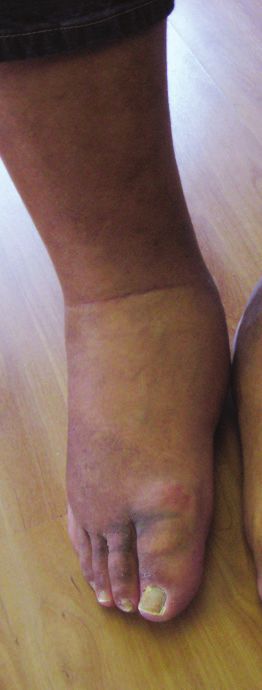
Important Facts about               An Alternative Scenario
CN/Diabetes                         The physician at the walk-in clinic was
• Patients with peripheral          aware of a rare condition called Charcot
  vascular disease are some-        foot. He was still concerned, however, that
  what protected from CN as         he would miss an infection in this patient
  vasodilation is part of the       with diabetes. He prescribed cephalexin
  pathogenesis.3,8                  but also advised the patient to remain
• Joints are the weak link in       non-weight bearing as if he had an
  the structure of the foot,        acute fracture. He sent him home with
  and therefore more sus-           an RCW, which was available in his phar-
  ceptible.                         macy, instructions to stop working, and
• The midfoot is most often         an urgent referral to a multidisciplinary
  affected as it is subjected       clinic that deals with diabetic foot issues.
  to more force during the          The following day the blood report was
  phases of walking. This is        obtained and indicated a normal CBC,
  the classic “rocker bottom”       creatinine, and CRP. His HbA1c was 9.6%.
  deformity. However, any
  joint of the foot can be
  affected.9                      but devices like a removable cast     imaging or laboratory. Early diag-
• Hyperglycemia causes            walker (RCW) are also commonly        nosis is important for leading to
  increased risk of ligament      used to offload the foot. Continue    early, appropriate management
  and tendon weakening.3,10       immobilization until lower            and prevention of further compli-
• Patients with diabetes          extremity edema and warmth            cations. CN should be suspected
  often have lower bone min-      resolve accompanied by evidence       in any patient over 40 years old
  eral densities, a factor for    of fracture consolidation.2,6         with peripheral neuropathy that
  development of CN. This is        In the subacute and chronic         presents with an acutely swol-
  more of an issue with type 1    stages, recommend devices             len foot with little or no known
  than type 2 diabetes.2          include the Charcot restraint
• Only a third of patients will
                                  orthotic walker (CROW) and the
  report trauma leading to                                               Key Points
                                  patellar tendon-bearing brace
  their symptoms.3
                                  (PTB). In the chronic stage, cus-      ✔✔ A high index of suspicion
                                  tom-made shoes are indicated.2,6           is required to correctly
                                    Surgery may be considered if             diagnosis CN in a timely
                                  conservative treatment fails to            manner.
                                  establish a plantigrade foot.          ✔✔ Develop an approach to the
                                    There is currently no evidence           red, hot, swollen foot with
                                  for the use of bisphosphonates             or without pain. Consider
                                                                             the possibility of infection,
                                  in managing CN.2,6
                                                                             which can co-exist with
                                                                             Charcot changes.
                                  Conclusion                             ✔✔ I f Charcot neuroarthrop-
                                  Charcot neuroarthropathy is a              athy is a concern, advise
                                  commonly missed diagnosis.                 the patient to remain
                                  It relies on an astute clinician,          non-weight bearing while
                                                                             appropriate referrals are
                                  because early physical findings
                                                                             arranged.
                                  can be subtle with little help from

46   Wound Care Canada                                                        Volume 17, Number 1 · Spring 2019
trauma. It is unclear why not all References 8. Strotman P, Reif T, Pinzur M. Charcot
arthropathy of the foot and ankle.
patients with diabetic neuroarth- 1. Schmidt B, Holmes C. Updates on
diabetic foot and Charcot osteopath- Foot Ankle Int. 2016;37(11):1255–63.
ropathy develop Charcot foot.
ic arthropathy. Curr Diabetes Rep. 9. Holmes C, Schmidt B, Munson M,
Inflammation seems to be at the 2018;18(10):74. Wrobel J. Charcot stage 0: A review
core of the process, and this may 2. Marmolejo V, Arnold J, Ponticello M, and considerations for making
be related to risk factors and gen- Andersen C. Charcot foot: Clinical the correct diagnosis early. J Clin
clues, diagnostic strategies, and treat- Endocrinol Diabetes. 2015;1(18).
etic predisposition.13
ment principles. Am Fam Physician. 10. Lipsky B, Berendt A, Cornia P, Pile J,
It can be difficult for health- 2018;97(9):594–99. Peters E, Armstrong D, et al. Infectious
care providers on the front line 3. Rogers L, Frykberg R, Armstrong D, Diseases Society of America clinical
to access the appropriate refer- Boulton A, Edmonds M, Ha Van G, et al. practice guideline for the diag-
The Charcot foot in diabetes. Diabetes nosis and treatment of diabetic
rals in a timely manner. Enlist
Care. 2011;34:2123–29. foot infections. Clin Infect Dis.
help from colleagues when 2012;54(12):e132–e173.
4. Dodd A, Daniels T. Charcot neuroar-
referral to a multidisciplinary thropathy of the foot and ankle. J 11. Holmes C, Schmidt B, Munson M,
team is not possible. An ortho- Bone Joint Surg Am. 2018;100:696– Wrobel JS. Charcot stage 0: A review
pedist, podiatrist or chiropodist 711. and consideratons for making the cor-
5. Mautone M, Naidoo P. Charcot rect diagnosis early. Clinicial Diabetes
should be able to help with and Endocrinology. 2015;1(18):12 pp.
neuroarthropathy: An imaging
these difficult cases. review. J Med Imaging Radiat Oncol. 12. Schmidt MB, Holmes CM. Updates on
Consider CN in the differen- 2015;59:395–402. diabetic foot and Charcot osteopathic
tial diagnosis of a red, swollen 6. La Fontaine J, Lavery L, Jude E. Current arthropathy. Current Diabetes Reports.
concepts of Charcot foot in diabetic 2018;18(74):11 pp.
foot to prevent the devastating
patients. The Foot. 2016;26:7–14. 13. Gökhan K, Birsel O, Güven MF, Öǧüt
consequence of a deformed foot
7. Jordan W. Neuritic manifestations in T. An overview of the Charcot foot
and long-term effects on quality diabetes mellitus. Arch Intern Med. pathophysiology. Diabet Foot Ankle.
of life, morbidity and mortality. 1936;57(2):307. 2013;4(10).
YOU HAVE THE POWER TO
1
DISRUPT AND DESTROY BIOFILM
2,3
TO ADVANCE HEALING
78% CHANCE
OF BIOFILM4
NON-HEALING WOUNDS
NEED MORE THAN SILVER™
Specifically developed to win the battle against biofilm,
MORE THAN SILVERTM technology harnesses the synergistic
power of three key components; ionic silver, a surfactant and a metal
chelating agent working together to deliver superior1 performance.*
1. Bowler PG, etVolume
al. Parsons, 17, Number
Wound 1 (2016)
Medicine 14 · Spring6–11. 2019
2. Metcalf DG et al. J. Wound Care 2016; Vol25, No3.
3. Metcalf DG, et al. Int Wound J 2017; 14: 203-213. 4. Malone M et al. 2017. JWC; 20-25.
+Wound Care=Canada 47
AQUACEL, Hydrofiber and MORE THAN SILVER are trademarks of ConvaTec Inc. ©2019 ConvaTec Inc. AP-019908-MM MORE THAN
SILVER™
* When compared to AQUACEL™ Ag Extra™ dressing and other silver-only competitor dressings: ACTICOAT™ 7 and SILVERCEL™ Non-Adherent dressings.A special invitation for members of
Wounds Canada
The Skin Spectrum Summit is a NEW LOCATION
Saturday April 6, 2019 | Toronto
landmark one-day event on Chestnut Conference Centre
ethnodermatology that takes place
Friday May 10, 2019 | Montreal
in three major Canadian cities. InterContinental Montreal
NEW LOCATION
Saturday June 1, 2019 Vancouver
Celebrating its fifth year, the Summit Coast Coal Harbour Hotel
is an educational congress of Gen-
eral Practitioners, Family Physicians, Toronto Chair:
nurses, nurse practitioners, and der- Dr. Gary Sibbald,
University of Toronto
matologists committed to providing
Montreal Chair:
better dermatological care for Dr. Danielle Marcoux,
Canada’s diverse and growing ethnic University of Montreal
communities, notably Fitzpatrick Vancouver Chair:
Dr. Jason Rivers,
Scale Skin Types III through VI. University of British Columbia
EDUCATIONAL OBJECTIVES
• Learn about the different skin conditions in Canada’s ethnic
• population including the manifestations of common dermatologi-
cal problems for persons with skin of colour
• Improve diagnostic practices of different skin conditions in the
growing ethnic population
• Learn strategies and tools to better manage patients with skin of
colour including potential unique challenges that they may face in
their treatment
Members of Wounds Canada
can register for only $79 + HST, regular $179 (a savings of $100)
Please use the discount code: WC20
For more information and to register please visit
www.skinspectrum.ca
or email us at health@chronicle.orgYou can also read