The effects of three different contrast agents (Gd-BOPTA, Gd-DTPA, and Gd-DOTA) on brachial plexus magnetic resonance imaging - Annals of ...
←
→
Page content transcription
If your browser does not render page correctly, please read the page content below
Original Article
                                                                                                                                    Page 1 of 8

The effects of three different contrast agents (Gd-BOPTA,
Gd-DTPA, and Gd-DOTA) on brachial plexus magnetic resonance
imaging
Xiaojing Zhang1,2#, Wensheng Wang3#, Tiefang Liu2, Yeqing Qi2, Lin Ma1,2
1
    Medical School of Chinese People’s Liberation Army, Beijing, China; 2Department of Radiology, The First Medical Centre, Chinese People’s
Liberation Army General Hospital, Beijing, China; 3Department of Radiology, Xiyuan Hospital, China Academy of Chinese Medical Sciences,
Beijing, China
Contributions: (I) Conception and design: X Zhang, L Ma; (II) Administrative support: W Wang; (III) Provision of study materials or patients:
X Zhang, T Liu; (IV) Collection and assembly of data: X Zhang, T Liu, Y Qi; (V) Data analysis and interpretation: X Zhang, L Ma; (VI) Manuscript
writing: All authors; (VII) Final approval of manuscript: All authors.
#
These authors contributed equally to this study.
Correspondence to: Lin Ma. Department of Radiology, The First Medical Centre, Chinese People’s Liberation Army General Hospital, Beijing 100853,
China. Email: cjr.malin@vip.163.com.

                  Background: MRI is very important for guiding the diagnosis and treatment of brachial plexus diseases.
                  The most used type of MRI brachial plexus imaging is the 3D Short Term Inversion Recovery (STIR)
                  sequence with contrast agent. This study aimed to investigate the effect of three contrast agents; gadobenate
                  dimeglumine (Gd-BOPTA), gadopentetate dimeglumine (Gd-DTPA), and Gadoteric Acid Meglumine (Gd-
                  DOTA) on brachial plexus magnetic resonance imaging (MRI).
                  Methods: We recruited 60 patients with suspected brachial plexus injury randomly into three groups. MRI
                  images were obtained from each patient. Prior to scanning, the first group was injected with GD-BOPTA,
                  the second group with Gd-DTPA, and the third with Gd-DOTA. The amount of contrast agent was
                  0.1 mmol/kg according to the weight of each patient, the injection rate was 1.5 mL/s, and 20 mL saline was
                  injected at the same rate with a high-pressure injector. Immediately after the injection of contrast agent and
                  saline, a 3D Sampling perfection with application optimized contrasts using different flip angle evolutions
                  (SPACE) STIR sequence was used for scanning. The Signal Intensity (SI) and Standard Deviation (SD) of
                  Maximal intensity projection (MIP) images for regions outside the anatomy (ROI background) with area
                  of 17 mm2 on both sides of the C6 peripheral nerves (ROI nerve), and tissue adjacent to the peripheral
                  nerves (ROI tissue) were obtained. Signal to noise ratio (SNR) and contrast to noise ratio (CNR) were then
                  calculated.
                  Results: The SNR was 40.66±25.27, 34.65±14.86, and 44.63±30.79 for Gd-BOPTA, Gd-DTPA, and Gd-
                  DOTA, respectively and the CNR was 20.24±15.17, 16.07±7.50, and 20.84±15.53 for Gd-BOPTA, Gd-
                  DTPA, and Gd-DOTA, respectively. In addition, there was no statistical difference in the SNR or CNR
                  of brachial plexus nerves using the three contrast agents to enhance the 3D SPACE sequence χ2=1.877,
                  P=0.391>0.05 and χ2=1.717, P=0.424, respectively.
                  Conclusions: There were no significant differences in the efficacy of three contrast agents in imaging the
                  brachial plexus.

                  Keywords: Magnetic resonance imaging (MRI); brachial plexus; contrast; signal to noise ratio (SNR); contrast to
                  noise ratio (CNR); Gd-BOPTA; Gd-DTPA; Gd-DOTA; 3D SPACE STIR

                  Submitted Nov 26, 2020. Accepted for publication Jan 29, 2021.
                  doi: 10.21037/atm-21-348
                  View this article at: http://dx.doi.org/10.21037/atm-21-348

© Annals of Translational Medicine. All rights reserved.                    Ann Transl Med 2021;9(4):344 | http://dx.doi.org/10.21037/atm-21-348
Page 2 of 8 Zhang et al. Contrast agents on brachial plexus MR Imaging
Introduction Methods
Attaining high-quality images of the brachial plexus has Participants
always been problematic for clinicians (1) and several
We enrolled 60 patients with suspected brachial plexus injury
different modalities have been used to address this
who were treated in our hospital from July 2016 to January
challenge in daily practice. Although it is safe, ultrasound
2017. The mean age of patients was 47.33±15.05 years
imaging is greatly affected by the operator, and by bone
and there were 32 males and 28 females. Using a random
and lung movement, so the visibility and quality of
number table, the 60 patients were randomly divided into
imaging is poor (2,3). Computed tomography (CT) lacks
three groups of 20. The first group was composed of 9 men
the contrast resolution required to adequately evaluate
and 11 women, the second group of 11 men and 9 women
soft tissues and the brachial plexus is iso-signal compared
and the third group of 12 men and 8 women. The inclusion
to the surrounding muscles and vasculature in images.
criteria of patients were as follows: (I) age ≥18 years; (II)
Sclerotic artifacts from adjacent bones can sometimes
interfere with imaging and reduce CT image quality (4) and patients with a glomerular filtration rate (GFR)Annals of Translational Medicine, Vol 9, No 4 February 2021 Page 3 of 8
Table 1 Scanning sequence of the brachial plexus TE =11 ms, TR =672 ms, slice thickness =4 mm, spacing
Serial =0 mm, bandwidth =347 Hz/Px, number of excitations =1,
Scanning sequence Position
number acquisition time = 3 minutes and 58 seconds. The sequence
1 Sag T2 Sagittal position T2 used was 3D SPACE STIR and the scanning parameters
were as follows: coronal position, FOV =32 cm × 32 cm ×
2 Sag T1 Sagittal position T1
6 cm, matrix =320×320, TE =131 ms, TR =3,500 ms, flipping
3 Sag T2 fs dixon Sagittal position T2 fat pressure response time TI =220 ms, slice thickness =1 mm, bandwidth
4 Tra T2 msma Horizontal axis position T2 =625 Hz/pixel, acquisition time = 8 minutes 33 seconds. The
5 3D SPACE STIR Coronal position 3D contrast medium dosage of the three groups was used in
SPACE STIR accordance with the principle of 0.1 mmol/kg. The contrast
medium was injected through the forearm cubital vein
6 3D SPACE STIR + C Coronal position 3D SPACE
STIR enhanced through a high-pressure syringe (Ulrich, Germany), and the
injection speed was 1.5 mL/s. An injection of 20 mL of saline
7 Sag T1 + C Sagittal axis position
T1 enhanced fat pressure
at a rate to flush the pipeline was used, and the 3D SPACE
STIR sequence scan was conducted immediately after the
9 Cor T1 + C Coronal position T1 enhanced
injection of contrast agent and saline. The first group of
fat pressure
patients received Gd-BOPTA (Bracco, Italy), the second
10 Tra T1 fs dixon + C Transverse position group Gd-DTPA (Bayer, Germany), and the third group Gd-
T1 enhanced fat pressure
DOTA (Jiangsu Hengrui Pharmaceutical Co., Ltd., China).
SPACE, Sampling Perfection with Application-optimized
Contrasts by using different flip angle Evolutions; STIR, Short
Term Inversion Recovery. Image processing and analysis
Images obtained were transmitted to the Siemens
echo time (TE) =107 ms, repeat time (TR) =3,500 ms, slice workstation (Siemens Numaris/4 Syngo MR B17) and
thickness =3 mm, spacing =0.5 mm, bandwidth =260 Hz/Px, the maximum intensity projection (MIP) was used to
reconstruct the three-dimensional image of the brachial
averages =1, acquisition time = 1 minute and 38 seconds.
plexus. Two senior radiologists measured the average signal
Sag T1: sagittal position, FOV = 26 cm × 26 cm, matrix
value (SI) and mean square deviation (SD) of the left and
=320×256, TE =9 ms, TR =600 ms, slice thickness =3 mm,
right C6 nerve and surrounding soft tissues. The region of
spacing =0.5 mm, bandwidth =260 Hz/Px, averages =1,
interest (ROI) size was 21 pixels. To calculate the average
acquisition time = 1 minute 41 seconds. Sag T2 fs dixon:
value, we tried to avoid bones, air, and other locations
sagittal position, FOV =26 cm × 26 cm, matrix =320×240,
during the measurement. After the ROI was placed, the
TE =112 ms, TR =4,300 ms, slice thickness =3 mm, spacing
system software automatically displayed the average SI
=0.5 mm, bandwidth =347 Hz/Px, number of excitations
value and the mean SD. The signal to noise ratio (SNR)
averages =1, acquisition time = 1 minute 39 seconds. Tra T2
and contrast to noise ratio (CNR) were calculated according
msma: transverse position, FOV =20 cm × 20 cm, matrix to Eqs. [1] and [2], respectively.
=320×256, TE =112 ms, TR = 6,033 ms, slice thickness =
4 mm, spacing =0 mm, bandwidth = 284 Hz/Px, averages SNR = SI nerve / SD background [1]
=2, acquisition time = 2 minutes and 08 seconds. Sag T1+C: CNR = (SI nerve - SD tissue)/SD tissue [2]
sagittal position, FOV = 26 cm × 26 cm, matrix =320×256,
TE =10 ms, TR =650 ms, slice thickness =3 mm, spacing =
0.5 mm, bandwidth =347 Hz/Px, number of excitations Statistical analysis
averages = 1, acquisition time = 1 minute and 50 seconds. SPSS20.0 software (IBM, Chicago, USA) was used for
Cor T1+C: coronal position, FOV =26 cm × 26 cm, matrix statistical analysis. The Kolmogorov-Smirnov test was
=320×256, TE =10 ms, TR =650 ms, slice thickness =3 mm, used to determine whether data conformed to a normal
spacing =0.5 mm, bandwidth= 347 Hz/Px, averages =1, distribution. If so, one-way Anova was used and if not, the
acquisition time= 1 minute 50 seconds. Tra T1 fs dixon + C: non-parametric Kruskal-Wallis (KW) test was used, and
transverse position, FOV =20 cm × 20 cm, matrix =320×256, PPage 4 of 8 Zhang et al. Contrast agents on brachial plexus MR Imaging
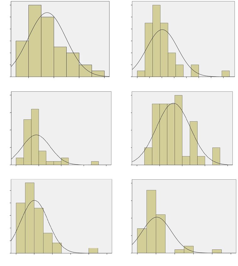
Results did not follow a normal distribution (Table 2 and Figure 1).
Normal distribution of the SNR and CNR of the brachial
plexus using three different contrast agents Comparison of the SNR and CNR of the brachial plexus
using three different contrast agents
Both the SNR (P=0.007Annals of Translational Medicine, Vol 9, No 4 February 2021 Page 5 of 8
Table 3 SNR and CNR of brachial plexus using different contrast States, more than 10 million patients undergo enhanced
agents magnetic resonance examination each year, accounting for
Contrast agents SNR CNR 40% to 50% of all magnetic resonance examinations (9) and
Gadobeme meglumine 40.66±25.27 20.24±15.17 both methods are becoming increasingly common in China.
Enhanced magnetic resonance examinations are based on
Gadopentetate meglumine 34.65±14.86 16.07±7.50
the characteristics of the high relaxation rate of gadolinium
Gadoterate meglumine 44.63±30.79 20.84±15.53 contrast agents. After the contrast agent is intravenously
χ2 value 1.877 1.717 injected it can not only highlight the shape and boundary of
P value 0.391 0.424 lesions but can also help clinically clarify disease based on
the enhanced characteristics of the lesion, greatly benefiting
SNR, signal to noise ratio; CNR, contrast to noise ratio.
differential diagnosis.
Since MRI contrast agents were first used in 1988,
more than 300 million examinations have taken place and
200
22 currently over 30 million are conducted annually worldwide.
On July 21, 2017, the European Medicines Agency (EMA)
150 formally banned the use of some linear gadolinium contrast
15
agents, namely gadopentetate meglumine, gadolinium
diamine, and gadolinium fuseamide. It also restricted the use
27 2
100 8 35 of gadolinium meglumine to liver imaging only. However,
18
one month earlier (May 22, 2017) the U.S. Food and
Drug Administration (FDA) issued a statement confirming
50
that the gadolinium deposition in the brain caused by the
current MRI gadolinium enhancement was not harmful and
did not restrict its use. The FDA statement confirmed it
0
would continue to evaluate the safety of gadolinium contrast
Gd-DTPA SNR Gd-BOPTA SNR Gd-DOTA SNR
agents, and the FDA National Toxicology Research Center
Figure 2 SNR box plot of the brachial plexus after using three was conducting further research.
contrast agents Gd-DTPA, Gd-BOPTA, and Gd-DOTA. *, The heavy metal gadolinium binds to a ligand (chelate).
Outliers. SNR, signal to noise ratio. Free Gd3+ is highly toxic and can replace the Ca + ions
on many peptides and biological enzymes in the human
body, thereby inhibiting their functions. To reduce its
100 toxicity, researchers have found ways to bind free Gd3+
15 22 with various ligands to form stable chelates, which are not
80 easily decomposed, thereby reducing toxicity. Gadolinium
contrast agents can be divided into two types, “linear”
60 and “large ring” based on their structure. Linear agents
27
18 2 were the first used magnetic resonance contrast agent.
35
Theoretically, the ligand in linear agents is “open ring” and
40 4
19 is easy to dissociate. This structure easily separates a certain
coordination site in the chelate, and further leads to the
20
sequential separation of other coordination sites, releasing
free gadolinium harmful Gd3+. In contrast, in macrocyclic
0
agents, Gd3+ is “fixed” around the ligand making this
Gd-DTPA SNR Gd-BOPTA SNR Gd-DOTA SNR structure highly stable and not easy to free from the
Figure 3 CNR box plot of the brachial plexus after using three chelating ring structure.
contrast agents Gd-DTPA, Gd-BOPTA, and Gd-DOTA. *, In this study, three representative magnetic resonance
Outliers. CNR, contrast to noise ratio. contrast agents were selected from several commonly used
© Annals of Translational Medicine. All rights reserved. Ann Transl Med 2021;9(4):344 | http://dx.doi.org/10.21037/atm-21-348Page 6 of 8 Zhang et al. Contrast agents on brachial plexus MR Imaging
A B C
Figure 4 The use of the three contrast agents on the enhanced 3D SPACE sequence. (A) A 39-year-old female patient who presented with a
left clavicle fracture and brachial plexus injury. The magnetic resonance contrast agent used during the examination was Gd-BOPTA. (B) A
49-year-old female patient with peripheral neuropathy and the magnetic resonance contrast agent used was gadopentetate meglumine. (C) A
29-year-old male patient presenting with upper limb numbness and the MRI contrast used was Gd-DOTA. It can be seen from the images
that the display of the brachial plexus by the three contrast agents is similar.
in our daily work. Gd-BOPTA is a linear contrast agent ability of 3D SPACE STIR sequencing to display the
with high gadolinium concentration and relaxation rate, brachial plexus, by significantly shortening the T1 relaxation
due to its non-specific distribution of the inter-cells space time of tissue and the T2 relaxation time will be shortened
and the characteristics of specific uptake by hepatocytes. accordingly (20-23). Due to the blood nerve barrier, the
When Gd-BOPTA is injected intravenously, 95% of the contrast agent does not easily enter the nerve sheath, so the
agent is metabolized out of the body by the kidneys and 5% normal magnetic resonance signal of the brachial plexus is
is excreted through the biliary tract (10), as it holds similar only slightly affected. Choosing the appropriate T1 imaging
characteristics, Gd-BOPTA can also be used as a specific parameter will also suppress the lymph nodes, fat, small
contrast agent for the liver imaging. Both Gd-BOPTA and blood vessels and muscle surrounding the brachial plexus
Gd-DTPA are ionic linear contrast agents excreted through improving its contrast with surrounding tissues.
the kidneys. Gd-DOTA is cyclic contrast agent. There The SNR and CNR of brachial plexus images after using
are reports that the stability of gadolinium-based contrast three contrast agents were compared, and the difference
agents can be assigned in the following order: cyclic was not statistically significant. Some scholars have reported
contrast agent > ionic linear contrast agent > nonionic linear that when undertaking MR brain examinations using the
contrast agent (11). While the phenomenon of gadolinium same dosage condition (24), gadobeme meglumine not only
deposition is relatively rare. Studies have shown that their made brain metastases clearer and more obvious, but also
use may result in their deposit in various parts of the human increased the detection rate of occult lesions and improved
body. As early as 2004, some scholars found that different clinical treatment program in comparison to gadopentetate
gadolinium contrast agents had different types of deposition meglumine. However, we found no advantages to applying
in bones (12) and the brain, especially in the globus pallidus, gadolinium meglumine to examine the brachial plexus,
thalamus, and dentate nucleus (13). Numerous studies when compared with gadopentetate meglumine and
have shown that non-ionic linear contrast agents have poor gadoterate meglumine although this may be related to the
stability and are more likely to cause gadolinium to deposit characteristics of the 3D SPACE STIR sequence.
in the skull (14-18). There are several limitations to this study. Firstly,
The combined use of contrast agents with 3D-T2- the sample size was small; secondly, only patients with
STIR imaging technology improves the evaluation of suspected brachial plexus injury were included; thirdly, the
arm plexus anatomy and pathology and contributes to the SNR and CNR of only the bilateral 6th cervical nerves were
future understanding of brachial plexus neuropathy and the measured; and finally, there is a lack of comparison between
development of surgical planning for surgery (19). machines from different manufacturers and different field
The injection of contrast agent can also improve the strengths. Further studies with larger sample sizes, with
© Annals of Translational Medicine. All rights reserved. Ann Transl Med 2021;9(4):344 | http://dx.doi.org/10.21037/atm-21-348Annals of Translational Medicine, Vol 9, No 4 February 2021 Page 7 of 8
clearly defined pathologies and evaluating a broader range License (CC BY-NC-ND 4.0), which permits the non-
of brachial plexus nerves using a range of imaging devices commercial replication and distribution of the article with
and parameters are required to confirm our results. the strict proviso that no changes or edits are made and the
original work is properly cited (including links to both the
formal publication through the relevant DOI and the license).
Conclusions
See: https://creativecommons.org/licenses/by-nc-nd/4.0/.
There was no significant difference in the efficacy of the
three contrast agents, Gd-BOPTA, Gd-DTPA and Gd-
References
DOTA to evaluate the brachial plexus using enhanced
MRI and no adverse reactions were associated with the 1. Rehman I, Chokshi FH, Khosa F. MR imaging of the
use of any agent. As the molecular structures of the three brachial plexus. Clin Neuroradiol 2014;24:207-16.
contrast agents are different, the stability of the molecular 2. Tagliafico A, Succio G, Serafini G, et al. Diagnostic
structure of gadolinium meglumine acid is better than that performance of ultrasound in patients with suspected
of the other two gadolinium contrast agents. Based on these brachial plexus lesions in adults: a multicenter retrospective
findings, we recommend the use of gadolinium meglumine study with MRI, surgical findings and clinical follow-up as
acid in enhanced MRI imaging. reference standard. Skeletal Radiol 2013;42:371-6.
3. Martinoli C, Gandolfo N, Perez MM, et al. Brachial plexus
and nerves about the shoulder. Semin Musculoskeletal
Acknowledgments
Radiol 2010;l5:523-46.
Funding: None. 4. Yoshikawa T, Hayashi N, Yamamoto S, et al. Brachial
plexus injury: clinical manifestations, conventional imaging
findings, and the latest imaging techniques. Radiographics
Footnote
2006;26:S133-S143.
Reporting Checklist: The authors have completed the MDAR 5. van Es HW, Bollen TL, van Heesewijk HP. MRI of
reporting checklist. Available at http://dx.doi.org/10.21037/ the brachial plexus: a pictorial review. Eur J Radiol
atm-21-348 2010;74:391-402.
6. Saifuddin A. Imaging tumours of the brachial plexus.
Data Sharing Statement: Available at http://dx.doi. Skeletal Radiol 2003;32:375-87.
org/10.21037/atm-21-348 7. Amrami KK, Port JD. Imaging the brachial plexus. Hand
Clin 2005;21:25-37.
Conflicts of Interest: All authors have completed the ICMJE 8. Ramalho J, Semelka RC, Ramalho M, et al. Gadolinium-
uniform disclosure form (available at http://dx.doi. based contrast agent accumulation and toxicity: an update.
org/10.21037/atm-21-348). The authors have no conflicts Am J Neuroradiol 2016;37:1192-8.
of interest to declare. 9. Zhou Z, Lu ZR. Gadolinium-based contrast agents for
magnetic resonance cancer imaging. Wiley Interdiscip Rev
Ethical Statement: The authors are accountable for all Nanomed Nanobiotechnol 2013;5:1-18.
aspects of the work in ensuring that questions related 10. Pasquini L, Napolitano A, Visconti E, et al. Gadolinium-
to the accuracy or integrity of any part of the work are Based Contrast Agent-Related Toxicities. CNS Drugs
appropriately investigated and resolved. This study was 2018;32:229-240.
approved by the Ethics Committee of the PLA General 11. Bhargava R, Hahn G, Hirsch W, et al. Contrast-enhanced
Hospital and all patients provided written informed magnetic resonance imaging in pediatric patients: review
consent. All procedures performed in this study involving and recommendations for current practice. Magn Reson
human participants were in accordance with the Declaration Insights 2013;6:95-111.
of Helsinki (as revised in 2013). 12. Wiginton CD, Kelly B, Oto A, et al. Gadolinium-based
contrast exposure, nephrogenic systemic fibrosis, and
Open Access Statement: This is an Open Access article gadolinium detection in tissue. AJR Am J Roentgenol
distributed in accordance with the Creative Commons 2008;190:1060-8.
Attribution-NonCommercial-NoDerivs 4.0 International 13. Zhang Y, Cao Y, Shih GL, et al. Extent of signal
© Annals of Translational Medicine. All rights reserved. Ann Transl Med 2021;9(4):344 | http://dx.doi.org/10.21037/atm-21-348Page 8 of 8 Zhang et al. Contrast agents on brachial plexus MR Imaging
hyperintensity on unenhanced T1-weighted brain 2015;276:836-44.
MR images after more than 35 administrations of 19. Chen WC, Tsai YH, Weng HH, et al. Value of
linear gadolinium-based contrast agents. Radiology enhancement technique in 3D-T2-STIR images of the
2017;282:516-25. brachial plexus. J Comput Assist Tomogr 2014;38:335-9.
14. Quattrocchi CC, Mallio CA, Errante Y, et al. Gadodiamide 20. Veronesi BA, Rodrigues MB, Sambuy MTC, et al. Use of
and dentate nucleus T1 hyperintensity in patients with magnetic resonance imaging to diagnose brachial plexus
meningioma evaluated by multiple follow-up contrast- injuries. Acta Ortop Bras 2018;26:131-4.
enhanced magnetic resonance examinations with no 21. Tagliafico A, Bignotti B, Tagliafico G, et al. Usefulness of
systemic interval therapy. Invest Radiol 2015;50:470-2. IDEAL T2 imaging for homogeneous fat suppression and
15. Kanda T, Matsuda M, Oba H, et al. High T1 signal reducing susceptibility artefacts in brachial plexus MRI at
intensity in dentate nucleus after multiple injections of 3.0 T. Radiol Med 2016;121:45-53.
linear gadolinium chelates response. Radiology 2015;1:617. 22. Tomura N, Saginoya T, Kokubun M, et al. T2-weighted
16. Weberling LD, Kieslich PJ, Kickingereder P, et al. IDEAL fast spin echo imaging of the brachial plexus:
Increased signal intensity in the dentate nucleus on comparison with STIR. Acta Radiol 2015;56:1242-7.
unenhanced T1-weighted images after gadobenate 23. Cejas C, Rollán C, Michelin G, et al. High resolution
dimeglumine administration. Invest Radiol 2015;50:743-8. neurography of the brachial plexus by 3 Tesla magnetic
17. Kanda T, Osawa M, Oba H, et al. High signal intensity in resonance imaging. Radiologia 2016;58:88-100.
dentate nucleus on unenhanced T1-weighted MR images: 24. Colosimo C, Demaerel P, Tortori-Donati P, et al.
association with linear versus macrocyclic gadolinium Comparison of gadobenate dimeglumine (Gd-BOPTA)
chelate administration. Radiology 2015;275:803-9. with gadopentetate dimeglumine (Gd-DTPA) for
18. Ramalho J, Castillo M, AlObaidy M, et al. High signal enhanced MR imaging of brain and spine tumours in
intensity in globus pallidus and dentate nucleus on children. Pediatr Radiol 2005;35:501-10.
unenhanced T1-weighted MR images: evaluation of
two linear gadolinium-based contrast agents. Radiology (English Language Editor: B. Draper)
Cite this article as: Zhang X, Wang W, Liu T, Qi Y, Ma L.
The effects of three different contrast agents (Gd-BOPTA, Gd-
DTPA, and Gd-DOTA) on brachial plexus magnetic resonance
imaging. Ann Transl Med 2021;9(4):344. doi: 10.21037/atm-21-
348
© Annals of Translational Medicine. All rights reserved. Ann Transl Med 2021;9(4):344 | http://dx.doi.org/10.21037/atm-21-348You can also read