Extensive skin necrosis after periprosthetic knee infection: a case that highlights the possibility of complications induced by ...
←
→
Page content transcription
If your browser does not render page correctly, please read the page content below

JBJI

                                                                                                                                                      Case report
J. Bone Joint Infect., 6, 235–240, 2021

                                                                                                                                        Open Access
https://doi.org/10.5194/jbji-6-235-2021
© Author(s) 2021. This work is distributed under                                                                    Journal of Bone
                                                                                                                  and Joint Infection
the Creative Commons Attribution 4.0 License.

           Extensive skin necrosis after periprosthetic knee
           infection: a case that highlights the possibility of
        complications induced by low-molecular-weight heparin
        Hélder Fonte1 , André Carvalho1 , João Rosa1 , Cláudia Pereira2,3 , Alexandre Pereira1 , Ricardo Sousa1,3 ,
                          and further members of the Porto Bone and Joint Infection Unit+
                   1 Departmentof Orthopaedics, Centro Hospitalar Universitário do Porto, Porto, Portugal
                 2 Department
                            of Internal Medicine, Centro Hospitalar Universitário do Porto, Porto, Portugal
           3 GRIP (Porto Bone and Joint Infection Unit), Centro Hospitalar Universitário do Porto, Porto, Portugal
                                   + A full list of authors appears at the end of the paper.

                                 Correspondence: Hélder Fonte (helderfonte14@gmail.com)

                 Received: 29 March 2021 – Revised: 8 June 2021 – Accepted: 17 June 2021 – Published: 28 June 2021

       Abstract. We describe a case of a patient with atrial fibrillation, anticoagulated with dabigatran, that developed
       severe knee skin necrosis in the setting of an acute periprosthetic knee infection, after initiating low-molecular-
       weight heparin. A wide range of etiology hypotheses was discussed within a multidisciplinary team. The complex
       approach consisted of treating the underlying infection, multiple types of soft-tissue management, and stopping
       enoxaparin.

1   Introduction                                                    and Raut, 2011). The authors describe a case of acute knee
                                                                    periprosthetic joint infection (PJI) complicated with severe
Severe wound complications following total knee arthro-             skin necrosis in an anticoagulated patient that stopped dabi-
plasty (TKA), though uncommon, are of major importance.             gatran before the surgery and started enoxaparin on the
Clinical presentation ranges from wound problems and su-            post-operative period. PJI treatment required a two-stage ap-
perficial infections to full-thickness skin necrosis (Galat,        proach with implant removal, medial gastrocnemius muscle
2009). One of the possible causes of skin necrosis is the ad-       flap, negative pressure dressing, hyperbaric oxygen cham-
ministration of low-molecular-weight heparin. This compli-          ber, and split-thickness skin graft. After laborious soft-tissue
cation, occurring at a distance from the injection site, has        management, the patient ultimately underwent a successful
been increasingly reported and more frequently observed             second-stage procedure, being able to walk autonomously
with subcutaneous therapy, though it is an extremely rare           and presenting a good knee range of motion, with complete
event. Female gender, high BMI (> 25 kg m−2 ), and long             wound healing.
duration of heparin therapy have been identified to be the
risk factors; however, little is known about the true incidence
(Schindewolf et al., 2009). These mechanisms are consid-            2   Report of the case
ered: immunologically mediated either via thrombosis result-
ing from heparin-induced immune aggregation of platelets            A 78-year-old woman, with a history of atrial fibrillation and
(heparin-induced thrombocytopenia syndrome, HIT) or a for-          hypertension, underwent left TKA for primary osteoarthri-
mation of antigen–antibody complexes in cutaneous blood             tis (Fig. 1a). Chronic medication included dabigatran,
vessels (type III hypersensitivity syndrome) (Handschin et          olmesartan–hydrochlorothiazide, and amlodipine. There was
al., 2005). Moreover, to the best of our knowledge, there is        no history of renal failure, diabetes mellitus, autoimmune
only one case reported in the English literature about tis-         disease, peripheral arterial or venous disease, or other rele-
sue necrosis occurring after knee arthroplasty (Karmegam            vant comorbidities (CHA2DS2-VASc = 4 points). Dabiga-

Published by Copernicus Publications on behalf of EBJIS and MSIS.
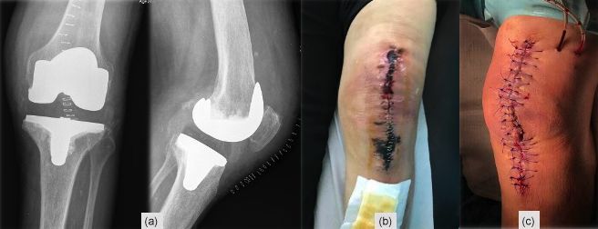
236                                                   H. Fonte et al.: Extensive skin necrosis after periprosthetic knee infection

Figure 1. (a) Post-operative TKA X-ray, (b) skin necrosis on the 18th post-operative day, and (c) surgical wound immediately after debride-
ment and antibiotics with implant retention (DAIR).

Figure 2. (a) Skin necrosis on the fifth day after DAIR, (b) X-ray after the implant removal and application of an antibiotic-loaded cement
spacer, and (c) medial gastrocnemius muscle flap for wound coverage.

tran was stopped 3 d before surgery. Throughout the pro-                plant an antibiotic-loaded handmade mobile spacer (with 4 g
cedure, a tourniquet was used (45 min with a pressure of                vancomycin and 2 g meropenem per bag of bone cement)
300 mm Hg), and a suction drain was in place for 24 h. Dur-             and perform a medial gastrocnemius muscle flap for wound
ing the post-operative period, therapeutic enoxaparin (60 mg            coverage (Fig. 2b–c). Microbiological samples, including
BID) was started. The patient was discharged on day five                implant sonication, isolated the same microorganisms, but
with a clean wound and recommendation to switch back to                 given the unfavourable clinical course, broad-spectrum an-
oral dabigatran. On the 18th post-operative day, a significant          tibiotics were maintained for 6 weeks.
wound skin necrosis was found with slight concurrent wound                 During the next weeks, an extensive superficial skin necro-
leakage (Fig. 1b), and despite C-reactive protein (CRP) levels          sis developed around the knee, extending to the lower leg
of 15.8 mg L−1 , there was a high suspicion of an underlying            around the incision made to harvest the muscle flap (Fig. 3).
acute periprosthetic joint infection. The patient was switched          An arteriography was performed, and occlusive arterial dis-
back to enoxaparin and was scheduled for surgery within                 ease was definitively ruled out. Skin biopsy revealed mi-
2 d (Fig. 1c). After surgical debridement with implant re-              crovascular thrombotic phenomena and ischemia without
tention (DAIR), broad-spectrum IV antibiotics were started              vasculitis. Complete blood count (CBC) revealed persistently
(vancomycin and piperacillin–tazobactam). Despite these ef-             normal platelet values and slight post-operative anaemia.
forts, skin necrosis progressed on the distal part of the wound         Erythrocyte sedimentation rate (ESR) and C-reactive protein
(Fig. 2a). Microbiological samples confirmed infection with             (CRP) levels were elevated but ran a favourable downward
Enterococcus faecalis and Staphylococcus haemolyticus in                trend. All coagulation parameters were normal, and autoim-
multiple samples. On the fifth day after DAIR, skin necro-              mune disease assessment was negative. A haematology con-
sis at the distal part of the wound progressed (Fig. 2a),               sult was also requested, thinking of low-molecular-weight
and a decision was made to remove the prosthesis and im-                heparin (LMWH)-induced skin necrosis, but given the lack

J. Bone Joint Infect., 6, 235–240, 2021                                                       https://doi.org/10.5194/jbji-6-235-2021
H. Fonte et al.: Extensive skin necrosis after periprosthetic knee infection                                                          237

Figure 3. (a–d) Skin necrosis progression after TKA revision, extending to the lower leg around the incision made to harvest the muscle
flap.

Figure 4. (a) A period of 3 weeks after revision surgery, new debridement of all necrotic areas, interruption of enoxaparin, negative pres-
sure dressings, and hyperbaric oxygen chamber treatment were performed. (b–c) Progressive healing of the wound. (d) New debridement.
(e) Split-thickness skin graft of the thigh for wound coverage.

of thrombocytopenia and atypical clinical presentation, a de-              Second-stage surgery was consecutively postponed, but
cision was made not to pursue more specific testing regard-             when satisfactory soft tissues were present (Fig. 6a) and af-
less of our suspicion.                                                  ter an adequate discussion with the patient, a decision to
   After 3 weeks, surgical debridement of the necrotic tissues          go ahead with the second stage was taken 1 year after the
was undertaken. With the lack of an alternative diagnosis, a            original surgery. Dabigatran was discontinued for 3 d before,
decision was made to discontinue enoxaparin and reinstate               without enoxaparin bridging, and immediately resumed af-
dabigatran. A negative-pressure dressing was applied, and               ter surgery. Intraoperatively, multiple deep tissue samples
complementary treatment on a hyperbaric oxygen chamber                  were collected and showed no bacterial growth. A period of
was also initiated. Necrosis stopped progressing (Fig. 4a–d),           2 years after the second stage, the patient is infection-free,
and eventually a split-thickness skin graft of the thigh for            with a painless, well-functioning knee arthroplasty, and no
wound coverage was performed (Fig. 4e).                                 systemic disorder was diagnosed (Fig. 6).
   During the outpatient follow-up, soft tissues showed slow
but progressive improvements and eventually healed com-                 3     Discussion
pletely. A few weeks after hospital discharge, an ulcer on
the lateral aspect of the leg (unrelated to previous inci-              Extensive skin necrosis can originate in a wide range of eti-
sions) developed and progressed (Fig. 5a–d). A biopsy was               ologies, detailed in the following subsections.
again performed that revealed unspecific findings, suggest-
ing an infected venous leg ulcer, and a 2-week course of oral
                                                                        3.1    Drug-induced skin necrosis
amoxicillin–clavulanate was instituted with excellent clini-
cal response (Fig. 5e–h). A new immunological assessment                Skin necrosis in response to anticoagulant treatment is a rare
showed positive ANA with a low value of anti-dsDNA, with-               adverse reaction and is even more uncommon in patients
out complement consumption and positive IgG anti-beta-2-                treated with heparin compared to oral anticoagulants (0.01 %
glycoprotein I (anti-cardiolipin and lupus anticoagulant re-            of patients) (Estébanez et al., 2019). There are some patho-
mained negative), but after 12 weeks they were back to nor-             physiological mechanisms that explain LMWH-induced skin
mal.                                                                    necrosis: first, it was found to be associated with an estab-
                                                                        lished heparin-induced thrombocytopenia (HIT) syndrome.

https://doi.org/10.5194/jbji-6-235-2021                                                       J. Bone Joint Infect., 6, 235–240, 2021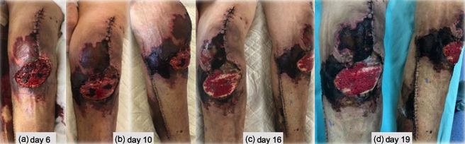
238                                                    H. Fonte et al.: Extensive skin necrosis after periprosthetic knee infection

Figure 5. (a–d) Lateral leg wound evolution and (e–h) response after institution of a 2-week course of oral amoxicillin–clavulanate.

Figure 6. (a) Skin before the second-stage surgery, (b) post-operative X-ray, (c) skin fully healed, (d1–d2) left knee range of motion in the
last evaluation, 0–115◦ .

J. Bone Joint Infect., 6, 235–240, 2021                                                         https://doi.org/10.5194/jbji-6-235-2021
H. Fonte et al.: Extensive skin necrosis after periprosthetic knee infection                                                239

Here, an antibody–platelet–heparin complex leads to an acti-             findings (cardinal finding) (Dahl et al., 2002). More-
vation of the coagulation cascade that results in microthrom-            over, the biopsy did not reveal any suggestive features.
bosis of dermal vessels and skin necrosis. Second, vasculitis
of dermal vessels induced by a type III hypersensitivity reac-      3. Although skin necrosis of the distal part of the wound
tion to the LMWH (Arthus phenomenon, with deposit of im-               after DAIR may have been caused by wound closure
munocomplexes on the endothelial structure) has been pro-              with inadequate skin tension, that would not explain the
posed as an alternative pathomechanism (Handschin et al.,              skin necrosis after the original procedure or after the
2005). It usually presents close to the injection site, although       first stage and muscle flap.
it has been described at distance on rarer occasions (Balestra
et al., 1994; Estébanez et al., 2019). To the best of our knowl-   3.3   Arterial disease or embolic phenomena
edge, there is only one reported case in the English litera-       Although skin necrosis is the final result of superficial mi-
ture about tissue necrosis occurring after knee arthroplasty       crovasculature occlusion, we worried that some kind of ma-
(Karmegam and Raut, 2011). Despite the atypical location of        jor arterial disease could be responsible for the exuberant
the necrotic lesions and the lack of classical thrombocytope-      clinical presentation. Blood vessel obstruction due to em-
nia and heparin–platelet factor 4 antibodies, we believe this      bolic phenomena was also a possible cause. The reasons that
is a case of LMWH-induced skin necrosis for the following          we believe skin necrosis was not caused by underlying arte-
reasons:                                                           rial disease or embolic phenomena are as follows:
 1. Skin necrosis stopped progressing after we decided to           1. The patient underwent a completely normal angiogra-
    discontinue enoxaparin and switch back to dabigatran.              phy.
 2. Although the mechanism is not as clear, heparin-                2. Completely normal laboratory coagulation parameters
    induced necrosis can occur in the absence of thrombo-              and continuing anticoagulation medication exclude a
    cytopenia and responsible antibodies.                              primary hypercoagulable condition as the cause for skin
                                                                       necrosis.
 3. This is indeed an exclusion diagnosis, but we believe all
    other possible diagnoses were thoroughly excluded.              3. Unlike other forms of necrosis, with embolic phenom-
                                                                       ena, the areas of involvement tend to be small, distal,
                                                                       and multiple.
3.2   Infection

We believe it is imperative to address periprosthetic joint in-    3.4   Autoimmune diseases
fection (PJI) early and aggressively to obtain good results
(Barros et al., 2019). This principle was also observed in         Several autoimmune diseases may present cutaneous in-
this case, and polymicrobial PJI was confirmed. Necrotiz-          volvement with skin necrosis. They could be associated with
ing fasciitis (NF) may also be caused by polymicrobial in-         a positive ANA test (like systemic lupus erythematosus –
fections (Steer et al., 2012) and was considered despite a         SLE – and scleroderma) or ANCA-positive and ANCA-
non-compatible clinical presentation. As such, despite the         negative vasculitis. Antiphospholipid syndrome (APS) is an
favourable course of blood inflammatory markers, we wor-           acquired thrombophilia caused by autoantibodies against
ried that some “occult” microorganism would be responsi-           phospholipids, causing arterial and venous thrombosis. Di-
ble for the ongoing skin necrosis and decided to keep broad-       agnosis of APS involves the presence of thrombotic clinical
spectrum antibiotics for the entire 6-week period. The rea-        events in addition to elevated autoantibodies on at least two
sons that we believe skin necrosis was not caused by the un-       occasions, 12 weeks apart (Frances, 2010). The reasons that
derlying infection (and no reason to prolong antibiotic ther-      we believe it is not an autoimmune disease are as follows:
apy was present) are as follows:                                    1. ANA and dsDNA were positive in the second study
                                                                       only (and not during acute post-operative skin necro-
 1. PJI was adequately addressed from the start. Debride-
                                                                       sis), without systemic symptoms or laboratory findings
    ment and antibiotics with implant retention were per-
                                                                       to support the diagnosis of SLE. In addition, immuno-
    formed timely, and even if a persistent infection could
                                                                       suppression was not used in the acute event, and 2 years
    be considered as a contributing cause in the early stages,
                                                                       later, no other manifestations occurred.
    it would certainly not be responsible for what happened
    after prosthesis removal and spacer implantation.               2. Vasculitis is completely absent in all biopsies taken.
 2. NF initial clinical presentation resembles cellulitis that      3. The absence of previous thrombotic events, other sug-
    rapidly progresses within 24 to 72 h (Steer et al., 2012).         gestive clinical features, and the presence of positive
    This was not the case here, nor was there ever dispro-             anti-beta2-glycoprotein in only one study do not sup-
    portionate pain and tenderness compared with physical              port the diagnosis of APS.

https://doi.org/10.5194/jbji-6-235-2021                                                 J. Bone Joint Infect., 6, 235–240, 2021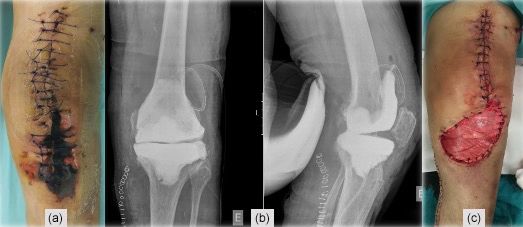
240                                                   H. Fonte et al.: Extensive skin necrosis after periprosthetic knee infection

3.5    Pyoderma gangrenosum                                          Review statement. This paper was edited by Parham Sendi and
                                                                     reviewed by two anonymous referees.
Pyoderma gangrenosum (PG) is an ulcerating neutrophilic
dermatosis that can occur in areas of trauma or following
surgical procedures. While the onset of PG is sudden, it tends       References
to remain a chronic ailment. Diagnosis is often by exclusion,
and although lesions appear, infected cultures are not useful        Ahronowitz, I., Harp, J., and Shinkai, K.: Etiology and
(Ahronowitz et al., 2012). The reasons that we believe it is            management of pyoderma gangrenosum: A compre-
not PG are as follows:                                                  hensive review, Am. J. Clin. Dermatol., 13, 191–211,
                                                                        https://doi.org/10.2165/11595240-000000000-00000, 2012.
                                                                     Barros, L. H., Barbosa, T. A., Esteves, J., Abreu, M., Soares,
  1. After surgical debridement of the skin necrosis, there             D., and Sousa, R.: Early Debridement, antibiotics and im-
     was no further progression of the ulcer. If PG was                 plant retention (DAIR) in patients with suspected acute infec-
     the culprit, one could expect post-surgical worsening,             tion after hip or knee arthroplasty - safe, effective and without
     known as the pathergy phenomenon (Duarte et al.,                   negative functional impact, J. Bone Joint Infect., 4, 300–305,
     2009), the reason that surgical debridement is usually             https://doi.org/10.7150/jbji.39168, 2019.
     contraindicated.                                                Balestra, B., Quadri, P., Dermarmels Biasiutti, F., Furlan, M., and
                                                                        Lammle, B.: Low molecular weight heparin-induced throm-
                                                                        bocytopenia and skin necrosis distant from injection sites,
  2. The spontaneous improvement despite the lack of                    Eur. J. Haematol., 53, 61–63, https://doi.org/10.1111/j.1600-
     corticosteroid/immunosuppressive therapy also speaks               0609.1994.tb00184.x, 1994.
     against this diagnosis, as does the good clinical outcome       Dahl, P. R., Perniciaro, C., Holmkvist, K. A., O’Connor,
                                                                        M. I., and Gibson, L. E.: Fulminant group A streptococ-
     after the second-stage revision surgery.
                                                                        cal necrotizing fasciitis: Clinical and pathologic findings in
                                                                        7 patients, J. Am. Acad. Dermatol., 47 (4), pp. 489–492,
   This report illustrates a challenging case of extensive              https://doi.org/10.1067/mid.2002.120536, 2002.
necrosis complicating a PJI. Difficulties around diagno-             Duarte, A. F., Nogueira, A., Lisboa, C., and Azevedo, F.: Pyo-
sis and treatment were numerous, even within a well-                    derma gangrenosum – clinical, laboratory and therapeutic ap-
trained multidisciplinary team. Despite a successful out-               proaches. Review of 28 cases, Dermatol. Online J., 15, 3,
come, the exact aetiology of the necrosis remains unproven,             PMID: 19903431, 2009.
but enoxaparin-induced skin necrosis emerges as a diagnos-           Estébanez, A. Silva, E., Cordero, P., and Martín, J. M.:
                                                                        Heparin-Induced Skin Necrosis Occurring at a Distance
tic of exclusion.
                                                                        From Injection Sites, Actas Dermo-Sifiliográficas, 110, 2–4,
                                                                        https://doi.org/10.1016/j.ad.2018.03.036, 2019.
                                                                     Frances, C.: Dermatological manifestations of Hughes an-
Ethical statement. The patient was informed that data from the          tiphospholipid antibody syndrome, Lupus, 19, 1071–1077,
case would be submitted for publication and gave their consent.         https://doi.org/10.1177/0961203310370343, 2010.
                                                                     Galat, D. D., McGovern, S. C., Larson, D. R., Harrington,
                                                                        J. R., Hanssen, A. D., and Clarke, H. D.: Surgical treat-
Data availability. The data used to support the findings of this        ment of early wound complications following primary total
study are included in the article.                                      knee arthroplasty, J. Bone Joint Surg. Am. A, 91, 48–54,
                                                                        https://doi.org/10.2106/JBJS.G.01371, 2009.
                                                                     Handschin, A. E., Trentz, O., Kock, H. J., and Wanner, G.
Team list. GRIP (Porto Bone and Joint Infection Unit) consists          A.: Low molecular weight heparin-induced skin necrosis – A
of the following members: Cláudia Pereira, Miguel Abreu, Daniel         systematic review, Langenbeck’s Arch. Surg., 390, 249–254,
Soares, Ernestina Reis, Ana Cláudia Santos, and Ricardo Sousa.          https://doi.org/10.1007/s00423-004-0522-7, 2005.
                                                                     Karmegam, A. and Raut, V.: Tissue necrosis after use of enoxaparin
                                                                        in total knee arthroplasty: a case report, Am. J. Orthop., 40, 152–
Author contributions. All authors discussed the results and con-        153, PMID: 22016872, 2011.
tributed to the final paper.                                         Schindewolf, M., Schwaner, S., and Wolter, M.: Incidence and
                                                                        causes of heparin-induced skin lesions, CMAJ, 181, 477–481,
                                                                        https://doi.org/10.1503/cmaj.081729, 2009.
Competing interests. The authors declare that they have no con-      Steer, A. C., Lamagni, T., Curtis, N., and Carapetis, J. R.:
flict of interest.                                                      Invasive group a streptococcal disease: Epidemiology,
                                                                        pathogenesis and management, Drugs, 72, 1213–1227,
                                                                        https://doi.org/10.2165/11634180-000000000-00000, 2012.
Disclaimer. Publisher’s note: Copernicus Publications remains
neutral with regard to jurisdictional claims in published maps and
institutional affiliations.

J. Bone Joint Infect., 6, 235–240, 2021                                                     https://doi.org/10.5194/jbji-6-235-2021
You can also read