Cerebral fat embolism syndrome after long bone fracture due to traffic accident: a case report
←
→
Page content transcription
If your browser does not render page correctly, please read the page content below
Chen et al. Neuroimmunol Neuroinflammation 2018;5:31
DOI: 10.20517/2347-8659.2018.23
                                                                                                     Neuroimmunology
                                                                                                 and Neuroinflammation

Case Report                                                                                                                    Open Access

Cerebral fat embolism syndrome after long bone
fracture due to traffic accident: a case report
Xing-Yong Chen1,#, Jian-Ming Fan2,#, Ming-Feng Deng2, Ting Jiang3, Feng Luo3
1
 Department of Neurology, Fujian Provincial Hospital, Fujian Medical University Provincial Clinical College, Fuzhou 350001, China.
2
  Intensive Care Unit, Fujian Provincial Hospital Wuyi Branch Hospital, Wuyishan City Hospital, Wuyishan 354300, China.
3
  Department of Image Diagnoses, Fujian Provincial Hospital Wuyi Branch Hospital, Wuyishan City Hospital, Wuyishan 354300, China.
#
  Authors contributed equally.

Correspondence to: Dr. Xing-Yong Chen, Department of Neurology, Fujian Provincial Hospital, Fujian Medical University
Provincial Clinical College, Fuzhou 350001, China. E-mail: cxyong77@163.com; Dr. Jian-Ming Fan, Intensive Care Unit, Fujian
Provincial Hospital Wuyi Branch Hospital, Wuyishan City Hospital, Wuyishan 354300, China. E-mail: 13706910979@139.com

How to cite this article: Chen XY, Fang JM, Deng MF, Jiang T, Luo F. Cerebral fat embolism syndrome after long bone fracture due
to traffic accident: a case report. Neuroimmunol Neuroinflammation 2018;5:31. http://dx.doi.org/10.20517/2347-8659.2018.23

Received: 26 Apr 2018 First Decision: 11 Jun 2018 Revised: 20 Jun 2018 Accepted: 20 Jun 2018 Published: 1 Aug 2018

Science Editor: Athanassios P. Kyritsis Copy Editor: Jun-Yao Li Production Editor: Cai-Hong Wang

Abstract
Cerebral fat embolism syndrome (CFES) is an uncommon but serious complication of long bone fracture. We reported
a 19-year-old male patient who sustained CFES due to multiple limbs long bone fractures after a traffic accident injury.
He gradually developed into coma within 24 h after his injury. The arterial blood gas analyses were normal. There was
a small amount of gas in the right pleural cavity on the thoracic computed tomography (CT). Although there were no
remarkable intracranial abnormalities on the initial brain CT findings, the typical brain magnetic resonance imaging (MRI)
findings of the starfield pattern and scattered foci were observed. Both T2-weighted imaging and diffusion weighted
imaging of MRI indicated multiple scattered lesions in the bilateral cerebrum hemisphere white matter, grey matter,
basal ganglia, corpus callosum and thalamus indicative of acute infarcts without microbleeding on the susceptibility-
weighted imaging sequences. With the above findings, the diagnosis of the case was cerebral fat embolism syndrome.
Although the patient was treated with comprehensive support in the intensive care unit, he remained unconscious and
was discharged after 7 days of hospitalization.

Keywords: Cerebral fat embolism, infarction, long bone fracture

INTRODUCTION
Fat embolism syndrome (FES) is a potentially fatal complication and occurs most commonly after long
bone fracture. Many cases occur as subclinical events and remain undiagnosed. The incidence of clinically
                       © The Author(s) 2018. Open Access This article is licensed under a Creative Commons Attribution 4.0
                       International License (https://creativecommons.org/licenses/by/4.0/), which permits unrestricted use,
    sharing, adaptation, distribution and reproduction in any medium or format, for any purpose, even commercially, as long
    as you give appropriate credit to the original author(s) and the source, provide a link to the Creative Commons license,
    and indicate if changes were made.

                                                                                                                               www.nnjournal.net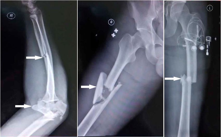
Page 2 of 7 Chen et al. Neuroimmunol Neuroinflammation 2018;5:31 I http://dx.doi.org/10.20517/2347-8659.2018.23
A B C
Figure 1. X-ray showed multiple long bone fractures, including right distal humerus distal, right ulna and its olecranon (A), bilateral femur
shafts (B, C) (arrow)
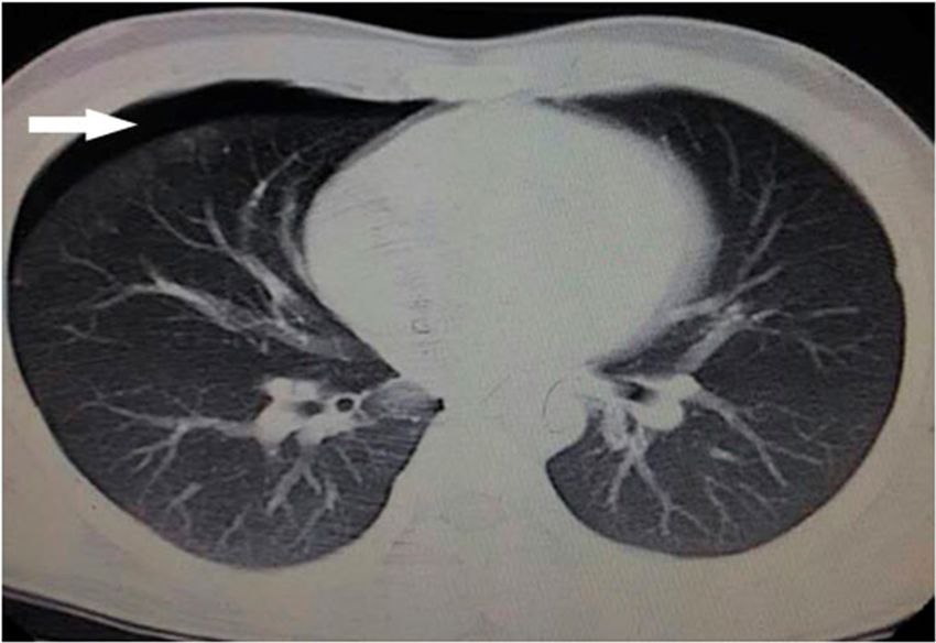
Figure 2. Thoracic computed tomography indicated a small amount of gas in the right pleural cavity (arrow)
significant FES occurs only in 0.9%-2.2% of long-bone fractures[1]. FES is characterized by various central
nervous system, respiratory, cutaneous, and hematological manifestations[2]. Cerebral fat embolism syndrome
(CFES) is a variant of FES. The neurological symptoms can be focal or diffuse, and most of the times exist
with respiratory symptoms. We here reported a case of cerebral fat embolism (CFE) due to multiple long
bone fractures of limbs after a traffic accident injury.
CASE REPORT
A 19-year-old male patient was admitted to the Emergency Department after a traffic accident injury
on May 19, 2016. He could not move his limbs except for the left upper limb due to bone fractures. He had
no history of loss of consciousness, ear, nose, and throat bleed, vomiting, or convulsion. He was conscious,
oriented with normal breathing and was hemodynamically stable. In the physical examination, body
temperature was 36.8 °C, pulse was 78/min, breath rate was 22/min, and blood pressure was 130/65 mmHg.
There were not positive neurologic dysfunctions. Electrocardiogram was normal. X-ray showed multiple long
bone fractures, including bilateral femurs, right distal humerus, right ulna and its olecranon [Figure 1]. There
was a small amount of gas in the right pleural cavity on the thoracic computed tomography (CT) [Figure 2].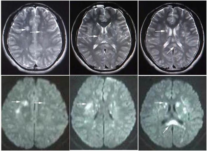
Chen et al. Neuroimmunol Neuroinflammation 2018;5:31 I http://dx.doi.org/10.20517/2347-8659.2018.23            Page 3 of 7

                 Figure 3. There was no remarkable abnormality on the brain computed tomography on admission

However, there were no remarkable abnormalities on abdominal CT and color Doppler ultrasound. The results
of hematologic and biochemical parameters of the patient were as follows at first admission: hemoglobine 149 g/L,
platelet count 187 × 109/L, procalcitonin 0.10 ng/mL, prothrombin time 13.7 s, alanine aminotransferase
196 U/L, aspartate aminotransferase 286 U/L, lactate dehydrogenase 739 U/L, creatine kinase 1092 U/L,
creativekinase MB 78 U/L, and cardiac troponin I 0.01 ng/mL. The patient did not have hypoxemia and
the arterial blood gas analyses were normal. He was treated in the intensive care unit. About fifteen hours
later, he became somnolence and gradually developed into a coma. Neurological examination revealed
bilateral Babinski sign. His pupils were isocoric and bilaterally responsive to light. There were not cutaneous
manifestations. Blood gas analysis was retested and normal. Echocardiogram showed normal ventricular
function without any thrombus or patent foramen ovale (PFO). Bilateral lower limb vascular color Doppler
ultrasound did not show any thrombus signs. Although in the present case there were no remarkable
intracranial abnormalities on the initial brain CT findings [Figure 3], the typical brain magnetic resonance
imaging (MRI) findings of starfield patterns were observed. Both T2-weighted imaging and diffusion
weighted imaging of MRI indicated multiple foci lesions in the bilateral cerebrum hemisphere white matter,
grey matter, basal ganglia, corpus callosum and thalamus indicative of acute infarcts [Figure 4]. However, there
was not any microbleeding on the susceptibility-weighted imaging sequences of brain MRI [Figure 4]. With
the above findings, the diagnosis of the case was cerebral fat embolism syndrome. The patient was treated
with methylprednisolone injection (80 mg, intravenous infusion, twice a day) and low molecular weight
heparin calcium injection (Dalteparin Sodium, Fragmin: 0.2 mL/5000 IU, subcutaneous injection, once a day),
antibiotics (Cefoperazone sodium and sulbactam sodium) and dehydrating drugs (such as mannitol and
human albumin solution). The bone fractures were firstly externally fixed upon admission into the hospital
and then internally fixed on fifth day during in-hospital stay. Unfortunately, the patient did not recover and
remained unconscious, and he was discharged after 7 days of hospitalization.

DISCUSSION
FES is a common clinical entity that can occasionally have significant neurological sequelae. Fat emolysm
syndrome has been reported not only after long-bone injuries, but also after rib or tarsal bone involvement[3,4].
The risk of fat emboli is highest within the first few days after trauma. Clinical symptoms of fat embolism usually
develop gradually within 24 to 72 h after injury[5], but in some cases, early manifestation occurs. Patients can
present with triad of varying severity of neurological, respiratory, and cutaneous manifestations, depending
on the embolic burden in the respective vasculature. CFES is a variant of FES characterized by a predominance
of neurologic manifestations often without the pulmonary or dermatologic findings seen in FES. The period
from the time of injury to the development of cerebral FES is typically between 12 h and 3 days[6].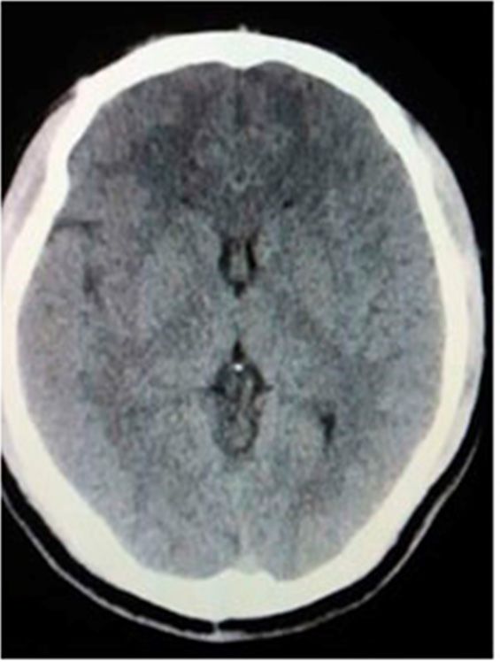
Page 4 of 7            Chen et al. Neuroimmunol Neuroinflammation 2018;5:31 I http://dx.doi.org/10.20517/2347-8659.2018.23

        MRI-T2

      MRI-DWI

       MRI-SWI

Figure 4. Both T2-weighted imaging and diffusion weighted imaging (DWI) of magnetic resonance imaging (MRI) indicated multiple foci
lesions in the bilateral cerebrum hemisphere white matter, grey matter, basal ganglia, corpus callosum and thalamus indicative of acute
infarcts (arrow). However, there was not any microbleeding on the susceptibility-weighted imaging sequences of brain MRI

CFES occurs after fat emboli enter the arterial circulation. However, the underlying mechanism remains
poorly understood. It was suggested that fat globules may enter the arterial circulation by two mechanisms[2,7].
Firstly, fat globules can enter the left atrium directly from the right heart through a shunt, such as a PFO
(paradoxical embolism), especially when the patient takes a deep breath or performs Valsalva maneuver
after trauma. Secondly, microglobules of fat may filter directly through the lung capillaries to reach the
arterial system. The diameter of these microemboli are small (7-10 μm in diameter) and malleable and may
not lead to significant pulmonary injury[7]. In our case, the absence of PFO and arrhythmia in this patient
with CFE supports the latter mechanism. However, the mechanisms underlying the pathophysiology of
CFE are complex. Great amounts of microglobules of fat may randomly enter the internal carotid artery
or/and vertebrobasilar artery, and usually cause distal vascular occlusion. Simple vascular occlusion with
ischemia probably does not fully explain the FES. Mechanical mobilization of fat globules from the bone
marrow to the intramedullary veins and pulmonary microcirculation, along with endothelial damage by fat
metabolism products and cytokines, has been proposed[8]. Such biochemical pathways of injury result from
the complex inflammatory response to free fatty acids released by the hydrolysis of embolized fat that has
entered blood vessels. This inflammatory response may have local effects on the brain and other tissues and
systemic consequences, including shock and anticoagulation[2,7].

Neurologic manifestations of CFES vary greatly, ranging from mild headache, diffuse encephalopathy,
and seizures to focal features, including pyramidal signs, pupillary paresis, and aphasia[1,2,7]. These signs
can occur in isolation or accompany with respiratory and cutaneous manifestations. Some patients do not
have hypoxemia, and these petechiae occur in only 20%-50% of patients and resolve quickly. Therefore, it
is difficult for us to diagnose the case. The Gurd's and Wilson's criteria are often used to make diagnoses[9].
Chen et al. Neuroimmunol Neuroinflammation 2018;5:31 I http://dx.doi.org/10.20517/2347-8659.2018.23   Page 5 of 7

One major and four minor criteria must be present to formally diagnose FES. Neither of these diagnostic
tools includes brain imaging, which seems to be the most specific. CFES is a clinical diagnosis, but specific
findings on neuroimaging studies can be strongly supportive. The purpose of a CT scan is to rule out certain
stroke mimics and detect hemorrhage, not necessarily to rule in the diagnosis of ischemic stroke. CT scans
may not be sensitive enough to detect an ischemic stroke, especially if it is small, acute (especially within
24 h of the stroke onset), or in the posterior fossa (i.e., brainstem and cerebellum areas). In other words, a
normal CT scan does not rule out the diagnosis of ischemic stroke. Noticeably, it is important to underline
that a careful examination of brain-CT findings, such as the topography and density measurements of round
lesions (such as round hypodense lesions, -40 HU)[3], were enough to confirm the clinical suspicion of CFE[3].
This is important because in some cases the MRI cannot be available or would be impossible to perform.
MRI is more sensitive and demonstrates multiple small hyperintense, intracerebral lesions. There were great
amounts of hyperintense lesions on MRI T2-weighted scans. The most characteristic MRI finding is the
starfield pattern, demonstrating scattered foci of high-intensity restricted diffusion on diffusion-weighted
imaging[7,10]. This is most apparent in the acute phase, from 4 h to the first few days from the time of injury.
Such widespread petechial hemorrhage and bland microinfarction have been demonstrated on autopsy[11]. In
our case, the initial brain-CT scan was normal, while MRI showed extensive cortical and subcortical regions
fat embolism which led to disturbance of consciousness. There are few differential diagnoses of disseminated
hyperintense lesions on T2-weighted scans which include diffuse axonal injury, areas of vasogenic edema
associated with microinfarcts, and demyelinating diseases[12] which were ruled out by history and clinical
scenario, in our case. Of note, in some patients who sustained severe trauma, both CFE and diffuse axonal
injury (DAI) could be the cause of altered consciousness in the absence of marked intracranial lesions in
cranial CT[13]. However, distinguishing CFE and DAI can be difficult clinically. Generally, DAI develops
immediately after the insult, whereas CFE occurs 24 to 72 h after the trauma and even after internal fixation
for the fractures[13]. It was reported that there was no significant difference between diagnostic performance
of diffusion tensor imaging (DTI) and conventional MRI in CFES, but a difference in directional diffusivities
was clearly identified between CFES and DAI.

There are currently no disease-specific treatment guidelines for FES or CFES other than supportive care to
address both intrinsic lung pathology and airway protection in the setting of neurological impairment[2].
Pharmacological intervention, including administration of heparin, dextran, aspirin, statin, albumin, and
steroids and glucose loading, proved to be ineffective[14,15]. Corticosteroids have been extensively studied with
variable results, and their use is controversial. In cases of fulminant FES, corticosteroids may be considered.
Gupta et al.[16] propose a regimen of methylprednisolone 1.5 mg/kg IV every 8 h for 6 doses in a select
group of patients with long bone or pelvic fractures at high risk of developing FES and without significant
contraindications. In our case, the patient was treated with methylprednisolone injection (80 mg twice a day).
The side effects of corticosteroids such as promoting coagulation and ulcer, disorder of electrolyte metabolism
should be emphasized. Anticoagulation has been shown to prevent stroke in patients with cardioembolic
and other noncardioembolic sources. However, the early use of anticoagulants has been associated with
hemorrhagic transformation. Hitherto, there is insufficient evidence to support that routine administration
of anticoagulation agent is effective and safe for FES. One possible benefit from anticoagulation for fat
embolism may potentially decrease the risk of deep vein thrombosis. Early surgical stabilization should
be considered. Early fixation of fractures within 24 h has been recommended to prevent further trauma at
the injury site, thus decreasing the incidence of FES. The prognosis of CFES is variable, depending on the
severity of the manifestations and on the quality and timing of treatment. Most of patients recovered fully
from this disease, and other survivors remained in cognitive disorder, and some even die[3,7,15].

In summary, we highlight that CFES could develop within hours after long bone fractures. Neurologic
manifestations of CFES vary greatly. Neuroimaging is critical in the diagnosis of CFES. The brain-CT scan
indicating the presence of round, hypodense lesions within the range of fat (-40 HU) suggests fat embolism.
Page 6 of 7            Chen et al. Neuroimmunol Neuroinflammation 2018;5:31 I http://dx.doi.org/10.20517/2347-8659.2018.23

The typical starfield pattern may be present on MRI in some cases of acute phase. Supportive respiratory
and neurological cares remain the mainstay of therapy. Prognosis is variable depending on the context of
concomitant illness and premorbid functional status.

DECLARATIONS
Authors’ contributions
Conception and design of study, first draft and revision of manuscript: Chen XY, Fan JM
Provided some clinical data: Deng MF
Provided the images data: Jiang T, Luo F
Read and approved the final manuscript: all authors

Availability of data and materials
The data and material could be available to readers upon request.

Financial support and sponsorship
This study was supported by the grants from the National Natural Science Foundation of China (81771250),
Fujian Province Natural Science Fund (2016J01432), Project for Scientific and Technological Innovation of
Fujian Province (2017Y9065), and Young and Middle-aged Talents Training Project of Health and Family
Planning Committee of Fujian Province (2015-ZQN-JC-5).

Conflicts of interest
All authors declare that there are no conflicts of interest.

Ethical approval and consent to participate
The study was approved by the local ethics committee and informed consent was obtained from the patient’s
guardian.

Consent for publication
Not applicable.

Copyright
© The Author(s) 2018.

REFERENCES
1.  Kosova E, Bergmark B, Piazza G. Fat embolism syndrome. Circulation 2015;131:317-20.
2.  Mijalski C, Lovett A, Mahajan R, Sundararajan S, Silverman S, Feske S. Cerebral fat embolism: a case of rapid-onset coma. Stroke
    2015;46:e251-3.
3. Scarpino M, Lanzo G, Moretti M, Olivo G, Amantini A, Grippo A. Delayed cerebral fat embolism occurring after off-pump coronary
    artery bypass grafting. Cardiol J 2018;25:155-7.
4. Kralovec ME, Houdek MT, Martin JR, Morrey ME, Cross WR. Atypical presentation of fat embolism syndrome after gunshot wound to
    the foot. Am J Orthop (Belle Mead NJ) 2015;44:E71-4.
5. Jorens PG, Van Marck E, Snoeckx A, Parizel PM. Nonthrombotic pulmonary embolism. Eur Respir J 2009;34:452-74.
6. Manousakis G, Han DY, Backonja M. Cognitive outcome of cerebral fat embolism. J Stroke Cerebrovasc Dis 2012;21:901-6.
7. Kellogg RG, Fontes RB, Lopes DK. Massive cerebral involvement in fat embolism syndrome and intracranial pressure management. J
    Neurosurg 2013;119:1263-70.
8. Taviloglu K, Yanar H. Fat embolism syndrome. Surg Today 2007;7:5-8.
9. Aggarwal R, Pal S, Soni KD, Gamangatti S. Massive cerebral fat embolism leading to brain death: a rare presentation. Indian J Crit
    Care Med 2015;19:687-9.
10. Herway ST, Slotto J, Harlan E, Newhouse B. Cerebral fat embolism syndrome. Anesthesiology 2016;124:1167.
11. Miller P, Prahlow JA. Autopsy diagnosis of fat embolism syndrome. Am J Forensic Med Pathol 2011;32:291-9.
12. Parizel PM, Demey HE, Veeckmans G, Verstreken F, Cras P, Jorens PG, De Schepper AM. Early diagnosis of cerebral fat embolism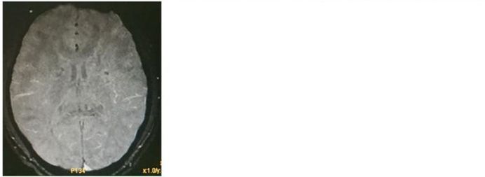
Chen et al. Neuroimmunol Neuroinflammation 2018;5:31 I http://dx.doi.org/10.20517/2347-8659.2018.23                    Page 7 of 7

    syndrome by diffusion-weighted MRI (starfield pattern). Stroke 2001;32:2942-4.
13. Chen PC, Hsu CW, Liao WI, Chen YL, Ho CH. Hyperacute cerebral fat embolism in a patient with femoral shaft fracture. Am J Emerg
    Med 2013;31:1420.e1-3.
14. Whalen LD, Khot SP, Standage SW. High-dose rosuvastatin treatment for multifocal stroke in trauma-induced cerebral fat embolism
    syndrome: a case report. Pediatr Neurol 2014;51:410-3.
15. Mijalski C, Lovett A, Mahajan R, Sundararajan S, Silverman S, Feske S. Cerebral fat embolism. Stroke 2015;46:e251-3.
16. Gupta A, Reilly CS. Fat embolism. CEACCP 2007;7:148-51.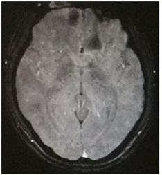
You can also read