Prognostic Value of a New Integrated Parameter-Both Collateral Circulation and Permeability Surface-in Hemorrhagic Transformation of Middle ...
←
→
Page content transcription
If your browser does not render page correctly, please read the page content below
ORIGINAL RESEARCH
                                                                                                                                         published: 25 August 2021
                                                                                                                                   doi: 10.3389/fnagi.2021.703734

                                            Prognostic Value of a New Integrated
                                            Parameter—Both Collateral
                                            Circulation and Permeability
                                            Surface—in Hemorrhagic
                                            Transformation of Middle Cerebral
                                            Artery Occlusion Acute Ischemic
                                            Stroke: Retrospective Cohort Study
                                            Chanchan Li 1 , Xiaozhu Hao 1 , Luyi Lin 1 , Chengfeng Sun 1 , Hai Yu 2 , Zhenwei Yao 1 ,
                                            Xiaoyuan Feng 1 and Yanmei Yang 1*
                                            1
                                             Department of Radiology, Huashan Hospital, Fudan University, Shanghai, China, 2 Department of Neurology, Huashan
                                            Hospital, Fudan University, Shanghai, China

                                            Background: Multimodal CT, including CT angiography (CTA) and CT perfusion
                                            (CTP), was increasingly used in stroke triage. This study was to determine the
                                            relationship between a new integrated parameter—both collateral circulation and relative
                                            permeability surface (PS)—and the hemorrhagic transformation (HT) in acute ischemic
                           Edited by:
               Rubem C. A. Guedes,          stroke (AIS) with middle cerebral artery occlusion (MCAO).
   Federal University of Pernambuco,
                                Brazil
                                            Methods: We retrospectively reviewed consecutive AIS patients with MCAO who
                      Reviewed by:
                                            underwent baseline CTA/CTP within 4 h of symptom onset and follow-up susceptibility-
              Norbert Nighoghossian,        weighted imaging (SWI) within 3 weeks. Collateral circulation was assessed on the
   Université Claude Bernard Lyon 1,
                                            baseline CTA. Baseline CTP data were postprocessed to generate PS parameter. The
                              France
                   Chushuang Chen,          patients with poor collateral circulation and at the same time with high relative PS were
The University of Newcastle, Australia      classified as the group of both poor collateral circulation and high relative PS. HT was
                 *Correspondence:           defined according to European Cooperative Acute Stroke Study II criteria on follow-up
                       Yanmei Yang
                yym9876@sohu.com
                                            SWI imaging. Multivariate logistic regression analysis was performed using HT as an
                                            outcome variable.
           Received: 30 April 2021
           Accepted: 26 July 2021           Results: The group of patients with both poor collateral circulation and high
         Published: 25 August 2021          relative PS was thirteen and thirty-three (52%) developed HT of the final cohort
                             Citation:      sixty-three AIS patients with MCAO. Multivariate logistic analysis revealed the new
      Li C, Hao X, Lin L, Sun C, Yu H,
    Yao Z, Feng X and Yang Y (2021)
                                            integrated parameter—both collateral circulation and relative PS (odds ratio, 16.59; 95%
Prognostic Value of a New Integrated        confidence interval, 13.09–19.10; P < 0.001) was independent predictor of HT. The
           Parameter—Both Collateral        area under the curve was 0.85 (95% confidence interval, 0.81–0.89). The sensitivity was
          Circulation and Permeability
             Surface—in Hemorrhagic         57%, specificity 97% and positive predictive value 92%, negative predictive value 58%.
   Transformation of Middle Cerebral
     Artery Occlusion Acute Ischemic
                                            Conclusions: For AIS patients with MCAO, these with poor collateral circulation on CTA
 Stroke: Retrospective Cohort Study.        and at the same time with high relative PS on CTP were at high risk for HT.
  Front. Aging Neurosci. 13:703734.
     doi: 10.3389/fnagi.2021.703734         Keywords: collateral circulation, stroke, angiography, perfusion imaging, cerebral hemorrhage

Frontiers in Aging Neuroscience | www.frontiersin.org                            1                                        August 2021 | Volume 13 | Article 703734
Li et al. Multimodal CT and Hemorrhagic Transformation
INTRODUCTION were excluded: (1) history of brain tumor or brain surgery; (2)
ischemic stroke in the bilateral cerebral hemispheres; (3) low
Hemorrhagic transformation (HT) is an unwanted complication imaging quality; (4) abnormal endovascular procedure including
of acute ischemic stroke (AIS) that may severely worsen the inappropriate thrombectomy and impaling vessels.
prognosis (Knight et al., 2019). Multimodal CT, including CT This study was approved by the Ethics Committee of Huashan
angiography (CTA) and CT perfusion (CTP), is a part of routine Hospital, Fudan University (2020-939) and was registered at the
acute stroke care in many stroke centers. Numerous studies have Chinese Clinical Trial Registry (ChiCTR2000035575). Informed
tried to determine clinical and imaging parameters associated consent was waived for collection and analysis of preexisting
with HT so as to identify patients at highest risk for thrombolytic data. This study was carried out in accordance with the
or endovascular therapies. Indeed, risks factors for HT have been Declaration of Helsinki.
investigated and included higher age, higher stroke severity, large
vessel occlusion and collateral score on CTA and permeability Clinical Variables
surface (PS) on CTP et al. (Hom et al., 2011; Bivard et al., 2016; The following clinical variables were collected for each
Marsh et al., 2016; Wang et al., 2016; Kalinin et al., 2017; Li patient: sex, age, admission NIHSS, risk factors (hypertension,
Q. et al., 2017; Li Y. et al., 2017; Liu et al., 2017; Puig et al., hyperlipidemia, diabetes mellitus, atrial fibrillation, previous
2017). However, many studies taken it for granted yet could stroke, and current smoking), recanalization therapy
not confirm the cause-and-effect relationship between collateral (intravenous thrombolysis and endovascular thrombectomy)
circulation and PS for these two parameters were simultaneously and time to recanalization therapy, time to imaging.
acquired (Li Q. et al., 2017; Puig et al., 2017; Horsch et al., Patients without contraindication to thrombolysis received
2018). Therefore, it was inappropriate to draw a conclusion that intravenous recombinant tissue plasminogen activator (rt-PA)
inadequate collateral circulation contributed to the increased PS treatment. Eligible patients without extensive early ischemic
or to identify collateral circulation as a confounding factor in the signs on pre-interventional scans received endovascular
HT prediction model. treatment. Patients with contraindications to thrombolysis
In this study, we integrated the collateral circulation and PS received supportive care.
into a new parameter—both collateral circulation and relative
PS. We hypothesized that this new parameter contributes to Image Acquisition
HT and we aimed to determine the relationship between this All patients underwent baseline NCCT/CTP/CTA within 4 h of
new parameter and HT in AIS with middle cerebral artery symptom onset and follow-up SWI within 3 weeks. Whole-brain
occlusion (MCAO). NCCT, CTP, and CTA were simultaneously performed on a 256-
section CT scanner (Brilliance iCT, Phillips Medical Systems,
Cleveland, OH, United States) as follows: Jog mode, 120 kVp,
MATERIALS AND METHODS 150 mAs, field of view 220 mm, matrix 512 × 512. A dual-
head power injector (Stellate Injection System, Indianola, PA,
Study Population and Design United States) was used to inject 40 ml of non-ionic contrast
A single-center retrospective cohort study was conducted in medium (Ultravist, iodine 370 mg I/ml; Bayer Healthcare, Berlin,
consecutive AIS patients, according to Guidelines for Diagnosis Germany) at 5 ml/s followed by 20 ml saline with 5-s (CTP)
and Treatment of Acute Ischemic Stroke in China 2014, and 8-s (CTA) delay. CTP included a 50-s scan reconstructed
presenting to the Stroke Unit of Huashan Hospital, Fudan at 0.4-s intervals to produce a series of 312 sequential images
University between January 2016 and December 2019. Patients for 13 sections, covering a total of 120 mm from the foramen
were included if the following inclusion criteria were met: (1) magnum to the lateral ventricles. SWI was performed on a 3.0T
age > 18 years and < 85 years; (2) National Institutes of superconducting magnetic resonance imaging (MRI) scanner
Health Stroke Scale (NIHSS) was 4 to 22 at admission; (3) (Verio, Siemens AG, Erlangen, Germany).
time from symptom onset to no-contrast CT (NCCT)/CTP/CTA
was within 4 h and before recanalization treatment; (4) AIS Image Analysis
with middle cerebral artery (MCA)-M1 occlusion documented Baseline CTA data were processed using the Phillips Brilliance
on CTA; (5) the admission NCCT showed no evidence of Workspace portal software (Vision 5.0.2), including 10-mm axial
intracerebral hemorrhage; (6) time from symptom onset to and multi-planar maximum intensity projection reconstructions.
follow-up susceptibility-weighted imaging (SWI) was within The leptomeningeal collateral circulation was graded according
3 weeks. Patients with one or more of the following conditions to a previously reported scoring system on a scale from 0 to 3:
0 = absent collateral, 1 = collateral filling < 50% of the occluded
territory, 2 = collateral filling > 50% < 100% of the occluded
Abbreviations: AIS, acute ischemic stroke; BBB, blood-brain barrier; CTA, CT
angiography; CTP, CT perfusion; HI, hemorrhagic infarction; HT, hemorrhagic territory, and 3 = collateral filling 100% of the occluded territory
transformation; MCA, middle cerebral artery; MCAO, middle cerebral artery (Figure 1). The results were dichotomized into poor collateral
occlusion; MRI, magnetic resonance imaging; NCCT, no-contrast CT; NIHSS, circulation (a score of 0 or 1) versus good collateral circulation (a
National Institutes of Health Stroke Scale; PH, parenchymal hematoma; PS,
score of 2 or 3) (Tan et al., 2009). MCA-M1 occlusion was defined
permeability surface; ROI, regions of interest; rt-PA, recombinant tissue
plasminogen activator; SICH, symptomatic intra cerebral hemorrhage; SWI, as a main MCA trunk occlusion before the bifurcation, with or
susceptibility-weighted imaging. without ipsilateral internal carotid artery occlusion.
Frontiers in Aging Neuroscience | www.frontiersin.org 2 August 2021 | Volume 13 | Article 703734Li et al. Multimodal CT and Hemorrhagic Transformation
FIGURE 1 | Examples of collateral circulation from 0 to 3 (white arrow). (A) 0 = absent collateral, (B) 1 = collateral filling < 50% of the occluded territory, (C)
2 = collateral filling > 50% < 100% of the occluded territory, and (D) 3 = collateral filling 100% of the occluded territory. All four patients were acute ischemic stroke
(AIS) with right middle cerebral artery (MCA)-M1 occlusion. AIS, acute ischemic stroke; MCA, middle cerebral artery.
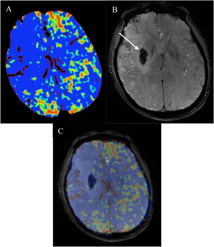
Postprocessing of baseline CTP source images was performed side were in the exact location where the HT happened. Relative
using Phillips brain perfusion software (Vision 5.0.2) to generate PS were calculated by normalizing the values in the HT region
parametric maps of PS (Figure 2A). The adiabatic approximation with those in the contralateral mirror region. The results were
tissue homogeneity model and a deconvolution technique were dichotomized into high relative PS (≥2.89) versus low relative PS
used to calculate CTP parameters. HT was defined as a (< 2.89) (Li Q. et al., 2017).
hypointense susceptibility effect-induced area in the ischemic Two radiologists (CL and XH) who were blinded to the
region on follow-up SWI (within 3 weeks) (Figure 2B). If study groups independently assessed the MCA-M1 status
present, HT was classified according to the European Cooperative and collateral circulation. Two radiologists (LL and CS)
Acute Stroke Study II classification into four subtypes, that is, assessed the follow-up SWI for HT and relative PS in
hemorrhagic infarction (HI) 1 and 2 and parenchymal hematoma selected ROIs. According to the new parameter—both collateral
(PH)1 and 2. Symptomatic intracerebral hemorrhage (SICH) circulation and relative PS, the patients with poor collateral
was identified as PH with worsening of neurological deficit ≥ 4 circulation and at the same time with high relative PS were
points on the NIHSS that was temporally related to its imaging classified as the group of both poor collateral circulation and
appearance (Larrue et al., 2001). Baseline PS maps and follow- high relative PS.
up SWI images were co-registered automatically using OpenCV
2.4.9 (Figure 2C). Then the regions of interest (ROIs) were drawn Statistical Analysis
within the HT region and the corresponding contralateral region. Statistical analyses were performed using SPSS 19.0 software
This method made the ROI on the PS maps on the affected (SPSS, Chicago, IL, United States). Quantitative variable was
Frontiers in Aging Neuroscience | www.frontiersin.org 3 August 2021 | Volume 13 | Article 703734Li et al. Multimodal CT and Hemorrhagic Transformation FIGURE 2 | (A) CT perfusion permeability surface (PS) map, follow-up (B) susceptibility-weighted imaging (SWI) in the same patient as in Figure 1B and (C) co-registered map. Obviously increased (A) PS on the affected side compared with the contralateral normal side. Follow-up (B) SWI showed hemorrhagic transformation (HT) (white arrow) which belonged to parenchymal hematoma (PH) 2. PS map and follow-up SWI image were co-registered automatically using OpenCV 2.4.9. Then the regions of interest (ROIs) were drawn within the HT region and the corresponding contralateral region. Relative PS were calculated by normalizing the values in the HT region with those in the contralateral mirror region. PS, permeability surface; SWI, susceptibility-weighted imaging; HT, hemorrhagic transformation; PH, parenchymal hematoma; ROIs, regions of interest. expressed as mean ± standard deviation and categorical RESULTS variable was expressed as proportion. Differences were assessed using the χ2 test (categorical variables), Student’s-t test Sixty-three patients were included in this study from January (variables with normal distribution), or Mann–Whitney rank 2016 to December 2019. Study flow chart was shown in test (variables without a normal distribution). Univariate Supplementary Figure I. The baseline characteristics had no and multivariate logistic regression analysis were performed significant difference between the patients involved and not using HT as an outcome variable. Because we had only a involved in this study (Supplementary Table I). Of the final limited number of outcomes and only three variables could cohort comprising sixty-three AIS patients with M1-MCA be selected maximally to have at least 10 outcomes per occlusion, thirteen was the group of both poor collateral variable. Variables analyzed were age, admission NIHSS, the circulation and high relative PS and fifty belonged to the other. new parameter—both collateral circulation and relative PS Thirty-three (52%) developed HT. ECASS II types were as (Horsch et al., 2018). Interrater reliability was evaluated using follows: fifteen HI1(45%), eleven HI2(33%), four PH1(12%), weighted k values for ordered categorical variables. All P < 0.05 three PH2(9%), and four SICH (12%). The mean time from was considered significant. symptom onset to HT was 5.21 ± 4.92 days. Interrater Reliability Frontiers in Aging Neuroscience | www.frontiersin.org 4 August 2021 | Volume 13 | Article 703734
Li et al.                                                                                                                 Multimodal CT and Hemorrhagic Transformation

TABLE 1 | Clinical characteristics of patients.

                                                                The patients with both poor                                The other                           P value
                                                                 collateral circulation and                                 patients
                                                                  high relative PS (n = 13)                                 (n = 50)

Male, n (%)                                                                   8 (62)                                        24 (48)                            0.744
Age, years                                                               62.12 ± 8.71                                    63.23 ± 10.82                         0.939
Admission NIHSS                                                          17.22 ± 3.54                                    15.43 ± 4.81                          0.331
Hypertension, n (%)                                                           8 (62)                                        29 (58)                            0.678
Hyperlipidemia, n (%)                                                         3 (23)                                        8 (16)%                            0.539
Diabetes mellitus, n (%)                                                     11 (87)                                        30 (60)                            0.729
Atrial fibrillation, n (%)                                                    1 (8)                                          1 (2)                             /
Previous stroke, n (%)                                                        1 (8)                                          4 (8)                             /
Current smoking, n (%)                                                        5 (38)                                        22 (44)                            0.723
Time to recanalization therapy, hours                                    4.42 ± 0.81                                      4.61 ± 0.65                          0.396
Recanalization therapy
Intravenous thrombolysis, n (%)                                               4 (31)                                        16 (32)                            0.788
Endovascular thrombectomy, n (%)                                              3 (23)                                        10 (20)                            0.532
Time to CTA/CTP, hours                                                   2.43 ± 0.31                                      2.64 ± 0.13                          0.623
Time to SWI, weeks                                                       2.22 ± 0.57                                      2.53 ± 0.46                          0.815
HT, n (%)                                                                    12 (92)                                        21 (42)
Li et al. Multimodal CT and Hemorrhagic Transformation
Wang et al., 2016; Li Q. et al., 2017; Li Y. et al., 2017; Puig CT parameter into the clinical decision-making algorithm
et al., 2017). Our study showed the poor collateral circulation may allow for a greater appreciation of the risk-benefit
and at the same time the high relative PS provided a key profile before decisions about thrombolytic therapy in AIS.
link to a higher incidence of HT. To our knowledge, this Further analysis with a larger number of patients and
integrated parameter—both collateral circulation and relative PS clinical end points in an independent data set is required
has not been reported previously. Our results supported the to optimize and validate the methodology and potentially
view that the extent of hemodynamic derangement can help make this multimodal image protocol more applicable in the
identify those at highest risk for HT and HT was the result clinical setting.
of severe ischemic damage to the vessel walls that leads to
BBB disruption (Hom et al., 2011; Wang et al., 2016; Li Q.
et al., 2017; Li Y. et al., 2017; Puig et al., 2017). This new DATA AVAILABILITY STATEMENT
integrated parameter showed 97% specificity and 92% positive
predictive value in predicting HT of AIS patients with MCA- The raw data supporting the conclusions of this article will be
M1 occlusion. For AIS patients with MCAO, these with poor made available by the authors, without undue reservation.
collateral circulation on CTA and at the same time with high
relative PS on CTP were at high risk for HT. It is the strength
of our study. This may help clinicians to tighten current rt-PA ETHICS STATEMENT
treatment indications to exclude patients who may previously
have been included. The studies involving human participants were reviewed and
CT angiography maximum intensity projection approved by the Ethics Committee of Huashan Hospital, Fudan
reconstruction is effective to evaluate leptomeningeal University. Written informed consent for participation was not
collateral flow. After MCA-M1 occlusion, leptomeningeal required for this study in accordance with the national legislation
collateral is rapidly recruited to supply the ischemic and the institutional requirements.
area (Li Q. et al., 2017). PS map on CTP is intended
to reflect the degree of BBB disruption, although it is
potentially fraught with substantial variability due to AUTHOR CONTRIBUTIONS
differences in acquisition and postprocessing techniques,
CL, ZY, XF, and YY were responsible for study concept and
including the size and molecular charge of the contrast
the design. CL, XH, LL, CS, and HY collected the data. CL,
agent used. To a certain extent, our findings prompted
XH, LL, and CS analyzed the data and conducted the statistical
that collateral blood flow may be a potential therapeutic
analysis. CL wrote the manuscript. ZY, XF, and YY supervised the
target for reducing the incidence of HT. However,
manuscript and provided technical or information support. All
there was one uncertainty that should be taken into
authors approved the final version of the manuscript.
consideration that our results could not confirm that
poor collateral circulation contributed to the increased
PS or good collateral circulation contributed to the
decreased PS for these two parameters were simultaneously
FUNDING
acquired in this study. This work was supported by grants from the National Natural
This study has many limitations. First, this is a single- Science Foundation of China (Grant numbers: 81771788 and
center study with a small sample size, that is, an insufficient 81801660). None of these funding sources were involved in the
number of patients with HT to stratify into symptomatic study design, data collection, data analysis or interpretation, or
versus asymptomatic groups, or PH versus HI groups. The the writing of the report and the decision to submit the article
parameters threshold may not be accurate. Second, this for publication.
study has the limitations and potential biases inherent in
a retrospective study. The duration of the CTP acquisition
is relatively short (50 s), which may make it questionable ACKNOWLEDGMENTS
if it is truly measuring BBB permeability because our
study is retrospectively conducted with imaging performed We would like to thank all the subjects for participating in
for clinical purposes rather than imaging optimized this study and the support by the Huashan Hospital, Fudan
for research purposes. Therefore, further prospective University. We would also like to thank Shengjie Zhang for the
research is required. help of image analysis.
CONCLUSION SUPPLEMENTARY MATERIAL
In conclusion, the new integrated parameter—both collateral The Supplementary Material for this article can be found online
circulation and relative PS predicts HT in AIS patients with at: https://www.frontiersin.org/articles/10.3389/fnagi.2021.
MCAO. The incorporation of readily available multimodal 703734/full#supplementary-material
Frontiers in Aging Neuroscience | www.frontiersin.org 6 August 2021 | Volume 13 | Article 703734Li et al. Multimodal CT and Hemorrhagic Transformation
REFERENCES tomography score in associated with hemorrhagic transformation after acute
cardioembolic stroke. Front. Neurol. 8:591. doi: 10.3389/fneur.2017.00591
Bivard, A., Cheng, X., Lin, L. T., Levi, C., Spratt, N., Kleinig, T., et al. (2016). Global Marsh, E. B., Llinas, R. H., Schneider, A. L. C., Hillis, A. E. H., Lawrence, E.,
white matter hypoperfusion on CT predicts larger infarcts and hemorrhagic Dziedzic, P., et al. (2016). Predicting hemorrhagic transformation of acute
transformation after acute ischemic. CNS Neurosci. Ther. 22, 238–243. doi: ischemic stroke: prospective validation of the HeRS score. Medicine 95:e2430.
10.1111/cns.12491 doi: 10.1097/MD.0000000000002430
Hom, J., Dankbaar, J. W., Soares, B. P., Schneider, T., Cheng, S. C., Bredno, Puig, J., Blasco, G., Daunis, P., van Eendendburg, C., Carrillo, M., Aboud,
J., et al. (2011). Blood-brain barrier permeability assessed by perfusion CT C., et al. (2017). High-permeability region size on perfusion CT predicts
predicts symptomatic hemorrhagic transformation and malignant edema in hemorrhagic transformation after intravenous thrombolysis in stroke. PLoS
acute ischemic stroke. Am. J. Neuroradiol. 32, 41–48. doi: 10.3174/ajnr.A2244 One. 12:e0188238. doi: 10.1371/journal.pone.0188238
Horsch, A. D., Bennink, E., van Seeters, T., Kappelle, L. J., Graaf, Y. V. D., Mali, Tan, I. Y. L., Demchuk, A. M., Hopyan, J., Zhang, J., Gladstone, D., Wong, K., et al.
W. P. T. M., et al. (2018). Computed tomography perfusion derived blood- (2009). CT angiography clot burden score and collateral score: correlation with
brain barrier permeability does not yet improve prediction of hemorrhagic clinical and radiologic outcomes in acute middle cerebral artery infarct. Am. J.
transformation. Cerebrovasc. Dis. 45, 26–32. doi: 10.1159/000485043 Neuroradiol. 30, 525–531. doi: 10.3174/ajnr.A1408
Kalinin, M. N., Khasanova, D. R., and Ibatullin, M. M. (2017). The hemorrhagic Wang, W., Li, M. C., Chen, Q. X., and Wang, J. (2016). Hemorrhagic
transformation index score: a prediction tool in middle cerebral artery ischemic transformation after tissue plasminogen activator reperfusion therapy for
stroke. BMC Neurol. 17:177. doi: 10.1186/s12883-017-0958-3 ischemic stroke: mechanisms, models, and biomarkers. Cell. Mol. Neurobiol. 52,
Knight, G. A., Nario, J. J. Q., and Gupta, A. (2019). Cause of acute stroke: a 1572–1579. doi: 10.1007/s12035-014-8952-x
patterned approach. Radiol. Clin. North Am. 57, 1093–1108. doi: 10.1016/j.rcl.
2019.07.007 Conflict of Interest: The authors declare that the research was conducted in the
Larrue, V., von Kummer, R., Muller, A., and Bluhmki, E. (2001). Risk factors absence of any commercial or financial relationships that could be construed as a
for severe hemorrhagic transformation in ischemic stroke patients treated potential conflict of interest.
with recombinant tissue plasminogen activator: a secondary analysis of the
European-Australasian acute stroke study (ECASS II). Stroke 32, 438–441. doi: Publisher’s Note: All claims expressed in this article are solely those of the authors
10.1161/01.str.32.2.438 and do not necessarily represent those of their affiliated organizations, or those of
Li, Q., Gao, X. Y., Yao, Z. W., Feng, X. Y., He, H. J., Xue, J., et al. (2017). Permeability the publisher, the editors and the reviewers. Any product that may be evaluated in
surface of deep middle cerebral artery territory on computed tomographic this article, or claim that may be made by its manufacturer, is not guaranteed or
perfusion predicts hemorrhagic transformation after stroke. Stroke 48, 2412– endorsed by the publisher.
2418. doi: 10.1161/STROKEAHA.117.017486
Li, Y., Xia, Y., Liu, N., Jackson, A., Wintermark, M., Zhang, Y., et al. (2017). Focal Copyright © 2021 Li, Hao, Lin, Sun, Yu, Yao, Feng and Yang. This is an open-access
low and global high permeability predict the possibility, risk, and location of article distributed under the terms of the Creative Commons Attribution License
hemorrhagic transformation following intra-arterial thrombolysis therapy in (CC BY). The use, distribution or reproduction in other forums is permitted, provided
acute stroke. Am. J. Neuroradiol. 38, 1730–1736. doi: 10.3174/ajnr.A5287 the original author(s) and the copyright owner(s) are credited and that the original
Liu, L., Wu, B., Zhao, J. L., Cao, Y. Y., Dedhia, N., Caplan, L. R., et al. (2017). publication in this journal is cited, in accordance with accepted academic practice. No
Computed tomography perfusion alberta stroke program early computed use, distribution or reproduction is permitted which does not comply with these terms.
Frontiers in Aging Neuroscience | www.frontiersin.org 7 August 2021 | Volume 13 | Article 703734You can also read