Results of Colostomy Use in Children with Anorectal Malformation
←
→
Page content transcription
If your browser does not render page correctly, please read the page content below
Results of Colostomy Use in Children with Anorectal
                                       Malformation

Temur T. Narbaev1; Mahmud M. Aliev2; Shovkat T. Bozorov3; Jasmin T. Turaeva4; Muzaffar A. Yuldashev5;
                                       Izzatullo Z. Sobitov6
     1
       Department of Faculty Pediatric Surgery, Tashkent Pediatric Medical Institute, Tashkent, Uzbekistan.
                                             1
                                               temur1972@inbox.ru
     2
       Department of Faculty Pediatric Surgery, Tashkent Pediatric Medical Institute, Tashkent, Uzbekistan.
     3
       Department of Faculty Pediatric Surgery, Tashkent Pediatric Medical Institute, Tashkent, Uzbekistan.
     4
       Department of Faculty Pediatric Surgery, Tashkent Pediatric Medical Institute, Tashkent, Uzbekistan.
     5
       Department of Faculty Pediatric Surgery, Tashkent Pediatric Medical Institute, Tashkent, Uzbekistan.
     6
       Department of Faculty Pediatric Surgery, Tashkent Pediatric Medical Institute, Tashkent, Uzbekistan.

   Abstract
   Introduction. Anorectal malformations in children are still one of the most challenging problems in
   pediatric coloproctology. The incidence of anorectal malformations in recent years has no tendency
   to decrease and, according to various authors, ranges from 1 in 4000-5000 live births. Most
   pediatric surgeons continue to adhere to the opinion about the advisability of preliminary colostomy
   and delayed proctoplasty at the age of 6-18 months or when the child reaches a certain body weight
   (8-10 kg.). They are motivating this tactic with the possibility of creating an optimal condition for
   performing a complex intervention, reducing anesthetic risk, avoiding technical errors.
   Objective is to improve treatment outcomes for anorectal malformations in children with prior
   colostomy.
   Materials and methods. The work is based on the results of treatment of 154 children with
   anorectal malformation, with preliminary colostomy in the clinic of the Tashkent Pediatric Medical
   Institute for the period from 2000 to 2020.
   Along with routine and general clinical examination methods, all children underwent: X-ray of the
   abdominal cavity, colostography, fistuloirrigography, excretory urography, cystography, ultrasound
   of the perineum (small pelvis), neurosonography (NSG) screening tests.
   Results. 154 (10*0%) children had colostomy as a palliative stage of treatment. Of these 117 (76%)
   children developed colostomy on the first day of life, with the development of intestinal obstruction.
   In 37 (24%) children, the formation of a colostomy was performed directly by us. 9 (5.8%) children
   as the first stage before primary radical correction with a high form of the defect and 10 (6.5%)
   children previously operated on and requiring re-corrective operations, 5 (3.2%) patients
   underwent colostomy after the development of complications in the early postoperative period. In 13
   (8.5%) cases, colostomies were formed with identified concomitant anomalies and defects that
   clinically "dominated" over anorectal malformation. In 2 (5.4%), a double sigmastoma was

  ISSN: 2237-0722                                                                                             17
  Vol. 11 No. 2 (2021)
  Received: 20.02.2021 – Accepted: 02.04.2021
imposed, in 2 (5.4%) a distal single-barreled sigmastoma, in 5 (13.5%) the Hartmann type terminal
sigmastoma.
Conclusion. The use of colostomy in children with anorectal malformation made it possible to carry
out the necessary surgical tactics in a timely and differentiated manner. To reduce the frequency,
nature of complications and early disability, to improve the quality of life and social adaptation of
patients.
Key-words: Anorectal Malformations, Colostomy, Surgical Treatment.
1. Introduction
Anorectal malformations in children are a special chapter of pediatric coloproctology.
Treatment of this pathology, which is very painful for children and their parents, requires a purely
individual approach. Colostomy - removal of a segment (s) of the colon or its loop on the anterior
abdominal wall. Although the first stoma was imposed for the treatment of anorectal malformation in
1783, to date, the place and type of stoma in the treatment of anorectal malformation remains an
important issue among pediatric surgeons [1, 2, 3].
In the specialized literature, you can find different judgments about the level of colostomy
imposition. Some authors recommend choosing the sigmoid colon for this purpose, others prefer the
transverse colon, others - the blind and ascending. This issue cannot be resolved unambiguously. First
of all, a colostomy should correspond to the main idea of treatment, and also not create any special
difficulties during subsequent operations [4, 5, 6].
Recent years have been marked by the possibility of correcting anorectal defect without using
a protective colostomy. Moreover, in most publications, preliminary stoma placement is considered
correct. At the same time, one cannot agree with the widespread replication of operations without
stomas, since this will increase the risk of complications [7, 8].
Clinical studies have shown a high rate of complications associated with neonatal colostomy,
in particular, transverse stoma has a higher complication rate than sigmostomy. However,
controversy continues over the type of split colostomy. This study compares the clinical outcomes of
loop and split colostomy superimposed for anorectal malformation. [9, 10, 11].
There is always a temptation to correct anorectal anomalies without a protective colostomy,
but complications associated with both the colostomy itself and its formation and subsequent closure
pose a serious threat to the normal functioning of both the intestine itself and the anal sphincter
apparatus [12, 13, 14].
The technique of performing the operative technique is very important - the imposition of an
intestinal stoma in order to prevent colostomy complications. It is not controversial that in children
ISSN: 2237-0722 18
Vol. 11 No. 2 (2021)
Received: 20.02.2021 – Accepted: 02.04.2021with high variants of malformations and cloaca, as well as before re-corrective operations, it is
necessary to perform a colostomy first, and after 2-3 months, a radical correction of the defect [15,
16, 17, 18].
Most pediatric surgeons continue to adhere to the opinion about the advisability of
preliminary colostomy and delayed proctoplasty at the age of 6-18 months or when the child reaches
a certain body weight (usually about 8-10 kg.), Motivating this tactic with the possibility of creating
the optimal conditions for performing a complex intervention, avoiding technical errors and excessive
trauma to the muscular structures of the pelvic floor, pelvic organs, vessels and nerves of the rectum
[19, 20, 21, 22].
2. Materials and Methods
In our work, we analyzed the use of 154 (100%) colostomies. In the period from 2000 to
2020, 117 (76%) children with and without fistulous, as well as with high fistulous forms of anorectal
malformation with an already formed preliminary colostomy in the Perinatal centers or in clinics at
the place of residence turned to the department of pediatric surgery of the clinic of the Tashkent
Pediatric Medical Institute. The age of the children ranged from 2 months to 14 years. The
distribution of patients by the type of colostomy and the form of anorectal malformations is presented
in Table 1.
Table 1 - Distribution of Patients Admitted with Colostomy by the Type of Colostomy and
the form of Anorectal Malformations
Colostomy types Right-handed Left-handed Total
Anorectal malformation form According to Suspended Cecostomy Suspended Double Distal
Girdaladze sigmastoma single-
barreled
sigmastoma
Vestibular fistula 7 - 1 (1%) - 1 (1%) - 5 (4,3%) 7 (6%)
Rectourethral fistula 15 2 (1,7%) 3 (2,6%) 2 (1,7%) 3 (2,6%) 2 (1,7%) 3 (2,6%) 15
(12,8%)
Rectovesical fistula 10 - 1 (1%) 1 (1%) 3 (2,6%) 1 (1%) 4 (3,4%) 10 (9%)
Atresia without fistula 52 4 (3,4%) 6 (5,1%) 7 (6%) 10 (9%) 2 (1,7%) 23 (19,6%) 52
(44,4%)
Cloaca 5 - 1 (1%) - 2 (1,7%) - 2 (1,7%) 5 (4,3%)
Rectal atresia 2 - - - 1 (1%) - 1 (1%) 2 (1,7%)
Rectovaginal fistula 17 3 (2,6%) - - 4 (3,4%) 2 (1,7%) 8 (6,8%) 17
(14,5%)
Rectal sac 2 1 (1%) - - - 1 (1%) - 2 (1,7%)
Vacterl Association 1 (1%) 2 (1,7%) - 1 (1%) - - 4 (3,4%)
4
Colon atresia 3 - 1 (1%) 2 (1,7%) - - - 3 (2,6%)
Total = 117 11 (9,4%) 15 12 (10,3%) 25 8 (6,8%) 46 (39%) 117
(12,8%) (21,4%) (100%)
P ≥0.5
ISSN: 2237-0722 19
Vol. 11 No. 2 (2021)
Received: 20.02.2021 – Accepted: 02.04.2021As can be seen from Table 1, out of 117 children admitted to us, in 7 (6%) patients,
preliminary colostomy was formed with vestibular, in 17 (14.5%) rectovaginal, in 15 (12.8%)
rectourethral and in 10 (9%) rectovesical fistulas. In 52 (44.4%) cases with atresia without fistula, 5
(4.3%) cloaca, 2 (1.7%) rectal atresia, 2 (1.7%) rectal sac, 4 (3.4%) Vacterl associations, 3 (2.6%)
atresia of the colon. Right-sided colostomy was applied in 38 (32.5%) patients: 11 of them (according
to Girdaladze) (9.4%), suspended 15 (12.8%), cecostomy 12 (10.3%). Left-sided colostomas were
applied in 24 (67.5%) patients: suspended 25 (21.4%), double sigmastoma 8 (6.8%), distal
single-barreled sigmastoma 46 (39.3%).
3. Results and Discussion
In 37 (24%) children, the formation of a colostomy (ilest) was performed directly by us. Of
these, 9 (5.8%) children as the first stage before primary radical correction with a high form of defect
and 10 (6.5%) children previously operated on one or more times with the development of gross
anatomical and functional disorders of the lowered intestine and perineum requiring
repeated -corrective surgeries, 5 (3.2%) patients underwent colostomy after the development of
complications in the early postoperative period, in 13 (8.5%) cases, colostomy was formed with
identified concomitant anomalies and defects that clinically "dominated" anorectal malformation. The
distribution of patients according to indications and type of colostomy formation is presented in Table
2.
Table 2 - Distribution of Patients According to Indications and Type of Colostomy Formation
Colostomy types Suspended Cecostomy Double Distal single- Hartmann Total
(ileostomy) sigmastoma barreled type
sigmastoma
Indications
As the first stage before - - 2 (5,4%) 2 (5,4%) 5 (13,5%) 9 (24,3%)
primary radical
correction
Before re-corrective 2 (5,4%) - 1 (2,7%) 3 (8,1%) 4 (10,8%) 10 (27%)
operations
In case of complications 2 (5,4%) 2 (5,4%) 1 (2,7%) - - 5 (13,5%)
in the early
postoperative period
With concomitant 3 (8,1%) 3 (8,1%) - 2 (5,4%) 5 (13,5%) 13 (35,1%)
anomalies and
malformations
Total = 37 7 (18,9%) 5 (13,5%) 4 (10,8%) 7 (18,9%) 14 (37,8%) 37 (100%)
P ≥0.5
ISSN: 2237-0722 20
Vol. 11 No. 2 (2021)
Received: 20.02.2021 – Accepted: 02.04.2021Of 9 (24.3%) children, we formed colostomy as the first stage before primary radical
correction, 2 (5.4%) had double sigmastoma, 2 (5.4%) had a distal single-barreled sigmastoma, 5
(13.5%) %) terminal sigmastoma of the Hartmann type (corrugation rule).
Of 10 (27%) children whose colostomy was formed before re-corrective operations,
previously operated on one or several times with the development of gross anatomical and functional
disorders of the reduced intestine and perineum, 2 (5.4%) had suspended (ileostomy), 1 (2.7%)
double sigmastoma, y3 (8.1%) distal single-barreled sigmastoma, y4 (10.8%) according to the
Hartmann type (corrugation rule).
Out of 5 (13.5%) children in whom colostomy was formed due to complications in the early
postoperative period (retraction of the lower bowel, necrosis of the stump, early adhesive obstruction,
etc.), 2 (5.4%) had suspended (ileostomy) 2 (5.4%) had cecostomas and 1 (2.7%) had double
sigmastomas.
Of 13 (35.1%) children whose colostomy was formed with concomitant anomalies and
malformations that did not allow one-stage radical surgery, 3 (8.1%) had suspended (ileostomy), 3
(8.1%) had cecostomy, 2 (5.4%) distal single-barreled sigmastoma, terminal sigmastoma of the
Hartmann type (by the corrugation rule) in 5 (13.5%).
Staged corrective surgeries were performed depending on the “clinical dominant” of this or
that pathology. In a number of cases, in relation to the revealed lesion of the urinary tract, it was
necessary to adhere to expectant tactics. This was done if information about a specific nosological
unit made it possible to predict the outcome of the operation in general terms, and there were also
burdensome moments.
Expectant tactics are forced with rectourethral and rectovaginal fistulas, neurogenic bladder.
For example, only after the elimination of the intestinal anastomosis is it permissible to intervene for
a megaureter, vesicoureteral reflux, hydronephrosis, etc.
Only upon further examination with atresia of the anus and rectum, with high fistulous forms
with an anastomosis in the bladder and urethra in boys, in 11 patients, the distal end of the atresized
intestine opened into the bladder in the projection of the Lieteau triangle, it should be noted that of
them 2 patients with rectovesical fistula-girls, which, according to the literature, is a very rare variant
of malformations. 4 children had rectourethral fistula.
Low forms of anorectal malformations can be treated without a split colostomy. Also, many
data have shown the reliability of a one-stage operation for medium and high atresia; the need for a
separate colostomy remains the first link in surgical correction. By itself, the imposition of a
ISSN: 2237-0722 21
Vol. 11 No. 2 (2021)
Received: 20.02.2021 – Accepted: 02.04.2021colostomy is a minor surgical operation, but it is fraught with serious complications. Complications
include, but are not limited to: retraction, prolapse, parastomal hernia, and bowel obstruction.
X-ray research methods carried out at different times after the imposition of a colostomy
showed that the decrease in the diameter of the disconnected part of the colon was directly
proportional to the life of the disconnected intestine. In 7 children with a high form of atresia, in
whom a colostomy was placed on the right sections of the large intestine in the form of double barrels
in the first days after birth, there was a decrease in the intestinal diameter to 1.0-1.5 cm within 6-8
months after the intervention.
I would like to note that in 3 patients at the place of residence, the high form of the defect was
regarded as low and an attempt was made to perineal lowering of the intestine, which subsequently
led to the discovery in two patients of an anastomosis with the bladder and urethra, respectively, with
the development of retraction of the reduced bowel and metabolic phenomena. Acidosis,
pyelonephritis and cystitis.
During repeated visits of initially operated children with retraction of the reduced bowel, with
gross cicatricial changes in the obturator of the rectum, recurrence of the disease, as well as with the
formation of complete perrectal fistulas, the first stage of re-corrective surgical treatment was always
colostomy imposition.
In three patients with rectovaginal fistula, an attempt at perineal correction of the defect led to
the development of retraction of the reduced bowel, anastomotic failure, and cicatricial deformity of
the anus. In the future, a preliminary formation of a single-barreled terminal sigmostoma was
performed, followed by repeated radical surgery.
Stoma level has been associated with complications. A sigmostomy is more appropriate, while
a transversal tube has a higher risk of complications. Prolapse in this case is the most numerous
complication. It has been found that loop colostomy has a higher complication rate and prolapse.
Neither height nor type of stoma was associated with complications.
This managed not only to avoid evagination of the stoma intestine, but also constant
contamination and maceration of the pericolostomy zone, since the created intestinal "accordion"
passed the intestinal contents after some accumulation and pressure at the colostomy mouth. The
pressure, in turn, prevented the development of "disconnected gut syndrome". So, in our studies, we
identified early and late complications due to the wrong choice of the type, level and technique of
colostomy placement. Table 3.
ISSN: 2237-0722 22
Vol. 11 No. 2 (2021)
Received: 20.02.2021 – Accepted: 02.04.2021Table 3 - The Nature and Frequency of Colostomy Complications
Stoma type Complications
Suppuration Stoma Parastomal Intestinal «Disconnected» Distal
and stenosis eventration evagination gut syndrome fecal
dehiscence of stones
the wound
edges
Right-handed
by 3 1 1 1 2 7
Girdaladze
Suspended 2 1 1 1 5
Cecostomy 3 2 2 - 1 2
Left-handed
Suspended 3 4 1 - - 5
Double 2 3 2 3 1 1
sigmastoma
Distal single- 3 2 2 1 - -
barreled
sigmastoma
Hartmann 1 1 - - - -
type
Total 17 14 8 6 5 20
Right-sided colostomas among the patients examined by us, according to the Girdaladze
method, accounted for 11 (9.4%) children. The Girdaladze method is based on the creation of an
unnatural anus, in which the spur is formed by two narrow muscular-aponeurotic flaps of the external
oblique muscle of the abdomen, held under the intestinal loop in a direction perpendicular to it and
sutured to the opposite parts of the aponeurosis.
From this group, 3 patients developed complications in the form of suppuration and
dehiscence of the wound edges. In our opinion, the reason for the development of this complication
was infection of the edges of the postoperative wound with intestinal discharge and technical errors in
the colostomy operation.
Stoma stenosis developed in 1 patient, which was the result of an insufficiently wide outlet on
the skin of the anterior abdominal wall. Parastomal eventration developed in 1 patient due to the
formation of a wide canal on the anterior abdominal wall. Intestinal evagination developed in 1
patient. The reason for the development of this complication was the leaving of a large segment of the
free loop of the large intestine in the abdominal cavity. Disconnected gut syndrome developed in 2
patients due to the lack of passage of intestinal contents through the intestinal lumen and further
"shutdown" from normal functioning.
ISSN: 2237-0722 23
Vol. 11 No. 2 (2021)
Received: 20.02.2021 – Accepted: 02.04.2021Fecal stones in the distal colon developed in 7 (12.5%) patients with suspended colostomy
and 2 (3.6%) children with cecostomy. In our opinion, the reason for the development of this
complication is insufficient evacuation of fecal contents from the suspended colostomy.
In children with an early cecostomy, 1 patient had a complication in the form of
“disconnected” bowel syndrome. A high colostomy imposition can be considered as the cause of this
complication, which led to the "shutdown" of the functioning of the distal colon and further
narrowing of the intestinal lumen. In 2 patients, we observed the presence of fecal stones in the distal
intestine.
During the formation of a suspended colostomy on the left in 2 patients, we noted suppuration
and dehiscence of the wound edges and evagination of the intestines in 1 child. During the formation
of a suspended colostomy and a cecostomy, one complication was noted in the form of stoma
stenosis.
Left-sided colostomas in children were characterized by suppuration and dehiscence of the
wound edges developed in 3 patients with a suspended colostomy, in 2 with a double sigmastoma, in
3 with a distal single-barreled sigmastoma, and in 1 with a Hartmann-type stoma, which is due to
infection of the edges of the postoperative wound with intestinal discharge due to improper ostomy
care and hygiene violations. This complication was stopped by the method of local antiseptic therapy
and the imposition of guiding sutures.
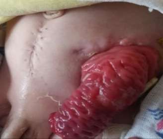
Stoma evagination was observed in 3 patients with double sigmastoma, and in 1 with distal
single-barreled sigmastoma. Fig. 1
Figure 1 - Evagination of the Large Intestine through a Colostomy
ISSN: 2237-0722 24
Vol. 11 No. 2 (2021)
Received: 20.02.2021 – Accepted: 02.04.2021Prolapse can occur with all types of stomas. The prolapse of the intestine through the stoma
was associated with the leaving in the abdominal cavity of a large free pstomal section of the
intestine, which is very mobile and can "turn inside out" through the colostomy opening. If a
colostomy is placed in a fixed part of the colon, prolapse will not occur. The following reasons were
predisposed to this: increased intra-abdominal pressure, the use of foods that increase peristalsis. In 2
patients, 4 cm of bowel prolapse was observed. These children underwent conservative therapy by
correcting nutrition and using a cotton gauze pad (pelot), pressing the surface of the colostomy. In 3
patients there was a prolapse of 12 cm of the intestine, with its infringement in the stoma opening. In
this group of children, we used surgical treatment - stoma reconstruction.
Because of the high incidence of prolapse, loop colostomy is associated with a higher
complication rate than split stoma. Other complications, including megarectum and UTIs, are
independent of the type of stoma. In general terms, the best kind of split sigmoidostomy has not yet
been determined.
Bowel eventration was noted in 1 patient with suspended colostomy, 2 with double
sigmastoma, and 2 with distal unilateral sigmastoma. The colon loops fell out through the incision
wound next to the withdrawn intestinal stoma. This complication developed when a too wide canal
was formed in the abdominal wall. In this situation, an emergency operation was carried out with the
elimination of the intestinal eventration and the reconstruction of the stoma by imposing a
single-barreled colostomy like Hartmann with corrugation of the intestine. The predisposing factors
for eventration were: an increase in intra-abdominal pressure, a change in the regenerative abilities of
tissues in the exhausted state of the patient (with hypotrophy, hypoproteinemia).
Stoma stenosis developed in 4 patients with suspended colostomy, 3 with double sigmastoma,
2 with distal single-barreled sigmastoma, and 1 with Hartmann-type stoma patients. The narrowing
was in the final section of the intestine, at the level of the skin and at a depth - at the level of the
dissected aponeurosis. The reasons for the development of stoma stenosis were: the imposition of
frequent interrupted sutures, which led to hypergranulation of the tissue around the sutures, followed
by impaired circulation of the stoma outlet; a too narrow opening was created in the
musculo-aponeurotic layer and there was a compression of the intestine, followed by its stenosis;
A long-standing stricture can lead to a suprastenotic expansion of the overlying sections with
the development of irreversible processes in the intestinal wall, in the future this can complicate
reconstructive operations on the intestine.
Disconnected bowel syndrome developed in 1 patient with double sigmastoma. The cause of
this complication was the wrong choice of the colostomy height, which led to the "disconnection" of
ISSN: 2237-0722 25
Vol. 11 No. 2 (2021)
Received: 20.02.2021 – Accepted: 02.04.2021the distal colon from normal functioning. This complication made it difficult to carry out further
surgical manipulation.
In 5 children with suspended and in 1 child with double sigmastoma, we observed the
presence of fecal stones in the distal part of the intestine, which is associated with the throwing of
fecal masses into the stoma discharge loop, in the distal part of the colon, fecal stones are formed,
which contributes to the preservation of suprasthenotic expansion and symptoms intoxication
When analyzing the advantages of any of the types of colostomy, I would like to note that
often a colostomy is the only way to create conditions for emptying the intestine against the
background of intestinal obstruction. It is possible to work without infection of surgical wounds,
which prevents the development of postoperative complications in the form of retraction, prolapse of
the rectal mucosa, stenosis of the anus and incontinence phenomena.
Overall, our results support the fact that sigmostomy is more favorable than transverse colon
stomas. Also, our studies show the presence of greater complications from loop stomas than from
separate ones; in particular, this applies to prolapse. It should be noted that the lack of information
from pediatric surgeons about other colostomy techniques also plays a huge role, and more often the
ostomy surgeon mainly uses the technique that he knows best. When forming a colostomy, it is
necessary not only to create an adequate emptying of the intestine, but also to plan the further stage of
the operation anatomically and physiologically in order to avoid complications associated with
incorrect determination of the type and level of colostomy.
4. Conclusion
1. All high forms of anorectal defects, as well as cloacal forms, require preliminary formation of
a "protective" colostomy;
2. Re-corrective operations in all cases of complications should be performed only under the
guise of a "protective" colostomy.
3. Preferably the formation of a single-barreled terminal colostomy (sigmostomy);
4. Colostomy must be taken out through a separate "window" outside the surgical incision;
5. Colostomy is necessary in case of identified concomitant anomalies and defects that clinically
"dominate" over anorectal malformation;
At the same time, it was possible to completely and adequately empty the intestine,
morphologically verify the defective segment of the intestine, avoid the "disconnected" bowel
ISSN: 2237-0722 26
Vol. 11 No. 2 (2021)
Received: 20.02.2021 – Accepted: 02.04.2021syndrome, infection of the urinary system and perform reconstructive surgery in the shortest possible
time and under aseptic conditions.
Acknowledgements
We are grateful to the staff members of Tashkent Pediatric Medical Institute for the
cooperation and support in our research. The participants kindly gave full written permission for this
report.
Consent
Written informed consent was obtained from all participants of the research for publication of
this paper and any accompanying information related to this study.
Conflict of Interest
The authors declare that they have no competing interests.
Funding
No funding sources to declare.
References
Smirnov, A.N., & Poddubnyĭ, I. V. (1990). Surgical treatment of anorectal developmental defects in
children (review of foreign literature). Surgery, (8), 149-155.
Bischoff, A., Levitt, M.A., & Peña, A. (2013). Update on the management of anorectal
malformations. Pediatric surgery international, 29(9), 899-904.
Puri, A., Chadha, R., Choudhury, S.R., & Garg, A. (2006). Congenital pouch colon: follow-up and
functional results after definitive surgery. Journal of pediatric surgery, 41(8), 1413-1419.
Narbaev, T.T., Aliev, M.M., Turaeva, N.N., & Ollabergenov, O.T. (2019). Results of surgical
treatment of anorectal malformation in children. Med. Scientific and Innovative. European Journal of
Pediatrics, 1(1), 136-143.
Aliev, M.M., Razumovsky, A.Y., & Narbaev, T.T. (2019). Modified method of perineal proctoplasty
with anorectal malformation in children. Russian Journal of Pediatric Surgery, Anesthesia and
Intensive Care, 9(3), 33–42.
Degtyareva, Y.G. (2017). Congenital malformations of the anorectal region. Monograph; Rep.
scientific and practical Center of Pediatr. Surg.- Minsk.
ISSN: 2237-0722 27
Vol. 11 No. 2 (2021)
Received: 20.02.2021 – Accepted: 02.04.2021Salamov, K.N. (1989). Anorectal anomalies in combination with malformations of other organs and systems. Probl. Proctology MIIOI Research Institute of Proctology M, 8, 51-54. Aliyev, M.M., Narbaev, T.T., Dzhalalov, M.D., & Kholmetov, Sh Sh. (2017). Concomitant anomalies and malformations with anorectal malformation in children. Scientific and practical Journal of Pediatrics, 3, 6-9. Lautz, T.B. (2015). Vacterl associations in children undergoing surgery for esophageal atresia and anorectal malformations: Implications for pediatric surgeons. Journal of Pediatric Surgery, 50(8), 1245-1250. Mittal, A., Airon, R.K., Magu, S., Rattan, K.N., & Ratan, S.K. (2004). Associated anomalies with anorectal malformation (ARM). The Indian Journal of Pediatrics, 71(6), 509-514. Krasovskaya, T.V., Kobzeva, T.N., Smirnov, A.N., & Novozhilov, V.A. (1989). A program for examining patients with malformations of the anorectal region. Vopr. protection of motherhood and childhood, 8, 25-28. Levin, M.D. (2013). Standardization of x-ray examination of the colon and anorectal zone / MD Levin, YuG Degtyarev, VI Averin, JF Abu-Varda, TM Bolbas. News surgery, 21(4), 90–98. Karsten, K. (2016). Voiding Cystourethrography in the Diagnosis of Anorectal Malformations. / Karsten K, Rothe K, Märzheuser S. European Journal of Pediatric Surgery, 26(6), 494-499. Cuschieri, A., & Eurocat Working Group. (2002). Anorectal anomalies associated with or as part of other anomalies. American journal of medical genetics, 110(2), 122-130. Lenyushkin, A.I., & Chuplak, II. (2001). Problems of combined proctourogenital pathologies in pediatric surgery. Pediatric Surgery, 1, 12-16. Narbayev, T.T., Tilavov, UKh., & Turaeva, N.N. (2018). Rehabilitation of Children with Anorectal Malformations. Journal of Progressing Aspects in Pediatrics and Neonatology, 1(5), 1-5. Ivanov, P.V., Kirgizov, I.V., Baranov, K.N., & Shishkin, I.A. (2010). Stage treatment of anorectal malformations in children. Med. Bulletin, 3, 88-89. Ivanov, V.V., Aksel’rov, M.A., Aksel’rov, V.M., Bel’kovich, S.V., Kostrygin, S.V., Tolkachev, R.A., & Svazyan, V.V. (2007). Colostomy as the first stage of surgical correction of malformations of the anorectal region in newborns. Pediatric surgery, (2), 6-8. Holschneider, A., Hutson, J., Peña, A., Beket, E., Chatterjee, S., Coran, A., & Kunst, M. (2005). Preliminary report on the International Conference for the Development of Standards for the Treatment of Anorectal Malformations. Journal of pediatric surgery, 40(10), 1521-1526. Aliyev, M.M., Narbayev, T.T., Turayeva, N.N., Bozorov, SH.T. (2011). Features of protective colostomy in children with anorectal malformation. Scientific and practical journal Pediatrics, 34-37. Aliyev, M.M., Narbayev, T.T., Turayeva, N.N., Chuliyev, M.S., & Nasyrov, M.M. (2016). Choice of method and level of colostomy in children with Hirschsprung's disease and anorectal malformation. Journal Bulletin of the Association of Doctors of Uzbekistan, 2, 95-99. Narbaev, T.T., Nosirov, A.A., Terebaev, B.A., Turaeva, N.N., Tilavov, U.K., Bayakhmedov, F.F., & Sobitov, I.Z. (2020). Assessment of the State of the Rectal Sphincter Apparatus in Anal Incontinence in Children after Surgery. Journal of Advanced Medical and Dental Sciences Research, 8(9), 69-73. ISSN: 2237-0722 28 Vol. 11 No. 2 (2021) Received: 20.02.2021 – Accepted: 02.04.2021
You can also read