Imaging findings of renal telangiectasia in a Maltese dog
←
→
Page content transcription
If your browser does not render page correctly, please read the page content below
Case Report                                                      Veterinarni Medicina, 65, 2020 (10): 457–463

                                                                      https://doi.org/10.17221/87/2020-VETMED

Imaging findings of renal telangiectasia
in a Maltese dog
Daji Noh1, Seoung-Woo Lee2, Donguk Jung3, Jin-Kyu Park2, Kija Lee1*
1
  Department of Veterinary Diagnostic Imaging, College of Veterinary Medicine,
  Kyungpook National University, Daegu, Republic of Korea
2
  Department of Veterinary Clinical Pathology, College of Veterinary Medicine,
  Kyungpook National University, Daegu, Republic of Korea
3
  Jung Animal Clinic, Daegu, Republic of Korea
*Corresponding author: leekj@knu.ac.kr

Citation: Noh D, Lee SW, Jung D, Park JK, Lee K (2020): Imaging findings of renal telangiectasia in a Maltese dog. Vet
Med-Czech 65, 457–463.

Abstract: A 13-year-old neutered male Maltese dog presented with a three-month history of haematuria. The abdomi-
nal ultrasound and computed tomography revealed a large, multifocally-calcified left renal mass that was disrupting
most of the renal parenchyma. Most of the areas of this mass did not show any contrast enhancement on the CT.
After a nephrectomy, renal telangiectasia was diagnosed on the histopathology. Renal telangiectasia is a heredi-
tary dysplastic blood vessel disease only previously reported in Pembroke Welsh Corgi. This is the first diagnostic
imaging report of renal telangiectasia and the first report in a Maltese. Renal telangiectasia should be considered
for the differential diagnosis of destructive renal masses with faint or no contrast enhancement on a CT, even
in cases of unilateral renal involvement in Maltese dogs or those in non-corgi breeds.

Keywords: canine; computed tomography; non-corgi breed; renal mass

  The term renal telangiectasia was first used in             To the best of our knowledge, this article repre-
1983 in the field of veterinary medicine to dif-            sents the first case report of renal telangiectasia
ferentiate it from renal haemangioma. Renal tel-            in a Maltese and is the first to describe the diag-
angiectasia is defined as a rare dysplastic blood           nostic imaging findings with radiographs, ultraso-
vessel disease in dogs (Moore and Thorton 1983).            nography, and computed tomography (CT).
A similar haemangiomatous disease in humans is
hereditary haemorrhagic telangiectasia charac-
terised as an autosomal dominant, multi-systemic            Case description
disorder of angiogenesis with severe and recurrent
haemorrhaging (Sadick et al. 2005). Renal telangi-            A 13-year-old neutered male Maltese dog pre-
ectasia in dogs is also thought to be a genetic dis-        sented with a history of haematuria for three
order, and, up to this point, has only been reported        months. There was no prior history of urinary dis-
in Pembroke Welsh Corgis. In a previous report              orders, and the owner had not seen stranguria or
on Pembroke Welsh Corgis, the histopathologi-               dysuria. The patient was alert at presentation, and
cal features of renal telangiectasia were described         there were no significant findings on the physical
in detail, but diagnostic imaging was limited to ra-        examination other than the marked, continuous
diographs only (Moore and Thorton 1983).                    haematuria when pressure was applied to the ab-

457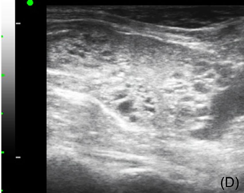
Case Report                                                      Veterinarni Medicina, 65, 2020 (10): 457–463

                                                                      https://doi.org/10.17221/87/2020-VETMED

domen. The complete blood count tests were un-               mulated in the renal pelvic region. Renal degenera-
remarkable and a serum chemistry panel revealed              tion of the right kidney, multiple septated anechoic
elevated blood urea nitrogen (570 mg/l; reference            hepatic cysts, and prostatomegaly with small-sized
range, 70–270 mg/l) and lactate (32.9 mg/l; refer-           cysts were detected (Figure 2A–2C).
ence range, 5–25 mg/l). The urine stick test was               Subsequent thoracic and abdominal CT images
not readable due to an excess of red blood cells             were acquired using a 32-slice CT scanner with-
in the sample.                                               out contrast (Alexion; Toshiba Medical Systems,
  Thoracic and abdominal radiographs were ac-                Ohtawara, Japan). The patient was positioned in
quired (AccuRay-603R; DK Medical Systems, Seoul,             ventral recumbency on the CT table under general
Republic of Korea). On the abdominal radiograph,             anaesthesia. The CT technical parameters were as
an irregularly shaped, soft tissue opacity in the left       follows: 120 kV, 150 mA, a 1.0 mm slice thickness,
upper abdomen was suspected to be associated with            and a 0.75 s rotation time. All the CT data were
the left kidney. A multifocal bone opacity material          reconstructed as cross-sectional in the transverse,
was sporadically distributed at the caudal portion           sagittal, and dorsal planes at 3 mm slice thickness.
of the mass. The contour of the right kidney was             Contrast studies were then performed with the in-
unclear to evaluate (Figure 1). No significant find-         travenous administration of iohexol (600 mg/kg;
ings were observed on the thoracic radiographs.              Omnipaque 300; GE Healthcare, Cork, Ireland)
  An abdominal ultrasound was performed (Pro-                for 20 s using an autoinjector (Medrad ®; Bayer
sound F75; Hitachi Aloka Medical, Tokyo, Japan)              HealthCare LLC, Whippany, USA). Three-phase
with a linear array (10–13 MHz). A large, left-side          post-contrast CT images were acquired 20, 30, and
renal mass with a heterogeneous echo pattern and             90 s after injection. The left renal mass (5.1 × 5.4 ×
multifocal mineralisation was visualised. The size           5.9 cm) was irregularly marginated and isoattenu-
measurement of the renal mass was confounded                 ating (46 HU) originating from the cranial renal
by its amorphous shape and poor distinction from             pole with parenchymal mineralisation in the cau-
the normal renal tissue. Anechoic fluid had accu-            dal part of the mass. The mass occupied most of

                                                                        (A)

                                                                                                                  (B)
Figure 1. Right lateral (A) and ventrodorsal (B) abdominal radiographs. A well-defined, irregularly shaped, soft tissue
mass (arrowheads) is visualised in the left upper abdomen. Multifocal bone opacity material is sporadically distrib-
uted at the caudal portion of the mass. Prostatomegaly and sacralisation of the seventh lumbar vertebrae are also
observed

458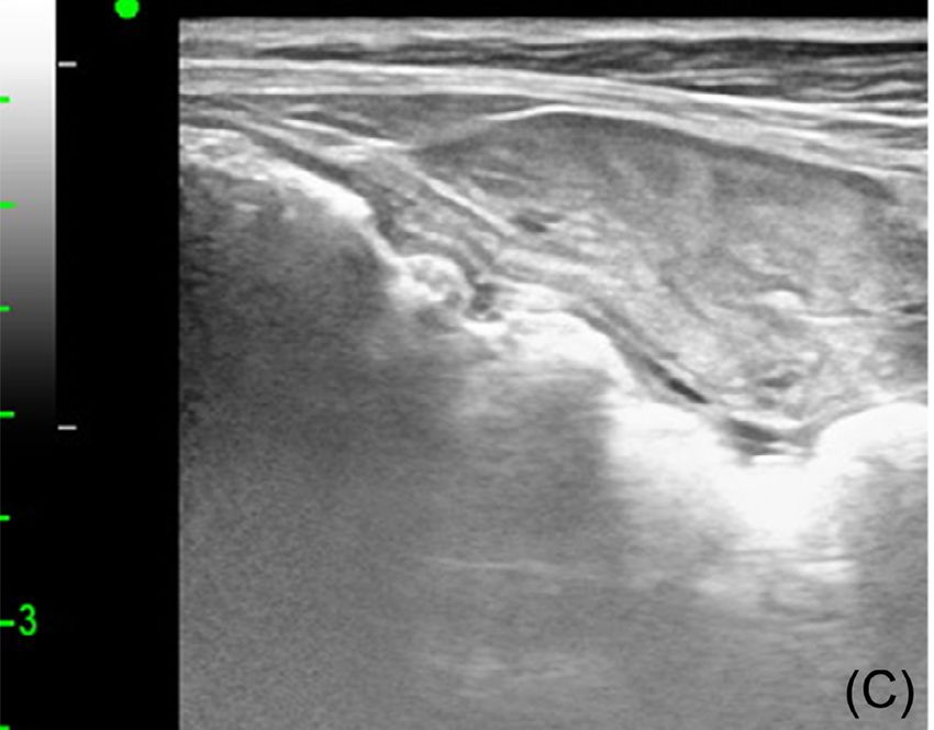
Case Report                                                     Veterinarni Medicina, 65, 2020 (10): 457–463

                                                                     https://doi.org/10.17221/87/2020-VETMED

                                                    (A)                                                        (B)

                                                               Figure 2D

                                                    (C)                                                         (D)
Figure 2. Ultrasonographic image of the renal mass (A), hepatic cysts (B), prostate at presentation (C), and prostate
one year after the nephrectomy (D). The normal renal structure is disrupted, and the renal mass contains multifocal
mineralisation and anechoic fluid (arrowheads) in the pelvic region (A). Septated anechoic hepatic cysts are shown
(B). Small-sized prostatic cysts at presentation (C) is changed to multiple newly formed, spoke-wheels shaped pros-
tatic cysts one year after nephrectomy (D)

the renal parenchyma and had invaded the left               Ingelheim, Germany) were administered intrave-
proximal ureter. Most of the renal mass was not             nously for prophylaxis and analgesia. Anaesthesia
contrast-enhanced, but the vessel and slight ring           was induced with propofol and maintained with
enhancement was shown (Figure 3). There was no              isoflurane (Ifran®; Hana Pharm., Seoul, Republic
apparent evidence of a pulmonary metastasis or              of Korea) and the dog was positioned in a dorsal re-
regional lymphadenopathy. Multiple hepatic cysts            cumbency. After routine preparation of the abdo-
(Figure 3) and prostatomegaly were detected.                men for aseptic surgery, a ventral midline incision
  From the above haematuria and diagnostic im-              was made. Upon surgery, the left kidney was easily
aging findings, a malignant renal tumour was                separated from the peritoneum. Overall, the left
at the top of the list of differential diagnoses, but       kidney was enlarged and well-capsulised without
differentiating the subtype of the renal tumour was         any peritoneal adherence containing an irregularly
not possible on the CT.                                     marginated brownish cranial pole.
  After obtaining the owner’s consent, the dog                Using a vascular clip, the kidney with capsule and
underwent a nephrectomy of the left kidney.                 ureter were removed together. Closure of the sur-
Cefazolin (20 mg/kg, Cefozol; Hankook Korus                 gical wound was performed in a routine fashion.
Pharm Co., Seoul, Republic of Korea) and meloxi-            The dog was hospitalised for 5 days for postopera-
cam (0.2 mg/kg, Metacam; Boehringer Ingelheim,              tive observation.

459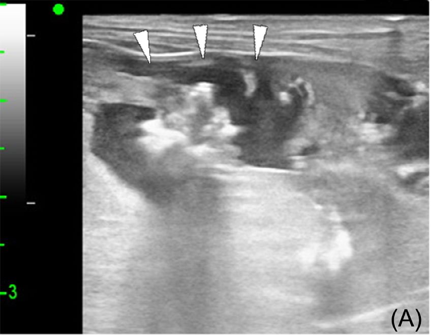
Case Report Veterinarni Medicina, 65, 2020 (10): 457–463
https://doi.org/10.17221/87/2020-VETMED
(A) (B) (C) (D)
(E) (F) (G) (H)
I (J) (K) (L)
Figure 3. CT images of the non-contrast (A, E, I, K), post-contrast corticomedullary (B, F), early (C, G), and late (D,
H, J, L) nephrographic phases in a soft tissue window setting (WW 450, WL 40). The left renal mass occupies most
of the renal parenchyma, while only a small portion of normal renal structure remains(white arrows). This renal
mass is shown as isoattenuating (46 HU) except for the mineralisation lesions (A). Most of this area does not con-
trast enhance (46, 45, and 46 HU on the corticomedullary, early, and late nephrographic phases, respectively), but
the vessel (white arrowheads) and slight ring enhancement are shown in the corticomedullary and nephrographic
phases (B–D, F–H). Furthermore, sporadic parenchymal mineralisations in the caudal part of the mass are shown
(E). This renal mass invades the left proximal ureter (black arrows; I, J). Multiple hepatic cysts (black arrowheads;
11 and 13 HU on the non- and post-contrast, respectively) are detected (K, L)
The postoperative histopathologic diagnosis of maturia. At the one-year follow-up, the dog was
the mass was renal telangiectasia (Figure 4). The still healthy without any clinical signs of urinary
patient recovered well and showed no further hae- disease. However, an abdominal ultrasound showed
multiple progressive cystic lesions in the liver and
newly formed, spoke-wheel shaped cysts in the
prostate gland (Figure 2D).
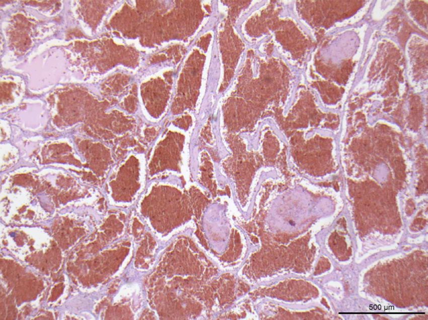
Figure 4. Histopathological section of the renal mass
showing large numbers of blood-filled cysts replacing
most of the renal parenchyma. The cysts are made up
of single-layered, well differentiated endothelial cells
and separated by thick, fibrous septa without neoplas-
tic changes. The dilated vessels contain large numbers
of red blood cells, fibrin thrombi, and acute inflamma-
tory cells such as neutrophils. The image also shows
diffuse interstitial renal fibrosis and multi-focal calcifi-
cation. Haematoxylin-eosin stain; Scale bar = 500 µm
460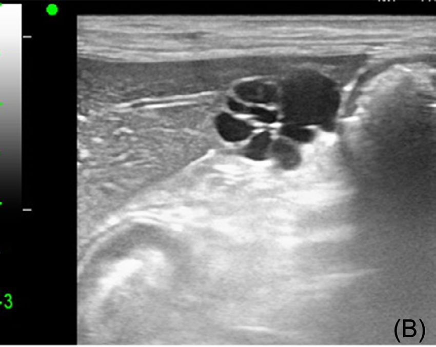
Case Report                                                 Veterinarni Medicina, 65, 2020 (10): 457–463

                                                                https://doi.org/10.17221/87/2020-VETMED

DISCUSSION AND CONCLUSIONS                              are frequently calcified, particularly in renal cell
                                                        carcinomas, while the calcification associated with
  Renal telangiectasia is a genetic, rare dysplas-      benign renal tumours are relatively rare (Weyman
tic blood vessel disease in dogs that has only been     et al. 1982). These reports suggest a tendency to-
reported in Pembroke Welsh Corgis (Moore and            wards calcification in malignant renal tumours,
Thorton 1983). In this case, a primary renal tu-        though comprehensive studies in dogs have yet
mour was at the top of the differential diagnosis       to be published. Considering that renal telangiec-
based on the clinical and diagnostic imaging find-      tasia can also be calcified, the presence of the cal-
ings, while the histopathologic diagnosis was renal     cification alone is insufficient to distinguish renal
telangiectasia. In general, differential diagnoses      telangiectasia from a renal malignant tumour
of unilateral renomegaly with contour distortions       on the diagnostic imaging.
include primary renal neoplasia, metastatic neo-          In this case, the large area of the renal mass did
plasia, abscesses, haematoma, granuloma, cysts,         not show any contrast enhancement on the CT
or hamartoma (Seiler 2018).                             that was different from other common renal tu-
  In cases of haematuria with a unilateral renal        mours, such as renal cell carcinoma, renal sarcoma,
mass and obliteration of the normal renal structure,    and haemangioma (Sheth et al. 2001; Katabathina
a primary renal tumour such as renal carcinoma,         et al. 2010). Because of the low incidence and
sarcoma, or nephroblastoma is often strongly sus-       lack of large-scale CT studies on canine primary
pected, and nephrectomy is indicated to improve         renal tumours, a potential correlation between
the likelihood of the survival (Nyland et al. 2002;     the malignancy and contrast enhancement has
Bryan et al. 2006; Seiler 2018).                        yet to be verified. Only two cases of non-contrast-
  In this case, these prior perspectives were not       enhanced renal masses have been reported in dogs,
sufficient to distinguish the renal telangiectasia      for renal fibrosarcoma and renal angiomyxoma
from a primary renal tumour. This unilateral re-        (Gajanayake et al. 2010; Park et al. 2015). The atten-
nal mass also differed from findings in a previ-        uation of the contrast enhancement also depends
ous renal telangiectasia study showing bilateral        on the amount of iodine deposition in the target
renal involvement (Moore and Thorton 1983).             organ, the organ’s intravascular blood volume,
Suppositions regarding this discrepancy include         the vascular resistance, the capillary or venous
that renal telangiectasia may only be apparent uni-     anatomy, and the intravascular or interstitial envi-
laterally, or that the second, mildly affected kidney   ronment (Herman 2004). A vascular malformation
may not show any structural changes that could          can cause increased interstitial pressure, hypoxia,
be detected on the imaging, necessitating a histo-      and large diffuse distances for the intravascular
pathology for confirmation. In either possibility,      molecules and the lack of contrast enhancement
renal telangiectasia should be considered in the dif-   in the renal mass in this dog with renal telangiecta-
ferential in cases of haematuria with a unilateral      sia may have resulted from dysplastic blood vessels
renal mass in the imaging, in both the Corgi and        (Jain and Kozak 2008).
Maltese breeds.                                           Although CT studies on canine renal tumours are
  The only imaging findings discussed in the pre-       scarce, the presence of a non-contrast-enhanced
vious report of renal telangiectasia were renal cal-    renal mass may help differentiate the renal telan-
culi and calcification on conventional radiographs      giectasia from other malignant renal tumours.
in a subset of dogs with haematuria that had per-         In this case, multiple progressive hepatic cysts
sisted for several years (Moore and Thorton 1983).      and prostatic cysts were also identified. In a previ-
These findings were consistent with the present         ous study of renal telangiectasia, multiple organs
case that showed sporadic calcification throughout      were found to be involved upon the histopatho-
the renal mass upon imaging.                            logical examination, but no vascular abnormalities
  Several case studies have reported malignant          related to the haemangiomatous syndrome were
renal tumours with calcification such as a renal        reported (Moore and Thorton 1983). In humans,
adenocarcinoma (Konde et al. 1985), a renal inter-      von Hippel-Lindau disease, a haemangiomatous
stitial cell tumour (Ditersand and Wells 1986), and     syndrome, is associated with the cyst forma-
a sarcomatoid renal cell carcinoma in dogs (Zini        tion in multiple organs and an over-production
et al. 2003). In humans, malignant renal tumours        of vascular endothelial growth factors affecting

461
Case Report Veterinarni Medicina, 65, 2020 (10): 457–463
https://doi.org/10.17221/87/2020-VETMED
angiogenesis (Moore and Thorton 1983; Na et al. Bryan JN, Henry CJ, Turnquist SE, Tyler JW, Liptak JM,
2003; Sadick et al. 2005). Recent human studies Rizzo SA, Sfiligoi G, Steinberg SJ, Smith AN, Jackson T.
have also revealed highly expressed vascular endo- Primary renal neoplasia of dogs. J Vet Intern Med. 2006
thelial growth factors and the therapeutic effects Sep-Oct;20(5):1155-60.
of anti-vascular endothelial growth factors in he- Cnossen WR, Drenth JP. Polycystic liver disease: An over-
reditary haemorrhagic telangiectasia (Sadick et al. view of pathogenesis, clinical manifestations and manage-
2005; Ardelean and Letarte 2015). Vascular endo- ment. Orphanet J Rare Dis. 2014 May 1;9:1-13.
thelial growth factors have also been implicated Diters RW, Wells M. Renal interstitial cell tumors in the dog.
in polycystic liver and kidney disease (Cnossen and Vet Pathol. 1986 Jan;23(1):74-6.
Drenth 2014). These findings suggest that the mul- Gajanayake I, Priestnall SL, Benigni L, English K, Summers
tiple progressive hepatic and prostatic cysts ob- BA, Garden OA. Paraneoplastic hypercalcemia in a dog
served in this case could be associated with renal with benign renal angiomyxoma. J Vet Diagn Invest. 2010
telangiectasia, although no empirical data therein Sep;22(5):775-80.
has been reported for dogs. Herman S. Computed tomography contrast enhancement
Further studies about the correlation of overex- principles and the use of high-concentration contrast
pressed vascular endothelial growth factors with media. J Comput Assist Tomogr. 2004 Jul-Aug;28:S7-11.
telangiectasia or a polycystic disease in canines are Jain RK, Kozak KR. Molecular pathophysiology of tumors.
needed to elucidate their pathology and uncover In: Halperin EC, Parez CA, Brady LW, editors. Principles
appropriate therapeutic modalities. and practice of radiation encology. Philadelphia, USA:
There were several limitations to this case that Lippincott Williams & Wilkins; 2008. p. 126-41.
must be acknowledged. First, a fine needle aspira- Katabathina VS, Vikram R, Nagar AM, Tamboli P, Menias
tion and tissue core biopsies of the mass were not CO, Prasad SR. Mesenchymal neoplasms of the kidney
performed before the nephrectomy. Second, the in adults: Imaging spectrum with radiologic-pathologic
optimal artery, corticomedullary, nephrographic, correlation. Radiographics. 2010 Oct;30(6):1525-40.
and excretory phases of the kidneys were not ac- Konde LJ, Wrigley RH, Park RD, Lebel JL. Sonographic ap-
quired in the contrast CT study because bolus- pearance of renal neoplasia in the dog. Vet Radiol. 1985
tracking was not performed. May;26(3):74-81.
However, this case is valuable in that it describes Moore FM, Thorton GW. Telangiectasia of Pembroke welsh
several imaging findings in renal telangiectasia that corgi dogs. Vet Pathol. 1983 Mar;20(2):203-8.
could be helpful in distinguishing it from a primary Na X, Wu G, Ryan CK, Schoen SR, di’Santagnese PA, Mess-
renal tumour. ing EM. Overproduction of vascular endothelial growth
This study also indicated that renal telangiectasia factor related to von Hippel-Lindau tumor suppressor
may appear unilaterally on the diagnostic imaging gene mutations and hypoxia-inducible factor-1 alpha
and is not limited to Pembroke Welsh Corgis, un- expression in renal cell carcinomas. J Urol. 2003 Aug;
like previous reported incidences of renal telan- 170:588-92.
giectasia. Nyland TG, Widmer WR, Mattoon JS. Urinary tract. In:
The findings also suggested that the renal telan- Mattoon JS. Nyland TG, editors. Small animal diagnostic
giectasia might be related to multiple cystic lesions ultrasound. St. Louis, USA: Elsevier Health Sciences;
in other organs. In conclusion, renal telangiectasia 2002. p. 557-607.
should be considered for the differential diagnosis Park HA, Jeong CW, Kim GS, Kim HJ, Do SH, Park HM.
of destructive renal masses with faint or no contrast Primary renal fibrosarcoma with local invasion into the
enhancement on a CT, even in cases of unilateral mesenteric membrane of a mongrel dog. Korean J Vet
renal involvement in Maltese or other non-Corgi Res. 2015 Mar;55(1):65-9.
breeds. Sadick H, Naim R, Gossler U, Hormann K, Riedel F. Angio-
genesis in hereditary hemorrhagic telangiectasia: VEGF165
plasma concentration in correlation to the VEGF expres-
REFERENCES sion and microvessel density. Int J Mol Med. 2005 Jan;
15(1):15-9.
Ardelean DS, Letarte M. Anti-angiogenic therapeutic strat- Seiler GS. Kidneys and ureters. In: Thrall DE, editor. Text-
egies in hereditary hemorrhagic telangiectasia. Front book of veterinary diagnostic radiology. St. Louis, US:
Genet. 2015 Feb 11;6:35. Elsevier Health Sciences; 2018. p. 823-45.
462Case Report Veterinarni Medicina, 65, 2020 (10): 457–463
https://doi.org/10.17221/87/2020-VETMED
Sheth S, Scatarige JC, Horton KM, Corl FM, Fishman EK. Zini E, Bovero A, Nigrisoli E, Ratto A, Rampazzo A, Zatelli A.
Current concepts in the diagnosis and management of re- Sarcomatoid renal cell carcinoma with osteogenic differ-
nal cell carcinoma: Role of multidetector CT and three- entiation and paraneoplastic hepatopathy in a dog, pos-
dimensional CT. Radiographics.2001 Oct;21:S237-54. sibly related to human Stauffer’s syndrome. J Comp Pathol.
Weyman PJ, McClennan BL, Lee JK, Stanley RJ. CT of cal- 2003 Nov;129(4):303-7.
cified renal masses. AJR Am J Roentgenol. 1982 Jun;138
(6):1095-9. Received: April 11, 2020
Accepted: August 26, 2020
463You can also read