Retroperitoneal Liposarcoma: A Case Report and Review of Literature - Global Journals
←
→
Page content transcription
If your browser does not render page correctly, please read the page content below

Global Journal of Medical Research: F
                          Diseases
                          Volume 21 Issue 3 Version 1.0 Year 2021
                          Type: Double Blind Peer Reviewed International Research Journal
                          Publisher: Global Journals
                          Online ISSN: 2249-4618 & Print ISSN: 0975-5888

Retroperitoneal Liposarcoma: A Case Report and Review of
Literature
                                                                             By Dr. Elize Isabella Wethmar
Introduction- Liposarcomas are rare malignant tumours of adipocytic differentiation and are
classified under the soft tissue sarcoma subtype histologically. Retroperitoneal liposarcoma
(RPL) has an incidence of 1 per 2.5 million individuals, and the retroperitoneum is the second
most common site for a liposarcoma, following the lower limb as the most common site. The
retroperitoneum is a space that can easily expand;therefore tumours grow in this space without
any symptoms until they are very large.
        This case study reports a patient diagnosed with a retroperitoneal liposarcoma treated
with primary radical surgery and the patient is currently being followed-up in our unit with close
monitoring and Computed Tomographic (CT) imaging. The patient consented to the reporting of
this case.
GJMR-F Classification: NLMC Code: WJ 768

RetroperitonealLiposarcomaACaseReportandReviewofLiterature

                                                             Strictly as per the compliance and regulations of:

© 2021. Dr. Elize Isabella Wethmar. This is a research/review paper, distributed under the terms of the Creative Commons
Attribution-Noncommercial 3.0 Unported License http://creativecommons.org/licenses/by-nc/3.0/), permitting all non-commercial
use, distribution, and reproduction in any medium, provided the original work is properly cited.
Retroperitoneal Liposarcoma: A Case Report
                and Review of Literature
                                                       Dr. Elize Isabella Wethmar

                  I.      Introduction                               offered to the patient. The patient consented to the
                                                                     procedure. A mid-line laparotomy was performed, and a

L
      iposarcomas are rare malignant tumours of                      large retroperitoneal mass was found. This mass was
      adipocytic differentiation and are classified under            adherent to the left ovary, left ureter, left psoas muscle
      the soft tissue sarcoma subtype histologically.                and external iliac vessels as well as the sigmoid colon.

                                                                                                                                            2021
Retroperitoneal liposarcoma (RPL) has an incidence of 1              With careful anatomical dissection the mass was
per 2.5 million individuals, and the retroperitoneum is the          resected, and a full staging laparotomy and lymph node

                                                                                                                                                      Year
second most common site for a liposarcoma, following                 sampling of the left pelvic and para-aortic lymphnodes
the lower limb as the most common site. The                          was performed. The specimen comprised of a 527g                                       1
retroperitoneum is a space that can easily                           encapsulated, lobulated portion of tissue, with a
expand;therefore tumours grow in this space without

                                                                                                                                           Global Journal of Medical Research ( FD ) Volume XXI Issue III Version I
                                                                     histological diagnosis of a well differentiated
any symptoms until they are very large.                              liposarcoma, sclerosing variant. No areas of
         This case study reports a patient diagnosed                 dedifferentiated liposarcoma were noted and the
with a retroperitoneal liposarcoma treated with primary              margins were clear of tumour. All resected lymph nodes
radical surgery and the patient is currently being                   were negative for metastatic disease. The patient had
followed-up in our unit with close monitoring and                    an uneventful post-operative recovery.
Computed Tomographic (CT) imaging. The patient                                Although the retroperitoneal liposarcoma
consented to the reporting of this case.                             appears to have been removed completely in this
                   II.     Case Report                               patient, local recurrence is likely. Since there is currently
                                                                     no evidence that radiotherapy or chemotherapy
         A 70-year-old female patient was referred to our            improves survival rates or recurrence rates of the
unit for assessment of anasymptomatic solid left sided               disease in the immediate adjuvant setting, the patient
adnexal mass seen on CT scan during evaluation for                   did not receive any adjuvant therapy and is being
aurological complaint.                                               monitored closely with clinical examinations and 3
         At the time of the referral the mass measured               monthly CT-scans to evaluate for any local or distant
5.5 x 4.9 x .7.2 cm with a macroscopic fat and a large               recurrent disease.
soft tissue component on CT-scan assessment. Tumour
markers were all essentially normal, CA 125 of 15 U/mL,                              III.    Discussion
CA 19.9 of U/mL, CEA of 2.8 ug/L and the AFP 13.4 k/U,
                                                                     a) Surgical and Anatomical features
which is a slightly raised level. During this assessment,
                                                                              The retroperitoneum and the preperitoneum
an excisional procedure was offered to the patient, but
                                                                     forms the extraperitoneal space, which is the portion of
due to the Covid 19 pandemic the patient opted to wait
                                                                     the pelvis and abdomen which does not lie within the
with a surgical procedure and follow-up at a later stage.
                                                                     peritoneal space. The retroperitoneal space is an almost
         The patient presented 9 months later for re-
                                                                     virtual and expandable space, defined anteriorly by the
assessment. During this time the mass had increased
                                                                     peritoneal extensions anchoring the transverse colon,
significantly in size and had become symptomatic and
                                                                     the small bowel as well as the 1 ascending and
palpable in the left lower quadrant of her abdomen. On
                                                                     descending colon, part of the duodenum, part of the
repeat CT-scan the mass appeared to have significantly
                                                                     pancreas and part of the liver1 2 3. The retroperitoneum
increased in size, measuring 12x 8 x 14 cm. The mass
                                                                     contains the kidneys, the adrenal glands, the pancreas,
still had a predominantly solid appearance and was
                                                                     part of the duodenum, ascending and descending
strongly associated to the left ovary and likely ovarian in
                                                                     colon, the abdominal aorta and vena cava (dividing into
origin. Ovarian cancer markers were repeated and as
                                                                     the common iliac, external and internal iliac arteries and
above stayed within normal limits. Due to the
                                                                     veins respectively), the abdominal lymphnodes groups
appearance of the lesion and the very rapid growth, a
                                                                     and tracts, six major nerves and the autonomic lumbar
non-benign lesion, possibly of ovarian origin was
                                                                     nerve chains and the connective tissue of fasciae, with
suspected and an explorative/staging laparotomy was
                                                                     the White line of Toldt as the fusion between the
Author: Life the Glynnwood Hospital, Benoni, South Africa.           mesocolon and the posterior retroperitoneum.
e-mail: elize.wethmar@gmail.com

                                                                                                                  © 2021 Global Journals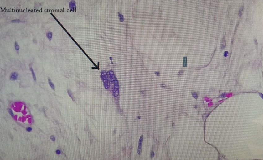
Retroperitoneal Liposarcoma: A Case Report and Review of Literature

                                                                                    Treatment of most conditions involving the             b) Histopathological features
                                                                           retroperitoneum requires surgical intervention. A                         Retroperitoneal liposarcomas usually consist of
                                                                           complete and thorough understanding of the anatomy              a large, well-circumscribed, lobulated mass. Variable
                                                                           of the structures involving the retroperitoneum is prudent      consistencies are present, from yellow to firm grey to
                                                                           in avoiding inadvertent damage to underlying structures.        gelatinous areas, depending on the proportion of fat,
                                                                           Complications may arise from inadvertent damage to              fibrous and myxoid components. Larger retroperitoneal
                                                                           structures located within the retroperitoneum during surgical   tumours appear more heterogeneous, often containing
                                                                           manipulation or instrumentation4.                               foci of fat necrosis and punctate haemorrhages5.
 2021      Year

                2
Global Journal of Medical Research ( FD ) Volume XXI Issue III Version I

                                                                                                    Figure 1: Macroscopic appearance of the excised retroperitoneal liposarcoma
                                                                                     As in many tumours, the histological                  present in various numbers, but the presence or
                                                                           classification of liposarcoma has evolved over the past         absence is not necessary for the diagnosis of a
                                                                           several decades, mostly owing to the advances in our            liposarcoma. Sclerosing atypical lipomatous tumour
                                                                           understanding of molecular genetics.                            presents second most frequently and is most often seen
                                                                                     The most recent World Health Organisation             in the retroperitoneum or spermatic cord. The most
                                                                           (WHO) classification system (2020) recognized five              important histological finding is scattered bizarre stromal
                                                                           different types of major liposarcoma subtypes,                  cells, showing marked nuclear hyperchromasia.
                                                                           differentiated by distinctive morphologies and unique           Inflammatory ALT represents the rarest subtype,
                                                                           genetic findings 5:                                             occurring most often in the reproperitoneum5.
                                                                           •     Atypical lipomatous tumour (ALT)/ well differentiated
                                                                                 liposarcoma (includes adipocytic [or lipoma like],
                                                                                 sclerosing and inflammatory variants)
                                                                           •     Dedifferentiated liposarcoma
                                                                           •     Myxoid liposarcoma
                                                                           •     Pleomorphic liposarcoma
                                                                           •     Myxoid pleomorphic liposarcoma
                                                                                   In atypical lipomatous tumour/well differentiated
                                                                           liposarcoma adipocytic variant consists out of cells that
                                                                           vary substantially in size as well as cells that have
                                                                           nuclear atypia in fat or spindle cells. Lipoblasts can be

                                                                           © 2021 Global Journals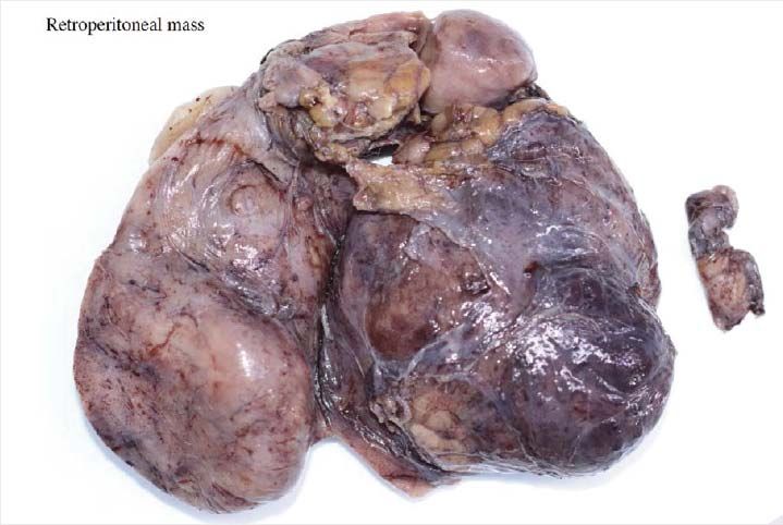
Retroperitoneal Liposarcoma: A Case Report and Review of Literature

                                                                                                                    2021      Year
                                                                                                                                   3

                                                                                                                   Global Journal of Medical Research ( FD ) Volume XXI Issue III Version I
     Figure 2: Low power microscopy view demonstrating a deifferentiated liposarcoma

Figure 3: A microscopic view of a lipoblast, which may or may not be present in a liposarcoma

                                                                                          © 2021 Global Journals
2021      Year                                                                                     Retroperitoneal Liposarcoma: A Case Report and Review of Literature

                4
Global Journal of Medical Research ( FD ) Volume XXI Issue III Version I

                                                                                   Figure 4: A multinucleated stromal cell which is present in a Sclerosing atypical lipomatous tumour during
                                                                                                                                   microscopy

                                                                                     Dedifferentiated liposarcoma can arise as a         presence of fat within a retroperitoneal lesion is helpful
                                                                           synchronous lesion in 90% of cases and as                     in refining the differential diagnosis 9. It is easy to
                                                                           metachronous lesion in 10% of cases 6. These tumours          recognize fat within a lesion due to its characteristic
                                                                           exhibit a wide morphological spectrum and                     imaging appearance:
                                                                           histologically show areas of high grade, poorly               •   On ultrasound appearance it is hyperechoic and
                                                                           differentiated     sarcoma      resembling   high-grade           may demonstrate posterior acoustic shadowing
                                                                           myxofibrosarcoma, fibrosarcoma, malignant solitary
                                                                                                                                         •   Computed Tomographic (CT) imaging derives
                                                                           fibrous tumour or pleomorphic sarcoma not otherwise
                                                                                                                                             contrast parameters predominantly from the
                                                                           specified. Dedifferentiated liposarcomas can be of
                                                                                                                                             physical properties of tissue – in combination with
                                                                           variable      histological    grade5.    Dedifferentiated
                                                                                                                                             high resolution spiral CT, this provides accurate
                                                                           liposarcomas is an aggressive disease, arising most
                                                                                                                                             attenuation measurement, with – 10 to -100
                                                                           commonly in the retroperitoneum and is associated with
                                                                                                                                             HU9corresponding to fat 10
                                                                           high rates of local and metastatic recurrence and
                                                                           disease specific mortality 7.                                 •   Magnetic resonance imaging (MRI) has lower
                                                                                                                                             spatial resolution than CT imaging, but better soft
                                                                                     Use of the term atypical lipomatous tumour is
                                                                                                                                             tissue contrast and greater sensitivity in detection of
                                                                           determined by tumour location and resectability. In
                                                                                                                                             microscopic fat. The two MRI techniques for the
                                                                           locations such as the retroperitoneum, it is usually
                                                                                                                                             identification of fat within a structure are fat
                                                                           impossible to obtain a wide tumour free surgical margin
                                                                                                                                             saturation and chemical shift imaging
                                                                           of more than 2cm, thus local recurrence is common and
                                                                           leads to mortality, seen in the absence of                              It can be difficult to localize large abdominal
                                                                           dedifferentiation or metastases 8.                            masses to an anatomical space and to accurately
                                                                                                                                         identify the organ of origin thus to determine whether the
                                                                           c) Radiological features                                      lesion arises from a retroperitoneal organ, or from the
                                                                                    The introduction of computed tomography (CT),        soft tissue10. If the mass is surrounded by the
                                                                           magnetic       resonance       imaging      (MRI),   and      parenchyma of the organ, it undoubtedly arises from the
                                                                           ultrasonography have greatly enhanced our capability to       organ, however if the mass originates at the margin of
                                                                           make the diagnosis of abdominopelvic neoplasms and            the organ, it is more difficult the determine the origin.
                                                                           determine and direct treatment, as well as observe the        The interface between the mass with the adjacent organ
                                                                           progress of the neoplasm and effect of treatment over         can provide insight as to whether it displaces the organ
                                                                           time. Identification of a retroperitoneal mass at imaging     or arises from the organ (Table 1).
                                                                           is a challenging task for radiologists, however the

                                                                           © 2021 Global Journals
Retroperitoneal Liposarcoma: A Case Report and Review of Literature

          Table 1: Positive signs indicating that a Retroperitoneal Tumour Arises from an Adjacent Organ9
                    Sign                                                 Definition
       Beak sign                        Sharp beak shape of organ of origin occurs at the edge of the interface with
                                        the tumour
       Embedded organ Sign              Organ of origin is encased by the tumour, without a sclerotic interface
       Phantom (invisible) organ sign   Organ of origin is obliterated by the tumour
       Prominent feeding artery sign    Large feeding arteries of a hypervascular tumour point to the organ of origin

         A CT image of a lipoma will reveal a well-            containing lesions with minimal soft tissue attenuation
defined homogenous mass with fat attenuation. Areas of         and commonly contain septa – the appearance may be
soft-tissue attenuation may be seen within the tumour          indistinguishable from a lipoma9 and therefore
and may represent fat necrosis, septa or normal                aretroperitoneal purely fatty lesion should be considered
adjacent structures. If a predominantly solid soft-tissue      a liposarcoma rather than a lipoma until proven
component or adjacent organ invasion is present, a             otherwise      with      histological     confirmation10 11.

                                                                                                                                       2021
liposarcoma should be suspected. The imaging                   Dedifferentiated liposarcomas appear remarkably
characteristics of liposarcomas differs, depending on          similar to well-differentiated liposarcomas of CT

                                                                                                                                                 Year
the      histological    subtype.     Well-differentiated      imaging, and dedifferentiation is suggested by focal
liposarcomas appear as well defined predominantly fat-         nodular non-lipomatous regions larger than 1 cm 12.                                    5

                                                                                                                                      Global Journal of Medical Research ( FD ) Volume XXI Issue III Version I

                           Figure 5: Transverse CT-scan image demonstrating a liposarcoma

                                                                                                             © 2021 Global Journals
2021 Year Retroperitoneal Liposarcoma: A Case Report and Review of Literature
6
Global Journal of Medical Research ( FD ) Volume XXI Issue III Version I
Figure 6: Coronal CT scan image demonstrating the liposarcoma
d) Treatment options leading to local recurrence in the abdomen, which
i. Surgery constitutes the cause of death in three out of four
Surgery is the mainstay of treatment for non- patients 15. High grade, dedifferentiated tumours are at a
metastatic retroperitoneal sarcoma8. If possible, higher risk to recur and spread systemically, so even if
macroscopically complete resection of tumour should extensive surgery with adequate margins is achieved,
be aimed for and this can lead to radical surgery the prognosis remains dismal, querying the fact whether
requiring en-bloc removal of adjacent structures. If the a patient should be exposed to the morbidity of
pre-treatment diagnosis can be made with certainty, extensive surgery if the mortality in dedifferentiated
based on radiologic and clinical findings and complete liposarcoma remains high irrespective of treatment.
resection is deemed possible, pre-treatment biopsy is ii. Radiation Therapy
not advised and has no value 13. If radiologic Currently there is no convincing evidence for the
investigations suggest a pathology that does not require role of radiotherapy (RT) in the adjuvant setting for the
primary surgery (e.g. lymphoma, Ewing Sarcoma, GIST) management of RLS. Several authors have analysed the
or the incomplete resection is expected, biopsy will be data from the surveillance, epidemiology, and end result
necessary to plan alternative treatment. Image guided (SEER) data base in order to define the role of adjuvant
core biopsy is advised and preferred over open or RT in RLS and as a general finding it did not improve
laparoscopic approaches, which may be associated survival or did so in a subgroup of patients with stage I
with tumour spillage and compromise future surgical disease only. To date, no randomised trials have been
strategy by altering tissue planes 14. completed or published comparing surgery alone with
The removal of the entire tumour with a margin combined surgery and RT. Pre-operative RT in certain
of normal tissue is usually not possible in large settings is showing some promise, however further
retroperitoneal liposarcomas due to the presence of studies and data would be needed.
adjacent large vessels, nerves and bony structures,
© 2021 Global JournalsRetroperitoneal Liposarcoma: A Case Report and Review of Literature
iii. Systemic therapy 7. Lee, A., Thway, K., Huang, P. and Jones, R., 2018.
Chemotherapy has an established role in the Clinical and Molecular Spectrum of Liposarcoma. Journal
palliative management of advanced or metastatic soft of Clinical Oncology, 36(2), pp.151-159.
tissue sarcoma8. Active agents include the 8. Matthyssens, L., Creytens, D. and Ceelen, W., 2015.
anthracyclines (doxorubicin and epirubicin) and the Retroperitoneal Liposarcoma: Current Insights in
Diagnosis and Treatment. Frontiers in Surgery,
alkylating agent ifosfomide 16. In patients with resistant
9. Shaaban, A., Rezvani, M., Tubay, M., Elsayes, K.,
disease, gemcitabine, docetaxel, trabectedin and
Woodward, P. and Menias, C., 2016. Fat-containing
pazopanib were established as effective second- or Retroperitoneal Lesions: Imaging Characteristics,
third-line options in the recent years 17. Localization, and Differential Diagnosis. RadioGraphics,
The response of liposarcoma to chemotherapy 36(3), pp.710-734.
differs according to histological subtype and grade8. 10. Pubs.rsna.org. 2021. Fat-containing Retroperitoneal
Well-differentiated and dedifferentiated liposarcoma Lesions: Imaging Characteristics, Localization, and
respond poorly to systemic therapy, therefore novel Differential Diagnosis | RadioGraphics. [online] Available
at:
2021
molecular targets will have to be identified to explorenew
possibilities for treatment. MDM2 and CDK4 targeted [Accessed 14 April 2021].
11. Song, T., Shen, J., Liang, B., Mai, W., Li, Y. and Guo, H.,
Year
therapy as well as the tyrosine kinase inhibitor Sunitinib
2007. Retroperitoneal liposarcoma: MR characteristics
is currently showing promise in treatment of RLS8.
and pathological correlative analysis. Abdominal Imaging,
7
32(5), pp.668-674.
IV. Conclusion 12. Murphey, M., Arcara, L. and Fanburg-Smith, J., 2005.
Global Journal of Medical Research ( FD ) Volume XXI Issue III Version I
Imaging of Musculoskeletal Liposarcoma with Radiologic-
Retroperitoneal liposarcoma is a rare Pathologic Correlation. Radio Graphics, 25(5), pp.
malignancy and challenging to diagnose, treat and 1371-1395.
monitor for recurrence. The presence of ahigh-grade 13. Chew, C., Reid, R. and O’Dwyer, P., 2006. Value of biopsy
dedifferentiated component does make the disease in the assessment of a retroperitoneal mass. The
more aggressive, but even well differentiated tumours Surgeon, 4(2), pp.79-81.
can be difficult to manage and recurrences can be 14. Hwang, S., Warrier, S., Thompson, S., Davidson, T., Yang,
widespread and higher grade than the primary tumour. J. and Crowe, P., 2013. Safety and accuracy of core
Even though the mainstay of treatment is biopsy in retroperitoneal sarcomas. Asia-Pacific Journal of
surgery, multi-disciplinary discussion is paramount, Clinical Oncology, 12(1), pp.e174-e178.
especially in view of anatomically irresectable sights and 15. Anaya, D., Lahat, G., Wang, X., Xiao, L., Tuvin, D., Pisters,
potential future benefits of non-surgical therapies. As P., Lev, D. and Pollock, R., 2008. Establishing Prognosis
in Retroperitoneal Sarcoma: A New Histology-Based
with any rare malignancy national and international
Paradigm. Annals of Surgical Oncology, 16(3), pp.
collaboration is encouraged to learn from each other 667-675.
and improve management of retroperitoneal 16. Krikelis, D. and Judson, I., 2010. Role of chemotherapy in
liposarcoma. the management of soft tissue sarcomas. Expert Review
of Anticancer Therapy, 10(2), pp.249-260.
References Références Referencias 17. Constantinidou, A., Pollack, S., Loggers, E., Rodler, E.
and Jones, R., 2013. The evolution of systemic therapy in
sarcoma. Expert Review of Anticancer Therapy, 13(2),
1. Mirilas, P. and Skandalakis, J., 2010. Surgical Anatomy of pp.211-223.
the Retroperitoneal Spaces Part II: The Architecture of the
Retroperitoneal Space. The American Surgeon, 76(1),
pp.33-42.
2. Mirilas, P. and Skandalakis, J., 2009. Surgical Anatomy of
the Retroperitoneal Spaces–Part I: Embryogenesis and
Anatomy. The American Surgeon, 75(11), pp.1091-1097.
3. Liles, J., Tzeng, C., Short, J., Kulesza, P. and Heslin, M.,
2009. Retroperitoneal and Intra-Abdominal Sarcoma.
Current Problems in Surgery, 46(6), pp.445-503.
4. Mirilas, P. and Skandalakis, J., 2010. Surgical Anatomy of
the Retroperitoneal Spaces, Part IV: Retroperitoneal
Nerves. The American Surgeon, 76(3), pp.253-262.
5. Anderson, W. and Doyle, L., 2021. Updates from the 2020
World Health Organization Classification of Soft Tissue
and Bone Tumours. Histopathology.
6. Coindre, J., Pédeutour, F. and Aurias, A., 2009. Well-
differentiated and dedifferentiated liposarcomas. Virchows
Archiv, 456(2), pp.167-179.
© 2021 Global JournalsYou can also read