Case Report Acute Complete Foot Drop Caused by Intraneural Ganglion Cyst without a Prior Traumatic Event
←
→
Page content transcription
If your browser does not render page correctly, please read the page content below
Hindawi
Case Reports in Orthopedics
Volume 2020, Article ID 1904595, 7 pages
https://doi.org/10.1155/2020/1904595

Case Report
Acute Complete Foot Drop Caused by Intraneural Ganglion Cyst
without a Prior Traumatic Event

          Stavros Stamiris ,1,2 Dimitrios Stamiris ,1,2 Athanasios Sarridimitriou,2
          Elissavet Anestiadou ,3 Christos Karampalis,2 and Vasileios Vrangalas2
          1
            Orthopaedic Department, Papageorgiou General Hospital, Ring Road West 56403, Nea Efkarpia, Thessaloniki, Greece
          2
            Orthopaedic Department, 424 General Military Hospital, Ring Road West 56429, Nea Efkarpia, Thessaloniki, Greece
          3
            Faculty of Medicine, Aristotle University, 54124 Thessaloniki, Greece

          Correspondence should be addressed to Stavros Stamiris; st.stamiris@hotmail.com

          Received 20 January 2020; Accepted 24 February 2020; Published 5 March 2020

          Academic Editor: Ali F. Ozer

          Copyright © 2020 Stavros Stamiris et al. This is an open access article distributed under the Creative Commons Attribution License,
          which permits unrestricted use, distribution, and reproduction in any medium, provided the original work is properly cited.

          Intraneural ganglion cysts are benign soft-tissue masses located in the epineurium of peripheral nerves. They originate from nearby
          joint connections via articular branches. Traumatic events seem to play a role in their pathogenesis as well. Clinical manifestations
          include pain over the area of the cyst, palpable tender mass, hypoesthesia, and muscle weakness depending on the affected nerve.
          Our case highlights an uncommon clinical manifestation of this entity with acute foot drop, as the primary symptom, without any
          previous traumatic event, enriching by this way the current diagnostic thinking process of clinical physicians. We report a case of a
          42-year-old military officer who presented to our emergency department with acute foot drop that appeared during a march.
          Initially, the common peroneal palsy was misdiagnosed as L5-S1 disc herniation, but investigation with lumbar MRI scan led to
          rejection of our primary diagnosis. After performing EMG of the lower extremity and knee MRI, an intraneural ganglion cyst of
          the common peroneal nerve was diagnosed. Patient was treated with surgical decompression of the cyst, followed by ligation
          and complete resection of the articular branch, as well as disarticulation of the superior tibiofibular joint. At a twelve-month
          follow-up, the patient showed significant functional recovery. This is, to the best of our knowledge, the first case of intraneural
          ganglion cyst manifested with an acute complete foot drop without a clear prior traumatic event. We underline the need for a
          high index of suspicion when dealing with cases of acute peroneal palsy without any accompanying symptoms.

1. Introduction                                                          genesis of an endoneural ganglion cyst [4]. According to their
                                                                         theory, the origin of the cyst is the nearby joint. Through a
Intraneural ganglion cysts represent rare benign cystic                  capsular defect (traumatic or degenerative), synovial fluid
lesions formed within the epineurium of peripheral nerves,               enters the articular branch via a one-way valve mechanism
near joints. The most common site affected is the common                  and tracks proximally, dissecting the epineurium, until it
peroneal nerve and its branches, while similar cysts of the              reaches the main trunk of the nerve [4].
ulnar, sciatic, and tibial nerves have also been reported [1, 2].
    Patients usually experience pain, numbness, hypoesthe-               2. Case Presentation
sia, and muscle weakness along the distribution of the
affected nerve. Positive Tinel’s sign and a palpable tender               This study was conducted in accordance with the ethical
mass around the area of the ganglion cyst are also common.               standards of the institutional review board of our hospital
The onset of symptoms can be gradual or, less commonly,                  and with the 1964 Helsinki Declaration and its later amend-
acute, usually exacerbated by a previous traumatic event [3].            ments or comparable ethical standards [5].
    The exact etiology of this condition remains unknown.                    A 42-year-old male presented to the emergency depart-
Spinner et al. proposed the unifying articular theory, which             ment of our military hospital with reported mild low back
highlights the key role of the articular branch in the patho-            pain, associated with numbness in his right leg below the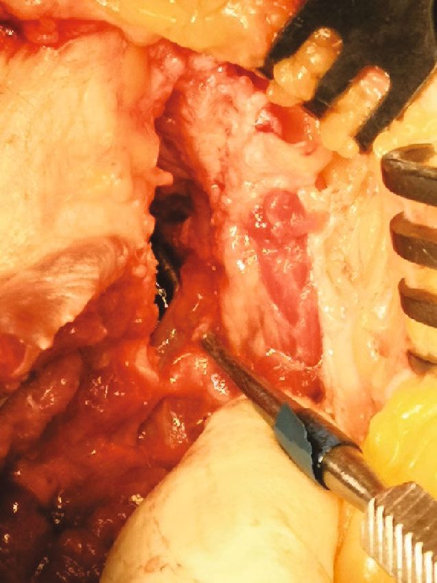
2                                                                                                  Case Reports in Orthopedics

knee, and ipsilateral complete foot drop that occurred dur-       surgery, clinical examination revealed functional recovery.
ing a military march. The onset of neurological symptoms          Following the aforementioned findings, the patient was
was acute, without any previous symptoms, reaching                allowed to return to his military duties.
immediately its full intensity and was perceived by the
patient as dragging of the foot. The patient had no history       3. Discussion
of lumbar spine disease. No traumatic event was reported
during the march.                                                 Intraneural ganglion cyst cases reported in the literature are
    Physical examination of the right lower extremity showed      rare. The most frequent site of occurrence is reported to be
negative Lasegue’s sign and straight leg raise test and           the common peroneal nerve, followed by the ulnar and tibial
hypoesthesia in the lateral aspect of the leg and dorsal aspect   nerves [6]. Although several theories have been proposed to
of the foot. Foot dorsiflexion and eversion and large toe dor-     interpret this pathology (recurrent trauma [3], intraneural
siflexion were severely impaired (tibialis anterior, extensor      hemorrhage [7], mucoid degeneration [8], and de novo for-
hallucis longus, extensor digitorum longus, and peroneus          mation from hamartomatous cell rests [9]), the articular the-
muscle strength assessment revealed a grade of 1/5 in the         ory, described by Spinner et al., is the most widely accepted.
MRC scale). There were no clinical signs of muscle atrophy        According to this theory, endoneural ganglion cysts originate
on the anterior and lateral compartments of the leg. Physical     from nearby joints (in the case of our patient, from the supe-
examination of the contralateral lower extremity was normal.      rior tibiofibular joint). Through a capsular defect, joint fluid
Knee and lumbar X-rays were normal.                               exits via a one-way mechanism and tracks along the epineu-
    Our initial therapeutic approach included rest, NSAIDs,       rium of the innervating articular branch following the path of
painkillers, and corticosteroid intramuscular injections. The     least resistance. In intraneural ganglion cysts of the common
patient was admitted to our clinic under observation status,      peroneal nerve, fluid originates from the superior tibiofibular
and lumbar spine MRI scan was ordered; however, results           joint [4]. In the present case, we were able to identify the
did not correspond to the patient’s symptomatology                articular branch during the surgery and resect it. Further-
(Figure 1). Moreover, the patient’s blood tests, including        more, Spinner et al. proposed dynamic aspects of cyst forma-
inflammatory markers (WBC, CRP, and ESR), were normal.             tion, according to which the various patterns of ascent,
    Following negative lumbar spine MRI results, a second         crossover, and descent down terminal nerve branches are
more thorough physical examination was conducted, which           attributed to intra-articular pressure fluctuation and
revealed positive Tinel’s sign in the area of the fibular neck,    dynamic pressure fluxes [10]. More recent literature has
shifting diagnostic thinking process towards peripheral neu-      incorporated the direct or indirect injury of the joint as a
ropathy. Subsequent EMG and NCS showed decreased con-             key component in the pathogenesis of an intraneural gan-
duction velocity and amplitude in the common peroneal             glion cyst, adding it up to the articular theory [11, 12].
nerve around the area of the popliteal fossa. Furthermore,            Common symptomatology of an endoneural ganglion
knee MRI showed a multilobulated lesion in the area around        cyst of the common peroneal nerve includes pain over the
the fibular head, in close proximity to the common peroneal        fibular head, with or without swelling, positive Tinel’s sign,
nerve (Figure 2).                                                 and paresthesia over the lateral surface of the tibia and dor-
    Surgical exploration under loop magnification was              sum of the foot. Some patients may present with gradual or
decided. The common peroneal nerve was identified and              acute weakness of the muscles located in the anterior and lat-
followed to its bifurcation. Common and deep peroneal             eral compartments of the leg. Muscle denervation and atro-
nerves appeared oedematous (Figure 3). The articular branch       phy have also been described [3, 13–16]. In our case, the
was also identified and surgically prepared from its origin        patient presented with an acute painless foot drop that
near deep peroneal nerve bifurcation, up to the superior          occurred during a military march and developed in a short
tibiofibular joint. The superior tibiofibular joint was disarti-    period of time with no obvious history of trauma. To the best
culated, and the articular branch was ligated and transected      of our knowledge, this is the first reported case of an intra-
(Figure 4). A small anatomic specimen of the peroneal artic-      neural ganglion cyst resulting in an acute foot drop without
ular branch was sent for histologic examination. An incision      a prior traumatic event. Additionally, a foot drop as the pri-
was made to the epineurium of the common peroneal nerve           mary symptom is an underreported manifestation of an
and mucoid material was evacuated from the ganglion cyst          intraneural ganglion cyst in the literature [14, 17]. Coexisting
(Figure 3).                                                       mild lumbar pain at first led us to a wrong initial assessment.
    Postoperatively, the patient was treated with a foot drop         MRI and/or ultrasound serve as useful tools of the diag-
polyethylene splint and physiotherapy. Histological exami-        nostic process. On ultrasonography, an endoneural ganglion
nation confirmed our diagnosis of an endoneural ganglion           cyst appears as a large well-circumscribed hypoechogenic
cyst (Figure 5). At a 3-month follow-up after the surgical        lesion [18], while on MRI it appears as a multilobulated
decompression, the patient showed clinical (tibialis anterior     lesion with low signal intensity on T1-weighted images and
4/5, extensor hallucis longus 2/5, extensor digitorum longus      high signal on T2-weighted images, oriented longitudinally
3/5, and peroneus muscles 4/5 in the MRC scale for muscle         along the course of the affected nerve. Furthermore, muscle
strength and complete return of sensation to the lateral          denervation oedema can be seen on T2-weighted images as
aspect of the leg and dorsal aspect of the foot) and EMG evi-     hyperintensity. Muscle atrophy is also characterized as
dence of recovery. Subsequent knee MRI showed no signs of         hyperintensity on T1-weighted images [19]. Recognition of
recurrence (Figure 6). At the last follow-up, 12 months after     the articular connection is a possible finding, but it is not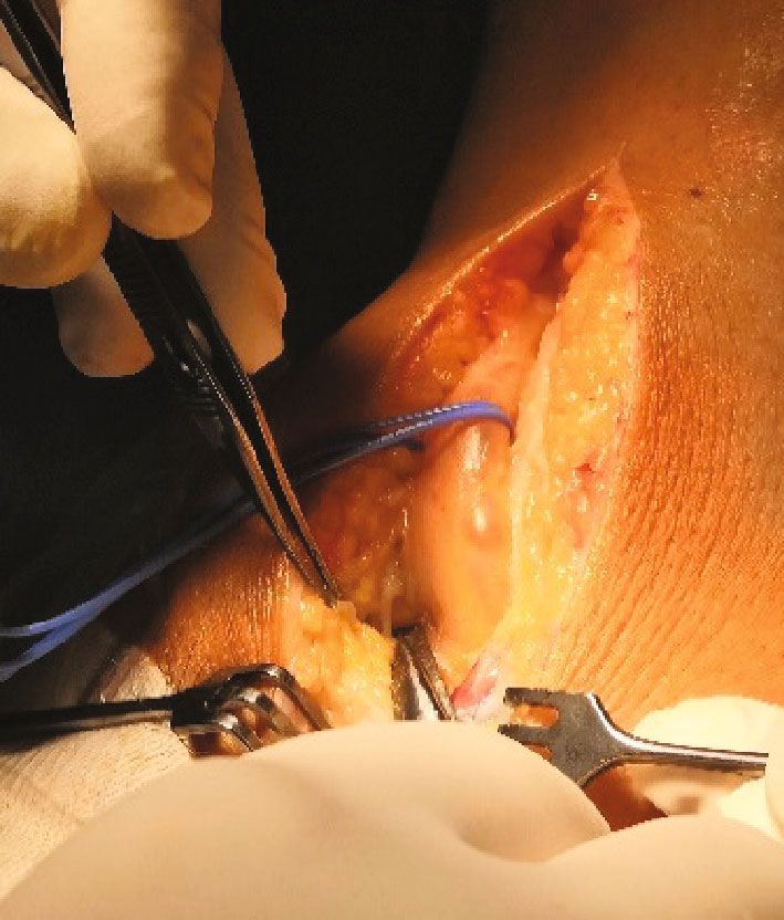
Case Reports in Orthopedics 3
(a)
(b) (c)
Figure 1: (a) Sagittal T2 lumbar spine MRI showing multiple disc bulges at L3/L4 and L4/L5. (b) Axial T2 at the L4/L5 level showing a mild
disc bulge narrowing the right nerve root foramen. (c) Axial T2 at the L5/S1 level showing mild right paracentral disc bulge.
(a) (b)
(c) (d)
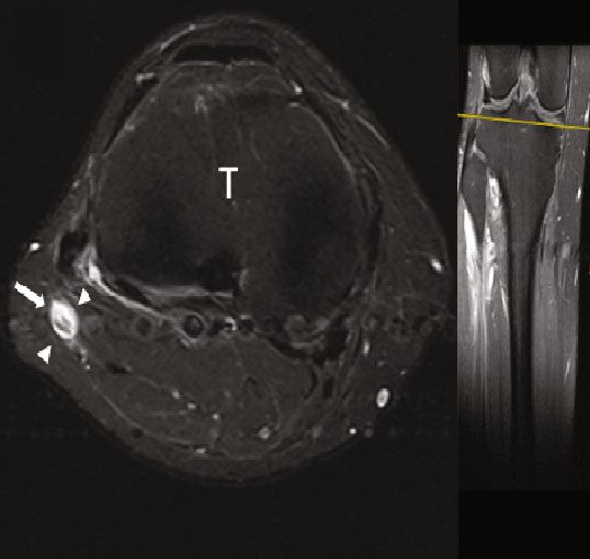
Figure 2: T2-weighted MR axial (a) and coronal (b) images of the knee at the level of the tibiofibular joint. White arrows point to the ganglion
cyst extending from the STF joint, and the black arrow points to the ascending oedematous CPN. T2-weighted axial image at the level of the
fibular neck (c). A horizontal, linear area of increased T2 signal along the course of the nerve represents the extension of the ING along the
transverse limb of the peroneal nerve articular branch (white arrow). T2-weighted axial image above the level of the fibular neck (d) showing
an intraneural ganglion cyst (white arrow) in which the tibial and peroneal divisions are separately contained (arrowheads) T: tibia; F: fibula.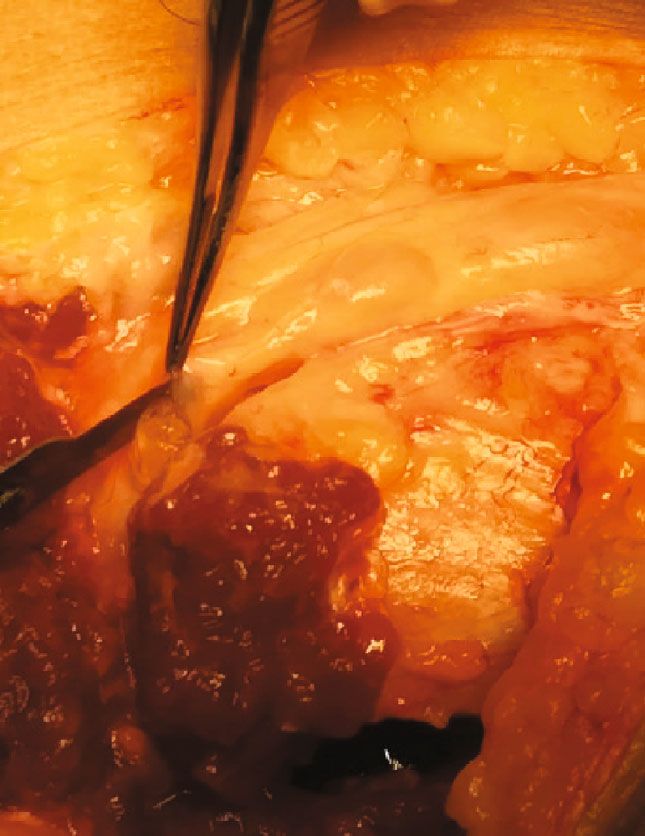
4 Case Reports in Orthopedics
⁎
⁎
(a) (b)
Figure 3: Photographs demonstrating the oedematous common peroneal nerve (black arrow) intraoperatively. In (b), an incision was made
in the epineurium to enable the evacuation of the mucoid content and the decompression of the nerve. Fibular head (black asterisk) and the
dissected peroneus longus muscle (white asterisk) can also be seen.
⁎
⁎
⁎
⁎
(a) (b)
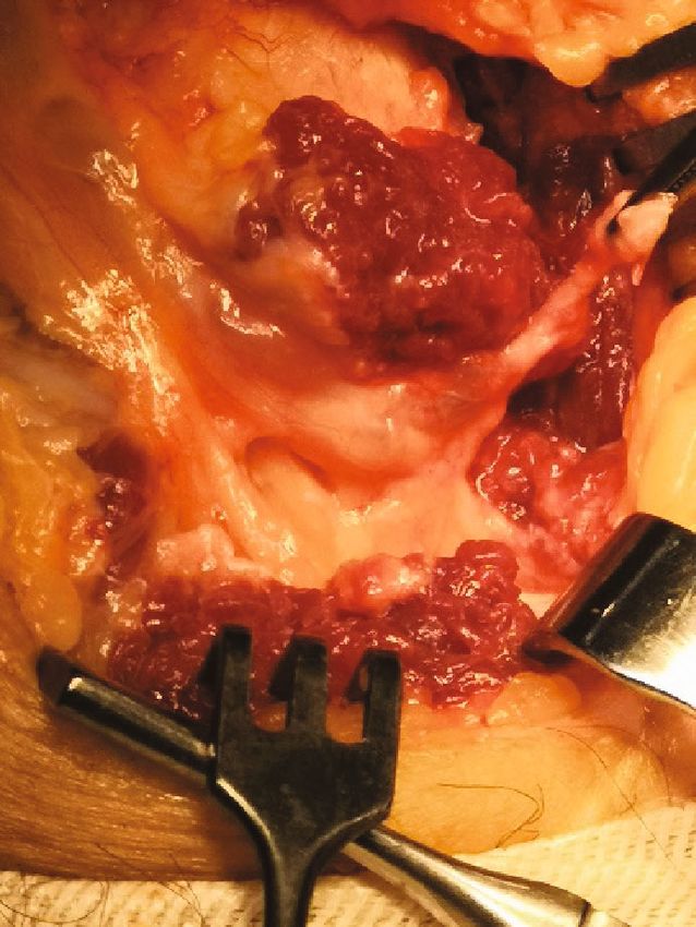
Figure 4: Photographs illustrating the articular branch ((a) black arrow) to the superior tibiofibular joint ((b) white arrow). Transection of
this branch after ligation alongside disarticulation of the superior TF joint is essential to prevent recurrence. In (a), deep peroneal nerve
(black asterisk) and the peroneus longus muscle (white asterisk) can also be recognized. The peroneus longus was dissected to allow better
view of the deep and superficial peroneal nerves, as well as the peroneal articular branch.
always easily detected on MRI. Spinner et al. demonstrated the neighboring anatomical structures as well as the articular
three reproducible MRI features that can provide aid in iden- connection and, unless a specialized radiologist in musculo-
tifying the joint connection (tail sign) and differentiating skeletal ultrasonography is available or MRI is contraindi-
between intraneural and extraneural ganglion cysts (trans- cated, intraneural ganglion cysts are best imaged with
verse limb sign, signet ring sign) [20]. magnetic resonance [21].
Ultrasonography is less time consuming than MRI and Standard treatment of intraneural ganglion cysts is surgi-
may be of value when guided percutaneous aspiration is cal excision of the ganglion and nerve decompression. An
decided, but it fails to illustrate the relation of the cyst with alternative minimally invasive treatment is decompression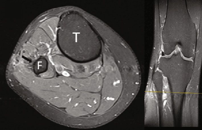
Case Reports in Orthopedics 5
Figure 5: Ganglion of nerve sheath with myxoid change and cystic degeneration seen in the connective tissue of the nerve (H&E ×10, ×40,
and ×400).
(a) (b)
(c) (d)
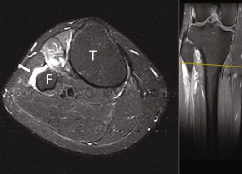
Figure 6: T2-weighted MR axial images of the knee at 6 months (a and b) and 10 months (c and d) postoperative in levels corresponding to
preoperative views. a and c are taken at a level above the fibular head. The absence of a high signal is noted in CPN (white arrow). b and d are
taken at the level in which a “transverse limb sign” was observed in preoperative MRI (black arrow). T: tibia; F: fibula.
by percutaneous aspiration of the ganglion under ultrasound tion of the articular branch as well as disarticulation of the
guidance, with or without corticosteroid injection [13]. Both involved joint at the cost of higher risk of iatrogenic nerve
these methods have reported high recurrence rates (38% and injury [6, 21]. Desy et al., in their review, advocated the sim-
50-70%, respectively). The best results in terms of recurrence ple incision and decompression of the cyst followed by STFJ
are obtained with surgical decompression and complete resection and ligation of the articular branch [6]. Our patient
removal of the cyst followed by ligation and complete resec- was treated with surgical decompression via a simple incision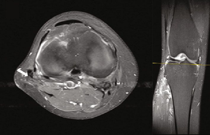
6                                                                                                      Case Reports in Orthopedics

followed by ligation and transection of the articular branch as    contributed to the initial drafting of the manuscript. All
well as disarticulation of the STFJ. At 12 months postopera-       authors read and approved the final manuscript.
tively, no recurrence was observed and the patient regained
functional recovery.
    This article demonstrates a rare case of an acute foot drop    References
caused by an intraneural ganglion cyst. To the best of our          [1] K. R. Swartz, D. Wilson, M. Boland, and D. B. Fee, “Proximal
knowledge, this is the first case of an intraneural ganglion             sciatic nerve intraneural ganglion cyst,” Case Reports in Medi-
cyst resulting in an acute foot drop without a prior traumatic          cine, vol. 2009, Article ID 810973, 4 pages, 2009.
event. Our hypothesis is that of an existing asymptomatic           [2] S. H. Colbert and M. C. H. Le, “Case report: intraneural gan-
intraneural ganglion cyst that deteriorated past the asymp-             glion cyst of the ulnar nerve at the wrist,” Hand, vol. 6, no. 3,
tomatic threshold point during the walk, possibly via an indi-          pp. 317–320, 2011.
rect mechanism, as proposed by Spinner et al. [11]. Although        [3] P. Patel and W. G. Schucany, “A rare case of intraneural gan-
a clear traumatic event was not reported, it is possible that a         glion cyst involving the tibial nerve,” Baylor University Medical
minor ankle torsional strain was transmitted through inter-             Center Proceedings, vol. 25, no. 2, pp. 132–135, 2012.
osseous membrane to STFJ, aggravating the preexisting gan-          [4] R. J. Spinner, J. L. D. Atkinson, and R. L. Tiel, “Peroneal
glion cyst [11]. Furthermore, this article provides additional          intraneural ganglia: the importance of the articular branch. A
support for the unifying articular theory, as proposed by               unifying theory,” Journal of Neurosurgery, vol. 99, no. 2,
Spinner et al. [4].                                                     pp. 330–343, 2003.
                                                                    [5] World Medical Association, “World Medical Association Dec-
                                                                        laration of Helsinki,” JAMA, vol. 310, no. 20, pp. 2191–2194,
4. Conclusion                                                           2013.
Intraneural ganglion cysts, although infrequent, are well           [6] N. M. Desy, H. Wang, M. A. I. Elshiekh et al., “Intraneural
established in the literature and should be considered in the           ganglion cysts: a systematic review and reinterpretation of
                                                                        the world’s literature,” Journal of Neurosurgery, vol. 125,
differential diagnosis of peripheral mononeuropathy, in
                                                                        no. 3, pp. 615–630, 2016.
order to avoid diagnostic pitfalls. Early diagnosis and surgical
                                                                    [7] E. S. Gurdjian, R. D. Larsen, and D. W. Lindner, “Intraneural
treatment with open decompression and concurrent address
                                                                        cyst of the peroneal and ulnar nerves. Report of two cases,”
of the articular branch are of paramount importance to                  Journal of Neurosurgery, vol. 23, no. 1, pp. 76–78, 1965.
obtain positive outcome and minimize recurrence risk.
                                                                    [8] P. F. Deluca and A. R. Bartolozzi, “Tibial neuroma presenting
                                                                        as a baker cyst. A case report,” The Journal of Bone & Joint Sur-
Abbreviations                                                           gery, vol. 81, no. 6, pp. 856–858, 1999.
                                                                    [9] B. M. Scherman, J. M. Bilbao, A. R. Hudson, and S. J. Briggs,
EMG:      Electromyogram test                                           “Intraneural ganglion: a case report with electron microscopic
NCS:      Nerve conduction study                                        observations,” Neurosurgery, vol. 8, no. 4, pp. 487–490, 1981.
NSAID:    Nonsteroid anti-inflammatory drugs                        [10] R. J. Spinner, K. K. Amrami, A. P. Wolanskyj et al., “Dynamic
MRI:      Magnetic resonance imaging                                    phases of peroneal and tibial intraneural ganglia formation: a
WBC:      White blood cell                                              new dimension added to the unifying articular theory,” Jour-
CRP:      C-reactive protein                                            nal of Neurosurgery, vol. 107, no. 2, pp. 296–307, 2007.
ESR:      Erythrocyte sedimentation rate                           [11] R. J. Spinner, M. A. Ibrahim Elshiekh, R. S. Tubbs, N. S. Turner
MRC:      Medical Research Council                                      III, and K. K. Amrami, “Posttraumatic torsional injury as an
STFJ:     Superior tibiofibular joint                                    indirect cause of fibular intraneural ganglion cysts: case
CPN:      Common peroneal nerve.                                        illustrations and potential mechanisms,” Clinical Anatomy,
                                                                        vol. 25, no. 5, pp. 641–646, 2012.
                                                                   [12] R. J. Spinner, F. Crnkovich, M. Ahmed Ibrahim Kobeal, and
Consent                                                                 K. K. Amrami, “Can trauma cause tibial intraneural ganglion
Written informed consent was obtained from the patient                  cysts at the superior tibiofibular joint?,” Clinical Anatomy,
                                                                        vol. 25, no. 6, pp. 785–787, 2012.
for publication of this case report and any accompanying
images.                                                            [13] T. Liang, A. Panu, S. Crowther, G. Low, and R. Lambert,
                                                                        “Ultrasound-guided aspiration and injection of an intraneural
                                                                        ganglion cyst of the common peroneal nerve,” HSS Journal,
Conflicts of Interest                                                   vol. 9, no. 3, pp. 270–274, 2013.
                                                                   [14] S. H. Coleman, P. K. Beredjeklian, and A. J. Weiland, “Intra-
The authors declare that they do not have any competing                 neural ganglion cyst of the peroneal nerve accompanied by
interests.                                                              complete foot drop. A case report,” The American Journal of
                                                                        Sports Medicine, vol. 29, no. 2, pp. 238–241, 2001.
Authors’ Contributions                                             [15] Y. S. Lee, J.-E. Kim, J. H. Kwak, I. W. Wang, and B. K. Lee,
                                                                        “Foot drop secondary to peroneal intraneural cyst arising
SS performed the literature search; drafted the manuscript;             from tibiofibular joint,” Knee Surgery, Sports Traumatology,
and designed, wrote, and revised the main body of the                   Arthroscopy, vol. 21, no. 9, pp. 2063–2065, 2013.
manuscript. DS, VV, AS, EA, and CK revised the final                [16] I. Ratanshi, T. A. Clark, and J. L. Giuffre, “Immediate nerve
manuscript, instructed the writing of the manuscript, and               transfer for treatment of peroneal nerve palsy secondary to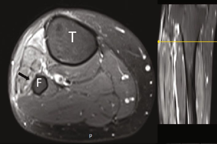
Case Reports in Orthopedics                                               7

       an intraneural ganglion: case report and review,” Plastic
       Surgery, vol. 26, no. 2, pp. 80–84, 2018.
[17]   A. Alsahhaf and W. Renno, “Ganglion cyst at the proximal
       tibiofibular joint in a patient with painless foot drop,” Pain
       Physician, vol. 19, no. 8, pp. E1147–E1160, 2016.
[18]   F. S. S. Leijten, W.-F. Arts, and J. B. C. M. Puylaert, “Ultra-
       sound diagnosis of an intraneural ganglion cyst of the peroneal
       nerve. Case report,” Journal of Neurosurgery, vol. 76, no. 3,
       pp. 538–540, 1992.
[19]   J. Panwar, A. Mathew, and B. P. Thomas, “Cystic lesions of
       peripheral nerves: are we missing the diagnosis of the intra-
       neural ganglion cyst?,” World Journal of Radiology, vol. 9,
       no. 5, pp. 230–244, 2017.
[20]   R. J. Spinner, N. M. Desy, and K. K. Amrami, “The cystic trans-
       verse limb of the articular branch: a pathognomonic sign for
       peroneal intraneural ganglia at the superior tibiofibular joint,”
       Neurosurgery, vol. 59, no. 1, pp. 157–166, 2006.
[21]   R. J. Spinner, N. M. Desy, M. G. Rock, and K. K. Amrami,
       “Peroneal intraneural ganglia. Part I. Techniques for successful
       diagnosis and treatment,” Neurosurgical Focus, vol. 22, no. 6,
       article E16, 2007.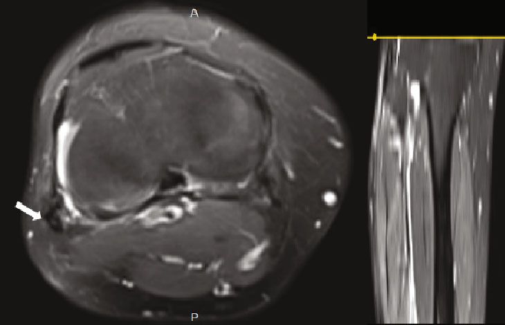
You can also read