CT-BASED TISSUE SEGMENTATION TO ASSESS KNEE JOINT INFLAMMATION AND REACTIVE BONE FORMATION ASSESSED BY F-FDG AND F-NAF PET/CT: EFFECTS OF AGE AND BMI
←
→
Page content transcription
If your browser does not render page correctly, please read the page content below
Original Article

CT-based tissue segmentation to assess knee joint
inflammation and reactive bone formation assessed by
18
  F-FDG and 18F-NaF PET/CT: Effects of age and BMI

Abdullah Al-Zaghal1 MD,              Abstract
                  1
                                     Objectives: This study was conducted to determine the role of computed tomography (CT)-based seg-
Dani P. Yellanki ,                   mentation methodology to semi-quantify the degree of in ammation and reactive bone formation in the
Cyrus Ayubcha1,                      knee joints by uorine-18- uorodeoxyglucose (18F-FDG) and 18F-sodium uoride positron emission tomo-
                                     graphy/CT (18F-NaF PET/CT) imaging, respectively. Furthermore, we assessed the impact of aging and body
Thomas J. Werner1 MSc,
                                     mass index (BMI) on these biological responses. Subjects and Methods: In this retrospective study, we
Poul F. Høilund-Carlsen2,3 MD,       examined a total of 97 subjects who had undergone both 18F-FDG and 18F-NaF PET/CT scanning. The mean
                                     age was 49.3±14.9 (21-75) and the mean BMI was 26.7±4.3 (17.7-42.0). Whole joint compartments and os-
DMSc,
                                     seous compartments were segmented on fused PET/CT images using a 3D-growing algorithm with an ad-
Abass Alavi1 MD (Hon), PhD           justable upper/lower Houns eld Units (HU) thresholds and manual tools. The metabolic activity and volu-
(Hon), DSc (Hon),                    me of each compartment was measured, values from the osseous compartment were subtracted from the
                                     whole joint to get the volume and metabolic activity of the soft tissue. The metabolic activity was correla-
                                     ted with age and BMI. Results: Fluorine-18-FDG uptake in the soft tissues surrounding the joint was
                                     0.35±0.07 while 0.19±0.04 in the osseous structures (P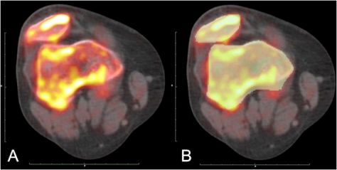
Original Article

two of the most important clinical risk factors for knee arthro-   Information regarding subjects with smoking habits, family
pathies: age and body mass index (BMI).                            history of CVD, and prescription medication were acquired
                                                                   through questionnaires. Fasting serum total cholesterol, se-
                                                                   rum low-density lipoprotein (LDL) cholesterol, serum high-
Patients and Methods                                               density lipoprotein (HDL) cholesterol, fasting plasma glu-
                                                                   cose and glycated hemoglobin (HbA1c) were measured. The
                                                                   estimated glomerular ltration rate (eGFR) was calculated
Fluorine-18-FDG and 18F-NaF PET/CT scans utilized in this          using the Modi cation of Diet and Renal Disease (MDRD)
retrospective study are part of the Cardiovascular Molecu-         equation. In each subject, the 10-year risk of developing CVD
lar Calci cation Assessed by 18F-NaF PET/CT (CAMONA)               was approximated using the FRS based on age, gender, sys-
protocol. CAMONA was a prospective study approved by               tolic blood pressure, total serum cholesterol, serum HDL cho-
the Danish National Committee on Biomedical Research Et-           lesterol, smoking habit, and management for hypertension.
hics, registered at ClinicalTrials.gov (NCT01724749), and
conducted from 2012 to 2016 in accordance with the Decla-           Table 1. Subjects' demographics
ration of Helsinki. Written informed consent was obtained
from all study subjects. Detailed description of the CAMO-                                                                    Total
NA study was previously published by Blomberg BA et al.                               Female        Male        P-value
                                                                                                                             (N=97)
(2017) [11]
                                                                                       Total        Total
Subjects selection                                                                    (n=48)       (n=49)
The CAMONA study consists of 139 volunteers; 89 healthy
subjects 50 subjects with history of chest pain. Healthy vo-        Age (Years)       51.01±        47.6±         0.25        49.3±
lunteers were recruited from the general population or                                 15.10         14.7                      14.9
from the blood bank of Odense University Hospital, Den-
mark. Subjects with a negative history of cardiovascular di-        Active                2            6                         8
                                                                    smoking
sease, oncologic disease, autoimmune disease, immunode-
  ciency syndromes, alcohol abuse, illicit drug use, or any         BP (mmHg)
prescription medication were considered as healthy volun-
teers. Framingham Risk Score was used to evaluate the mo-           Systolic          126.0±       132.1±         0.07        129.1±
di able cardiovascular risk factors and only subjects with                             17.8         16.0                       17.1
score below the upper limits of the recommended levels
were included [12]. Pregnant women were not included wit-           Diastolic          75.5±        79.1±         0.05        77.4±
hin the study population.                                                               9.9          8.3                       9.2
   Subjects with history of chest pain were recruited from
those whom were referred to the radiology department for            WBC               5.8±1.7      6.3±1.6        0.16       6.1±1.7
a coronary CT-angiography. Only patients with a 10 years            (109/L)
risk for fatal cardiovascular disease equal to or above 1%, as
                                                                    BMI (Kg/m2)        25.4±        27.9±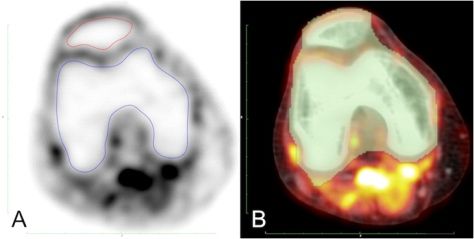
Original Article

  The imaging protocol of the CAMONA study was previo-             equation:
usly published by Blomberg BA et al. (2014) [13, 14]. In sum-
       18           18
mary, F-FDG and F-NaF PET/CT imaging were performed
on hybrid PET/CT systems (GE Discovery STE, VCT, RX, and
                                                                             18
690/710 systems (General Electric, Chicago, Illinois, USA)).         Total F-FDG metabolism in the bone compartment was
                                                                                               18
Fluorine-18-FDG PET/CT imaging was performed 180 minu-             subtracted from the total F-FDG metabolism in the whole
tes after intravenous administration of 4.0MBq/kg, after an        joint to get the uptake in the soft tissue. Volume of the bone
overnight fast of at least 8 hours and a con rmed blood glu-       ROI was subtracted from the volume of the whole joint ROI
cose concentration of below 8mmol/L. On average, F-NaF
                                                       18
                                                                   to get the volume of the soft tissue.
PET/CT imaging was performed within 14 days of F-FDG
                                                      18
                                                                     Averaged SUVmean was used for the semi-quanti cation
PET/CT imaging. Fluorine-18-sodium uoride PET/CT ima-              of 18F-FDG and 18F-NaF according to the following equation:
ging was performed 90min after intravenous administration
                 18
of 2.2MBq/Kg of F-NaF. Positron emission tomography ima-
ges were corrected for scatter, attenuation, random coinci-
dences, and scanner dead time. Low-dose CT imaging (140                                                                              18
kV, 30-110mA, noise index 25, 0.8 second/rotation, slice thic-        To decrease the impact of adipose tissue on F-FDG bio-
kness 3.75mm) was performed for attenuation correction             distribution during fasting state, SUVmean was corrected
and anatomical orientation. The e ective radiation dose re-        for lean body mass (SULmean). Tahari et al. (2014) reported
ceived from the entire imaging protocol was approximately          that using Janmahasatian formulation annuls the bias ca-
                                                                                           18
14mSv.                                                             used by body weight on F-FDG uptake by non-adipose tis-
                                                                   sue in females and decreases it in males [15]. Lean body
Image analysis                                                     mass (LBM) was calculated using Janmahasatian formula-
OsirX MD v.9.0 (DICOM viewer and image-analysis program,           tion [16]:
Pixmeo SARL; Bernex, Switzerland) was used for image ana-
lysis. Regions of interest (ROI) were manually assigned ac-
cording to predetermined anatomical criteria. On a 3D ma-
ximum intensity projection (MIP) coronal view, midpoint
between the lateral and the medial intercondylar tubercles
was used as a landmark, then 2 lines parallel to the axial
                                                                     Averaged SULmean was calculated according to the fol-
plane were drawn 4cm above and below to de ne the
                                                                   lowing equation:
upper and lower borders of the knee joint, respectively.
   Fluorine-18-FDG PET/CT images were segmented to me-
asure the metabolic activity of the boney and surrounding
soft tissue structures. For the whole joint segmentation, a
3D growing region algorithm with a lower Houns eld Unit
(HU) threshold of 90 was assigned on fused PET/CT images,
followed by a morphological closing and dilatation algorit-
hms with a structuring element radius of 20 and 5 units, res-
pectively. The ROI included the femur, patella, and tibia
along with the joint space, synovium and surrounding soft
tissue (Figure 1). For the segmentation of the osseous part,
ROI were manually assigned using a closed polygon tool
and included the femur, patella, and tibia. The metabolic          Figure 1. A) ROI manually assigned to include the femur and patella on an axial 18F-
activity of the soft tissue was measured by subtracting the        FDG PET image using closed polygon tool. B) ROI of the whole joint on an axial fused
                                                                   18
metabolic activity of the osseous part from the whole joint          F-FDG PET/CT after applying a 3D growing algorithm with a lower threshold of
activity.                                                          90HU followed by a morphological closing and dilatation algorithms with a strutu-
   Only the osseous compartment was segmented on F-
                                                          18       ring element radius of 20 units and 5 units, respectively.
NaF PET/CT images. A 3D growing region algorithm with a
lower threshold of 150HU was applied, followed by a mor-
phological closing algorithm with a structuring element ra-
dius of 15 units (Figure 2).
   Mean standardized uptake value (SUVmean) and ROIvolume
of both knees were measured for each trans-axial slice and
exported to a CSV le by Osirix. Total metabolic activity of
each slice was calculated by multiplying the slice SUVmean
by the slice ROIvolume. The total metabolism of all slices         Figure 2. A) Trans-axial fused 18F-NaF PET/CT images of the knee joint. B) Bone ROI
was summed up to measure the total metabolic activity of           highlighted after applying a 3D growing region algorithm with a lower threshold of
the investigated tissue compartment, as in the following           150HU and a morphological closing algorithm with a structuring element radius of 15.

www.nuclmed.gr                                                   Hellenic Journal of Nuclear Medicine • May-August 2018                         104
                                                                                                                                                 93
Original Article
Inter-operator agreement
Scans were independently analyzed by two investigators.
Bland Altman plot was used to assess inter-operator agre-
ement.
Statistical analysis
One-way ANOVA test, Pearson's correlation test, and linear
regression analysis were used for parametric analysis. Statis-
tical analysis was conducted using IBM SPSS Statistics ver-
sion 25.0 (IBM Corp. Released 2017. IBM SPSS Statistics for Figure 4. A) No association was found between the metabolic activity of 18F-NaF in
Macintosh, Version 25.0. Armonk, NY: IBM Corp). the knee joint and aging, B) Signi cant positive correlation was found between 18F-
NaF uptake in the knee joint with BMI.
The association between the di erences of measurements
Results conducted by both operators was not signi cant for 18F-FDG
activity in the soft tissue (r=0.14, P=0.15, range -0.04 to 0.05)
and 18F-NaF activity in the joint (r=0.11, P=0.28, range -0.18 to
18
For F-FDG , mean of the averaged SUVmean was 0.35±0.07 0.12) (Figure 6). These ndings indicate a strong inter-opera-
in the whole joint, 0.19±0.04 in the osseous segment and tor agreement.
0.43±0.09 for in the soft tissues (POriginal Article
18
ammatory activity (as noted by the degree of F-FDG up- In conclusion, this scienti c communication describes the
take) of both bone and soft tissue. role of PET-based molecular imaging and segmentation tec-
Musculoskeletal disorders pose a source of major health hniques in determining the metabolic activity of the knee
disabilities in the elderly. Osteoarthritis (OA) is a multi-facto- joint compartments and the changes that occur due to aging
rial joint disorder that is related to both mechanical and in- and BMI. In particular, we are reporting an association
ammatory factors and is a major cause of morbidity in el- between BMI and the in ammatory activity of the di erent
derly subjects. Previously published studies have reported knee joint compartments as well as bone turnover. Aging
an association between knee joint in ammation and aging was also associated with an increase in the in ammatory of
[17, 18]. the knee soft tissue compartment. Our ndings are consis-
Recent studies have described the in uence of body weig- tent with the previously published studies regarding the im-
ht in inducing and stimulating in ammatory reaction in the pact of aging as well as BMI on the knee joint.
weight-bearing joints of obese subjects [19]. Chondrocytes
have mechanoreceptors that sense changes in the exerted Financial disclosure
mechanical stress and translate it into biochemical signals, This study was funded by the Anna Marie and Christian Ras-
which consequently regulate the production of pro-in am- mussen's Memorial Foundation, University of Southern Den-
matory mediators [20, 21]. Mechanical strain and shear forces mark, Odense, Denmark, and the Jørgen and Gisela Thrane's
were also found to induce the production of COX-2, IL-2 and Philanthropic Research Foundation, Broager, Denmark.
PGE2 in broblast-like synoviocytes [22]. Increasing compres-
sive forces on subchondral bone was also associated with an Acknowledgment
increase in the expression of IL-6, COX-2 and IL-8 [23]. Infra- We thank the sta of the CAMONA study and the study parti-
patellar fat pad also contributes to joint in ammation by the cipants for their valuable contributions
secretion of adipokines into the synovium [24]. The increase
in the pro-in ammatory mediators keeps the joint in an ac- The authors declare that they have no con icts of interest.
tive low-grade state of in ammation and continuous gene-
ration of reactive oxygen species (ROS) [25]. Bibliography
Adipose tissue secretes in obese subjects also secretes cy- 1. Flandry F, Hommel G. Normal anatomy and biomechanics of the knee.
tokines as TNF-a, leptin, IL-1, and IL-6 into systemic circula- Sports Med Arthrosc Rev 2011; 19: 82-92.
2. Oeppen J, Vaupel JW. Broken limits to life expectancy. Science 2002;
tion [26]. Serum and synovial levels of leptin and IL-6 are as-
296: 1029-31.
sociated with OA synovitis and positively correlated with the 3. Fransen M, Simic M, Harmer AR. Determinants of MSK health and disa-
severity of degenerative changes on radiographs [27-29]. Os- bility: lifestyle determinants of symptomatic osteoarthritis. Best Pract
teophytes and chondrocytes were found to secrete leptin Res Clin Rheumatol 2014; 28: 435-60.
4. Nguyen U-SD, Zhang Y, Zhu Y et al. Increasing prevalence of knee pain
and IL-6 in advanced stages of OA [30].
and symptomatic knee osteoarthritis: survey and cohort data. Ann In-
Sodium uoride uptake depends on the availability of hyd- tern ed 2011; 155: 725-32.
roxyl ions (OH-) on the surface of hydroxyl-apatite (Ca10[PO4 5. Luyten FP, Denti M, Filardo G et al. De nition and classi cation of early
]6[OH]2) in bone matrix and the suppling blood ow [31]. Con- osteoarthritis of the knee. Knee Surg Sports Traumatol Arthrosc 2012;
20: 401-6.
ditions that alter bone metabolism whether by promoting 6. Kremers HM, Larson DR, Crowson CS et al. Prevalence of total hip and
bone formation or resorption lead to an increase in the sur- knee replacement in the United States. J Bone Joint Surg Am 2015; 97:
face area exposed to blood ow, thus, increasing 18F-NaF up- 1386-97.
take [32]. The increase in mechanical loading is associated 7. Guermazi A, Alizai H, Crema M et al. Compositional MRI techniques for
evaluation of cartilage degeneration in osteoarthritis. Osteoarthritis
with an increase in the proliferation and di erentiation of Cartilage 2015; 23: 1639-53.
osteoblasts and osteocytes, in turn increasing bone turnover 8. Jadvar H, Desai B, Conti PS. Sodium 18F- uoride PET/CT of bone, joint,
[33-36]. and other disorders. Semin Nucl Med 2015; 45: 58-65.
Bone mass density declines with aging [37], and this a ects 9. Raynor W, Houshmand S, Gholami S et al. Evolving role of molecular
imaging with 18F-sodium uoride PET as a biomarker for calcium meta-
the amount of hydroxyl-apatite in bone matrix, consequ- bolism. Curr Osteoporos Rep 2016; 14: 115-25.
ently decreasing the exposed surface area and binding sites 10. Kobayashi N, Inaba Y, Tateishi U et al. Comparison of 18F- uoride posit-
18
available to react with F-NaF. This might explain the absen- ron emission tomography and magnetic resonance imaging in evalu-
18
ce of a correlation between aging and the activity of F-NaF ating early-stage osteoarthritis of the hip. Nucl Med Commun 2015; 36:
84-9.
although it is common to nd an increase in metabolic acti- 11. Blomberg BA, De Jong PA, Thomassen A et al. Thoracic aorta calci -
vity at site of new bone formation (osteophytes) in elderly. cation but not in ammation is associated with increased cardiovas-
The limitation of the study was due to a lack of relevant cli- cular disease risk: results of the CAMONA study. Eur J Nucl Med Mol
nical information about subjects' history of knee pain or ar- Imag 2017; 44: 249-58.
12. D'Agostino RB, Vasan RS, Pencina MJ et al. General cardiovascular risk
thropathy. However, the main purpose of this research was to pro le for use in primary care: the Framingham Heart Study. Circula-
develop an analysis scheme for quantifying knee disorders tion 2008; 117: 743-53.
with PET. As such, we are presenting a methodology to seg- 13. Blomberg BA, Thomassen A, Takx RA et al. Delayed 18F- uorodeoxyglu-
ment the knee joint compartments to assess various knee cose PET/CT imaging improves quantitation of atherosclerotic plaque
in ammation: results from the CAMONA study. J Nucl Cardiol 2014; 21:
pathologies, aid in achieving an earlier disease diagnosis and 588-97.
provide an objective tool to follow disease activity as well as 14. Blomberg BA, Thomassen A, Takx RA et al. Delayed sodium 18F- uoride
treatment response. PET/CT imaging does not improve quanti cation of vascular calci ca-
www.nuclmed.gr Hellenic Journal of Nuclear Medicine • May-August 2018 106
93Original Article
tion metabolism: Results from the CAMONA study. J Nucl Cardiol 2014; 26. Greene MA, Loeser RF. Aging-related in ammation in osteoarthritis.
21: 293-04. Osteoarthritis Cartilage 2015; 23: 1966-71.
15. Tahari AK, Chien D, Azadi JR, Wahl RL. Optimum lean body formulation 27. Eaton CB. Obesity as a risk factor for osteoarthritis: mechanical versus
for correction of standardized uptake value in PET imaging. J Nucl Med metabolic. Med Health R I 2004; 87: 201-4.
2014; 55: 1481-4. 28. Dumond H, Presle N, Terlain B et al. Evidence for a key role of leptin in
16. Janmahasatian S, Du ull SB, Ash S et al. Quanti cation of lean bo- osteoarthritis. Arthritis Rheumatol 2003; 48: 3118-29.
dyweight. Clin Pharmacokinet 2005; 44: 1051-65. 29. Ku JH, Lee CK, Joo BS et al. Correlation of synovial uid leptin concen-
17. Saboury B, Parsons MA, Moghbel M et al. Quanti cation of aging e ec- trations with the severity of osteoarthritis. Clin Rheumatol 2009; 28:
ts upon global knee in ammation by 18F-FDG -PET. Nucl Med Commun 1431-5.
2016; 37: 254-8. 30. Stannus OP, Jones G, Quinn SJ et al. The association between leptin, in-
18. Hong YH, Kong EJ. (18 F) Fluoro-deoxy-D-glucose uptake of knee joints terleukin-6, and hip radiographic osteoarthritis in older people: a cross-
in the aspect of age-related osteoarthritis: a case-control study. BMC sectional study. Arthritis Res Ther 2010; 12: R95.
Musculoskelet Disord 2013; 14: 141. 31. Costeas A, Woodard HQ, Laughlin JS. Depletion of 18F from blood
19. Berenbaum F, Eymard F, Houard X. Osteoarthritis, in ammation and owing through bone. J Nucl Med 1970; 11: 43-5.
obesity. Curr Opin Rheumatol 2013; 25: 114-8. 32. Bastawrous S, Bhargava P, Behnia F et al. Newer PET application with an
20. Guilak F. Biomechanical factors in osteoarthritis. Best Pract Res Clin old tracer: role of 18F-NaF skeletal PET/CT in oncologic practice. Radi-
Rheumatol 2011; 25: 815-23. ographics 2014; 34: 1295-316.
21. Knapik DM, Perera P, Nam J et al. Mechanosignaling in bone health, tra- 33. Cao JJ. E ects of obesity on bone metabolism. J Orthop Surg Res 2011;
uma and in ammation. Antioxid Redox Signal 2014; 20: 970-85. 6: 30.
22. Takao M, Okinaga T, Ariyoshi W et al. Role of heme oxygenase-1 in in- 34. Markou P, Chatzopoulos D. Yttrium-90 silicate radiosynovectomy treat-
ammatory response induced by mechanical stretch in synovial cells. ment of painful synovitis in knee osteoarthritis. Results after 6 months.
In amm Res 2011; 60: 861-7. Hell J Nucl Med 2009; 12: 33-6.
23. Sanchez C, Pesesse L, Gabay O et al. Regulation of subchondral bone os- 35. Chatzopoulos D, Markou P, Iakovou I. Scintigraphic imaging of knee
osteoblast metabolism by cyclic compression. Arthritis Rheumatol 2012; synovitis in osteoarthritis after intra-articular injection of technetium-
64: 1193-203. 99m pertechnetate in the unilateral knee. Hell J Nucl Med 2006; 9: 69-71.
24. Ioan-Facsinay A, Kloppenburg M. An emerging player in knee osteoar- 36. Sojan S, Bartholomeusz D. Cutaneous radiation necrosis as a com-
thritis: the infrapatellar fat pad. Arthritis Research & Therapy 2013; 15: 225. plication of yttrium-90 synovectomy. Hell J Nucl Med 2005; 8: 58-9.
25. Sellam J, Berenbaum F. Is osteoarthritis a metabolic disease? Joint Bone 37. Black DM, Rosen CJ. Postmenopausal osteoporosis. N Engl J Med 2016;
Spine 2013; 80: 568-73. 374: 254-62.
Pablo Picasso. Child with a dove (1901). Oil in canvas. 73x54cm.
107
93 Hellenic Journal of Nuclear Medicine • May-August 2018 www.nuclmed.gr
9You can also read