Case Report: Infected primary hydatid cyst of the thigh version 1; peer review: awaiting peer review - F1000Research
←
→
Page content transcription
If your browser does not render page correctly, please read the page content below
F1000Research 2021, 10:786 Last updated: 23 AUG 2021

CASE REPORT

Case Report: Infected primary hydatid cyst of the thigh
[version 1; peer review: awaiting peer review]
Myriam Jrad, Haifa Zlitni                  , Hakim Zouari, Miriam Boumediene, Ines Soussi,
Meriem Bouzrara
Department of Radiology, Charles Nicolle University Hospital, 9 April Bd, Bab Souika, Tunis, 1006, Tunisia

v1   First published: 10 Aug 2021, 10:786                                          Open Peer Review
     https://doi.org/10.12688/f1000research.53651.1
     Latest published: 10 Aug 2021, 10:786
     https://doi.org/10.12688/f1000research.53651.1                                Reviewer Status AWAITING PEER REVIEW

                                                                                   Any reports and responses or comments on the

Abstract                                                                           article can be found at the end of the article.
Hydatic cyst may occur in many organs such as the liver, lung, brain or
heart with radiologic features of liver and lung involvement being well
known. The musculo-skeletal site is infrequent accounting for 0.7–3%
cases of all cases resulting from direct implantation of oncospheres
more often than hematic dissemination.
We report the case of an 18-year-old female student who visited our
hospital because of a swelling in the posteroexternal aspect of the left
thigh that had grown during the previous six months and had become
tender in the previous month with setup of fever three days before
admission. Superficial ultrasound and magnetic resonance imaging
demonstrated a cystic mass of the posterior compartment of the thigh
developed within the short chief of the biceps femoris. Serology for
hydatid cyst was positive. The diagnosis of an infected hydatid cyst
was suspected preoperatively, and the patient was given antibiotics
and anthelminthic treatment. The cyst was then completely excised
and the histopathologic exam confirmed the hydatic origin. The
patient was put on oral anti-helminthics and has been on regular
follow up for last twelve months with no evidence of recurrence.
Hydatidosis rarely occurs in the soft tissues and the diagnosis is
challenging particularly when it is secondary infected. Hydatid
serology provides certainty in the diagnosis of echinococcosis when it
is positive. When it’s negative, imaging (Ultrasound, Computed
tomography (CT) and Magnetic resonance imaging (MRI)) may be an
approach for making the diagnosis revealing the most characteristic
features of hydatid cyst.

Keywords
hydatidosis, cyst, ultrasound, MRI, surgery

                                                                                                                              Page 1 of 8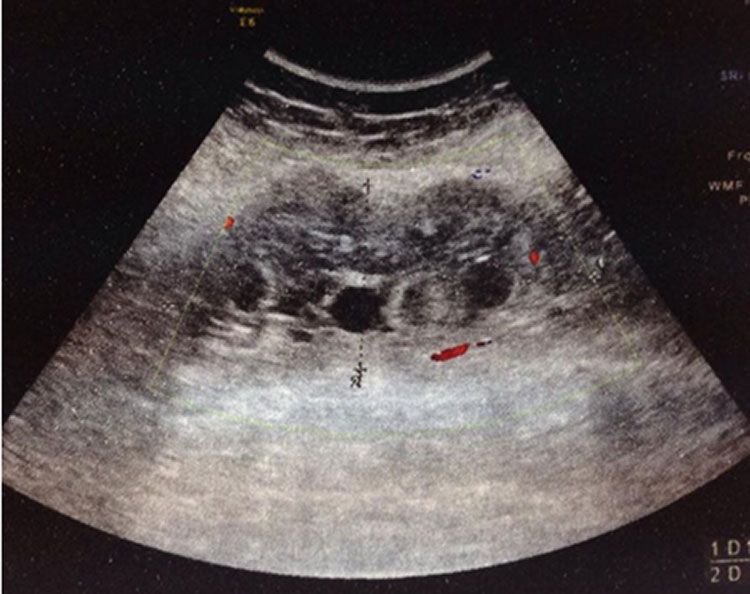
F1000Research 2021, 10:786 Last updated: 23 AUG 2021

Corresponding author: Haifa Zlitni (zlitnihaifa03@gmail.com)
Author roles: Jrad M: Conceptualization, Supervision, Validation, Writing – Review & Editing; Zlitni H: Methodology, Writing – Original
Draft Preparation; Zouari H: Conceptualization; Boumediene M: Investigation; Soussi I: Project Administration; Bouzrara M: Formal
Analysis
Competing interests: No competing interests were disclosed.
Grant information: The author(s) declared that no grants were involved in supporting this work.
Copyright: © 2021 Jrad M et al. This is an open access article distributed under the terms of the Creative Commons Attribution License,
which permits unrestricted use, distribution, and reproduction in any medium, provided the original work is properly cited.
How to cite this article: Jrad M, Zlitni H, Zouari H et al. Case Report: Infected primary hydatid cyst of the thigh [version 1; peer
review: awaiting peer review] F1000Research 2021, 10:786 https://doi.org/10.12688/f1000research.53651.1
First published: 10 Aug 2021, 10:786 https://doi.org/10.12688/f1000research.53651.1

                                                                                                                                  Page 2 of 8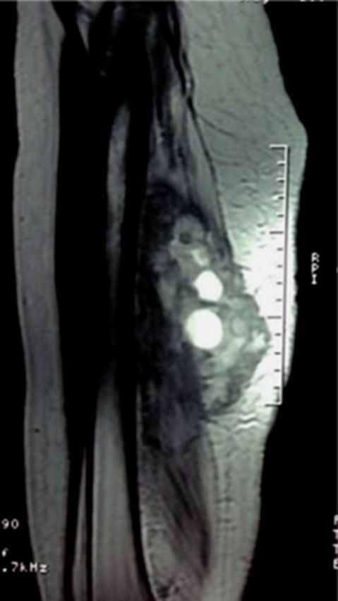
F1000Research 2021, 10:786 Last updated: 23 AUG 2021

Case presentation
An 18-year-old Tunisian female student presented to the orthopedics department of Charles Nicolle Hospital of Tunis,
Tunisia on January 15, 2019 with a lump in the posteroexternal aspect of the left thigh. She had noticed the swelling on her
thigh six months before visiting the hospital. She was without history of trauma, surgery or any additional disease. The
swelling had become painless during the six months prior to her visit but it had become tender within the previous month
with the setup of fever three days prior. On examination, the patient was febrile (38.5° Celsius) with normal vital
parameters. There was a tender, indurate, non-moveable lump on the posteroexternal aspect of the middle one-third of the
left thigh measuring about 12 cm  5 cm. The overlying skin was erythematous without any punctum or discharge. The
knee and leg movements were normal.

Laboratory investigations on January 15, 2019 showed a biological inflammatory syndrome with elevated white blood
cell count (12,000/mm3) and C-reactive protein. Conventional radiography of the left femur showed a thickening of the
soft tissues of the middle and posterior region of the thigh with integrity of the bone (Figure 1).

Superficial Doppler ultrasound performed the second day of hospitalization showed an ill-defined multilocular cystic
mass of the middle one-third of the posterior compartment of the left thigh measuring 13 cm  5.5 cm and containing an
echogenic peripheral portion that was finely vascularized on color Doppler (Figure 2). Magnetic resonance imaging
(MRI) performed two days later demonstrated a large intramuscular cystic mass of the middle one-third of the posterior
compartment of the left thigh (Figure 3) within the biceps femoris muscle measuring 10 cm  4 cm. This mass was
delimited by a discontinuous rim of low T2 and high T1 signal “rim sign” and contained multiple well defined cystic
lesions of more intense high T2 and low T1 signal corresponding to daughter cysts with a “cyst within a cyst appearance”.
This cystic mass was surrounded by an edematous infiltration of the adjacent muscles with low T1 and high T2 signal and
avid enhancement after contrast administration predominant in the posterior compartment. Enhancement of the muscular
fascia and of the subcutaneous fat of the posterior aspect of the thigh was noticed. The mass repressed the sciatica nerve
without invading it and was distant from the profound and superficial femoral pedicles. A low T1 signal of the spongy
bone enhanced after contrast administration was noticed (Figure 4).

The enzyme-linked immune-absorbent assay (ELISA) was positive for the Echinococcal granulosis antigens (40 U/ml).

The diagnosis of an infected hydatid cyst was suspected perioperatively and the patient was given antibiotics and anthelminthic
treatment (Albendazole 400 mg Per Os twice daily for 28 days). The patient didn't have any history of hydatidosis and hydatid
cysts were not detected in any other organ on preoperative computed tomography (CT) of the abdomen and thorax. The
surgical exploration found a firm oblong mass within the short chief of left biceps femoris densely adherent to the surrounding
muscles and abutting the femur cortex. The mass was widely excised. The surgeon then performed an irrigation with Povidone
iodine and hypertonic saline solutions and closed the wound over a negative suction drain. The macroscopic examination of the
lesion revealed multiple daughter cysts and the histopathological exam confirmed the hydatic origin.

Discussion
Echinococcosis is a cosmopolitan helminthic infection caused by the tapeworm Echinococcus granulosus and it affects
humans and many mammals.1 This tapeworm species is endemic in the Mediterranean region, Australia, Argentina,

Figure 1. Conventional radiography of the left femur showing a thickening of the soft tissues of the middle
and posterior region of the thigh with integrity of the bone.

                                                                                                                                  Page 3 of 8
F1000Research 2021, 10:786 Last updated: 23 AUG 2021
Figure 2. Superficial Doppler ultrasound in the transversal plane (A) and longitudinal plane (B) of the thigh
shows an ill-defined multilocular cystic mass of the middle one-third of the posterior compartment of the
thigh measuring 13 cm 5.5 cm and containing an echogenic peripheral portion that is finely vascularized on
color Doppler.
Figure 3. MRI of the left thigh: Sagittal TSE T2-weighted image shows the multiloculated cystic lesion with
multiple daughter cysts in the middle one-third of the posterior compartment of thigh.
Africa, Eastern Europe and the Middle East. The dog is a definitive host, but this situation is shared by the wolf and some
species of jackal.2
The dog infestation is through the digestive track and is believed to be secondary to the consumption of parasite viscera
especially the liver and the lungs of the sheep as an intermediate host.3 The latter, constituting the main reservoir of
Echinococcus tapeworm, becomes infected by eating grass soiled by the dog’s droppings containing the eggs of the
Page 4 of 8F1000Research 2021, 10:786 Last updated: 23 AUG 2021
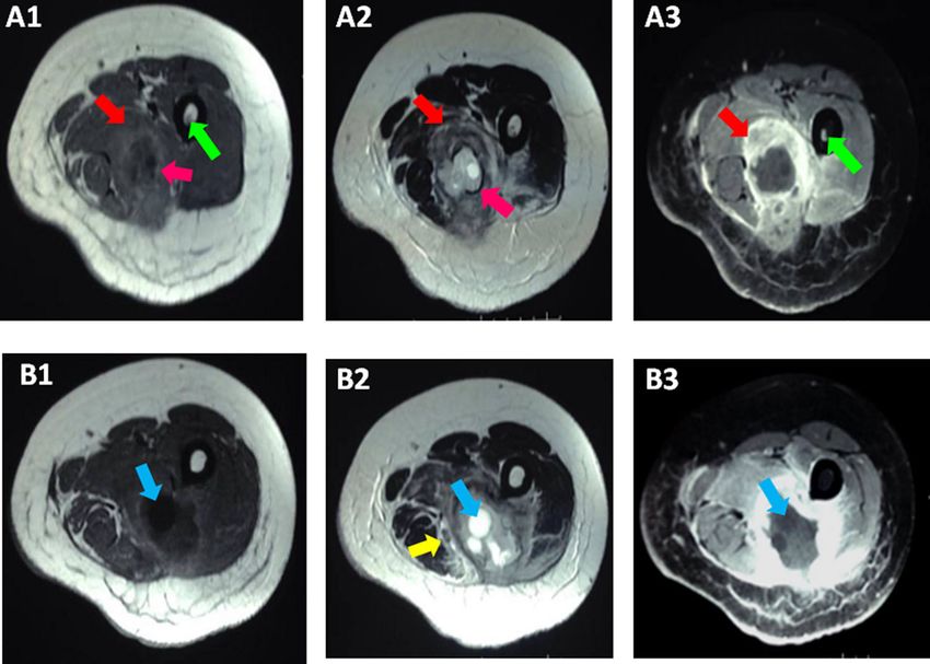
Figure 4. MRI of the left thigh in two different cutting levels (A1+A2+A3) and (B1+B2+B3). Axial TSE T1-weighted
images (A1+B1), TSE T2-weighted images (A2+B2) and contrast-enhanced fat-suppressed TSE T1-weighted images
demonstrated an intramuscular cystic mass of the middle one-third of the posterior compartment of the left thigh
within the biceps femoris muscle. This mass is delimited by a discontinuous rim of low T2 and high T1 signal "rim
sign"(pink arrow) and contains multiple well defined cystic lesions of more intense high T2 and low T1 signal
corresponding to daughter cysts (blue arrow) with a "cyst within a cyst” appearance. This cystic mass is surrounded
by an edematous infiltration of the adjacent muscles with low T1 and high T2 signal and avid enhancement after
contrast administration (red arrows). Enhancement of the muscular fascia and of the subcutaneous fat of the
posterior aspect of thigh. The mass represses the sciatica nerve without invading it (yellow arrow) and is distant from
the profound and superficial femoral pedicles. A low T1 signal of the spongiest bone enhanced after contrast
administration was noted (green arrow).
parasite.4 Humans are only an intermediate host and an epidemiological impasse of the parasite. They become infected
either through direct contact with parasitized dogs or indirectly through ingestion of contaminated food.3–5
Muscular localization of hydatid cyst is rare varying from 1 to 5.4 % of all hydatid locations.6 It’s the third localization
after the lungs and the liver. For some, involvement of the spleen must precede that of muscle since it is estimated at 8%.1,7
Several arguments have been put forward to explain the scarcity of muscle localization: the efficiency of hepatic and
pulmonary barriers that opposes the migration of the parasite into the systemic circulation; the muscular environment’s
hostility to the growth of hydatid larvae due to the production of lactic acid and the alternation of contraction–relaxation
inhibiting uniform vascularization.8,9 The muscle localization of echinococcosis seems to be mostly primary and affects
mainly proximal muscles of the lower limbs, very probably due to the importance of irrigation of these.10 Daali and
Hssaida reported 10 cases out of 15 of deep muscular location involving the diaphragm and psoas.11
Diagnosis of echinococcosis must be suspected when a patient from a rural area is presenting with slowly growing soft
tissue mass and it should be included in the differential diagnosis of limb masses: abscess, malignant or benign tumor,
calcified hematoma or lipoma.12 The diagnosis of echinococcosis should be considered before surgical biopsy in order to
prevent the risk of anaphylaxis.13
Ultrasound is a non-irradiating, accessible, and non-expensive exam, which can be used as a first line approach for
making the diagnosis revealing the most characteristic features of hydatid cyst: daughter cysts, detached membranes and
double line sign. Cysts may be classified according to the ultrasound criteria of Gharbi.14 Atalar et al. reported a
sensitivity of 95%, increasing to 100% in the presence of vesicular fibrils.15 In our case, superficial Doppler ultrasound
showed a multilocular cystic mass (type III) of the middle one-third of the posterior compartment of the left thigh. The
mass was ill-defined and containing an echogenic peripheral portion finely vascularized on color Doppler probably
Page 5 of 8F1000Research 2021, 10:786 Last updated: 23 AUG 2021
related to the secondary infection of the cyst. However, in non-endemic areas, ultrasound can be misleading with soft
tissue tumors especially in deep locations.16
Computed tomography (CT) radiologic features vary from a unilocular or multilocular cyst, with or without septas, debris
or wall calcifications, to a complex or solid mass without enhancement on intravenous contrast.13 The appearance of
muscular hydatidosis is unoften typical and the multivesicular form is specific as is reflects multiple daughter cysts within
the parent cyst. Since bony invasion and relationship of the cyst with adjacent organs is essential to describe, computed
tomography (CT) must be a part of the screening protocol.17
Although, magnetic resonance imaging (MRI) characteristics of liver hydatid cyst are detailed in the literature, the diagnosis is
challenging in the soft tissue of the musculoskeletal system because the magnetic resonance imaging (MRI) features are not
well labelled. Magnetic resonance imaging (MRI) is the gold standard imaging test in the identification of soft-tissue masses
including hydatid disease thanks to its capacity to establish most of its features, with the exception of calcifications.
Performing magnetic resonance imaging (MRI) requires the use of a surface antenna depending on the concerned part of the
body, the use of a large field of view allowing the inclusion of the neighboring joint, a section thickness of 3 to 7 mm, and an
inter-cut space of 0 to 2 mm. Acquisitions are performed in the axial plan, sagittal plan for anterior or posterior lesions, and
coronal plan for lateral or medial lesions. The sequences must include T1-weighted sequence in the axial plane and T2 and
T1-weighted sequences after fat saturation before and after injection of gadolinium in two orthogonal planes.
The classic magnetic resonance imaging (MRI) findings include a unilocular or multilocular cyst with a low-intensity rim
("rim sign") or detached membrane on T2-weighted images without enhancement after contrast injection.18 “The rim
sign” corresponds to the pericyst that is a collagen reaction generated by the host. The most pathognomonic sign is that of
daughter cysts within a larger cyst.19 The rim sign is a characteristic sign in muscular hydatidosis that is uncommon in
hydatic cyst located elsewhere in the body. Magnetic resonance imaging (MRI) of our patient demonstrated a cystic mass
containing multiple well-defined cysts corresponding to daughter cysts with a “cyst within a cyst appearance” delimited
by a discontinuous “rim sign”.20 This lesion was surrounded by an edematous infiltration with avid enhancement of the
muscular environment and the spongy bone in contact after contrast related to the secondary infection of the cyst.19–21
Hydatid serology provides certainty in the diagnosis of echinococcosis when it is positive. However, there is a significant
proportion of false negatives, variable depending on the location of the cyst. Lamine et al. reported 80% of false
negatives.9 The enzyme-linked immune-absorbent assay (ELISA) was positive for the E. granulosis antigens in our case.
Hypereosinophilia is not specific and inconstant and is of interest only in the orientation of the diagnosis, ultrasound, and
nowadays magnetic resonance imaging (MRI) can confirm the diagnosis.9,19
Surgical excision of the cyst is the treatment of choice. For non-surgical cysts, anthelminthic chemotherapy with or
without percutaneous aspiration-injection re-aspiration (PAIR) is an alternative option for the treatment.21,22 Our patient
was given antibiotics and anthelminthic treatment (Albendazole 400 mg Per Os twice daily for 28 days). Thereafter, the
mass was widely excised, and the patient was put on oral anthelminthics after surgery. Percutaneous drainage echo guided
without re-aspiration is simple, easy to apply, low cost, repeatable, and does not require hospitalization.21,22
Hydatidosis rarely occurs in the soft tissues and the diagnosis is challenging particularly when it is secondary infected.
Hydatid serology provides certainty in the diagnosis of echinococcosis when it is positive. When it’s negative, ultrasound
is an accessible way to approach the diagnosis, computed tomography (CT) may help to evaluate the surrounding tissues
and find calcifications and magnetic resonance imaging (MRI) provides imaging characteristics of hydatic cyst. Open
surgery is the gold standard of the treatment of muscular hydatidosis while ambulatory percutaneous techniques are
gaining scale.
Data availability
All data underlying the results are available as part of the article and no additional source data are required.
Consent
Written informed consent for publication of clinical details and clinical images was obtained from the patient.
Page 6 of 8F1000Research 2021, 10:786 Last updated: 23 AUG 2021
References
1. Abi F, El Fares F, KAIS D: Les localisations inhabituelles du kyste 13. Tekin R, Avci A, Tekin RC, et al.: Hydatid cysts in muscles: clinical
hydatique a propos de 40 cas. J Chir. 1989; 126: 307–12. manifestations, diagnosis, and management of this atypical
2. McManus DP, Yang YR: Helminthic Diseases Echinococcosis. In: presentation. Revista da Sociedade Brasileira de Medicina Tropical.
Reference Module in Biomedical Sciences. 2014. 2015 Oct; 48(5): 594–8.
PubMed Abstract|Publisher Full Text
3. Gougoulias NE, Varitimidis SE, Bargiotas KA, et al.: Skeletal muscle
hydatid cysts presenting as soft tissue masses. Hippokratia. 2010; 14. Atalar MH, Cankorkmaz L, Koyluoglu G, et al.: Imaging
14(2): 126–30. characteristics of three primary muscular hydatid cyst cases
PubMed Abstract|Free Full Text with various patterns. Kafkas J Med Sci. 2012; 2(2): 74–6.
Publisher Full Text
4. Mseddi M, Mtaoumi M, Dahmene J, et al.: Kyste hydatique
musculaire. Revue de Chirurgie Orthopédique et Réparatrice de 15. Benhaddoua H, Margib M, Kissrab M, et al.: Le kyste hydatiqueu du
l’Appareil Moteur. 2005 May; 91(3): 267–71. muscle trapezius: Une localisation inhabituelle. Archives de
Publisher Full Text pédiatrie. 2012: 263–5.
5. Hmidi M, Touiheme N, Rbai M, et al.: Isolated hydatid cyst of the 16. Mughal A, Saeed Minhas M, Bhatti A, et al.: Hydatid Cyst Of Skeletal
neck: An unusual site. Eur Ann Otorhinolaryngol Head Neck Dis. 2012 Muscle Presenting As Soft Tissue Tumour. J Coll Physicians Surg
Apr; 129(2): 108–10. Pak. 2018 Feb 26; 28(3): S51–3.
PubMed Abstract|Publisher Full Text PubMed Abstract|Publisher Full Text
6. Lamine A, Fikry T, Zryouil B: L’hydatidose primitive des muscles 17. Garcia Diez A, Ros Mondoza L, Villacampa V, et al.: MRI evaluation of
périphériques. A propos de 7 cas. Acta Orthop. 1993; 59: 184–7. soft tissue hydatid disease. Eur J Radiol. 2000; (10): 462–6.
PubMed Abstract|Publisher Full Text
7. Bendib A, Bendib S, Benmamar L, et al.: Tomodensitométrie du
kyste hydatique du foie: sémiologie et classification à propos de 18. Alexiadis G, Lambropoulou M, Deftereos S, et al.: Primary muscular
157 cas dont 146 vérifiés chirurgicalement. J Radiol. 1985; 66: hydatitosis. US, CT and MR findings. Acta Radiol. 2002; 43: 428–30.
367–75. PubMed Abstract|Publisher Full Text
8. Kehila M, Allegue M, Letaief R, et al.: Le kyste hydatique du muscle 19. Comert R, Aydingoz U, Ucaner A, et al. : Waterlily sign on MR
psoas: A propos d’un cas. J radiol. 1987; 68: 265–8. imaging of primary intramuscular hydatidosis of sartorius
muscle. Skel Radiol. 2003; 32: 420–3.
9. Essadki O, Elhajjam M, Kadiri R: Kyste hydatique des parties PubMed Abstract|Publisher Full Text
molles, aspect radiologique. Ann Radiol. 1996; 39: 135–41.
20. Bayram M, Sirikci A: Hydatic cyst located intermuscular area of
10. Daali M, Hssaida R: [Muscle hydatidosis. 15 cases]. Presse Med. the forearm: MR imaging findings. Eur J Radiol. 2000 Dec; 36(3):
2000 Jun 17; 29(21): 1166–9. 130–2.
PubMed Abstract PubMed Abstract|Publisher Full Text
11. Bourree P: Vers un traitement médical de l’hydatidose. Rev Prat. 21. Örmeci N, Idilman R, Akyar S, et al.: Hydatid cysts in muscle: a
1978; 28: 2879–900. modified percutaneous treatment approach. Int J Infect Dis.
12. Orhan Z, Kara H, Tuzuner T, et al. : Primary subcutaneous cyst 2007 May; 11(3): 204–8.
hydatic disease in proximal thigh: an unusual localisation: a PubMed Abstract|Publisher Full Text
case report. BMC Musculoskelet Disord. 2003 Nov 7; 4: 25. 22. Guillaureau P, Deunier B, Levet Y: Hydatidose musculaire à
PubMed Abstract|Publisher Full Text|Free Full Text localisation masseterine. 1986; 1049; 15.
Page 7 of 8F1000Research 2021, 10:786 Last updated: 23 AUG 2021
The benefits of publishing with F1000Research:
• Your article is published within days, with no editorial bias
• You can publish traditional articles, null/negative results, case reports, data notes and more
• The peer review process is transparent and collaborative
• Your article is indexed in PubMed after passing peer review
• Dedicated customer support at every stage
For pre-submission enquiries, contact research@f1000.com
Page 8 of 8You can also read