Comparison of imaging methods in POEMS syndrome
←
→
Page content transcription
If your browser does not render page correctly, please read the page content below
Biomed Pap Med Fac Univ Palacky Olomouc Czech Repub. 2012 Mar; 156(1):52–57.

                      Comparison of imaging methods in POEMS syndrome
     Jiri Minarika, Vlastimil Scudlaa, Jaroslav Bacovskya, Tomas Pikaa, Filip Ctvrtlikb, Iva Metelkovac, Miroslav Myslivecekc

Background. POEMS syndrome is a clinical condition with a very heterogeneous clinical manifestation. Its presenta-
tion as well as monitoring is complex and dependent on the clinician´s experience. One of the leading presenting
symptoms is based on evaluation of skeletal damage with typical osteosclerotic or mixed lesions.
Aims and methods. Our aim was to compare the usefulness of different imaging methods in the diagnostics of POEMS
syndrome, such as conventional radiography, densitometry, technetium scintigraphy, PET/CT scan, MRI and angiog-
raphy on a series of three patients with POEMS syndrome with different clinical manifestations and course of disease.
Results. Our series demonstrates different types of skeletal involvement in POEMS syndrome. Although conventional
X-ray is the imaging method mostly used for the evaluation, its sensitivity and specificity is low. Under specific condi-
tions, other imaging methods should be considered, giving a more complex outlook of the disease’s skeletal involve-
ment. Nevertheless, FDG-PET/CT confirmed its superiority in defining both skeletal lesions as well as the activity of
the neoplastic process.
Conclusions. We conclude that the different manifestation of the disease implies the necessity of a complex evaluation
of imaging methods in mutual concordance. FDG-PET/CT emerges as the most contributive method for the evaluation
of both the extent and activity of the disease.

Key words: POEMS syndrome, imaging, PET/CT, conventional radiography, computed tomography, magnetic reso-
nance

Received: June 4, 2011; Accepted: October 6, 2011; Available online: November 8, 2011
http://dx.doi.org/10.5507/bp.2011.053
a
  Department of Internal Medicine III – Nephrology, Rheumatology and Endocrinology, University Hospital Olomouc, Czech Republic
b
  Department of Radiology, University Hospital Olomouc
c
 Department of Nuclear Medicine, University Hospital Olomouc
Corresponding author: Jiri Minarik, e-mail: abretina@email.cz

INTRODUCTION                                                              Imaging methods play a crucial role in the diagnostics
                                                                      as well as in the monitoring of the disease, although there
    POEMS syndrome is a rare hematological disorder                   is no consensus on which of the methods should be rou-
belonging to plasma cell dyscrasias1. It is defined as a              tinely used in clinical practice. Our paper presents three
monoclonal plasma cell disorder together with periph-                 different manifestations of the disease with stress put on
eral neuropathy and variable presence of other organ or               the role of imaging methods, and their contribution to the
tissue impairment. The acronym represents characteris-                diagnostics of this clinical condition.
tic features of the syndrome (P = polyneuropathy, O =
organomegaly, E = endocrinopathy, M = monoclonal                      Case report 1
gammopathy, S = skin lesions). There have been several                    An 81 old male, originally with a neurological diag-
attempts at the definition of diagnostic criteria of the dis-         nosis of CIDP (Chronic Inflammatory Demyelinating
ease. However, most of them have a limited specificity                Polyneuropathy) was presented at our hematology depart-
and are unable to separate the syndrome itself from the               ment in October, 1998. He had severe polyneuropathy
coincidence of individual symptoms. The current opin-                 with both axon and myelin damage (according to electro-
ion on POEMS syndrome has been recently published as                  myography - EMG), and was unable to walk.
Mayo clinic criteria (Tab. 1) that reflect clinical as well as            Due to the presenting symptoms we aimed our diag-
pathobiological characteristics of the syndrome2.                     nostic approach from the beginning at POEMS syndrome
    Unlike multiple myeloma (MM), it is usually manifest-             as a majority of the findings fulfilled the diagnostic crite-
ed in younger patients, and its course is less aggressive3.           ria: We found several skin changes - hyperpigmentation
The presence of monoclonal plasma cells and chronic                   of the chest and back, hypertrichosis, thickening of the
progressive polyneuropathy is obligatory; however, only a             skin and several angiomas both on the body and the limbs.
few patients develop larger skeletal damage as seen in MM             In his armpits there were enlarged lymph nodes, and the
(ref.1). Moreover, the clinical manifestation can be very             histobiopsy confirmed Castleman´s disease. In both the
variable. The presence of all attributes of the acronym is            serum and spinal fluid we found monoclonal protein IgG
sometimes quite difficult to unravel, and most patients are           lambda, and the blood count showed slight thrombocyto-
usually treated for other (usually neurological) disorders.           sis (485x109/l). Within the endocrinology screening, we

                                                                 52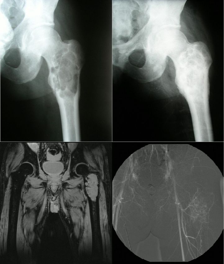
Biomed Pap Med Fac Univ Palacky Olomouc Czech Repub. 2012 Mar; 156(1):52–57.
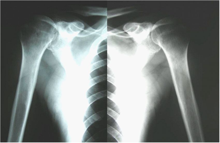
Fig. 1. Osteosclerotic
“thickening” of the shaft
of both proximal arm
bones in a patient with
POEMS syndrome.
found elevated levels of prolactin, FSH, urine cortisol, also considered as a possible cause. The polyneuropathy
blood glucose and C-peptide. In the bone marrow there was originally thought to be caused by long lasting type 2
was slightly increased number of monoclonal lymphoplas- diabetes. She had slightly enlarged liver on abdominal so-
mocytic elements (5.2%). The definitive diagnosis was nography (steatosis), and a few hemangiomas of the body
supported by the finding of osteosclerotic lesions, which and the limbs. Except for glucose metabolism (increased
were present on both of the proximal arm bones on the insulin, C-peptide), there was no pathology in the endo-
X-ray scans (Fig. 1), and the patient had higher density crine spectrum. We found no enlarged lymph nodes and
indices in his arms using densitometry by DEXA. the blood count as well as the biochemistry showed no
After the diagnosis we used actinotherapy for the os- significant abnormalities. In the bone marrow examina-
teosclerotic lesions and axillar lymph nodes enlargement, tion we could not find clonal plasmocytes, and there were
and conventional chemotherapy with pulses of alkylating no osteolytic nor osteosclerotic lesions on conventional X
agent (melphalan) and steroids (prednisone). The patient ray scan. At that time, the patient was diagnosed as having
improved with increased muscle strength, restitution of two concomitant clinical conditions – CIDP and MGUS
walking and disappearance of the lymph nodes. We could (monoclonal gammopathy of undetermined significance).
trace the normalization of blood count and endocrine Due to continuous worsening of the symptoms, newly
parameters, the decrease in monoclonal immunoglobulin diagnosed anemia and no effect of immunomodulatory
and bone marrow plasma cells. The clinical assessment therapy, the patient was re-assessed at a one year interval.
as well as EMG confirmed the symptoms of lower limb At that time we found focal accumulation of clonal plas-
neuropathy, however, they remained stable with no im- ma cells in the bone marrow, and histobiopsy confirmed
provement. myeloma. The X ray scan was still not very persuasive
The course of this POEMS syndrome was quite typical with several suspect osteolytic lesions of the skull, but the
with several subsequent progressions (usually defined as crucial finding was due to conventional 99mTc scintigra-
a slight increase in monoclonal immunoglobulin together phy (not MIBI), which found a small locus of osteoplas-
with worsened clinical state) followed by remission of the tic remodelation in the distal sternum. The same locus
disease. In September 2008, the patient died of an un- (very likely hidden in the summation of the projection of
related condition (unmanageable cardiac failure due to spine on classical X-ray scan) was clearly visible on sub-
severe mitral insufficiency with valve replacement and sequent CT scan (Fig. 2). These findings supported our
subsequent inoperable leak). hypothesis of osteosclerotic myeloma (POEMS syndrome
respectively).
Case report 2 The patient was treated with radiotherapy of the ster-
A 43 years old woman was diagnosed as a CIDP num followed by high-dose chemotherapy and autolo-
(chronic inflammatory demyelinizating polyneuropa- gous stem cell transplantation. Chemotherapy led to the
thy), treated by azathioprin and prednisone. She was normalization of blood count, a decrease in monoclonal
investigated in our department in 2004 for a small peak immunoglobulin and bone marrow plasma cell involve-
of monoclonal immunoglobulin IgG lambda (7.2 g/l). ment. The neuropathy of the limbs remained unimproved,
The presenting symptoms were, however, obscure due to apparently due to the presence of concomitant diabetic
several co-morbidities, although POEMS syndrome was neuropathy.
53Biomed Pap Med Fac Univ Palacky Olomouc Czech Repub. 2012 Mar; 156(1):52–57.
Fig. 2. Osteosclerotic lesion in distal sternum of a patient with POEMS syndrome. Focal accumulation of 99mTc on conventional
technetium scintigram (left). Osteosclerosis visible as “thickening” of sternum on CT scan (right).
The course of the disease was characterized by two Case report 3
subsequent progressions followed by conventional che- A 25 year old man with an 8-month history of poly-
motherapy regimens (melphalan plus prednisone and radiculoneuritis was treated with azathioprin and meth-
cyclophosphamide plus dexamethasone) with no signifi- ylprednisolone in the neurological department. He
cant change in overall patient status. The duration of the temporarily improved and started rehabilitation with
disease remission was about 9-12 months. At present, the the interruption of immunosuppressive treatment. This
patient is progressing with the eruption of multiple skin treatment-free period was complicated within 3 months by
hemangiomas and an increase of paraprotein. She is be- the development of polyserositis with lower limb edema,
ing treated with chemotherapy using lenalidomide and pleural and cardiac effusions, ascites and paraparesis of
prednisone. lower limbs, still with no pain or signs of bone damage.
He lost 25 kg in 8 months and developed overall weakness
Table 1. Diagnostic criteria of POEMS syndrome – Mayo clinic criteria from 2007(ref.2)
“Mayo clinic criteria 2007”*
Major criteria Minor criteria Other signs and symptoms
Polyneuropathy Organomegaly (splenomegaly, Clubbing
hepatomegaly, adenomegaly)
Monoclonal gammopathy Edema, pleural effusion, ascites Weight loss
Sclerotic bone lesions Endocrinopathy (adrenal, thyroid, Hyperhidrosis
pituitary, gonadal, parathyroid,
pancreatic)
Castleman disease Skin changes (hyperpigmentation, Pulmonary hypertension/restrictive
hypertrichosis, hemangioms, flush- lung disease
ing, acrocyanosis, white nails)
Elevation of Vascular Endothelial Papilledema Diarrhea
Growth Factor (VEGF)
Thrombocytosis or polycytemia Low vitamin B12 level
* Polyneuropathy with monoclonal gammopathy or Castleman disease must be present in all patients. For the diagnosis at least one more
major and one minor criterion is required.
54Biomed Pap Med Fac Univ Palacky Olomouc Czech Repub. 2012 Mar; 156(1):52–57.
Fig. 3. Large osteolytic le-
sion with osteosclerotic rims
and pathological vascularisa-
tion in a patient with POEMS
syndrome.
Conventional radiography
presents large osteolytic tumor
of proximal femur (A) with
subsequent recalcification
after successful therapy (B).
The tumor consists of mixed
osteolytic and osteosclerotic
involvement seen on MRI
(C), and has pathological vas-
cularisation visible on digital
subtraction angiography (D).
and dyspnea. The initial screening for possible infectious, teolytic/osteosclerotic lesions with FDG activity on clav-
malignant or systemic cause was, however, negative, with icles, vertebral bodies and ribs. The definitive diagnosis
normal biochemistry including CRP, negative infectious was confirmed by the finding of a compact infiltrate of
disease screening, normal oncomarkers, immunologi- monoclonal plasmocytes from targeted biopsy of one of
cal screening and endocrine tests (repeatedly). He had the larger lesions (right clavicle).
only substantial thrombocytosis (1016x109/l) and the After successful diagnosis the treatment started with
presence of monoclonal immunoglobulin IgG lambda radiotherapy of the hip, systemic chemotherapy (cyclo-
(up to 10 g/l). Ultrasonography confirmed ascites, mild phosphamide and dexamethasone), and preparation for
hepatomegaly, pleural and cardiac effusions with no oth- autologous transplant. The stem cell harvest was, however,
er pathology on parenchymatous organs. Tests done on unsuccessful (twice in a 2 month interval), probably due
the aspirate of both pleural effusion and ascites showed to long-term non-specific pretreatment. The following
transudate with no biochemical or cytological pathology, therapy consisted of conventional drugs and led to the
assessment of liquor was negative as well as the result of normalization of laboratory findings, to the disappear-
bone marrow trephine biopsy including immunopheno- ance of effusions and improvement of overall condition
typing. including substantial regression of peripheral neuropa-
Examination of bone X-ray (suspicion of myeloma) thy, recalcification of the osteolytic lesions (Fig. 3), and
showed a large cystic lesion 10x5 cm in the left proximal restitution of activities of daily living including normal
femur with MRI verified mixed osteolytic and osteoscle- walking.
rotic involvement and pathological vascularisation on The patient is at the moment in remission with no
angiography (Fig. 3). The most contributive approach chemotherapy.
was FDG-PET/CT which unraveled multiple mixed os-
55Biomed Pap Med Fac Univ Palacky Olomouc Czech Repub. 2012 Mar; 156(1):52–57.
DISCUSSION 2 we presented a patient with positive bone scintigraphy
based on which the diagnostic discretion was aimed at
POEMS syndrome represents a very heterogeneous this clinical condition.
disease with variable presence of accompanying symp- CT scan is more informative, although still not rou-
toms. Except for characteristic polyneuropathy and tinely used for whole body assessment. We found just
monoclonal gammopathy, the other associated symptoms two recent papers which demonstrated the potential of
might not be evident at the onset of the disease. Imaging FDG-PET/CT scan in the evaluation of POEMS (ref.17,18).
methods are therefore very useful in the diagnostics as Although quite expensive, PET/CT seems to be a very
well as in the assessment of bone involvement in POEMS convenient method for detection of the activity as well as
syndrome. Still, there is no standard algorithm for choice the extent and localization of bone and/or lymph node
of specific method, and the usefulness of different ap- lesions. Also, our findings in case report 3 support the use
proaches varies throughout the literature. of PET/CT especially in patients with diagnostic complex-
Conventional radiography might be contributive: ity. Despite the negative laboratory findings and missing
However, the bone lesions can be confused with other clinical symptoms of bone damage, PET/CT unraveled
conditions such as bone cysts, nonossifying fibromas, vast skeletal damage and also extraskeletal localization
benign bone islands or fibrous dysplasia1. There are two of the disease.
types of skeletal manifestations in POEMS syndrome: The present paper aims to demonstrate the complexity
proliferative changes and focal bone lesions4. Although of imaging in POEMS syndrome. As the condition is quite
approximately 95% of patients with POEMS syndrome rare, there are no recommendations on which of the imag-
have osteosclerotic lesions, some of the lesions can be ing methods are the most suitable. The assessed groups of
lytic with a sclerotic rim or mixed with different propor- patients are usually too small for generalization. Still, the
tion of sclerotic and lytic involvement1. Focal lesions can heterogeneity and different manifestation of the disease
be solitary or multiple, usually found in the axial bones4. foreshadow the necessity of a complex evaluation of dif-
The present paper accounts for all the manifestations – as ferent imaging methods in mutual concordance. We also
a “solitary osteosclerotic lesion“ in case report 2 (Fig. 2), show the increasing role of FDG-PET/CT which might
“oligo-focal“ proliferative changes in case report 1 (Fig. 1), have the ability to reflect the extent of POEMS syndrome,
and “multiple mixed osteolytic lesions with osteosclerotic especially in X-ray negative patients, as well as the pos-
component“ in case report 3 (Fig. 3). sible extra-osseal localization of the disease.
The extent of the lesions does not necessarily corre-
spond to the severity of the disease but the presence of
bone involvement is pathognomonic. Interestingly, we AKNOWLEDGEMENT
found increased bone density in the areas with osteoscle-
rotic involvement, which contributed to the evaluation Supported by VVZ MSM 6198959205.
of the lesion. DEXA evaluation might therefore be an
inexpensive complementary imaging method, although
contributive only in selected cases due to the character REFFERENCES
and variable localization of sclerotic lesions.
There have been just a few reports on the contribu- 1. Dispenzieri A. Diagnosis and treatment of POEMS syndrome. In
Rajkumar SV, Kyle RA, Treatment of multiple myeloma and related
tion of MRI in the diagnostics of POEMS syndrome5-9. disorders. Cambridge university press, New York, NY, USA, 2009: 182-
The MRI imaging in POEMS syndrome is quite rare as 195. ISBN 13: 9780521515030.
the diagnosis is usually based on clinical symptoms, and 2. Dispenzieri A. Mayo Clinic Criteria for the Diagnosis of
the MRI scans are mostly performed in situations of di- Polyneuropathy, Organomegaly, Endocrinopathy, Monoclonal
Gammopathy, and Skin Changes Syndrome. Note. From "POEMS
agnostic uncertainty. On the other hand, the character Syndrome". Blood Reviews 2007,21:287.
on T1 and T2-weighted sequences strongly illustrates the 3. Rajkumar SV, Kyle RA, Suarez GA, Dispenzieri A. Neuropathy asso-
different types of bone involvement5. ciated with plasma cell proliferative disorders. In Gertz MA, Greipp
The contribution of angiography is dubious as the le- PR. Multiple myeloma and related plasma cell disorders. Mayo
Foundation for medical education and research, Rochester, MN,
sions are usually not of such an extent as seen in case USA, 2004:35-52. ISBN 3-540-00811-X.
report 3. On the other hand, a recent studies of POEMS 4. Narvaez JA, Majos C, Narvaez J, Valls C, Fernandez-Cabrera L. POEMS
syndrome that focus on the role of angiogenic cytokines, syndrome: unusual radiographic, scintigraphic and CT features. Eur
especially the vascular endothelial growth factor (VEGF) Radiol 1998,8:134-6.
5. Chong ST, Beasley HS, Daffner RH. POEMS syndrome: radiographic
confirmed their elevated expression in POEMS syndrome, appearance with MRI correlation. Skeletal Radiol, 2006,35:690-695.
and their possible responsibility for the onset of the dis- 6. Michel JL, Gaucher-Hugel AS, Reynier C, Lhoste A, Philippe P. POEMS
ease1,10-13. From this point of view, a search for increased syndrome: imaging of skeletal manifestations, a study of 8 cases. J
neovascularisation could be challenging in the assessment Radiol 2003,84:393–7.
7. Kim JW, Lee SK, HA KM, Kim KH, Joh GY, Kim HJ, Yang SO. POEMS
of the extent of disease even before actual bone damage. syndrome: a case report. J Korean Med Sci 1992;7:79–84.
The osteosclerotic lesions are usually referred with 8. Furuzono H, Moritoyo T, Yamada H, Sugihara R, Nagamatsu K. A case
negative radionuclide bone scan, although some previous of Crow-Fukase syndrome which developed seven years following
reports have registered local increased uptake of radio- myelopathy of unknown origin. Rinsho Shinkeigu 1993;33:56–60.
9. Brazis PW, Liesegang TJ, Bolling JP, Kashii S, Trachtman M, Burde RM.
tracer4,14-16, usually attributed to focal cortical expansion, When do optic disc edema and peripheral neuropathy constitute
periostitis and sclerotic reaction14. Similarly, in case report poetry? Surv Ophthalmol 1990;35:219–25.
56Biomed Pap Med Fac Univ Palacky Olomouc Czech Repub. 2012 Mar; 156(1):52–57.
10. Watanabe O, Maruyama I, Arimura K, Kitajima I, Arimura H, Hanatani 14. Bessler W, Antonucci F, Stamm B, Stuckmann G, Vollrath T. Case re-
M, Matsuo K, Arisato T,Osame M. Overproduction of vascular en- port 646. POEMS syndrome. Skeletal Radiol 1991,20:212-5.
dothelial growth factor/vascular permeability factor is causative in 15. Mertens I, Vandeputte L, Van Haecke P, Thomas J, Samson I, Lateur
Crow-Fukase (POEMS) syndrome. Muscle Nerve 1998;21:1390-7. L, De Wolf-Peeters C, Paridaens R. Sclerotic IgA myeloma and poly-
11. Scarlato M, Previtali SC, Carpo M, Pareyson D, Briani C, Del Bo R, neuropathy: The POEMS syndrome. Ann Oncol 1995,6:731-2.
Nobile-Orazio E, Quattrini A, Comi GP. Polyneuropathy in POEMS 16. Jin S-A,Baek S-W, Song I-Ch,Yun G-W,Yang Y-J,Lee H-J,Yun H-J, Kim
syndrome: role of angiogenic factors in the pathogenesis. Brain J-M, Jo D-Y,Kim S. A case of multiple myeloma associated with mul-
2005,128:1911-20. tifocal osteosclerosis (multiple myeloma with osteosclerosis).Korean
12. Soubrier M, Dubost JJ, Serre AF, Ristori JM, Sauvezie B, Cathebras J Hematol 2009,44:188-92.
P, Piette JC, Chapman A, Authier FJ, Gherardi RK. Growth factors in 17. Alberti MA, Martinez-Yelamos S, Fernandez A, Vidaller A, Narvaez
POEMS syndrome: evidence for a marked increase in circulating JA, Cano LM, Gamez C, Martinez Matos JA. 18-F-FDG PET/CT in the
vascular endothelial growth factor. Arhritis Rheum 1997,40:786-7. evaluation of POEMS syndrome. Eur J Radiol 2010,76:180-2.
13. Gherardi RK, Belec L, Soubrier M, Malapert D, Zuber M, Viard JP. 18. An YS, Yoon JK, Hong SP, Joh CW, Yoon SN. 18F-FDG PET/CT in POEMS
Overproduction of proinflammatory cytokines imbalanced by their Syndrome. Nucl Med Mol Imaging 2007,41:66-7.
antagonists in POEMS syndrome. Blood 1996,87:1458-65.
57You can also read