Unveiling the Molecular Basis of the Noonan Syndrome-Causing Mutation T42A of SHP2 - MDPI
←
→
Page content transcription
If your browser does not render page correctly, please read the page content below
International Journal of
            Molecular Sciences

Communication
Unveiling the Molecular Basis of the Noonan
Syndrome-Causing Mutation T42A of SHP2
Angelo Toto *, Francesca Malagrinò, Lorenzo Visconti, Francesca Troilo and Stefano Gianni *
 Istituto Pasteur-Fondazione Cenci Bolognetti, Dipartimento di Scienze Biochimiche “A. Rossi Fanelli” and
 Istituto di Biologia e Patologia Molecolari del CNR, Sapienza Università di Roma, 00185 Rome, Italy;
 francescamalagrino@gmail.com (F.M.); lorenzo.visconti@uniroma1.it (L.V.); francesca.troilo@uniroma1.it (F.T.)
 * Correspondence: angelo.toto@uniroma1.it (A.T.); stefano.gianni@uniroma1.it (S.G.)

 Received: 12 December 2019; Accepted: 9 January 2020; Published: 10 January 2020

 Abstract: Noonan syndrome (NS) is a genetic disorder caused by the hyperactivation of the
 RAS-MAPK molecular pathway. About 50% of NS cases are caused by mutations affecting the SHP2
 protein, a multi-domain phosphatase with a fundamental role in the regulation of the RAS-MAPK
 pathway. Most NS-causing mutations influence the stability of the inactive form of SHP2. However,
 one NS-causing mutation, namely T42A, occurs in the binding pocket of the N-SH2 domain of the
 protein. Here, we present a quantitative characterization of the effect of the T42A mutation on the
 binding of the N-terminal SH2 domain of SHP2 with a peptide mimicking Gab2, a fundamental
 interaction that triggers the activation of the phosphatase in the cellular environment. Our results
 show that whilst the T42A mutation does not affect the association rate constant with the ligand,
 it causes a dramatic increase of the affinity for Gab2. This effect is due to a remarkable decrease
 of the microscopic dissociation rate constant of over two orders of magnitudes. In an effort to
 investigate the molecular basis of the T42A mutation in causing Noonan syndrome, we also compare
 the experimental results with a more conservative variant, T42S. Our findings are discussed in the
 context of the structural data available on SHP2.

 Keywords: protein binding; kinetics; mutagenesis

1. Introduction
     Noonan syndrome (NS) is an autosomal dominant multisystem disorder with a prevalence of
1/1000–1/2500, characterized by cardiac defects, skeletal malformations, typical facial features, short
stature, mental retardation, and cryptorchidism [1,2]. Cardiovascular defects are the most common
feature of NS, present in about 90% of patients. Only Down syndrome exceeds NS in prevalence of
congenital heart disease [3]. In the last few decades, NS has been extensively studied in order to
understand the genetic causes of this disease. It has been discovered that the RAS-MAPK molecular
pathway is hyper-activated in NS patients, and consequently several genes involved in its regulation
have a role in the onset of NS, such as PTPN11, SOS1, RAF1, etc. [2,4–6]. Missense mutations affecting
the PTPN11 gene are the major cause of NS, accounting for ~50% of the cases [7–9].
     PTPN11 gene encodes for Src homology region 2-containing protein tyrosine phosphatase 2 (SHP2),
a ubiquitous protein with a major role in the positive regulation of the RAS-MAPK pathway [10–12].
The three-dimensional structure of SHP2 is characterized by the presence of two SH2 domains (N-SH2
and C-SH2) located at the N-terminal portion of the protein, and a PTP domain, which exerts the
catalytic functions [12]. SH2 domains have the role of recognizing and binding short specific portions
of proteins characterized by the presence of a phosphor-tyrosine. The presence of SH2 domains allows
SHP2 to interact with specific partners, such as Grb2-associated binder (Gab) family adapter proteins,
allowing a correct signal transduction [11,13].

Int. J. Mol. Sci. 2020, 21, 461; doi:10.3390/ijms21020461                            www.mdpi.com/journal/ijms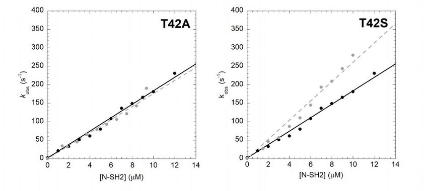
Int. J. Mol. Sci. 2020, 21, 461                                                                        2 of 7

     The catalytic activity of SHP2 is regulated by an auto-inhibition mechanism that involves the
interaction between the N-SH2 and PTP domain. In the absence of binding, N-SH2 physically blocks
the active site of the PTP domain, leading to the inhibition of its catalytic activity. On the contrary, when
N-SH2 binds, a ligand undergoes a major structural rearrangement that activates the phosphatase [12].
A number of mutations affecting SHP2 have been connected with the onset of NS. Whilst the majority
of these mutations concern residues at the interface between the N-SH2 and PTP domain (for example,
D61G, A72S, A72G, E76D, I282V, N308D, M504V) [7,14,15] destabilizing the catalytically inactive form
of SHP2 [16], one mutation, namely T42A, occurs in the N-SH2 domain far from the PTP domain.
Interestingly, in the latter case, a comparison by ITC of the binding properties of wild-type and mutated
N-SH2 revealed an increased capability of the naturally occurring variant; an observation which
demands additional experiments.
     In this communication, by characterizing the binding properties of the T42A variant in comparison
to that of wild-type N-SH2, we investigate the molecular basis by which the mutation leads to the
disease. In particular, by following the change in fluorescence signal due to Förster resonance energy
transfer between a tryptophan residue naturally present in the N-SH2 domain and a dansyl group
covalently attached to the peptide mimicking Gab2, a physiological ligand of SHP2, we could measure
the microscopic association and dissociation rate constants of binding. The comparison of kinetic
data obtained with wild-type and mutated N-SH2 revealed a dramatic increase in the affinity for
Gab2 in the mutant, due to a pronounced decrease of the microscopic dissociation rate constant. Our
data are briefly discussed on the basis of available structural data and previous works on the T42A
NS-causing mutation.

2. Results
      In order to unveil the effect of T42A mutation on the binding properties of the N-SH2 domain, we
resorted to performing kinetic binding experiments. In analogy to our previous work [17], the binding
between N-SH2 and Gab2 was measured by using a dansylated peptide mimicking the region of Gab2
ranging between residues 608 and 620 (Gab2608−620 ), presenting a phospho-tyrosine in position 614,
which is specifically recognized by N-SH2. The binding reaction was measured by stopped-flow mixing
experiments, by following the change in FRET signal occurring between residue tryptophan 6 of N-SH2
and a dansyl group covalently linked to the N-terminus of Gab2608−620 . Pseudo-first-order binding
experiments were performed by rapidly mixing a constant concentration of dansylated Gab2608−620
(1 µM) versus different concentrations of N-SH2 T42A, ranging from 1 to 12 µM. All traces followed
a single exponential decay. A comparison between the observed rate constants obtained for N-SH2
T42A to those of the wild-type proteins at the same experimental conditions is reported in Figure 1.
Both experiments were consistent with a linear dependence of pseudo-first-order kinetics. It is evident
that the T42A mutation has no effect on the recognition event of Gab2608−620 , the calculated kon values
(kon T42A = 17.6 ± 0.6 µM−1 s−1 ) being very similar to the one obtained for N-SH2 wt (kon wt = 18.2 ±
0.3 µM−1 s−1 ).
      In theory, analysis of the dependence of the observed rate constants under pseudo-first-order
conditions would allow the microscopic dissociation rate constants (koff ) to be calculated by extrapolating
at 0 [N-SH2]. However, because of the low values of koff , the high experimental error that arises
from this procedure jeopardizes a valuable calculation of koff and, therefore, demands a different
approach. To accurately determine koff values, we resorted to performing displacement experiments,
in which a pre-incubated complex of dansylated Gab2608−620 and N-SH2 (at 1:1 stoichiometric ratio)
were rapidly mixed with different concentrations, in high excess, of nondansylated Gab2608−620 .
In agreement with theory [18], when the concentration of the displacer was in high excess compared to
that of the pre-incubated complex, the observed rate constant was found to be insensitive to reactant
concentrations. The displacement time courses for N-SH2 T42A and wild-type N-SH2 are reported in
Figure 2. In both cases, data were satisfactorily fitted with a single exponential equation. Interestingly,
the T42A mutation caused a remarkable effect on koff , being 100 times lower compared to the one
D off on
SH2 T42A and Gab2. The affinity for the ligand was dramatically increased upon mutation, with a
KD = 1.7 ± 0.1 nM calculated for N-SH2 T42A compared to the KD = 100 ± 10 nM obtained under the
same experimental conditions for wild type N-SH2.
In order to obtain more detailed information about the role of T42 residue in the binding reaction
with
Int. J. Mol. Gab2,
Sci. 2020, we designed a more conservative N-SH2 variant where the threonine was replaced with3 of 7
21, 461
a serine. This variant retains the hydroxyl group while removing an electron donor methyl group
from the side chain. In analogy to the T42A mutant, we performed binding (Figure 1) and
obtained displacement (FigureN-SH2
for wild-type 2) kinetic experiments
under the same on experimental
the T42S mutantconditions
and compared wt =to1.85
(koffthem the s−1 ;
those±of0.10
koff T42A = 0.031
wild-type protein.
± 0.005 −1
Theskon).value
Thecalculated from fitting was
direct measurement of kcomparable with the one obtained for the
off allowed us to obtain the equilibrium
binding with N-SH2 wt (k on T42S = 26.1 ± 0.7 μM−1 s−1), indicating that the T42S mutation also does
dissociation rate constant (KD = koff /kon ) for the interaction between N-SH2 T42A and Gab2. The
not affect the recognition of Gab2608−620. On the other hand, the value of koff appears to be affected by
affinity for the ligand was dramatically increased upon mutation, with a KD = 1.7 ± 0.1 nM calculated
T42S mutation by a factor of 10 compared to that of the wild type, with a calculated value of 0.14 ±
for N-SH2 T42A compared to the KD = 100 ± 10 nM obtained under the same experimental conditions
0.02 s−1, and consequently an equilibrium dissociation constant of 5.3 ± 0.2 nM.
for wild type N-SH2.
Figure 1. Kinetics of binding between Gab2608−620 and N-SH2 wt (black circles, black continuous line)
and itsFigure
variants (gray circles,
1. Kinetics graybetween
of binding brokenGab2
lines)608−620
in buffer Hepes
and N-SH2 wt50 mM circles,
(black pH 7.2,black
NaClcontinuous at 10 ◦ C.
300 mM, line)
Int. J. Mol. Sci. 2019, 20, x FOR PEER REVIEW 4 of 8
wt data and its variants
were taken from(gray[17].
circles, grayare
Lines broken lines)
the best fitintobuffer Hepes
a linear 50 mM pH 7.2, NaCl 300 mM, at 10
equation.
°C. wt data were taken from [17]. Lines are the best fit to a linear equation.
To test the effects of the T42S and T42A mutations of the structure of N-SH2, we carried out a
complete folding characterization of these two variants, in comparison to the wild-type protein. In
particular, we subjected the mutants to equilibrium and kinetic unfolding experiments. The
experiments were carried out following the same procedures described previously for N-SH2 [17,19].
The folding and unfolding data obtained for T42S and T42A in comparison to wild-type N-SH2 are
reported in Figure 3. It is evident that, whilst the T42S mutant contributes to a minor destabilization
of the native state, in both cases the folding pathway is essentially unaffected by the mutations, as
proved by the apparent co-operativity of the equilibrium transitions as well as by the dependence of
the folding and unfolding rate constants. These findings suggest that the mutations do not
significantly alter the folding of N-SH2.
Figure 2. Displacement of the complex N-SH2:dansyl-Gab2608−620
Figure 2. Displacement of the complex N-SH2:dansyl-Gab2608−620
by a large excess of non-dansylated
by a large excess of non-dansylated
Gab2Gab2
608−620 . Lines represent the best fit to a single exponential equation.
608−620. Lines represent the best fit to a single exponential equation.
In order to obtain more detailed information about the role of T42 residue in the binding reaction
with Gab2, we designed a more conservative N-SH2 variant where the threonine was replaced with aInt. J. Mol. Sci. 2020, 21, 461 4 of 7
serine. This variant retains the hydroxyl group while removing an electron donor methyl group from
the side chain. In analogy to the T42A mutant, we performed binding (Figure 1) and displacement
(Figure 2) kinetic experiments on the T42S mutant and compared them to those of the wild-type protein.
The kon value calculated from fitting was comparable with the one obtained for the binding with
N-SH2 wt (kon T42S = 26.1 ± 0.7 µM−1 s−1 ), indicating that the T42S mutation also does not affect the
recognition of Gab2608−620 . On the other hand, the value of koff appears to be affected by T42S mutation
by a factor of 10 compared to that of the wild type, with a calculated value of 0.14 ± 0.02 s−1 , and
consequently an equilibrium dissociation constant of 5.3 ± 0.2 nM.
To test the effects of the T42S and T42A mutations of the structure of N-SH2, we carried out
a complete folding characterization of these two variants, in comparison to the wild-type protein.
In particular, we subjected the mutants to equilibrium and kinetic unfolding experiments. The
experiments were carried out following the same procedures described previously for N-SH2 [17,19].
The folding and unfolding data obtained for T42S and T42A in comparison to wild-type N-SH2 are
reported in Figure 3. It is evident that, whilst the T42S mutant contributes to a minor destabilization
of the native state, in both cases the folding pathway is essentially unaffected by the mutations, as
proved by the apparent co-operativity of the equilibrium transitions as well as by the dependence of
the folding and unfolding rate constants. These findings suggest that the mutations do not significantly
Int. folding
alter the J. Mol. Sci. 2019, 20, x FOR PEER REVIEW
of N-SH2. 5 of 8
Figure 3. Plot of the log10 of the (un)folding observed rate constants measured at different urea
Figure 3. Plot of the log10 of the (un)folding observed rate constants measured at different urea
concentrations for N-SH2 WT (black circles), N-SH2 T42A (dark gray circles), and N-SH2 T42S (light
concentrations for N-SH2 WT (black circles), N-SH2 T42A (dark gray circles), and N-SH2 T42S (light
gray circles) measured at the stopped-flow apparatus in buffer Tris-HCl 50 mM, pH 8.0, at 25 ◦ C.
gray circles) measured at the stopped-flow apparatus in buffer Tris-HCl 50 mM, pH 8.0, at 25 °C.
Lines represent the the
Lines represent bestbest
fit fit
to toananequation describing
equation describing a three-state
a three-state folding
folding mechanism
mechanism (see text(see
for text for
details). Inset Inset
details). panel: analysis
panel: of the
analysis amplitudes
of the amplitudesobtained
obtained in unfolding
unfoldingexperiments
experiments as aasfunction
a function
of of urea
urea concentration.
concentration. Lines are Lines
the bestare fit
thetobest fit to a sigmoid
a sigmoid equation. equation. It is evident
It is evident that
that the the mutations
mutations contribute to
a minorcontribute to a minorwithout
destabilization destabilization without altering
significantly significantly
thealtering
foldingtheoffolding
N-SH2.of N-SH2.
3. Discussion
3. Discussion
The collaborative efforts between structural biology and enzymatic essays on SHP2 has
The collaborative efforts between structural biology and enzymatic essays on SHP2 has provided
provided compelling evidence in favor of an auto-inhibition model at the basis of the regulation of
compelling evidence
this critical enzymein favor
[12,20]. In of
thisan auto-inhibition
scenario, model
the N-SH2 domain hasat thedepicted
been basis of
as athe regulation of this
conformational
switch—either it blocks the catalytic activity of the PTP or, when bound to a pY-containing sequence,
it releases the PTP domain, thereby tuning its activity. In this context, it is of particular interest to
characterize the only NS-causing mutation that does not occur at the interface between the N-SH2
and PTP domain, T42A. Phosphatase assays conducted on SHP2 protein [20] revealed that T42A
SHP2 displays a dramatically enhanced activity when in the presence of a phospho-tyrosine
containing peptide, compared to wild-type SHP2 and other variants not associated with NS, whilstInt. J. Mol. Sci. 2020, 21, 461 5 of 7
critical enzyme [12,20]. In this scenario, the N-SH2 domain has been depicted as a conformational
switch—either it blocks the catalytic activity of the PTP or, when bound to a pY-containing sequence,
it releases the PTP domain, thereby tuning its activity. In this context, it is of particular interest to
characterize the only NS-causing mutation that does not occur at the interface between the N-SH2 and
PTP domain, T42A. Phosphatase assays conducted on SHP2 protein [20] revealed that T42A SHP2
displays a dramatically enhanced activity when in the presence of a phospho-tyrosine containing
peptide, compared to wild-type SHP2 and other variants not associated with NS, whilst its basal
activity was comparable with the wild-type form. It has also been demonstrated that T42A mutation
causes an impairment of the binding capabilities of SHP2 [21], however, no information is available
about the details of this mutation’s effect on the interaction of the enzyme with its natural binding
partners. In this work, we provide a kinetic characterization of the effects of the NS-causing T42A
mutation on the binding of the N-SH2 domain with a peptide mimicking the scaffolding protein Gab2,
an interaction naturally occurring in the cellular environment that triggers the activation of SHP2.
Interestingly, while the mutation has no effect on the recognition of the ligand, with a conserved value
of kon , the complex appears to be strongly stabilized upon T42A mutation. In fact, the dramatic effect
observed on koff demonstrates that T42A mutation causes a less efficient release of the ligand from the
Int. J. Mol. Sci. 2019, 20, x FOR PEER REVIEW 6 of 8
N-SH2. Thus, in agreement with the auto-inhibition model, on the basis of our data we conclude that
T42A themutation
ligand fromlimitsthetheN-SH2.
abilityThus,
of N-SH2 to act aswith
in agreement a conformational
the auto-inhibition switch,
model,promoting
on the basistheofactivated
our
form ofdataSHP2 at lowerthat
we conclude ligandT42A concentrations.
mutation limits the ability of N-SH2 to act as a conformational switch,
Itpromoting
is of interest to analyze
the activated form our kinetic
of SHP2 dataligand
at lower in theconcentrations.
light of the three-dimensional structure of
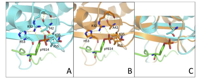
It is of interest to analyze our kinetic
N-SH2 (PDB: 4QSY). In fact, the structure shows that the hydroxyldata in the light of the three-dimensional structureisofinN-
group of T42 residue direct
SH2 (PDB: 4QSY). In fact, the structure shows that the hydroxyl
contact with the phosphate group of the phospho-tyrosine (Figure 4). Our results showed group of T42 residue is in direct
how a more
contact with
conservative the phosphate
mutation, T42S, whichgroupmaintains
of the phospho-tyrosine (Figure 4).
the polar properties ofOur
the results showed
side chain whilehow a more an
removing
conservative mutation, T42S, which maintains the polar properties of the side chain while removing
electron-donor methyl group, causes a less pronounced effect on the binding affinity. Of additional
an electron-donor methyl group, causes a less pronounced effect on the binding affinity. Of additional
interest, it may be noted that previous work based on molecular dynamics highlighted the presence
interest, it may be noted that previous work based on molecular dynamics highlighted the presence
of a conformational
of a conformational switchswitchin in
N-SH2,
N-SH2,primarily
primarily mediated
mediated by byTyr66,
Tyr66,Asp40,
Asp40, Lys55,
Lys55, andandGln57Gln57
[22]. [22].
Importantly,
Importantly,out ofoutthese of residues, Lys55,Lys55,
these residues, lies in the
lies proximity of T42. On
in the proximity the basis
of T42. On theof these
basis observations,
of these
we conclude that the hydroxyl group of T42 can have a role in shielding
observations, we conclude that the hydroxyl group of T42 can have a role in shielding the the negative charges
negative of the
phospho-tyrosine,
charges of the as well as in mediating
phospho-tyrosine, as welltheas role of Lys55 the
in mediating in an
roleopening-closing motion associated
of Lys55 in an opening-closing
motion binding,
with ligand associatedthereby
with ligand binding,
tuning thereby tuning the
the physiologically physiologically
correct correctof
rate of release rate
theofligand.
release of
Taken
the ligand.
together, Takensuggest
our results together, ourthe
that results
T42A suggest that the
mutation T42A
found inmutation
NS patientsfound in NS patients
impairs impairs of
this mechanism
ligandthis mechanism
release of ligand release
and consequently and consequently
promotes promotes aform
a hyper-activated hyper-activated
of SHP2, whichform ofisSHP2, which
the basis of the
is the basis of the onset of the disease.
onset of the disease.
4. (A,B)
FigureFigure Three-dimensional
4. (A,B) Three-dimensional structure ofof
structure the N-SH2
the N-SH2domain
domain wt
wt (in cyan) and
(in cyan) andT42A
T42A(in(inorange)
orange) in
complex with Gab1
in complex (inGab1
with green)
(in(PDB:
green)4QSY).
(PDB: Residues R32, K35,
4QSY). Residues T42/A42,
R32, H53, and
K35, T42/A42, K55and
H53, are K55
highlighted
are
with sticks and H-bonds
highlighted are represented
with sticks and H-bonds byare
yellow broken lines.
represented (C) Homology
by yellow modelling
broken lines. of the T42A
(C) Homology
variant of the N-SH2
modelling domain
of the T42A (inoforange)
variant computed
the N-SH2 with
domain (in SWISS-MODEL
orange) computed with web-based
SWISS-MODELservice,
web-using
based as
PDB 4QSY service, usingstructure
template PDB 4QSY(in as cyan).
template structure (inof
Side-chains cyan). Side-chains
T42/A42 of T42/A42with
are highlighted are highlighted
sticks.
with sticks.
4. Materials and Methods
4.1. Protein Expression and Purification
N-SH2 wild-type and the site-directed variants T42A and T42S were expressed and purified asInt. J. Mol. Sci. 2020, 21, 461 6 of 7
4. Materials and Methods
4.1. Protein Expression and Purification
N-SH2 wild-type and the site-directed variants T42A and T42S were expressed and purified as
described previously [17]. The variants were obtained using a QuickChange Lightning Site-Directed
Mutagenesis kit (Agilent Technologies, Santa Clara, California, United States), accordingly to the
manufacturer’s instructions.
4.2. Stopped-Flow Experiments
Binding kinetics experiments were performed using a single-mixing SX-18 stopped-flow
instrument (Applied Photophysics, Leatherhead, Surrey, UK,); binding experiments were conducted in
buffer Hepes 50 mM pH 7.2, NaCl 300 mM, at 10 ◦ C. Pseudo-first-order binding experiments were
performed by mixing a constant concentration of dansylated Gab2608−620 (1 µM) versus N-SH2 wt
and its variants at concentrations ranging from 1.5 µM to 12 µM. Displacement experiments were
performed by mixing the pre-incubated complex N-SH2:Gab2608−620 at the concentration of 1 µM with
the non-dansylated form of Gab2608−620 at increasing concentrations, ranging from 30 µM to 60 µM.
The excitation wavelength for all the experiments was 280 nm, and emission was collected using a
475-nm cut-off glass filter. For each acquisition, the average of at least five independent experiments
was fitted to a single exponential equation. Kinetic traces are provided as supplemental information
(Figure S1).
Unfolding and refolding kinetic experiments were conducted using a single-mixing SX-18
stopped-flow. Excitation wavelength was 280 nm and change in tryptophan fluorescence emission
was followed by using a 320 nm cut-off filter. Experiments were performed in buffer Tris-HCl 50 mM,
pH 8.0, at 25 ◦ C. Protein concentration was 3 µM.
4.3. Structure Homology Modelling
The three-dimensional structure of the T42A variant of the N-SH2 domain was computed
with SWISS-MODEL web-based service (https://swissmodel.expasy.org/), by using PDB 4QSY as the
template structure.
Supplementary Materials: Supplementary materials can be found at http://www.mdpi.com/1422-0067/21/2/461/s1.
Author Contributions: A.T. and S.G. designed the research. A.T., F.M., L.V., and F.T. performed the research.
All of the authors analyzed data. A.T. wrote the first version of the manuscript. All of the authors revised the
manuscript. All authors have read and agreed to the published version of the manuscript.
Funding: Work was partly supported by grants from the Italian Ministero dell’Istruzione dell’Università e
della Ricerca (Progetto di Interesse ‘Invecchiamento’ to S.G.), Sapienza University of Rome (B52F16003410005,
RP11715C34AEAC9B, and RM1181641C2C24B9 to S.G.), the Associazione Italiana per la Ricerca sul Cancro
(Individual Grant-MFAG 2016, 18701 to S.G.), and the Istituto Pasteur Italia (Teresa Ariaudo Research Project 2018,
to A.T.).
Conflicts of Interest: The authors declare no conflict of interest.
References
1. Noonan, J.A. Hypertelorism with Turner phenotype. A new syndrome with associated congenital heart
disease. Am. J. Dis. Child. 1968, 116, 373–380. [CrossRef] [PubMed]
2. Roberts, A.E.; Allanson, J.E.; Tartaglia, M.; Gelb, B.D. Noonan syndrome. Lancet 2013, 381, 333–342. [CrossRef]
3. Marino, B.; Digilio, M.C.; Toscano, A.; Giannotti, A.; Dallapiccola, B. Congenital heart diseases in children
with Noonan syndrome: An expanded cardiac spectrum with high prevalence of atrioventricular canal. J.
Pediatr. 1999, 135, 703–706. [CrossRef]
4. Carta, C.; Pantaleoni, F.; Bocchinfuso, G.; Stella, L.; Vasta, I.; Sarkozy, A.; Digilio, C.; Palleschi, A.; Pizzuti, A.;
Grammatico, P.; et al. Germline missense mutations affecting KRAS Isoform B are associated with a severe
Noonan syndrome phenotype. Am. J. Hum. Genet. 2006, 79, 129–135. [CrossRef] [PubMed]Int. J. Mol. Sci. 2020, 21, 461 7 of 7
5. Roberts, A.E.; Araki, T.; Swanson, K.D.; Montgomery, K.T.; Schiripo, T.A.; Joshi, V.A.; Li, L.; Yassin, Y.;
Tamburino, A.M.; Neel, B.G.; et al. Germline gain-of-function mutations in SOS1 cause Noonan syndrome.
Nat. Genet. 2007, 39, 70–74. [CrossRef] [PubMed]
6. Pandit, B.; Sarkozy, A.; Pennacchio, L.A.; Carta, C.; Oishi, K.; Martinelli, S.; Pogna, E.A.; Schackwitz, W.;
Ustaszewska, A.; Landstrom, A.; et al. Gain-of-function RAF1 mutations cause Noonan and LEOPARD
syndromes with hypertrophic cardiomyopathy. Nat. Genet. 2007, 39, 1007–1012. [CrossRef]
7. Tartaglia, M.; Mehler, E.L.; Goldberg, R.; Zampino, G.; Brunner, H.G.; Kremer, H.; van der Burgt, I.;
Crosby, A.H.; Ion, A.; Jeffery, S.; et al. Mutations in PTPN11, encoding the protein tyrosine phosphatase
SHP-2, cause Noonan syndrome. Nat. Genet. 2001, 29, 465–468. [CrossRef]
8. Gelb, B.D.; Tartaglia, M. Noonan syndrome and related disorders: Dysregulated RAS-mitogen activated
protein kinase signal transduction. Hum. Mol. Genet. 2006, 15, R220–R226. [CrossRef]
9. Tartaglia, M.; Gelb, B.D. Noonan syndrome and related disorders: Genetics and pathogenesis. Annu. Rev.
Genom. Hum. Genet. 2005, 6, 45–68. [CrossRef]
10. Scott, L.M.; Chen, L.; Daniel, K.G.; Brooks, W.H.; Guida, W.C.; Lawrence, H.R.; Sebti, S.M.; Lawrence, N.J.;
Wu, J. Shp2 protein tyrosine phosphatase inhibitor activity of estramustine phosphate and its triterpenoid
analogs. Bioorg. Med. Chem. Lett. 2011, 21, 730–733. [CrossRef]
11. Neel, B.G.; Gu, H.; Pao, L. The ’Shp’ing news: SH2 domain-containing tyrosine phosphatases in cell signaling.
Trends Biochem. Sci. 2003, 28, 284–293. [CrossRef]
12. Hof, P.; Pluskey, S.; Dhe-Paganon, S.; Eck, M.J.; Shoelson, S.E. Crystal structure of the tyrosine phosphatase
SHP-2. Cell 1998, 92, 441–450. [CrossRef]
13. Cunnick, J.M.; Dorsey, J.F.; Munoz-Antonia, T.; Mei, L.; Wu, J. Requirement of SHP2 binding to Grb2-associated
binder-1 for mitogen-activated protein kinase activation in response to lysophosphatidic acid and epidermal
growth factor. J. Biol. Chem. 2000, 275, 13842–13848. [CrossRef] [PubMed]
14. Tartaglia, M.; Niemeyer, C.M.; Fragale, A.; Song, X.; Buechner, J.; Jung, A.; Hählen, K.; Hasle, H.; Licht, J.D.;
Gelb, B.D. Somatic mutations in PTPN11 in juvenile myelomonocytic leukemia, myelodysplastic syndromes
and acute myeloid leukemia. Nat. Genet. 2003, 34, 148–150. [CrossRef]
15. Kosaki, K.; Suzuki, T.; Muroya, K.; Hasegawa, T.; Sato, S.; Matsuo, N.; Kosaki, R.; Nagai, T.; Hasegawa, Y.;
Ogata, T. PTPN11 (protein-tyrosine phosphatase, nonreceptor-type 11) mutations in seven Japanese patients
with Noonan syndrome. J. Clin. Endocrinol. Metab. 2002, 87, 3529–3533. [CrossRef]
16. Keilhack, H.; David, F.S.; McGregor, M.; Cantley, L.C.; Neel, B.G. Diverse biochemical properties of Shp2
mutants. Implications for disease phenotypes. J. Biol. Chem. 2005, 280, 30984–30993. [CrossRef]
17. Bonetti, D.; Troilo, F.; Toto, A.; Travaglini-Allocatelli, C.; Brunori, M.; Gianni, S. Mechanism of Folding and
Binding of the N-Terminal SH2 Domain from SHP2. J. Phys. Chem. B 2018, 122, 11108–11114. [CrossRef]
18. Antonini, E.; Brunori, M. Hemoglobin and Myoglobin in Their Reactions with Ligands; North-Holland: Amsterdam,
The Netherlands, 1971.
19. Visconti, L.; Malagrinò, F.; Gianni, S.; Toto, A. Structural characterization of an on-pathway intermediate
and transition state in the folding of the N-terminal SH2 domain from SHP2. FEBS J. 2019, 286, 4769–4777.
[CrossRef]
20. Martinelli, S.; Torreri, P.; Tinti, M.; Stella, L.; Bocchinfuso, G.; Flex, E.; Grottesi, A.; Ceccarini, M.; Palleschi, A.;
Cesareni, G.; et al. Diverse driving forces underlie the invariant occurrence of the T42A, E139D, I282V and
T468M SHP2 amino acid substitutions causing Noonan and LEOPARD syndromes. Hum. Mol. Genet. 2008,
17, 2018–2029. [CrossRef]
21. Müller, P.J.; Rigbolt, K.T.G.; Paterok, D.; Piehler, J.; Vanselow, J.; Lasonder, E.; Andersen, J.S.; Schaper, F.;
Sobota, R.M. Protein tyrosine phosphatase SHP2/PTPN11 mistargeting as a consequence of SH2-domain
point mutations associated with Noonan Syndrome and leukemia. J. Proteom. 2013, 84, 132–147. [CrossRef]
22. Guvench, O.; Qu, C.-K.; MacKerell, A.D. Tyr66 acts as a conformational switch in the closed-to-open transition
of the SHP-2 N-SH2-domain phosphotyrosine-peptide binding cleft. BMC Struct. Biol. 2007, 7, 14. [CrossRef]
[PubMed]
© 2020 by the authors. Licensee MDPI, Basel, Switzerland. This article is an open access
article distributed under the terms and conditions of the Creative Commons Attribution
(CC BY) license (http://creativecommons.org/licenses/by/4.0/).You can also read