RSC Chemical Biology COMMUNICATION
←
→
Page content transcription
If your browser does not render page correctly, please read the page content below
RSC
Chemical Biology
This article is licensed under a Creative Commons Attribution-NonCommercial 3.0 Unported Licence.
View Article Online
COMMUNICATION View Journal
A self-labeling protein based on the small
ultra-red fluorescent protein, smURFP†
Cite this: DOI: 10.1039/d1cb00127b
Open Access Article. Published on 29 June 2021. Downloaded on 7/28/2021 11:51:58 PM.
John-Hanson Machado,a Richard Ting,bc John Y. Lind and Erik A. Rodriguez *a
Received 7th June 2021,
Accepted 24th June 2021
DOI: 10.1039/d1cb00127b
rsc.li/rsc-chembio
Self-labeling proteins have revolutionized super-resolution and imagination. There are currently three self-labeling proteins
sensor imaging. Tags recognize a bioorthogonal substrate for known as Halo-, SNAP-, and CLIP-tags. Halo-tag (33 kDa) was
covalent attachment. We show the small Ultra-Red Fluorescent evolved from a bacterial haloalkane dehalogenase and
Protein (smURFP) is a self-labeling protein. The substrate is fluoro- covalently attaches an aliphatic hydrocarbon with a terminal
genic, fluoresces when attached, and quenches fluorescent cargo. chlorine with a cargo molecule.21,22 SNAP-tag (20 kDa) is a
The smURFP-tag has novel properties for tool development. mutant of the human O6-alkylguanine-DNA-alkyltransferase,
and the substrate molecule is O6-benzylguanine linked to a
Fluorescent proteins and genetically encoded sensors have cargo molecule.23 The CLIP-tag (20 kDa) was also derived from
revolutionized fluorescence imaging in living organisms but the human O6-alkylguanine-DNA-alkyltransferase, and the
are limited by the endogenous genetic code and available substrate is O2-benzylcytosine with a cargo.24 These self-
amino acids. Fluorescent proteins are typically less photostable labeling proteins and substrates are commercially available
than spectrally similar fluorescent small molecules commonly from Promega and New England Biolabs.
used for super-resolution microscopy.1–8 The endogenous The most common use for self-labeling proteins is the
amino acids allow for chemical modification9,10 but are not attachment of small molecule chemical dyes as the cargo for
specific for an individual protein of interest. Unnatural nucleic increased fluorescent intensity, greater photostability, temporal
acids and amino acids allow for the expansion of the genetic attachment of different color dyes, and fluorescence persists in
code and incorporation of L-unnatural amino acids tolerated by living and fixed cells.25–28 These tags allow for protein localiza-
biological ribosomes, including fluorescent unnatural amino tion beyond the diffraction limit of light, temporal labeling
acids for high photostability and single-molecule imaging.11–20 faster than fluorescent protein chromophore maturation, and
Unnatural amino acid incorporation remains limited by the protein–protein interactions. Biotin,29 halogenated fluoro-
ribosome for L-enantiomers and the evolution of aminoacyl- phores for correlative light and electron microscopy,30 and
tRNA synthetases that recognize unnatural amino acids. biorthogonal reactive molecules31 can serve as cargo molecules.
Self-labeling proteins are genetically encoded and fused to Halo-tag was used to create sensitive voltage sensors of multiple
proteins of interest to allow for the covalent attachment of a colours for fluorescence imaging in living cells and entire
bioorthogonal chemical substrate that is linked to a cargo organisms.32 All three self-labeling protein substrates only
molecule. Self-labeling proteins go beyond these limitations serve as recognition molecules for covalent attachment.
by covalently attaching bioorthogonal modified substrates that Fluorescence tracking of a non-fluorescent small-molecule
bear cargo molecules of larger size than tolerated by the cargo would require the attachment of a second fluorescent
ribosome, any enantiomer, and limited only by the chemists’ dye or genetic attachment to a fluorescent protein.
The small Ultra-Red Fluorescent Protein (smURFP, 32 kDa
a
Department of Chemistry, The George Washington University,
dimer) was evolved from a light-harvesting phycobilisome
Washington, DC 20052, USA. E-mail: erik_rodriguez@gwu.edu protein a-allophycocyanin.7 The evolution involved the manual
b
Department of Radiology, Molecular Imaging Innovations Institute (MI3), selection of 4106 E. coli colonies starting with autocatalytic
Weill Cornell Medicine, New York, NY, 10065, USA covalent attachment of phycocyanobilin, changing the sub-
c
Antelope Surgical, Biolabs@NYULangone, New York, NY, 10014, USA
d
strate to biliverdin IXa (BV), and evolving high expression
Tasmanian School of Medicine, University of Tasmania, Hobart, Tasmania 7000,
Australia
and stability. During this evolution, smURFP evolved a highly
† Electronic supplementary information (ESI) available. See DOI: 10.1039/ unusual recognition of BV. From crystal structures, most BV
d1cb00127b binding proteins and fluorescent proteins (iRFPs and IFPs)
© 2021 The Author(s). Published by the Royal Society of Chemistry RSC Chem. Biol.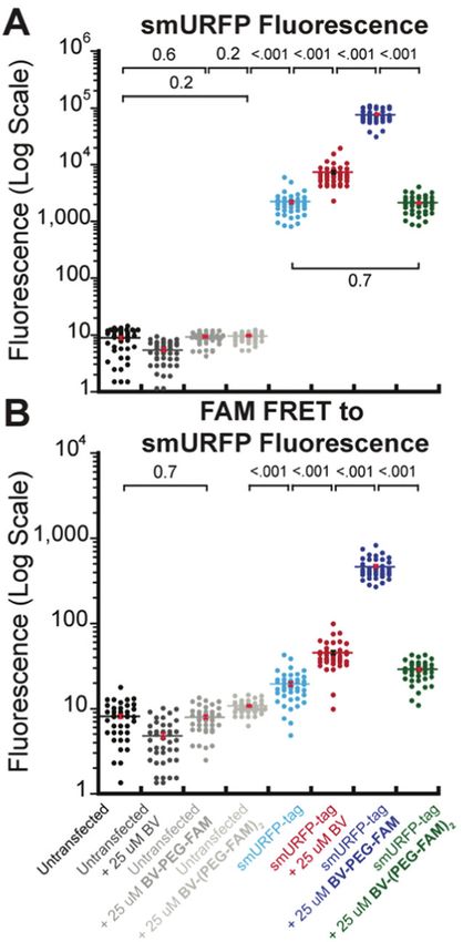
View Article Online
Communication RSC Chemical Biology
recognize the carboxylates on BV for binding or covalent
attachment.33–38 The crystal structure of a smURFP mutant
showed no recognition of the BV carboxylates.39 We showed
that methylation of the carboxylates created biliverdin dimethyl
This article is licensed under a Creative Commons Attribution-NonCommercial 3.0 Unported Licence.
ester (BVMe2) and is covalently attached to smURFP in human
embryonic kidney (HEK) 293A cells.7 smURFP lacks recognition
of the BV carboxylates.
We hypothesized that smURFP could serve as a self-labeling
protein and named the smURFP-tag. Biliverdin was modified
with a polyethylene glycol (PEG) linker to attach a cargo
molecule (Fig. 1). The PEG linker was chosen as a linear
Open Access Article. Published on 29 June 2021. Downloaded on 7/28/2021 11:51:58 PM.
molecule to mimic biliverdin dimethyl ester. The BV recogni-
tion molecule is a substrate for covalent attachment and is a
fluorogenic molecule. Upon covalent attachment of BV to
Fig. 1 smURFP-tag recognition of biliverdin (BV) modified substrate
smURFP, BV fluorescence is turned ‘‘on’’ by 410 000-fold
and experimental design. (A) The self-labeling protein, smURFP-tag, is
genetically fused to a protein of interest by an amino acid linker and enhancement of the quantum yield and emits far-red
is non-fluorescent. Modified BV substrate is added to the media and is fluorescence.7 The smURFP-tag is unique in that the recogni-
covalently attached to the smURFP-tag with the cargo molecule and tion molecule allows covalent attachment and far-red fluores-
becomes fluorescent in the far-red. (B) Hoechst 3342 labels DNA in cence tracking. To test this hypothesis, we targeted the
nuclei of all cells. The modified BV substrate (BV-PEG-FAM) is a
smURFP-tag to the nucleus to test if the BV substrate can pass
polyethylene glycol (PEG) linker, and the cargo molecule is
5(6)-carboxyfluorescein (FAM). Untransfected HEK293A cells serve as through the outer cell membrane and the nuclear envelope
a control and should be non-fluorescent. HEK293A cells are (Fig. 1B).
transfected with the smURFP-tag in the nucleus and become The BV substrate synthesis is relatively simple and uses
fluorescent. amide bond formation for stability. For attachment of a single
Scheme 1 Synthesis of the biliverdin (BV) substrates. A polyethylene glycol (PEG) linker separates the cargo molecule, R2, from the recognition
molecule, BV, to allow covalent attachment to the genetically encoded protein, smURFP-tag. R2 is 5(6)-carboxyfluorescein (FAM) in this study. (top)
BV-PEG-FAM is synthesized by amide bond coupling of PEG and FAM (ESI† Scheme S1) followed by amide bond attachment to BV. Steric hindrance
yields BV-PEG-FAM as the major product. (bottom) To avoid steric hindrance, BV-(PEG-FAM)2 is synthesized by amide coupling of PEG to BV followed by
amide bond formation between BV-(PEG)2 and FAM. The synthesis allows for simple and stable synthesis of the BV substrates.
RSC Chem. Biol. © 2021 The Author(s). Published by the Royal Society of Chemistry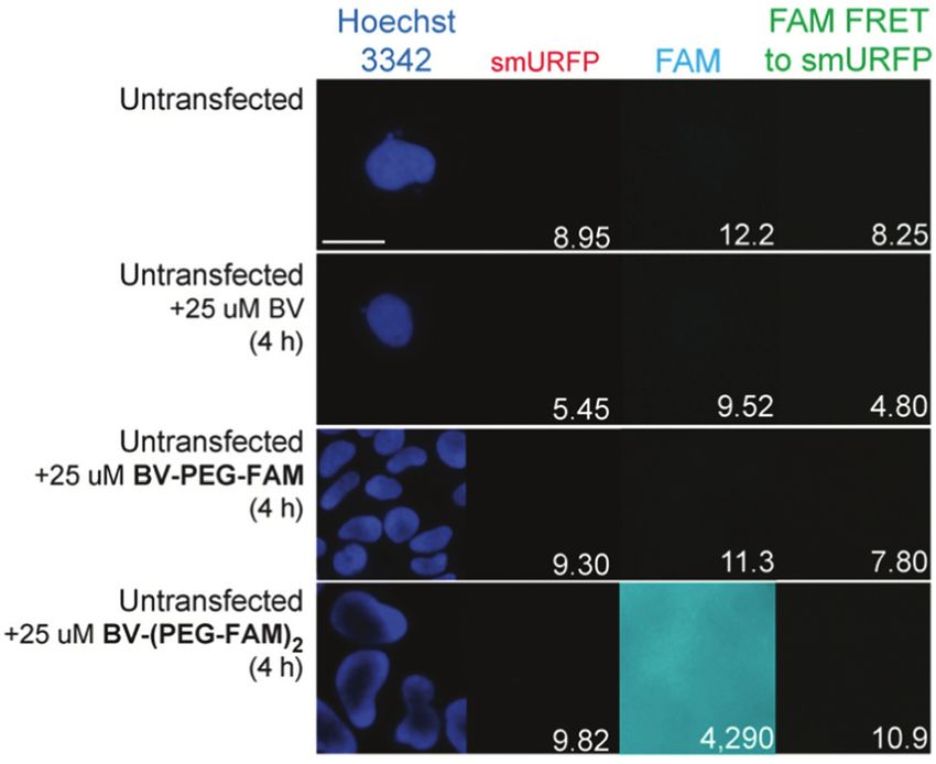
View Article Online
RSC Chemical Biology Communication
cargo, BV is reacted with H2N-PEG-R2 (Scheme 1). R2, in theory,
can be any small molecule and is tracked by far-red fluores-
cence of BV when covalently attached to smURFP-tag. Steric
hindrance limited the formation of BV with two cargo mole-
This article is licensed under a Creative Commons Attribution-NonCommercial 3.0 Unported Licence.
cules. To attach two cargo molecules, BV was first reacted with
H2N-PEG-NH2 in excess, followed by amide bond formation
with cargo molecule (R2) to couple two cargo molecules. In this
study, R2 is 5(6)-carboxyfluorescein (FAM) to allow for detection
by fluorescence imaging to confirm that the cargo is not
removed within the cell. The BV substrates are BV-PEG-FAM
and BV-(PEG-FAM)2 for the single and double cargo molecules,
Open Access Article. Published on 29 June 2021. Downloaded on 7/28/2021 11:51:58 PM.
respectively (Scheme 1).
BV-PEG-FAM was synthesized by amide bond formation
between 5(6)-carboxyfluorescein (FAM) and 2,2 0 -(ethylenedioxy)
bis(ethylamine) (PEG) to create PEG-FAM (ESI† Scheme S1).
Analytical HPLC–MS of PEG-FAM showed the major product at
11 minutes with FAM absorbance at 492 nm, and MS confirmed Fig. 2 Representative images of BV substrate addition to untransfected
the molecular weight (ESI† Fig. S1). PEG-FAM was coupled to cells. BV substrates were added for 4 h, and HEK293A cells were imaged
the BV carboxylates to create BV-PEG-FAM (ESI† Scheme S2). without removal. Lack of fluorescence is expected (Fig. 1B). Scale bar is
Analytical HPLC–MS of BV-PEG-FAM showed a significant peak 10 mm and numbers are mean fluorescence intensity of 40 cells.
at 16 minutes with BV absorbance at 645 and 780 nm and
correct mass (ESI† Fig. S2). Proton NMR of starting material, without BV substrate removal. A substrate concentration of
BV, and BV-PEG-FAM confirmed the structure (ESI† Fig. S3). 25 mM for BV, BV-PEG-FAM, and BV-(PEG-FAM)2 was chosen
The synthesis of BV-PEG-FAM showed no di-substituted because BV and BV dimethyl ester saturated smURFP-tag at
BV-(PEG-FAM)2 due to steric hindrance. 25 mM. The labeling time was 4 hours because fluorescence rise
The synthesis of BV-(PEG-FAM)2 used an alternative strategy fit a first-order, exponential rate equation, and BV saturation is
to avoid steric hindrance (Scheme 1). BV was reacted with PEG expected after r1.3 hours.7 Four hours ensures ample time to
to obtain amide bond attachment to both BV carboxylates (ESI† saturate the smURFP-tag with the substrates. Hoechst 3342 was
Scheme S3). Analytical HPLC–MS of BV-(PEG)2 confirmed used to visualize the DNA in the nucleus and showed all the
synthesis (ESI† Fig. S4). BV-(PEG)2 was reacted with FAM, and cells within an image (Fig. 2). smURFP, FAM, and FAM Förster
the doubly substitute, BV-(PEG-FAM)2 was purified (ESI† Resonance Energy Transfer (FRET) to smURFP fluorescence
Scheme S4). Analytical HPLC–MS of BV-(PEG-FAM)2 confirmed were imaged. HEK293A cells without transfection or untrans-
the molecule at 16 minutes by detecting the doubly charged fected showed minimal background fluorescence as expected
species by MS (ESI† Fig. S5). Proton NMR of BV-(PEG-FAM)2 (Fig. 1B). Addition of 25 mM BV or BV-PEG-FAM showed no
confirmed the structure of the molecule (ESI† Fig. S6). Attach- significant increase in fluorescence signal. The lack of FAM
ing unmodified PEG to BV removed steric hindrance to allow fluorescence from BV-PEG-FAM is due to the quenching by the
the attachment of two cargo, FAM molecules. The synthesis of recognition molecule BV. The FAM fluorescence increases with
the mono- and di-substituted BV-PEG-R2 and BV-(PEG-R2)2 the di-substituted BV-(PEG-FAM)2, and the single BV cannot
molecules are general strategies and should allow the attach- quench both FAMs. The lack of significant fluorescence on
ment of any cargo molecules. The amide bond is exceptionally untransfected cells is desired to only image the smURFP-tag
stable, surviving for B270 years in water at pH = 7,10 and fluorescence.
should survive the cellular environment. HEK293A cells were transiently transfected with smURFP-
BV substrates were added to purified smURFP-tag without tag localized within the nucleus by a nuclear localization
chromophore to demonstrate covalent attachment in vitro. Ten- sequence. We choose this location to test the ability of the BV
fold excess of BV, BV-PEG-FAM, and BV-(PEG-FAM)2 were added substrates to pass the outer and nuclear envelope membranes
to smURFP-tag overnight, and the free BV substrates were (Fig. 1). Previously, we showed that the negatively charged BV
removed by nickel column purification. Absorbance showed carboxylates limited membrane permeability and utility in cell
smURFP-tag covalently attached BV by the characteristic Soret culture and mice.7 Modifying the carboxylates is predicted to
and Q band at 385 and 642 nm, respectively (ESI† Fig. S7). enhance permeability to HEK293A cell membranes to increase
BV-PEG-FAM was covalently attached to the smURFP-tag and the smURFP fluorescence relative to BV. Without BV addition,
retained the absorbance of FAM at 492 nm in vitro. smURFP-tag smURFP-tag is fluorescent due to BV present in the fetal bovine
did not covalently attach BV-(PEG-FAM)2 in vitro by lack of serum (FBS). There is slightly greater fluorescence in the FAM
absorbance from 340–700 nm. FRET to smURFP channel because BV covalently attached to
BV substrates were initially added to HEK293A cells (Ther- smURFP-tag is somewhat excited by the blue excitation light
moFisher, R70507) that lacked any genetic modification (Fig. 3). The addition of BV and BV-PEG-FAM shows a 3.3- and
to visualize background fluorescence. Cells were imaged 34-fold, respectively, increase in smURFP fluorescence. FAM
© 2021 The Author(s). Published by the Royal Society of Chemistry RSC Chem. Biol.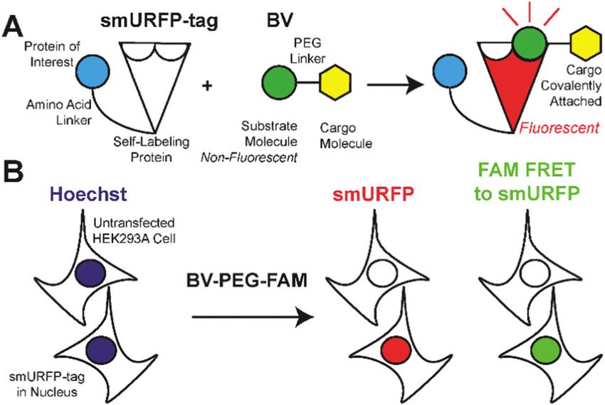
View Article Online
Communication RSC Chemical Biology
fluorescence was slightly increased for BV-PEG-FAM, and FAM
fluorescence was seen as FRET to smURFP. BV-(PEG-FAM)2
showed no increase in smURFP fluorescence, and the BV is
not covalently attached in vivo, which agrees with the lack of
This article is licensed under a Creative Commons Attribution-NonCommercial 3.0 Unported Licence.
in vitro attachment. The FAM fluorescence increases on the
di-substituted BV-(PEG-FAM)2 because BV does not quench
both FAMs. This experiment shows that BV-PEG-FAM can pass
the cell outer and nuclear membranes to deliver the cargo
(FAM) for covalent attachment to the smURFP-tag.
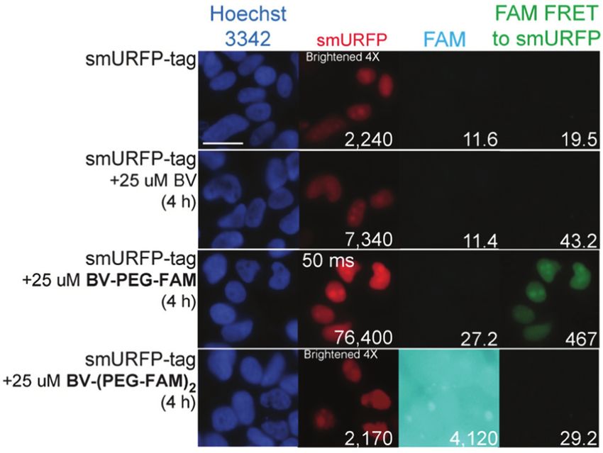
A region of interest was drawn on 40 cells for each sample,
and the mean fluorescence intensity was calculated. The
Open Access Article. Published on 29 June 2021. Downloaded on 7/28/2021 11:51:58 PM.
40 measurements are plotted with statistical comparison in
Fig. 4. Untransfected cells shown minimal smURFP fluores-
cence and are plotted on a Y-axis log scale to visualize points
(Fig. 4A). HEK293A cells with smURFP-tag within the nucleus
Fig. 3 Representative images of BV substrates added to HEK293A cells show a statistically significant increase in fluorescence.
transfected with nucleus localized smURFP-tag. BV substrates were added smURFP-tag without substrate and with BV-(PEG-FAM)2
for 4 h, and cells were imaged without removal. Scale bar is 10 mm and show the same amount of fluorescence and confirms that
numbers are mean fluorescence intensity of 40 cells. BV-(PEG-FAM)2 is not attached in vivo. The addition of
BV and BV-PEG-FAM show a statistically significant increase
in fluorescence. BV membrane permeability is limited by the
negatively charged carboxylates. BV-PEG-FAM masks a single
carboxylate to create a molecule with an overall neutral charge
(Scheme 1) that allows the molecule to pass the outer and
nuclear membranes to increase the fluorescence of smURFP-
tag by 34-fold.
Unexpectedly, FAM fluorescence was seen almost entirely as
FRET to smURFP. The FRET is due to the proximity of FAM to
smURFP-tag and the substantial extinction coefficient of
180 000 M1 cm1 for smURFP. Untransfected cells showed a
minimal increase in FAM FRET to smURFP fluorescence, and
BV-(PEG-FAM)2 showed the greatest increase due to the lack of
quenching of both FAM molecules. smURFP-tag without sub-
strate and BV show slightly increased signal relative to the
untransfected cells due to slight excitation of smURFP by the
blue light. The largest FAM FRET to smURFP signal is seen with
the BV-PEG-FAM and confirms that the cargo molecule is
delivered to the smURFP-tag and remains covalently attached
inside the nucleus.
Conclusions
The small Ultra-Red Fluorescent Protein (smURFP) is also a self-
labeling protein capable of recognizing and covalently attaching
carboxylate modified BV substrates. The smURFP-tag is unique
compared to Halo-, SNAP-, and CLIP-tags because BV serves as a
recognition molecule for covalent attachment, is a fluorogenic dye
for fluorescence tracking of covalently attached cargo, and
Fig. 4 Comparison of data points and mean fluorescence intensity of (A) quenches a fluorescent dye for imaging without substrate
smURFP and (B) FAM FRET to smURFP from Fig. 2 and 3. The Y-axis is removal. The smURFP-tag within the nucleus could covalently
shown on a log scale to visualize untransfected HEK293A cell data points. attach the mono-substituted BV-PEG-FAM, which was cell and
Mean fluorescence intensity is written in Fig. 2 and 3 and indicated by a
nuclear membrane permeant. BV-(PEG-FAM)2 did not covalently
horizontal bar. A one-way ANOVA with a = 0.05 and a post hoc Tukey honestly
significant difference (HSD) compared samples. All statistically insignificant
attach to the smURFP-tag in vitro and in vivo. Further evolution
comparisons are written, and all unwritten comparisons are p o 001. n = 40 of the smURFP-tag binding pocket is necessary to tolerate
cells and error bars are standard error of the mean (SEM). di-substituted BV substrates. In the presence of ten molar
RSC Chem. Biol. © 2021 The Author(s). Published by the Royal Society of ChemistryView Article Online
RSC Chemical Biology Communication
equivalents of the chromophore, smURFP-tag covalently attached 6 B. N. Giepmans, S. R. Adams, M. H. Ellisman and
two biliverdin but could only covalently attach a single biliverdin R. Y. Tsien, Science, 2006, 312, 217–224.
dimethyl ester by mass spectrometry and biophysical properties.7 7 E. A. Rodriguez, G. N. Tran, L. A. Gross, J. L. Crisp, X. Shu,
BV-PEG-FAM increases steric hindrance, and the stoichiometry is J. Y. Lin and R. Y. Tsien, Nat. Methods, 2016, 13,
This article is licensed under a Creative Commons Attribution-NonCommercial 3.0 Unported Licence.
expected to be a single BV-PEG-FAM per smURFP-tag, which is 763–769.
desired for increased brightness without diminishing the quan- 8 E. A. Rodriguez, J. Y. Lin, R. Ting, G. N. Tran and R. Y. Tsien,
tum yield. The unique properties of the smURFP-tag are useful for US Pat., US20180201655A1, 2019.
genetically targeting small molecules to proteins of interest with 9 O. Boutureira and G. J. Bernardes, Chem. Rev., 2015, 115,
far-red fluorescence tracking without the need to add a chemical 2174–2195.
dye or genetically fuse to a fluorescent protein. Live-cell super- 10 S. Mahesh, K. C. Tang and M. Raj, Molecules, 2018, 23, 2615.
resolution microscopy will utilize the smURFP-tag for imaging 11 R. Pantoja, E. A. Rodriguez, M. I. Dibas, D. A. Dougherty and
Open Access Article. Published on 29 June 2021. Downloaded on 7/28/2021 11:51:58 PM.
small-molecule fluorophores without substrate washing due to H. A. Lester, Biophys. J., 2009, 96, 226–237.
biliverdin quenching to ensure saturation of newly translated 12 E. A. Rodriguez, H. A. Lester and D. A. Dougherty, Proc. Natl.
proteins in the presence excess of substrate for the maximum Acad. Sci. U. S. A., 2006, 103, 8650–8655.
signal with low background fluorescence. The smURFP-tag should 13 E. A. Rodriguez, H. A. Lester and D. A. Dougherty,
find further use for the development of novel biosensors that RNA, 2007, 13, 1703–1714.
utilize the smURFP-tag fluorescence with chemical and protein 14 E. A. Rodriguez, H. A. Lester and D. A. Dougherty,
sensors3 that recognize small molecules, peptides, and other RNA, 2007, 13, 1715–1722.
biomolecules. smURFP-tag exogenous labelling of proteins with 15 C. J. Noren, S. J. Anthony-Cahill, M. C. Griffith and
sortase-mediated attachment,40 inclusion into virus particles,41 P. G. Schultz, Science, 1989, 244, 182–188.
and smURFP-tag nanoparticles42 will allow for bioorthogonal, 16 D. R. Liu, T. J. Magliery, M. Pastrnak and P. G. Schultz,
site-specific attachment of molecules without misfolding or opti- Proc. Natl. Acad. Sci. U. S. A., 1997, 94, 10092–10097.
mization of reaction conditions for targeted or biosensor imaging 17 L. Wang, A. Brock, B. Herberich and P. G. Schultz,
in entire living animals. Science, 2001, 292, 498–500.
18 D. L. Beene, D. A. Dougherty and H. A. Lester, Curr. Opin.
Neurobiol., 2003, 13, 264–270.
Author contributions 19 D. D. Young and P. G. Schultz, ACS Chem. Biol., 2018, 13,
854–870.
EAR conceptualized the methodology, designed experiments,
20 S. Hoshika, N. A. Leal, M. J. Kim, M. S. Kim, N. B. Karalkar,
and analysed fluorescence images. JHM, EAR, and RT synthe-
H. J. Kim, A. M. Bates, N. E. Watkins, Jr., H. A. SantaLucia,
sized and characterized molecules. All authors performed
A. J. Meyer, S. DasGupta, J. A. Piccirilli, A. D. Ellington,
experiments, wrote the paper, created figures, and reviewed
J. SantaLucia, Jr., M. M. Georgiadis and S. A. Benner,
the final manuscript.
Science, 2019, 363, 884–887.
21 G. V. Los and K. Wood, Methods Mol. Biol., 2007, 356,
Conflicts of interest 195–208.
22 G. V. Los, L. P. Encell, M. G. McDougall, D. D. Hartzell,
The described technology is patented (US20180201655A1).8 N. Karassina, C. Zimprich, M. G. Wood, R. Learish,
R. F. Ohana, M. Urh, D. Simpson, J. Mendez,
K. Zimmerman, P. Otto, G. Vidugiris, J. Zhu, A. Darzins,
Acknowledgements D. H. Klaubert, R. F. Bulleit and K. V. Wood, ACS Chem.
This project was funded by startup funds provided by The Biol., 2008, 3, 373–382.
George Washington University to EAR and US National Insti- 23 A. Keppler, S. Gendreizig, T. Gronemeyer, H. Pick, H. Vogel
tutes of Health grant U01NS090590 to JYL. and K. Johnsson, Nat. Biotechnol., 2003, 21, 86–89.
24 A. Gautier, A. Juillerat, C. Heinis, I. R. Correa, Jr.,
Notes and references M. Kindermann, F. Beaufils and K. Johnsson, Chem. Biol.,
2008, 15, 128–136.
1 E. A. Rodriguez, R. E. Campbell, J. Y. Lin, M. Z. Lin, 25 R. S. Erdmann, S. W. Baguley, J. H. Richens, R. F. Wissner,
A. Miyawaki, A. E. Palmer, X. Shu, J. Zhang and Z. Xi, E. S. Allgeyer, S. Zhong, A. D. Thompson, N. Lowe,
R. Y. Tsien, Trends Biochem. Sci., 2017, 42, 111–129. R. Butler, J. Bewersdorf, J. E. Rothman, D. St Johnston,
2 F. Montecinos-Franjola, J. Y. Lin and E. A. Rodriguez, A. Schepartz and D. Toomre, Cell Chem. Biol., 2019, 26(584–
Biochem. Soc. Trans., 2020, 48, 2657–2667. 592), e586.
3 G. C. H. Mo, C. Posner, E. A. Rodriguez, T. Sun and J. Zhang, 26 B. Barlag, O. Beutel, D. Janning, F. Czarniak, C. P. Richter,
Nat. Commun., 2020, 11, 1848. C. Kommnick, V. Goser, R. Kurre, F. Fabiani, M. Erhardt,
4 R. Y. Tsien, Annu. Rev. Biochem., 1998, 67, 509–544. J. Piehler and M. Hensel, Sci. Rep., 2016, 6, 31601.
5 N. C. Shaner, P. A. Steinbach and R. Y. Tsien, Nat. Methods, 27 F. Stagge, G. Y. Mitronova, V. N. Belov, C. A. Wurm and
2005, 2, 905–909. S. Jakobs, PLoS One, 2013, 8, e78745.
© 2021 The Author(s). Published by the Royal Society of Chemistry RSC Chem. Biol.View Article Online
Communication RSC Chemical Biology
28 J. B. Grimm, B. P. English, J. Chen, J. P. Slaughter, Z. Zhang, 35 D. Yu, W. C. Gustafson, C. Han, C. Lafaye, M. Noirclerc-
A. Revyakin, R. Patel, J. J. Macklin, D. Normanno, Savoye, W. P. Ge, D. A. Thayer, H. Huang, T. B. Kornberg,
R. H. Singer, T. Lionnet and L. D. Lavis, Nat. Methods, A. Royant, L. Y. Jan, Y. N. Jan, W. A. Weiss and X. Shu,
2015, 12, 244–250. Nat. Commun., 2014, 5, 3626.
This article is licensed under a Creative Commons Attribution-NonCommercial 3.0 Unported Licence.
29 S. Svendsen, C. Zimprich, M. G. McDougall, D. H. Klaubert 36 L. Lad, J. Friedman, H. Li, B. Bhaskar, P. R. Ortiz de
and G. V. Los, BMC Cell Biol., 2008, 9, 17. Montellano and T. L. Poulos, Biochemistry, 2004, 43,
30 V. Liss, B. Barlag, M. Nietschke and M. Hensel, Sci. Rep., 3793–3801.
2015, 5, 17740. 37 O. Cunningham, A. Dunne, P. Sabido, D. Lightner and
31 H. E. Murrey, J. C. Judkins, C. W. Am Ende, T. E. Ballard, T. J. Mantle, J. Biol. Chem., 2000, 275, 19009–19017.
Y. Fang, K. Riccardi, L. Di, E. R. Guilmette, J. W. Schwartz, 38 P. J. Pereira, S. Macedo-Ribeiro, A. Parraga, R. Perez-Luque,
J. M. Fox and D. S. Johnson, J. Am. Chem. Soc., 2015, 137, O. Cunningham, K. Darcy, T. J. Mantle and M. Coll,
Open Access Article. Published on 29 June 2021. Downloaded on 7/28/2021 11:51:58 PM.
11461–11475. Nat. Struct. Biol., 2001, 8, 215–220.
32 A. S. Abdelfattah, T. Kawashima, A. Singh, O. Novak, H. Liu, 39 J. P. Fuenzalida-Werner, R. Janowski, K. Mishra,
Y. Shuai, Y. C. Huang, L. Campagnola, S. C. Seeman, J. Yu, I. Weidenfeld, D. Niessing, V. Ntziachristos and A. C. Stiel,
J. Zheng, J. B. Grimm, R. Patel, J. Friedrich, B. D. Mensh, J. Struct. Biol., 2018, 204, 519–522.
L. Paninski, J. J. Macklin, G. J. Murphy, K. Podgorski, 40 I. Deshpande, J. Liang, D. Hedeen, K. J. Roberts, Y. Zhang,
B. J. Lin, T. W. Chen, G. C. Turner, Z. Liu, M. Koyama, B. Ha, N. R. Latorraca, B. Faust, R. O. Dror, P. A. Beachy,
K. Svoboda, M. B. Ahrens, L. D. Lavis and E. R. Schreiter, B. R. Myers and A. Manglik, Nature, 2019, 571, 284–288.
Science, 2019, 365, 699–704. 41 F. C. Herbert, O. R. Brohlin, T. Galbraith, C. Benjamin,
33 J. R. Wagner, J. Zhang, J. S. Brunzelle, R. D. Vierstra and C. A. Reyes, M. A. Luzuriaga, A. Shahrivarkevishahi and
K. T. Forest, J. Biol. Chem., 2007, 282, 12298–12309. J. J. Gassensmith, Bioconjugate Chem., 2020, 31, 1529–1536.
34 S. Ghosh, C. L. Yu, D. J. Ferraro, S. Sudha, S. K. Pal, 42 F. An, N. Chen, W. J. Conlon, J. S. Hachey, J. Xin, O. Aras,
W. F. Schaefer, D. T. Gibson and S. Ramaswamy, Proc. Natl. E. A. Rodriguez and R. Ting, Int. J. Biol. Macromol., 2020,
Acad. Sci. U. S. A., 2016, 113, 11513–11518. 153, 100–106.
RSC Chem. Biol. © 2021 The Author(s). Published by the Royal Society of ChemistryYou can also read