Ospite - e-learning "Sapienza"
←
→
Page content transcription
If your browser does not render page correctly, please read the page content below
Patogeno                     interazione
                                                            Ospite

                                     Attivazione del sistema
                                           immunitario
  Rapida e completa
  espulsione del patogeno

                                                        Persistenza del patogeno
              Infezione acuta                           (latenza e/o induzione di
              e successiva eliminazione                 una patologia cronica)
              del patogeno

    +/-                  ++
Pressione selettiva esercitata dal sistema immunitario sul patogeno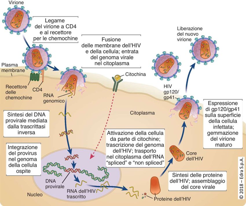
I patogeni hanno acquisito strategie per evadere l’immunità innata
ed adattativa, nel corso della co-evoluzione con l’ospite

  Esempi di strategie:

  “Dormancy” per rendersi “poco visibili”

  -produzione minima di proteine (herpesvirus)

  -integrazione del DNA nel genoma dell’ospite (retrovirus)

  -quiescenza dei micobatteri
I patogeni hanno “imparato” ad evadere l’immunità innata ed acquisita nel
corso della co-evoluzione con l’ospite

Esempi di strategie:

   “Sequestration” ovvero occupare “siti speciali”

   -invasione dei globuli rossi da parte del Plasmodio della malaria

   -colonizzazione della cistifellea da parte di S. enterica Typhi

   -uso da parte di HIV delle cellule dendritiche per il trasporto dalle mucose ai
   linfonodi
HIV usa le cellule dendritiche come cavallo di Troia per il trasporto dalle
                        mucose ai tessuti linfoidi
                             Legame di gp120 a DC-SIGN
                             Internalizzazione e persistenza nel
                             compartimento endosomale

   DC immatura

                                 Trans-infezione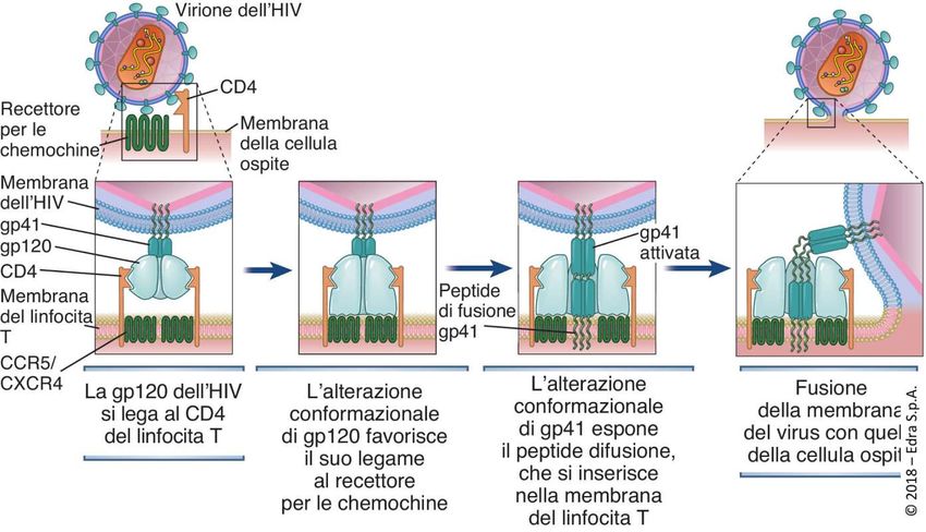
Struttura di HIV ed infezione cellulare
polimorfismi dell’envelope e corecettori utilizzati

Varianti R5 (M-tropiche) usano come corecettore CCR5 ed
infettano DC, macrofagi e cellule T CD4+

Varianti X4 (T-tropiche) usano come corecettore CXCR4 ed
infettano preferibilmente i linfociti T CD4+
HIV: penetrazione nella cellula bersaglio

In the model depicted, sequential conformational changes in gp120 and gp41 are induced by
binding to CD4. These changes promote binding of the virus to the coreceptor (a chemokine
receptor) and fusion of the HIV-1 and host cell membranes. The fusion peptide of activated gp41
contains hydrophobic amino acid residues that mediate insertion into the host cell plasma
HIV: ciclo biologico
Ciclo vitale
di HIV
L’interazione dei patogeni con le DC è mediato da recettori di vario tipo e promuove le
     risposte immunitarie ma alcuni patogeni possono sabotare i pathways di attivazione e
     maturazione

           DC immatura

C-type lectins and Toll-like receptors: pathogen receptors on dendritic cells. For the recognition of
microorganisms, immature dendritic cells (DCs) express Toll-like receptors (TLRs) and C-type lectins that bind
specific pathogen components and carbohydrate structures, respectively. After recognition by TLRs a signal-
transduction cascade is induced, which through the activation of nuclear factor-B (NF-B) results in the
upregulation of expression of co-stimulatory molecules and adhesion molecules, and the production of
cytokines, leading to DC maturation. The recognition of pathogens by C-type lectins leads to internalization of
pathogens and intracellular processing for presentation by MHC class I and II molecules to T cells. TCR, T-cell
receptor.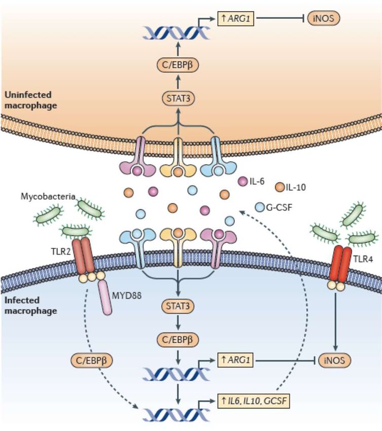
DC-SIGN: recettore lectinico di tipo C espresso da DC e macrofagi
DC-SIGN lega:
-HIV (gp120)                                             These receptors permit interactions with
-Virus Dengue                                            pathogens and endogenous soluble
-HCV                                                     proteins as well as cell-surface ligands
-Virus Ebola                                             expressed by T cells and anatomically
-CMV                                                     distinct endothelial cells, such as those
-Mycobacterium Tuberculosis                              found in secondary lymphoid organs.
-Candida Albicans                                        Langerin, a type-II C-type lectin
-Helicobacter pylori                                     exclusively expressed by Langerhans
                                                         cells, binds mannose-type ligands and
                                                         delivers material to unique Birbeck
                                                         granules.
                                                         DC-SIGN another type-II C-type lectin
                                                         is expressed by both macrophages and
                                                         DCs. It interacts with endogenous
                                                         molecules, such as ICAM-2, on
                                                         endothelial cells as well as ICAM-3 on T-
                                                         cells, mediating intercellular adhesion. In
                                                         addition, DC-SIGN binds pathogen-
                                                         associated mannose-type carbohydrates
                                                         found on viruses, bacteria and fungi.
                                                         Multimerisation of DC-SIGN and other
                                                         such receptors at the cell surface might
                                                         facilitate high-affinity ligand binding.

   Questi recettori appartengono alla famiglia dei PRR
   (pathogen recognition receptor)
Struttura di DC-SIGN
(carbohydrate recognition domain)
C-type lectins are transmembrane proteins that act as cell-adhesion receptors, are involved in
the regulation of signalling pathways and recognize specific carbohydrate structures that are
present on self antigens and pathogens.
DC-SIGN=Dendritic Cell-Specific Intercellular adhesion molecule-3-Grabbing Non-integrinReceptors and pathways implicated in the entry of HIV-1 into DCs
HIV-1 binds several different
DCs surface receptors, which
determines the fate of the
virus. Binding to conventional
HIV-1 receptor CD4, or DC-
specific DCIR leads to
productive infection of the cell
in a very small percentage of
DCs. In most cases, HIV-1
enters the DC via endocytosis
after binding DC-SIGN or
other receptors, namely,
Syndecan-3 or Siglec-1.
Binding to these receptors
can lead to trans-infection or
immune recognition and
consequent T cell activation.
Siglec-1 facilitates HIV-1
infection of antigen
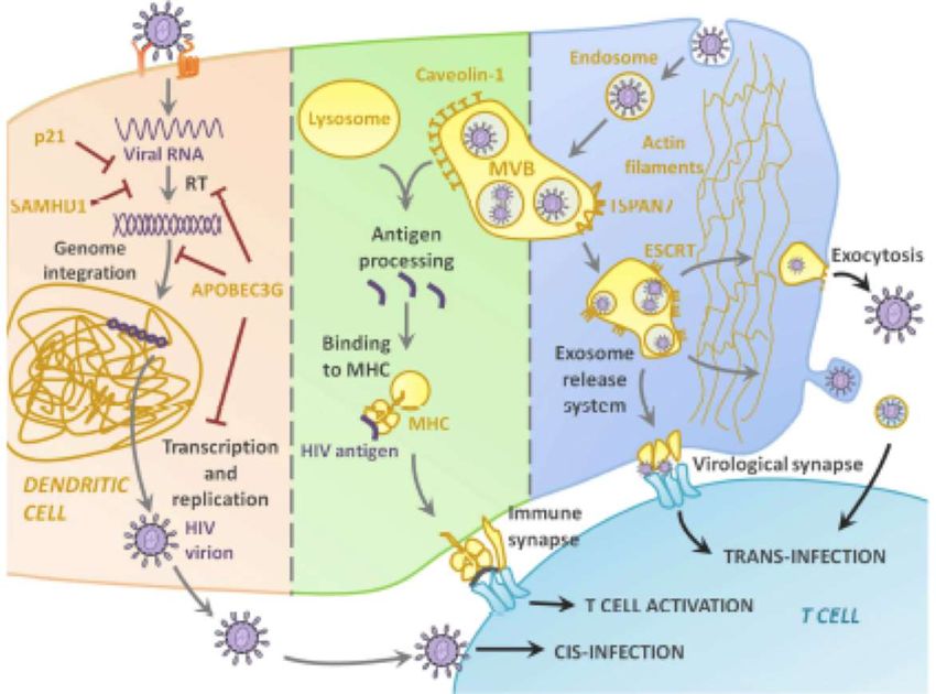
presenting cellsHIV usa DC-SIGN per penetrare nelle DC immature senza essere degradato e favorire la
trasmissione ai linfociti T HIV-1 subverts intracellular processing by
dendritic cells through DC-SIGN. a | DC-
SIGN (dendritic cell (DC)-specific intercellular
adhesion molecule-grabbing nonintegrin) is
expressed by immature DCs in mucosal tissues
and lymph nodes, and by DC precursors in the
blood. HIV-1 is captured by DC-SIGN that is
expressed by DC precursors in the blood after
infection or by immature DCs at mucosal entry
sites during sexual transmission. DC-SIGN-
bound HIV-1 enters the cell, but escapes
internalization into lysosomal compartments and
recycles back to the cell surface. By hiding
intracellularly in DCs, HIV-1 is protected during
migration to the lymphoid tissues. On arrival at
lymphoid tissues, DCs transmit HIV-1 to CD4+
T cells in trans, resulting in productive HIV-1
infection of CD4+ T cells.
b | High concentrations of HIV-1 allow viral
infection of DCs that results in the production of
HIV-1 by DCs, which subsequently infect T cells.
Sequestration of HIV-1 by DC-SIGN can allow
cis-infection of DCs by presenting the
infectious virus to CD4 and co-receptors to allow
efficient infection of DCs.
c | C-type lectins function as antigen receptors
to internalize antigen into lysosomes to enhance
antigen presentation by MHC class I and II
molecules. It remains to be determined whether
capture of HIV-1 by C-type lectins results in the
activation of DCs and presentation of viral
antigen by MHC molecules. CCR5, CC chemokine
receptor 5; TCR, T-cell receptor.Intracellular pathways and molecular partakers of HIV-1 trip inside DC
Only a small percentage of
HIV-DC interactions lead to
productive infection,
thanks to intracellular
molecular defense at
different stages of the
infection, including and
highlighting the antiviral
effect of p21, SAMHD1,
and APOBEC3G. Most of
the times, however, the
virus enters the DC by
endocytosis and
accumulates in multi-
vesicular bodies (MVB). If
the MVB fuses with the
lysosome, the virus is
recognized as antigenic and
processed, resulting in viral
peptides binding to MHC
and showing in the
membrane for AG
presentation to T cells.
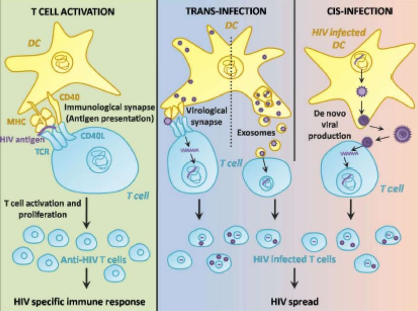
The key and most interesting role of DCs in HIV-1 infection is their function as Trojan Horse, as the virus in
the endosome can use the cells as a mean of transportation to the lymph nodes, and then be released
either through the virological synapse, or via exocytosis.Two-faced role of DCs in HIV-1 infection after T cell contact Two-faced role of DCs in HIV-1 infection after T cell contact. DC contact with T cells through the immunological synapse results in Ag presentation and a specific HIV-1 immune response. However, the DC-T cell interaction may also facilitate the transmission of the virus either from an infected DC cell to the surrounding T cells in the lymph nodes, or from a “carrier” DC via exosome release or infectious synapse.
Langerina e DC-SIGN hanno un ruolo opposto nella trasmissione di HIV
dati controversi sul ruolo protettivo nei confronti di HIV
Cellula di Langerhans
HIV-1 binding to Langerin favors the
Epidermica (LC)
routening of HIV-1 into the human
TRIM5α-mediated restriction pathway.
TRIM5α induces the assembly of an
autophagy-activating scaffold to Langerin,
which targets HIV-1 for autophagic
degradation and prevents infection of LCs.
Cellula dendritica-DC-SIGN+
Localizzazione delle cellule di Langerhans (LCs) e
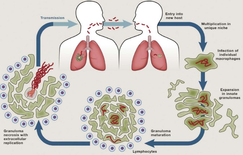
delle DC-SIGN+ negli epiteli delle mucose genitaliPathogenic life cycle of M. tuberculosis
Invasion of the host, replication and transmission of infection
Infection in the
lower airwayM. tuberculosis Evades Commensal Bacteria to Infect Its Host
Manipulation of macrophage
recruitment through
coordinated use of membrane
Microbicidal
macrophage lipids
M. tuberculosis avoids the
recruitment of microbicidal
macrophages to the site of
infection by masking its PAMPs
with the PDIM lipid. A related
surface lipid PGL recruits
permissive macrophages that can
transport the bacteria into deeper
tissues. However, the upper
airways are colonized by resident
microorganisms whose PAMPs
recruit microbicidal macrophages.
Therefore, this mycobacterial
Permissive
macrophage strategy to evade microbicidal
macrophages is only effective if
infection is initiated in the
relatively sterile lower lung.
PMID = phthiocerol dimycoceroserate
PGL = phenolic glycolipid
Cambier CJ et al. 2014 Cell 159:1497-1509Fit for consumption: zebrafish as a model for tuberculosis
Modeling mycobacterial infection in larval and adult zebrafish.
Modeling mycobacterial infection in
larval and adult zebrafish. (A) From top
– brightfield image of a zebrafish larva.
Middle – an approximate timeline of
progression of infection in zebrafish
larvae, in days post-infection (dpi).
Bottom – confocal images of zebrafish
lines, with fluorescently labeled
macrophages shown in red and
infecting fluorescent mycobacteria
visualized in green. Representative
images display from left – scattered
infected macrophages; center –
macrophages aggregated into
granulomas; right – the appearance of
extracellular bacteria as containment
fails at isolated granulomas. (B) Top –
image of an adult zebrafish. Bottom – a
timeline of infection progression in
weeks post-infection (wpi) in the adult
persistent infection model (based on
the work of Parikka et al., 2012).
Mark R. Cronan, and David M. Tobin Dis. Model. Mech.
2014;7:777-784M. tuberculosis evade la risposta immunitaria utilizzando diversi meccanismi
E B
A
PknG
C
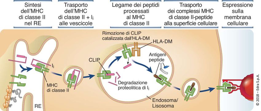
DFunzione della catena invariante e delle molecole HLA-DM nella presentazione
antigenica di classe II
catepsine (fondamentale l’azione della catepsina S)Intracellular Niches of M. tuberculosis The observed intracellular niches of M. tuberculosis within macrophages are shown with other pathogens occupying those niches also listed. Confirmed trafficking pathways are indicated with continuous arrows and putative ones with dashed arrows. Pathways dependent on the mycobacterial ESX1 secretion system are indicated.
Mycobacteria Exploit the Granuloma to
Expand Their Numbers in early infection
Mycobacteria within infected macrophages induce in an
ESX1-dependent fashion MMP9 expression in epithelial
cells surrounding the nascent granuloma.
MMP9 stimulates the recruitment of new macrophages to
the granuloma. Multiple new arrivals phagocytose the
bacterial contents of a given dying infected macrophage,
thus spreading the bacteria to new macrophages and
providing them new expansion niches.
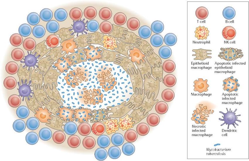
A classical tuberculosis
granuloma. The hallmark
tuberculosis granuloma is a
highly organized collection of
immune cells that aggregate
around a central necrotic core.(a) Infecting mycobacteria taken up
Persistenza del mycobacterium tuberculosis by alveolar macrophages in the lung
resist killing by subverting
phagosome maturation and by the
Th1 protective effect of their lipid-rich
cell wall. (b) Inflammatory signaling
in response to mycobacterial
Th17 components results in the
recruitment of TH1 T cells. T cell-
mediated activation of macrophages
enhances their ability to control
mycobacteria. (c) Remaining viable
mycobacteria are sequestered within
a granuloma made up of
macrophages and a variety of T cell
subsets. (d) M. tuberculosis is able
to persist in an asymptomatic form
within the host over many decades.
It is likely that mycobacteria are
held in a nondividing or slowly
dividing state in open lesions or in
IL2 closed lesions walled-off by fibrosis.
gd T cells (e) Reduced immunity is associated
with reactivation disease. Lack of
immunosurveillance may stimulate
renewed replication of mycobacteria
in open lesions. For mycobacteria in
closed lesions, liquefaction and
CD1-restricted
breakdown in the absence of
T cells
immune surveillance will result in
disease. Once the disease process is
underway, immunopathology
contributes to tissue damage and
ultimately efficient aerosol
transmission to a new host.DC-SIGN signaling modulates TLR signaling during M. tuberculosis infection
La produzione simultanea di IL10 e IL12 da parte delle
DC disturba il differenziamento dei linfociti T naive
verso il fenotipo Th1 ma induce Th producenti IL10
S276
Nucleus
IL6
IL10
IL12
DC-SIGN and TLR4 might inhibit each other
after the recognition of Mycobacterium DC-SIGN binds ligands such as mycobacteria. DC-SIGN induces RAF1
tuberculosis. Ligation of DC-SIGN by the M. phosphorylation, which modulates TLR induced NF-κB activation. Upon TLR
tuberculosis virulence factor mannose- stimulation the canonical NF-κB subunit p65 is released from its inhibitor and
capped cell-wall component lipoarabino- translocates to the nucleus. Phosphorylated RAF1 induces p65
mannan (ManLAM) reduces TLR4-triggered phosphorylation at Ser276 that functions as a binding site for the histone
DC maturation and enhances the production acetylase CBP. Acetylation of p65 induces enhanced and prolonged Il6, Il10 and
of interleukin-10 (IL-10) disfavouring a TH1- Il12ab transcription. CBP, CREB binding protein; DC-SIGN, DC-specific ICAM3-
cell-mediated response and survival of the grabbing nonintegrin; MyD88, myeloid differentiation primary response
pathogen. protein 88; NF-κB, nuclear factor κ B; IκBα, inhibitor of NF-κBα;
TLR, Toll-like receptor.Il micobatterio induce produzione di arginasi sia in cellule infettate che
non infettate inibendo la sintesi di ossido nitrico
MyD88-dependent arginase induction
Data from mouse prevents nitric oxide production in both
mycobacteria-infected and -uninfected
models
macrophages Activation of myeloid
differentiation primary response protein 88
(MYD88) signalling by mycobacteria (at least
in part through TLR2) induces
CCAAT/enhancer-binding protein β (C/EBPβ)-
mediated induction of interleukin-6 (IL-6), IL-
10, and granulocyte colony-stimulating factor
(GCSF) production. These signal transducer
and activator of transcription-3 (STAT3)-
activating cytokines act in both autocrine and
paracrine manners to induce arginase 1
(Arg1) expression which is partially
dependent on C/EBPβ. The produced
arginase can inhibit inducible nitric oxide
synthase (iNOS) activity through competition
for the common substrate arginine. The
MYD88 pathway for arginase production was
shown to confer a survival benefit for
mycobacteria in vivo and is thought to
counteract pathways that activate nitric oxide
production, such as TLR4.
Nat Rev Immunol. 2011 Mar;11(3):187-200Meccanismi di immunoevasione e immunosoppressione del Mycobacterium tuberculosis mediante: v Blocco della maturazione e dell’acidificazione del fagolisosoma v Inibizione della produzione di ROS (specie reattive dell’ossigeno) e resistenza all’ossido nitrico e agli intermedi reattivi dell’azoto v Manipolazione dell’apoptosi e dell’autofagia v Inibizione della maturazione delle cellule dendritiche v Interferenza con la presentazione antigenica mediata dalle molecole MHC di classe II e classe I
Strategie adottate da S. enterica Typhi per la persistenza nell’ospite e differenze con S.
enterica Typhimurium
Local acute inflammation
with gastroenteritis and Systemic infection
diarrhea
reservoir
On the left of the figure, the acute infection (human gastroenteritis) mediated by S. enterica Typhimurium
involves bacteria replicating freely in the intestine lumen and using multiple attachment factors
(represented by multicolored tips or fimbriae). S. enterica Typhimurium targets both enterocytes and M
cells for invasion, but is stopped at the mesenteric lymph nodes. Neutrophils are quickly attracted to the
invasion site and inflammation follows, leading to diarrhea. S. enterica Typhi has dispensed with many
attachment and shedding factors and may preferentially target a limited number of host cell types that
favor dissemination to deeper tissues. S. enterica Typhi can persist in the bone marrow for extended
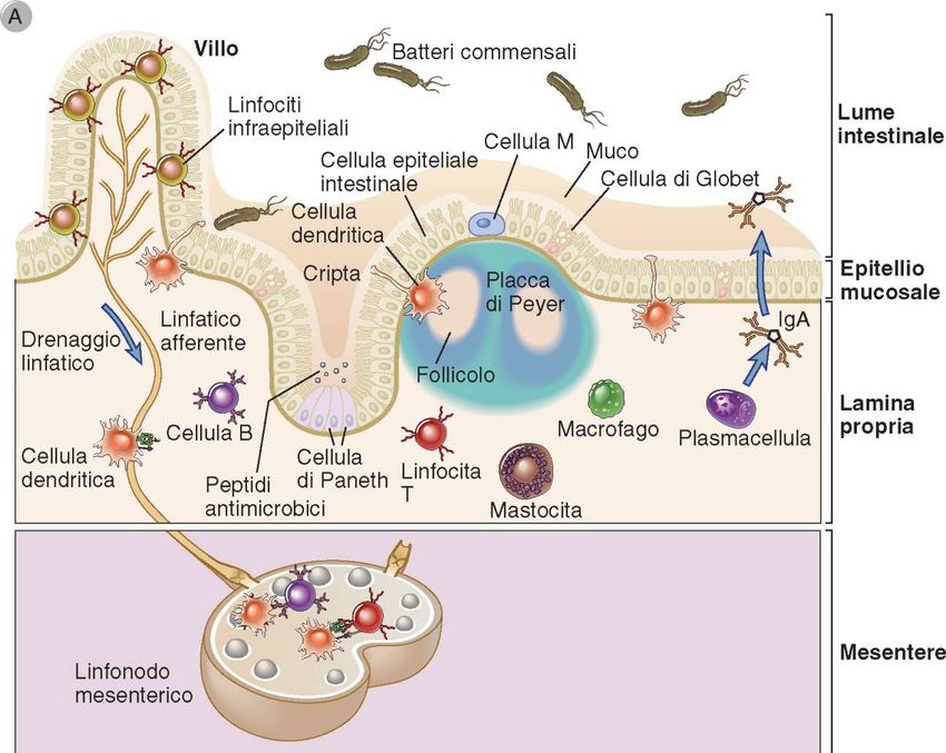
periods and in the gall bladder for life. T, T cell; B, B cell.Tessuti linfoidi associati alle mucose: placche del Peyer nell’intestino tenue
Le cellule M sono cellule specializzate
dell’epitelio intestinale
S. enterica Typhimurium
1   2   3   oltrepassa la barriera
            intestinale utilizzando
            più vie e determina una forte
            risposta infiammatoria locale

            TLR5       NF-kB

                       citochine e
                       chemochine che
                       sostengono un
                       processo
                       Infiammatorio
                       acuto

            Invece, S. enterica Typhi
            non infetta gli enterociti e
            non provoca una risposta
            infiammatoria locale ma supera
            i linfonodi mesenterici e causa
            infezione sistemica
Le cellule epiteliali giocano un ruolo cruciale nella difesa innata contro i patogeni
Variazioni antigeniche
Meccanismo adottato da virus (HIV, virus dell’influenza), batteri
(streptococco, stafilococco, meningococco, borrelia burgdorferi,
bordetella pertussis, neisseria gonorrhea) e protozoi
(tripanosomi)

Effetti:
Evasione della risposta umorale per
-mancato riconoscimento da parte degli anticorpi

Evasione della risposta cellulo-mediata per
-interferenza con la proteolisi e con la generazione dell’antigene
-mancato legame alle molecole MHC
-mancato riconoscimento da parte del TCR dei linfociti T
-creazione di APL (altered peptide ligand)
Molteplici sierotipi di Streptococcus pneumoniae:
            un caso di variazione antigenica

84 diversi sierotipi di Streptococcus pneumoniae agente della polmonite batterica, della
bronchite, l'otite media, la setticemia e la meningite.
Variazioni antigeniche del virus Antigenic drift Antigenic shift
dell’influenza di tipo A (mutazioni puntiformi) (riarrangiamento)
Anticorpi neutralizzanti contro la La variazione antigenica si ha quando
emoagglutinina bloccano il legame con sono scambiati segmenti di RNA tra ceppi
la cellula virali in un ospite intermedio
Le glicoproteine coinvolte
sono:
l’emoagglutinina
la neuroaminidasi
(sialidasi)
Lipid envelope
Nessuna immunità crociata protettiva
Le mutazioni alterano epitopi
verso il virus che esprime una nuova
dell’emoagglutinina che non sono più
emoagglutinina
riconosciuti dagli anticorpi neutralizzanti
antigenic drift
antigenic shift
small mutations new strainYou can also read