Occurrence of gastrointestinal parasites in camels in the Tianshan Mountains pastoral area in China - Sciendo
←
→
Page content transcription
If your browser does not render page correctly, please read the page content below
J Vet Res 64, 2020
DOI:10.2478/jvetres-2020-0071
Occurrence of gastrointestinal parasites in camels
in the Tianshan Mountains pastoral area in China
Zhang Guowu1* , Zhang Kai1*, Wang Xifeng1*, Ji Chunhui1, Ning Chengcheng1,
Zhao Yue1, Qiao Jun1, Meng Qingling1*, Zhang Xingxing2, Cai Kuojun3,
Zhang Jinsheng4, Zhang Zaichao5, Cai Xuepeng6
1
College of Animal Science and Technology, Shihezi University, Shihezi, Xinjiang, 832003, China
2
Institute of Animal Science and Veterinary Research,
Xinjiang Academy of Agricultural and Reclamation Science, Shihezi, Xinjiang, 832000, China
3
Center for Animal Disease Prevention and Control, Urumqi, Xinjiang, 830000, China
4
Center for Animal Disease Prevention and Control, Tacheng, Xinjiang, 834700, China
5
Center for Animal Disease Prevention and Control, Changji, Xinjiang, 831500, China
6
State Key Lab of Veterinary Etiological Biology, Lanzhou Veterinary Research Institute,
Chinese Academy of Agricultural Sciences, Lanzhou, Gansu, 730046, China
xjmqlqj@sina.com
Received: April 21, 2020 Accepted: October 12, 2020
Abstract
Introduction: Gastrointestinal parasites are some of the most common pathogens which are seriously harmful to the camel’s
health. The infection status of gastrointestinal parasites in camels (Camelus bactrianus) in the Tianshan Mountains pastoral area in
China is still unclear. The aim of this study was to investigate the species and infection intensity of gastrointestinal tract parasites
in local camels. Material and Methods: A total of 362 fresh faecal samples were collected and examined for parasite eggs using
the saturated saline floating and natural sedimentation method. The parasite eggs were subjected to morphological and molecular
examination and identification, and the infection rate and mean intensity of the parasites were analysed. Results: A total of 15
gastrointestinal tract parasite species’ eggs were identified, with a detection rate of 100%. Ostertagia spp. (100%) and
Trichostrongylus spp. (98.1%) were dominant. Camels were often coinfected by 5–14 species. The average number of eggs per
gram of faeces was higher for Ostertagia spp. (298), Haemonchus contortus (176) and Nematodirus spp. (138). The number of
species of parasites infecting young camels was significantly lower than that of adult camels, but the infection intensity in young
camels was significantly higher. Conclusion: Gastrointestinal parasites were highly prevalent in camels from the Tianshan
Mountains pastoral area in China. This finding provides important epidemiological data for the prevention and control of associated
infections in camels.
Keywords: Camelus bactrianus, gastrointestinal parasites, Ostertagia spp., Tianshan Mountains pastoral area in
China, Trichostronglyus spp.
Introduction Mountains pastoral area, and besides this it also has high
nutritional value. Furthermore, camel milk has health
With a camel population of about 280,000, China protection effects (2, 11, 22, 25), and camel hair can be
has an abundance of the Camelus bactrianus species and used as an important textile material. Accordingly, the
ranks second in the world for head of these ungulates. camel breeding industry has become a cornerstone of the
The Tianshan Mountains pastoral area in Xinjiang, local economy for herders.
China is a major distribution region of camels, owing to Gastrointestinal parasites are one of the most
its vast grasslands particularly suitable for extensive common pathogens, and can not only lead to nutritional
animal breeding, and accounts for about 20% of the total and immune inadequacy as well as stunted growth and
camels in the country. As the “ship of the desert”, the delayed development (4–8, 23), but also have adverse
camel is an important transportation tool in the Tianshan effects on the quality of camel meat and milk (18, 21).
© 2020 Guowu Zhang et al. This is an open access article distributed under the Creative Commons Attribution-
NonCommercial-NoDerivs license (http://creativecommons.org/licenses/by-nc-nd/3.0/)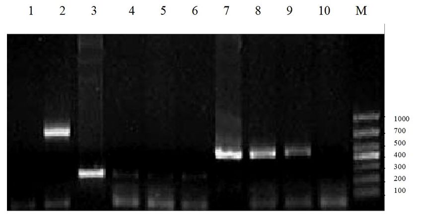
Guowu Zhang et al./J Vet Res/64 (2020)
These consequences of parasitical infection are potential The purpose of this study is to investigate the
threats to food safety and the health of the majority of species and infection intensity as well as to identify the
herders (1, 9, 12, 13, 27, 28). In recent years, with the dominant parasites infecting the gastrointestinal tract of
rapid development of the Chinese camel breeding camels in the studied area, which will provide useful
industry in the Tianshan Mountains pastoral area, the epidemiological data for preventing and controlling the
number of animals has reached more than 40,000. parasitic diseases of camels.
Nevertheless, due to the fact that this region’s camels are
mainly fed by grassland grazing and due to the weak
veterinary epidemic prevention and control measures, Results
they are often infected by a variety of gastrointestinal
tract parasites, which causes serious financial losses. Overview of the surveyed area’s climate and
However, the species and infection intensity of vegetation. The Tianshan Mountains pastoral area in
gastrointestinal tract parasites in camels in this region Xinjiang has a temperate continental arid and semi-arid
are still unknown. climate. The average annual temperature in northern
The purpose of this study is to investigate the Xinjiang is 4–9°C. Its annual precipitation is above
species and infection intensity as well as to identify the 150–200 mm and its annual frost-free period is 140–185
dominant parasites infecting the gastrointestinal tract of days. Considering the southern region, the average
camels in the studied area, which will provide useful annual temperature is 7–14°C. Its annual precipitation is
epidemiological data for preventing and controlling the 25–100 mm, and its annual frost-free period is 180–220
parasitic diseases of camels. days. The Tianshan grassland has abundant water
sources and a wide variety of pastures; different regions:
the high mountain areas are mainly covered with
Material and Methods Artemisia and Cyperaceae and partly with
miscellaneous grasses, the forest grass lands are mainly
With a camel population of about 280,000, China covered with leguminous plants and weedy grasses and
has an abundance of the Camelus bactrianus species and the low mountainous areas are mainly covered with
ranks second in the world for head of these ungulates. Bromus pauciflorus, Achnatherum sibiricum, Artemisia,
The Tianshan Mountains pastoral area in Xinjiang, Astragalus, and other grasses.
China is a major distribution region of camels, owing to Collection of camel faeces. During the period of
its vast grasslands particularly suitable for extensive 2016–2018, a total of 362 fresh faecal samples were
animal breeding, and accounts for about 20% of the total collected from camels (Camelus bactrianus) in the
camels in the country. As the “ship of the desert”, the Tianshan pastoral area of Xinjiang, China. Among them,
camel is an important transportation tool in the Tianshan 106 were from camels younger than two years, 139 were
Mountains pastoral area, and besides this it also has high collected from camels aged between two and six years,
nutritional value. Furthermore, camel milk has health and 117 were collected from older camels. These fresh
protection effects (2, 11, 22, 25), and camel hair can be faecal samples were collected from the rectum and
used as an important textile material. Accordingly, the sealed in 50 mL plastic bottles, stored at 4°C, and sent
camel breeding industry has become a cornerstone of the to the Xinjiang Key Laboratory of Animal Disease
local economy for herders. Prevention and Control. During the collection period,
Gastrointestinal parasites are one of the most information on clinical manifestations in the camels and
common pathogens, and can not only lead to nutritional deworming records were also collected from local
and immune inadequacy as well as stunted growth and veterinarians or those tending the grazing animals.
delayed development (4–8, 23), but also have adverse Morphological examination and identification
effects on the quality of camel meat and milk (18, 21). of parasite eggs in camel faeces. Eggs were first
These consequences of parasitical infection are potential examined using the saturated saline floating method. In
threats to food safety and the health of the majority of brief, 10 g of fresh faecal samples were crushed with
herders (1, 9, 12, 13, 27, 28). In recent years, with the a glass rod and washed 10 times with saturated saline
rapid development of the Chinese camel breeding solution. Then, the mixture was filtered with a 50-mesh
industry in the Tianshan Mountains pastoral area, the copper sieve and collected in a 100 mL conical beaker.
number of animals has reached more than 40,000. After clarification for 20 min, a wire ring with a diameter
Nevertheless, due to the fact that this region’s camels are of 10 mm was used to collect the film from the surface
mainly fed by grassland grazing and due to the weak of the liquid by parallel contact with the liquid surface
veterinary epidemic prevention and control measures, and release of the ring’s contents onto a glass slide.
they are often infected by a variety of gastrointestinal Simultaneously, parasite eggs in faeces were also
tract parasites, which causes serious financial losses. collected by the natural sedimentation method. The
However, the species and infection intensity of shape and colour of the parasite eggs were observed and
gastrointestinal tract parasites in camels in this region examined under the microscope, and the short and long
are still unknown. diameters of the eggs were measured and photographed.
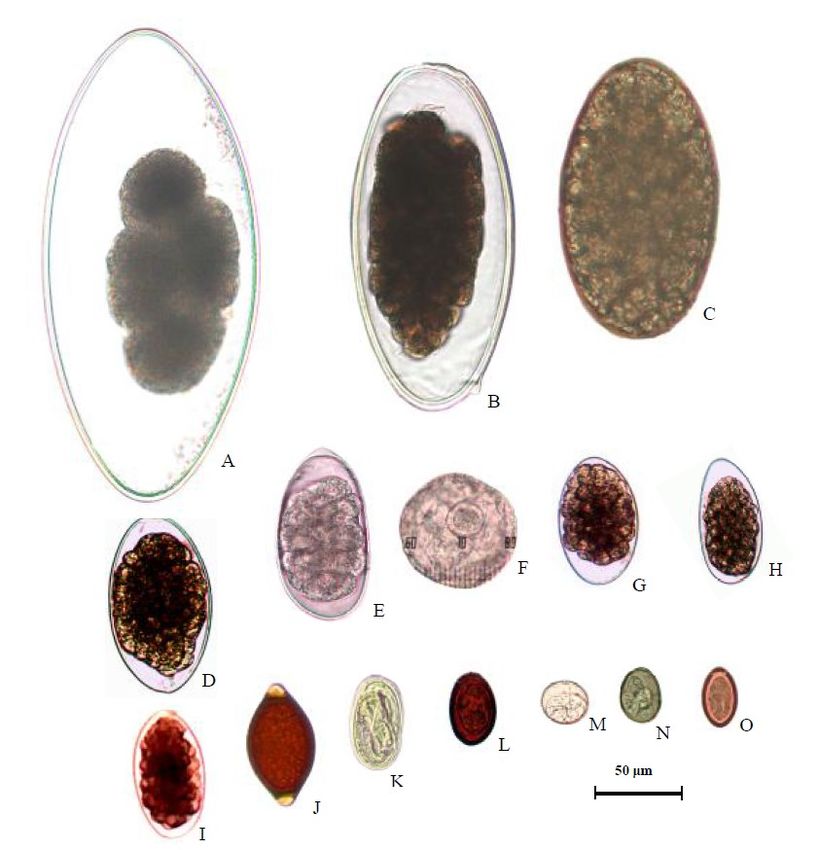
The morphological characteristics of each egg were usedGuowu Zhang et al./J Vet Res/64 (2020)
to identify the parasites according to Georgis’ continuous variables were compared using an F-test.
Parasitology for Veterinarians (3), Veterinary Clinical Data were expressed as mean ± standard deviation (SD).
Parasitology (26), and Veterinary Parasitology (23). A P value less than 0.05 was considered significantly
Molecular detection and identification of different, while a P value less than 0.01 was considered
parasite eggs in faeces. Nematode eggs with similar extremely significantly different.
morphological structure were identified molecularly. All of the 362 camels were infected with different
Briefly, the eggs collected from the faeces were species of parasite, as detected in their faeces. Based on
processed with a grinder and DNA was extracted using the morphological (Fig.1) and molecular examinations
the MiniBEST Universal Genomic DNA Extraction Kit (Fig. 2), a total of 15 species of parasites eggs were
(TaKaRa, Japan). Using the extracted DNA as detected and identified in the gastrointestinal tracts of
a template, PCR amplification was performed with the camels (Table 2), including those of nine species of
specific primers (Table 1) targeting a nematode nematode, three species of trematode, two species of
ribosomal DNA sequence which harbours 18S tapeworm, and one species of coccidia.
ribosomal RNA, internal transcribed spacer 1 and 5.8S Among the 15 species of parasite, the particularly
ribosomal RNA. The reaction conditions were as highly infective ones were Ostertagia spp. (100%,
follows: pre-denaturation at 95°C for 2 min, 362/362), Trichostrongylus spp. (98.1%, 355/362),
denaturation at 94°C for 50 s, annealing at 50°C for Haemonchus contortus (88.1%, 319/362), and
45 s, and extension at 72°C for 30–70 s in 35 cycles. The Nematodirus spp. (87.3%, 316/362), their prevalences
PCR products were analysed by electrophoresis on 1.5% qualifying Ostertagia spp. and Trichostrongylus spp. as
agarose gel. Then they were recovered with the dominant species of infecting parasites (Table 2).
MiniBEST Agarose Gel DNA Extraction Kit (TaKaRa, All the investigated camels were co-infected with
Japan) and sent to Shanghai Bioengineering Technology 5–14 different species of parasites (Fig. 3). The number
Co. Ltd. (Sangon, China) for sequencing. Each sample of cases of simultaneous infection with nine species of
was sequenced three times. Sequence homology was parasites was the highest, accounting for 23.8% (86/362),
evaluated in GenBank with Blast N software, and the followed in turn by cases of infection with 5, 6, 7, 8, 10,
species of the eggs were identified according to the 11, 12, and 13 different species of parasites, accounting
homology found. for 6.1% (22/362), 5.0% (18/362), 9.9% (36/362), 9.9%
Determination of parasitic infection intensity in (36/362), 20.7% (75/362), 9.7% (35/362), 6.9%
faeces. Infection intensity was determined using Stoll’s (25/362), and 5.8% (21/362), respectively, while the
dilution method. Briefly, a small conical beaker was number of 14-species infections was the lowest,
marked at the capacity of 56 mL and 60 mL, and 4% accounting for 2.2% (8/362) (Fig. 3).
NaOH solution were added to the 56 mL mark. Then, The average number of infectious parasite species
about 4 g of chopped faeces were added slowly until the in young camels was significantly lower than that in
liquid level reached the 60 mL mark. Next, more than 10 adult ones, but the mean intensity of parasitic infection
glass beads were added and the solution was shaken to in this age group shown as EPG of faeces was
make a fine and uniform faecal suspension. A 200 µL significantly higher (P < 0.05). The infection intensity in
volume of solution was collected on a glass slide, and juvenile camels was between that in young and adult
covered with a rectangle of glass of not less than 22 mm animals (Table 3), indicating that parasitic infection has
× 40 mm. Finally, the total egg numbers were counted a certain correlation with host age. However, there was
under a microscope. The number multiplied by 100 is no significant difference in the average number of
the number of eggs per gram of faeces (EPG). infecting parasite species and infection intensity
Statistical analysis of data. All data were analysed between male and female camels (P > 0.05), indicating
using SPSS 18 software (IBM SPSS, USA). The no correlation between parasitic infection and gender.
Table 1. Primers used in the study
Size of amplified product
Primer Nucleotide sequence (5′ to 3′) Target gene
(bp)
FP1 AGGTATCTGTAGGTGAACCTGC
Ribosomal ITS1 of Haemonchus contortus 810
RP1 ATACAAATGATAAAAGAACATC
FP2 GAGAGGACTGCGGACTGCTGTA
Ribosomal ITS1 of Trichostrongylus spp. 240
RP2 CTCACACACAGAGCTCTAACGG
FR3 CTGCGGAAGGATCATTGTCGAA
Ribosomal ITS1 of Chabertia ovina 475
RP3 ACTCTAAGCGTCTGCAATTCGT
FR4 TGTACACACCGCCCGTCGCTGT
Ribosomal ITS1 of Ostertagia spp. 475
RP4 TGACAACCAGGTACCGTACACA
FR5 GGACTGCGGACTGCTGTATCGA
Ribosomal ITS1 of Bunostomum spp. 475
RP5 TGTTAAACGTAAAAAATTGGTTGuowu Zhang et al./J Vet Res/64 (2020) Fig. 1. Morphological characteristics of eggs of different parasitic worms in the faeces of camels in the Tianshan Mountains pastoral area A – Nematodirus spp.; B – Marshallagia spp.; C – Fasciola hepatica; D – Chabertia ovina; E – Bunostomum spp.; F – Moniezia expansa; G – Haemonchus contortus; H – Trichostrongylus spp.; I – Ostertagia spp.; J – Trichuris spp.; K – Strongyloides papillosus; L – Dicrocoelium spp.; M – Eimeria spp.; N – Thysaniezia ovilla; O – Hasstilesia ovis Fig. 2. Molecular detection of nematode eggs with similar morphological structure in camel faeces M – DNA maker DL1000 (1000, 700, 500, 400, 300, 200, 100 bp); Lanes 1, 4, 5, 6, and 10 – negative results; Lane 2 – Haemonchus contortus; Lane 3 – Trichostrongylus spp.; Lane 7 – Chabertia ovina; Lane 8 – Ostertagia spp.; Lane 9 – Bunostomum spp.
Guowu Zhang et al./J Vet Res/64 (2020)

Table 2. Summary statistics of gastrointestinal parasites in camels in the Tianshan Mountains pastoral area

                                                        Infection rate          Mean EPG              Length of egg          Width of egg
 Parasite species
                                                              (%)                                          (μm)                  (μm)
 Ostertagia spp.                                       100 (362/362)            298 ± 57.1             71.25 ± 9.12          36.44 ± 4.57
 Trichostrongylus spp.                                 98.1 (355/362)           105 ± 36.2            82.24 ± 10.83          39.63 ± 3.53
 Haemonchus contortus                                  88.1 (319/362)           176 ± 62.5             75.65 ± 5.78          46.35 ± 7.88
 Nematodirus spp.                                      87.3 (316/362)           138 ± 41.8            241.36 ± 23.14        111.93 ± 18.51
 Marshallagia spp.                                     80.4 (291/362)           129 ± 38.7            182.89 ± 11.90         81.56 ± 7.42
 Trichuris spp.                                        77.3 (281/362)            93 ± 22.9             75.42 ± 6.57          35.27 ± 3.46
 Chabertia ovina                                        18.2 (66/362)            71 ± 10.4             89.24 ± 8.33          52.26 ± 5.72
 Bunostomum spp.                                         9.9 (36/362)            45 ± 14.8             88.67 ± 7.91          48.22 ± 2.35
 Strongyloides papillosus                              33.1 (120/362)            113 ± 8.7             51.78 ± 8.75          30.58 ± 5.67
 Thysaniezia ovilla                                    60.8 (220/362)           126 ± 29.4             25.18 ± 3.15          22.34 ± 2.78
 Moniezia expansa                                      42.3 (153/362)           103 ± 41.1             63.13 ± 4.32          46.86 ± 5.31
 Dicrocoelium spp.                                     62.4 (226/362)            82 ± 6.8              39.19 ± 4.74          31.25 ± 3.16
 Fasciola hepatica                                      24.6 (89/362)            20 ± 13.5            140.55 ± 7.68          84.54 ± 8.12
 Hasstilesia ovis                                      91.7(332/362)             56 ± 17.5             30.12 ± 3.81          18.32 ± 1.72
 Eimeria spp.                                          73.5 (266/362)            94 ± 16.2             34.18 ± 2.32          22.17 ± 1.72

Table 3. Prevalence of gastrointestinal parasites in camels in the Tianshan Mountains pastoral area

                                                                                                       Mean number of
                                                                                   Number of
                                  Potential propensity factor                                         parasite species in      Mean EPG
                                                                                examined camels
                                                                                                         co-infection
                                      Young camels (>2 years)                         106                  6 ± 1.6a            1046 ± 87.2a
                            Age       Sub-adult camels (2–6 years)                    139                  7 ± 1.8a             779 ± 39.6a
                                      Adult camels (>6 years)                         117                 11 ± 1.5b            462 ± 40.7b
                                      Male                                            206                  9 ± 1.2a             901 ± 85.5a
                            Sex
                                      Female                                          156                 10 ± 1.9a           1027 ± 101.2a

Different letters in same column mean significant difference (P < 0.05)

                            35

                            30
 Mixed infection rate (%)

                            25

                            20

                            15

                            10

                             5

                             0
                                  5          6         7         8        9         10           11       12           13    14
                                                                Number of parasite species
Fig. 3. Mixed infection with different species of gastrointestinal parasite in camels in the Tianshan Mountains pastoral area

Discussion                                                                                   majority of herders (9, 21, 22). The documents on camel
                                                                                             parasitic diseases in Iran showed that 48 helminth
     Parasitic disease is a disease that seriously                                           species were detected in the digestive system (21).
endangers the development of the camel industry (18, 20,                                     Therefore, the investigation of the prevalence of
22, 24) and that not only impedes the growth and                                             gastrointestinal parasites in camels in the Tianshan
development of camels (14, 17, 19), but is also one of                                       pastoral area is of great significance for improving the
the important reasons for the peak in camel deaths in the                                    economic output of the camel industry and preventing
spring, resulting in great economic losses (15, 16, 21).                                     the spread of parasitic zoonosis.
What is more, many zoonotic parasites (1, 9, 10, 12, 13,                                          Many studies reported that the helminth is
28) could contaminate camel milk (18) and meat, posing                                       particularly rich in the digestive tract of camelids, which
potential threats to food safety and the health of the                                       is more than 40 species (17, 21). Among them,
Guowu Zhang et al./J Vet Res/64 (2020)
Trichostrongylus spp. and Haemonchus contortus are project (No. XJ2017G085) and the International Science
the most common species (21). In our study, we and Technology Cooperation Program Project of XPCC
systematically investigated the infection status of camel (No. 2016AH006).
gastrointestinal parasites in the Tianshan Mountains
pastoral area. A total of 15 species of camel Animal Rights Statement: The authors declare that the
gastrointestinal parasite were identified, including experiments on animals were approved by the Research
9 species of nematodes, 3 species of tapeworm, 2 species and Ethical Committee of Shihezi University
of trematode, and 1 species of coccidian. All camels (RECSHZ2020166).
suffered from mixed infections of 5–14 parasite species.
Among them, Ostertagia spp. and Trichostrongylus spp. Acknowledgments: We thank the field staff who
were the dominant species. The average number of provided the samples for this study.
infected parasites in young camels was significantly
lower than in adult camels, but the young camel EPG
was significantly higher than that of adult camels, which References
may be related to the weak immune system and
incomplete establishment of parasite immunity in 1. Alam-Eldin Y.H., Abdel Aaty H.E., Ahmed M.A.: Molecular
juvenile camels. Our epidemiological surveys also characterization of cystic echinococcosis: First record of G7 in
Egypt and G1 in Yemen. Acta Parasitol 2015, 60, 662–665.
suggested that irregular deworming and being grazed 2. Badawy A.A., El-Magd M.A., AlSadrah S.A.: Therapeutic effect
and housed together with cattle and sheep were partially of camel milk and its exosomes on MCF7 cells in vitro and in vivo.
responsible for high prevalence of parasites in camels. Integr Cancer Ther 2018, 17, 1235–1246.
In addition, this survey also showed that mixed 3. Bowman D.D.: Georgis’ Parasitology for Veterinarians, Saunders,
parasitic infections were very common in camels, which St. Louis, 2009.
4. Cafrune M.M., Aguirre D.H., Rickard L.G.: Recovery of
was consistent with the clinical manifestations (diarrhoea, Trichuris tenuis Chandler, 1930, from camelids (Lama glama and
hair loss, reproductive rate decline, and high mortality) Vicugna vicugna) in Argentina. J Parasitol 1999, 85, 961–962.
of the camel population under investigation. We found 5. Dubey J.P., Hilali M., Van Wilpe E., Calero-Bernal R.,
that the pathogenicity of a parasite was related to the Verma S.K., Abbas I.E.: A review of sarcocystosis in camels and
level of infection, while characteristics of individual redescription of Sarcocystis cameli and Sarcocystis ippeni
sarcocysts from the one-humped camel (Camelus dromedarius).
species seemed to play a lesser role in pathogenicity. Parasitol 2015, 142, 1481–1492.
This survey also found a large number of eggs of 6. Dubey J.P., Schuster R.K.: A review of coccidiosis in Old World
nematodes in the Ostertagia, Trichostrongylidae, camels. Vet Parasitol 2018, 262, 75–83.
Haemonchus, and Bunostomum genera. These eggs were 7. Dubey J.P., Schuster R.K., Kinne J.: Gametogony of Eimeria
difficult to distinguish and identify at the level of species, cameli in the small intestine of one-humped camel (Camelus
dromedarius). Parasitol Res 2018, 117, 3633–3638.
and so they were further subjected to molecular 8. Ederli N.B., de Oliveira F.C.: Gastrointestinal nematodes in
identification. ostriches, Struthio camelus, in different regions of the state of Rio
To our knowledge, this was the first systematic de Janeiro, Brazil. Rev Bra Parasitol Vet 2015, 24, 168–173.
survey on the prevalence of gastrointestinal parasites in 9. Fatima T., Mehnaz S., Wang M., Yang J., Sajid M.S., Shen B.,
C. bactrianus in the Tianshan Mountains pastoral area in Zhao J.: Seroprevalence of Toxoplasma gondii in one-humped
camels (Camelus dromedarius) of Thal and Cholistan deserts,
China, and it offers useful epidemiological data for the Punjab, Pakistan. Parasitol Res 2019, 118, 307–316.
prevention and control of gastrointestinal parasite 10. Feng Y., Lu Y., Wang Y., Zhang L., Yang Y.: Toxoplasma gondii
infection in camels. In conclusion, gastrointestinal and Neospora caninum in farm-reared ostriches (Struthio camelus)
parasite infection in camels was very common in the in China. BMC Vet Res 2017, 13, 301.
Tianshan pasture. Therefore, it is necessary to take 11. Ibrahim H.R., Isono H., Miyata T.: Potential antioxidant bioactive
peptides from camel milk proteins. Anim Nutr 2018, 4, 273–280.
effective measures such as deworming and rotational 12. Jarvinen J.A., Dubey J.P., Althouse G.C.: Clinical and serologic
grazing to prevent and control the parasite infection in evaluation of two llamas (Lama glama) infected with Toxoplasma
camels. gondii during gestation. J Parasitol 1999, 85, 142–144.
13. Li Y., Yang J., Chen Z., Qin G., Li Y., Li Q., Liu J., Liu Z.,
*These authors contributed equally to the article and Guan G., Yin H., Luo J., Zhang L.: Anaplasma infection of
Bactrian camels (Camelus bactrianus) and ticks in Xinjiang,
should be considered co-first authors. China. Parasit Vectors 2015, 8, 313.
14. Metwally D.M., Qassim L.E., Al-Turaiki I.M., Almeer R.S.,
Conflict of Interests: The authors declare that there is El-Khadragy M.F.: Gene-based molecular analysis of COX1 in
no conflict of interests regarding the publication of this Echinococcus granulosus cysts isolated from naturally infected
article. livestock in Riyadh, Saudi Arabia. PLoS One 2018, 13, e0195016.
15. Mirshekar F., Yakhchali M., Shariati-Sharifi F.: Trypanosoma
evansi infection and major risk factors for Iranian one-humped
Financial Disclosure Statement: This work was camels (Camelus dromedarius). J Parasitic Dis 2017, 41,
supported by the National Key Research and 854–858.
Development Program (No. 2017YFD0501200), the 16. Mirzaei M., Ghahvei Y., Lefoulon E., Lia R.P., Otranto D.,
National International Science and Technology Martin C., Sazmand A.: Morphological and molecular
characterization of Onchocerca fasciata (Nematoda,
Cooperation Exchange Project (No. CK 07-11), the Onchocercidae) from dromedary camels (Camelus dromedarius)
Xinjiang Autonomous Region Graduate Innovation in Iran. Parasite 2018, 25, 50.Guowu Zhang et al./J Vet Res/64 (2020)
17. Pathak K.M., Chhabra M.B.: Parasites and parasitic diseases of 23. Taylor M.A., Coop R.L., Wall R.L.: Veterinary Parasitology,
the camel in India: a review. Indian J Anim Sci 2010, 80, Blackwell, Oxford, 2007.
699–706. 24. Ukashatu S., Saulawa M.A., Magaji A.A.: Epidemiology of
18. Saad N.M., Hussein A.A.A., Ewida R.M.: Occurrence of gastrointestinal parasites of one-humped camel (Camelus
Toxoplasma gondii in raw goat, sheep, and camel milk in Upper dromedarius) slaughtered in Sokoto central abattoir, Sokoto state,
Egypt. Vet World 2018, 11, 1262–1265. Nigeria. Sci J Vet Adv 2013, 1, 723–832.
19. Saeed M.A., Vaughan J.L., Jabbar A.: An update on sarcocystosis 25. Wang Z., Zhang W., Wang B., Zhang F., Shao Y.: Influence of
in one-humped camels (Camelus dromedarius). Parasitol 2018, Bactrian camel milk on the gut microbiota. J Dairy Sci 2018, 101,
145, 1367–1377. 5758–5769.
20. Sazmand A., Eigner B., Mirzaei M., Hekmatimoghaddam S.H., 26. Zajac A.M., Conboy G.A.: Veterinary Clinical Parasitology,
Harl J., Duscher G.G., Fuehrer H.P., Joachim A.: Molecular Wiley Blackwell, Ames, 2006.
identification of hemoprotozoan parasites in camels (Camelus 27. Zhai B., Niu Q., Liu Z., Yang J., Pan Y., Li Y., Zhao H., Luo J.,
dromedarius) of Iran. Iran J Parasitol 2016, 11, 568–573. Yin H.: First detection and molecular identification of Borrelia
21. Sazmand A., Joachim A.: Parasitic diseases of camels in Iran species in Bactrian camel (Camelus bactrianus) from Northwest
(1931–2017) – a literature review. Parasite 2017, 24, 21. China. Infect Genet Evol 2018, 64, 149–155.
22. Sazmand A., Joachim A., Otranto D.: Zoonotic parasites of 28. Zhu S., Zimmerman D., Deem S.L.: A review of zoonotic
dromedary camels: so important, so ignored. Parasit Vectors 2019, pathogens of dromedary camels. Ecohealth 2019, 16, 356–377.
12, 610.You can also read