Persistent mammalian orthoreovirus, coxsackievirus and adenovirus co-infection in a child with a primary immunodeficiency detected by metagenomic ...
←
→
Page content transcription
If your browser does not render page correctly, please read the page content below
Zurich Open Repository and
                                                                          Archive
                                                                          University of Zurich
                                                                          Main Library
                                                                          Strickhofstrasse 39
                                                                          CH-8057 Zurich
                                                                          www.zora.uzh.ch

 Year: 2018

      Persistent mammalian orthoreovirus, coxsackievirus and adenovirus
      co-infection in a child with a primary immunodeficiency detected by
                      metagenomic sequencing: a case report

    Lewandowska, Dagmara W; Capaul, Riccarda; Prader, Seraina; Zagordi, Osvaldo; Geissberger,
 Fabienne-Desirée; Kügler, Martin; Knorr, Marcus; Berger, Christoph; Güngor, Tayfun; Reichenbach,
Janine; Shah, Cyril; Böni, Jürg; Zbinden, Andrea; Trkola, Alexandra; Pachlopnik Schmid, Jana; Huber,
                                               Michael

Abstract: Background: We report a rare case of Mammalian orthoreovirus (MRV) infection in a child with
a primary immunodeficiency (PID). Infections with Mammalian orthoreovirus are very rare and probably
of zoonotic origin. Only a few cases have been described so far, including one with similar pathogenesis
as in our case. Case presentation: The patient, age 11, presented with flu-like symptoms and persistent
severe diarrhea. Enterovirus has been detected over several months, however, exact typing of a positive cell
culture remained inconclusive. Unbiased metagenomic sequencing then detected MRV in stool samples
from several time points. The sequencing approach further revealed co-infection with a recombinant
Coxsackievirus and Adenovirus. MRV-specific antibodies detected by immunofluorescence proved that
the patient seroconverted. Conclusion: This case highlights the potential of unbiased metagenomic
sequencing in supplementing routine diagnostic methods, especially in situations of chronic infection
with multiple viruses as seen here in an immunocompromised host. The origin, transmission routes and
implications of MRV infection in humans merit further investigation.

DOI: https://doi.org/10.1186/s12879-018-2946-7

Posted at the Zurich Open Repository and Archive, University of Zurich
ZORA URL: https://doi.org/10.5167/uzh-148244
Journal Article
Published Version

The following work is licensed under a Creative Commons: Attribution 4.0 International (CC BY 4.0)
License.

Originally published at:
Lewandowska, Dagmara W; Capaul, Riccarda; Prader, Seraina; Zagordi, Osvaldo; Geissberger, Fabienne-
Desirée; Kügler, Martin; Knorr, Marcus; Berger, Christoph; Güngor, Tayfun; Reichenbach, Janine; Shah,
Cyril; Böni, Jürg; Zbinden, Andrea; Trkola, Alexandra; Pachlopnik Schmid, Jana; Huber, Michael (2018).
Persistent mammalian orthoreovirus, coxsackievirus and adenovirus co-infection in a child with a primary
immunodeficiency detected by metagenomic sequencing: a case report. BMC Infectious Diseases, 18:33.
DOI: https://doi.org/10.1186/s12879-018-2946-7
Lewandowska et al. BMC Infectious Diseases (2018) 18:33
DOI 10.1186/s12879-018-2946-7
CASE REPORT Open Access
Persistent mammalian orthoreovirus,
coxsackievirus and adenovirus co-infection
in a child with a primary immunodeficiency
detected by metagenomic sequencing: a
case report
Dagmara W. Lewandowska1†, Riccarda Capaul1†, Seraina Prader2, Osvaldo Zagordi1, Fabienne-Desirée Geissberger1,
Martin Kügler1,5, Marcus Knorr1, Christoph Berger3, Tayfun Güngör4, Janine Reichenbach2, Cyril Shah1, Jürg Böni1,
Andrea Zbinden1, Alexandra Trkola1, Jana Pachlopnik Schmid2† and Michael Huber1*†
Abstract
Background: We report a rare case of Mammalian orthoreovirus (MRV) infection in a child with a primary
immunodeficiency (PID). Infections with Mammalian orthoreovirus are very rare and probably of zoonotic
origin. Only a few cases have been described so far, including one with similar pathogenesis as in our case.
Case presentation: The patient, age 11, presented with flu-like symptoms and persistent severe diarrhea.
Enterovirus has been detected over several months, however, exact typing of a positive cell culture remained
inconclusive. Unbiased metagenomic sequencing then detected MRV in stool samples from several time points. The
sequencing approach further revealed co-infection with a recombinant Coxsackievirus and Adenovirus. MRV-specific
antibodies detected by immunofluorescence proved that the patient seroconverted.
Conclusion: This case highlights the potential of unbiased metagenomic sequencing in supplementing routine
diagnostic methods, especially in situations of chronic infection with multiple viruses as seen here in an
immunocompromised host. The origin, transmission routes and implications of MRV infection in humans merit further
investigation.
Keywords: Orthoreovirus, Coxsackievirus, Adenovirus, Primary immunodeficiency, Metagenomic sequencing
Background Rapid diagnosis of viral infections is crucial in immuno-
Individuals suffering from primary immunodeficiencies compromised patients. While a range of established mo-
(PIDs) are prone to a variety of infections, and some types lecular tests can detect specific viruses, high-throughput
of PIDs can predispose the affected individuals to particu- metagenomic sequencing is based on virus-sequence inde-
lar pathogens. Infections that are usually controlled and pendent amplification of nucleic acids isolated directly
asymptomatic in immunocompetent individuals often from clinical samples; as such, it has the potential to
cause chronic, active disease in immunocompromised identify any virus in an “open diagnostics” approach [2, 3].
individuals [1]. Hence, this approach can detect rare viruses that are not
included in routine diagnostic panels and viruses with
sequence variations that would otherwise be missed [4].
* Correspondence: huber.michael@virology.uzh.ch
Here we used metagenomic sequencing to complement
†
Equal contributors routine diagnostic methods in a child with a PID suffering
1
Institute of Medical Virology, University of Zurich, Winterthurerstrasse 190, from persistent diarrhea.
8057 Zurich, Switzerland
Full list of author information is available at the end of the article
© The Author(s). 2018 Open Access This article is distributed under the terms of the Creative Commons Attribution 4.0
International License (http://creativecommons.org/licenses/by/4.0/), which permits unrestricted use, distribution, and
reproduction in any medium, provided you give appropriate credit to the original author(s) and the source, provide a link to
the Creative Commons license, and indicate if changes were made. The Creative Commons Public Domain Dedication waiver
(http://creativecommons.org/publicdomain/zero/1.0/) applies to the data made available in this article, unless otherwise stated.Lewandowska et al. BMC Infectious Diseases (2018) 18:33 Page 2 of 5
Case presentation sequenced both the Caco-2 culture supernatant and the
We report on a female child living in Switzerland with a original stool suspension. We detected many reads of
combined B- and T-cell immunodeficiency, hypogamma- Mammalian orthoreovirus 3 (MRV-3) in the cell culture
globulinaemia and autoimmunity (diabetes mellitus) supernatant and reads of Coxsackievirus A (CV-A) in the
under immunoglobulin replacement therapy. In early original stool suspension (Table 1). No virus reads were
February 2014, the patient, aged 11 at that time, pre- detected in the supernatant of a non-infected Caco-2 cell
sented with flu-like symptoms with cough, headache, culture used as a negative control.
fever and severe diarrhea. Although the other symptoms To trace the timing of these two virus infections we con-
resolved, the diarrhea persisted. In view of low initial ducted a retrospective analysis of the available stool sam-
calprotectin levels (100–400 μg/g), a fecal marker for in- ples by both cell culture and metagenomic sequencing
testinal inflammation, the diarrhea was considered as (Table 1). In cell cultures, a CPE was visible after about 14
caused by the underlying PID and not by an infectious days of culturing. By sequencing the cell culture superna-
agent. However, a rise in calprotectin level to 4000 μg/g tants, we found numerous reads for Human adenovirus C
in March 2015 prompted us to initiate virological investi- (HAdV-C) in these earlier time points, but none for MRV-
gations. Enterovirus had been detected over several months 3. By sequencing original stool suspensions, we identified
using a specific RT-PCR (Additional file 1: Table S1) and reads corresponding to the previously identified CV-A.
was thus considered as causative agent although other We verified all the metagenomics analyses by specific
viruses were not tested for during this time period. PCRs designed for the isolates in this study (Table 1). In
As symptoms continued, additional testing was per- addition to confirming MRV-3 in the three cell cultures
formed. A stool sample (November 3rd 2015) was positive supernatants with MRV-3 reads, we also detected MRV-
in Caco-2 cell culture showing a cytopathic effect (CPE) 3 in two corresponding stool suspensions by specific
after 9 days of incubation. The culture sample stained PCR. CV-A was confirmed in all stool suspensions and
positive with a pool of anti-enterovirus monoclonal anti- in one of two colon biopsies; HAdV-C was confirmed in
bodies, but we did not succeed to further subtype the sus- all tested cell cultures and stool suspensions (Table 1).
pected enterovirus using routine immunostaining To define the MRV-3 infection in more detail, we
methods. In order to identify the virus amplified in the combined sequencing reads from all time points and re-
Caco-2 cell culture, we applied unbiased metagenomic constructed a consensus sequence that covered the full
sequencing as previously described [4]. For three sampling coding sequences in all 10 segments of the MRV-3 iso-
time points in September and November 2015, we late (GenBank KX932029 - KX932038). Phylogenetic
Table 1 Retrospective viral diagnostics using cell culture, metagenomic sequencing and specific PCRs
Cell Metagenomic sequencing Specific PCR (threshold cycle)
culture (number of reads)
(CPE)
Date Sample Original Original material Cell culture MRV-3 MRV-3 cell CV-A HAdV HAdV cell
material supernatant original culture original original culture
material supernatant material material supernatant
25.09.2014 colon biopsy na na na undet na 37.3 undet na
02.01.2015 stool negative CV-A (9′618) na undet na 20.1 33.0 na
29.04.2015 colon biopsy na na na undet na undet undet na
30.04.2015 stool positive CV-A (555) HAdV-C (328′173) undet undet 23.6 35.3 12.0
26.05.2015 stool positive CV-A (746′862) HAdV-C (228′718) undet undet 28.0 40.5 13.8
HAdV-C (6′131)
30.06.2015 stool positive na HAdV-C (439′345) undet undet 24.0 37.0 14.6
14.07.2015 stool positive na HAdV-C (452′131) undet undet 23.5 33.3 12.2
10.09.2015 stool positive CV-A (195′249) MRV-3 (6′580) undet 21.6 20.1 undet na
3.11.2015 stool positive CV-A (414) MRV-3 (2′742) 35.8 22.2 27.3 undet na
25.11.2015 stool positive CV-A (1′549’977) MRV-3 (850) 37.3 24.0 21.2 undet undet
HAdV-C (102)
09.02.2016 stool na na na undet na 14.5 undet na
18.03.2016 stool negative na na undet na undet undet na
04.05.2016 stool negative na na undet na 23.5 38.24 na
na not done or not available, undet undetermined threshold cycle (> 45), cell culture (CPE) was tested on Caco-2 cellsLewandowska et al. BMC Infectious Diseases (2018) 18:33 Page 3 of 5
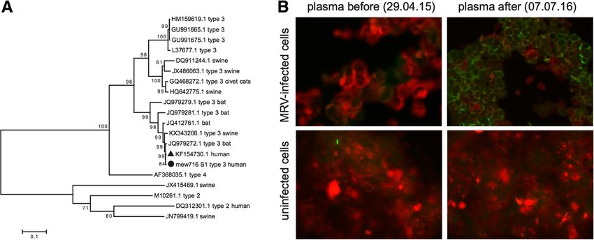
analyses showed that the MRV-3 sequence isolated in The most unexpected finding was the infection with
this study was most related in all 10 segments to an iso- MRV-3, proven by metagenomic sequencing and ser-
late from a child in Slovenia [5] and further clustered ology of samples from several time points. MRVs belong
with isolates from bats in Germany [6] and pigs in Italy to the Reoviridae family, a group of non-enveloped
[7] (Fig. 1a and Additional file 1: Figures S1-S9). dsRNA viruses with 10 genome segments and have been
In order to proof active replication and diagnosis of isolated from a wide range of mammalian hosts and in a
MRV infection, we performed immunofluorescence for variety of clinical contexts [5–7]. To date, the diseases
the detection of MRV-specific antibodies of the patient. associated with MRV infections in humans include
Patient plasma from time points after MRV detection respiratory disease [8], meningitis [9–11], acute necrotiz-
were positive for anti-MRV IgG antibodies showing that ing encephalopathy [12] and (in a similar case involving
the patient seroconverted (Fig. 1b). a child living in Slovenia) acute gastroenteritis [5]. Zoo-
We were able to reconstruct the full-length coxsackie- notic transmission is often suspected [6, 13, 14]. Our
virus genome present in this patient using additional patient lives close to a farm and might have come in
sequence information from sequencing with CV-A22 contact with pets, farm animals and animal feces making
serotype consensus primers. Genotyping and bootscan a zoonotic infection conceivable, however, due to the lack
analysis revealed that the isolate (GenBank KX932039) of data on MRV distribution and appropriate samples we
probably resulted from recombination between CV-A19 can only hypothesize on the transmission route [7]. Stool
and CV-A22 (Additional file 1: Figures S10 and S11). samples from 16 children with suspicion of gastrointes-
The detected HAdV-C was type 2, strain human/ARG/ tinal infection treated at the same hospital during 2015 all
A15932/2002/2[P2H2F2] (JX173079.1). tested negative with qPCR (data not shown).
The virus growing in the initial Caco-2 cell culture
was therefore MRV-3 and not an enterovirus as
Discussion and conclusions suggested by the serotyping assay. In fact, the manufac-
Using a metagenomic sequencing approach, we detected turer’s datasheet for the reagent used states that there is
multiple virus infections in a child with PID and persis- potential for cross-reaction with Hepatitis A, Reovirus 3,
tant diarrhea. Although routine PCR methods detected and some Rhinovirus and Astrovirus strains. The latter
Enterovirus in stool samples for a prolonged period of highlights the genuine difficulty in accurate detection of
time, an attempt to subtype the virus after cell culturing highly diverse virus families with numerous genotypes
was not successful. In order to resolve this, we per- such as Enteroviruses where typing by specific PCR can
formed metagenomic sequencing of cell culture superna- be challenging due to the high susceptibility to recom-
tants and stool suspensions. bination and the emergence of novel strains [15].
Fig. 1 Mammalian orthoreovirus infection confirmed by phylogenetic analysis and immunofluorescence staining. a Phylogenetic analysis of Mammalian
orthoreovirus segment S1 isolated in this study (circle, mew716_S1_type_3_human) reveals a close relationship with previously described isolates
identified in a child in Slovenia (triangle, KF154730), bats in Germany (JQ412761), and pigs in Italy (KX343206). Phylogenetic trees were constructed in
MEGA7 using the Maximum Likelihood method based on the Kimura 2-parameter model. Bootstrap values from 1000 tries are shown. MRV-type and host
species are depicted if available. b Anti-MRV-3 immunofluorescence of patient plasma before and after seroconversion on MRV-3-infected and uninfected
Caco-2 cellsLewandowska et al. BMC Infectious Diseases (2018) 18:33 Page 4 of 5
Notably, CV-A was detected in stool suspensions but canton of Zurich approved the study and written informed consent was
not in cell culture supernatants. While cell culturing was obtained.
critical for MRV-3 detection, CV-A, in line with a
Consent for publication
general difficulty to culture coxsackieviruses [16], did Written informed consent for the publication of this case report was
not infect Caco-2 cells. The reason that MRV-3 was not obtained from both parents of the child.
detected by metagenomic sequencing in the original
Competing interests
stool suspension, but only after amplification in cell The authors declare that they have no competing interests.
culture, is likely because it was at levels too low to be
detected with the applied depth of sequencing.
Publisher’s Note
In summary, this case highlights the complexity of infec- Springer Nature remains neutral with regard to jurisdictional claims in
tions in immunocompromised hosts and reveals limita- published maps and institutional affiliations.
tions of routine diagnostic methods. A combination of
Author details
traditional cell culture, metagenomic sequencing, verifica- 1
Institute of Medical Virology, University of Zurich, Winterthurerstrasse 190,
tion by specific PCRs and serology proved key to detect a 8057 Zurich, Switzerland. 2Division of Immunology, University Children’s
persistent co-infection with three clinically relevant viruses. Hospital Zurich, Steinwiesstrasse 75, 8032 Zurich, Switzerland. 3Division of
Infectious Diseases and Hospital Epidemiology, University Children’s Hospital
While the presence of these viruses in stool specimen Zurich, Steinwiesstrasse 75, 8032 Zurich, Switzerland. 4Division of Stem Cell
suggests a link with the observed pathogenesis, it cannot Transplantation, University Children’s Hospital Zurich, Rämistrasse 100, 8091
be defined to what extent each of the viruses contributed Zurich, Switzerland. 5Present address: Unilabs, Ringstrasse 12, 8600
Dübendorf, Switzerland.
to the initial flu-like symptoms and prolonged diarrhea.
Based on the timing of virus detection, the earlier detected Received: 12 April 2017 Accepted: 4 January 2018
CV-A and HAdV-C are more likely to be involved in the
child’s condition than the later emerging MRV-3.
References
1. Dropulic L, Cohen J. Severe viral infections and primary immunodeficiencies.
Additional file Clin Infect Dis. 2011;53(9):897–909.
2. Delwart EL. Viral metagenomics. Rev Med Virol. 2007;17(2):115–31.
3. Mokili JL, Rohwer F, Dutilh BE. Metagenomics and future perspectives in
Additional file 1: Portable Document Format. Table S1. Routine virus discovery. Current Opinion in Virology. 2012;2(1):63–77.
screening results for Enterovirus. Figures S1 to S9. Phylogenetic analyses of 4. Lewandowska DW, Zagordi O, Zbinden A, Schuurmans MM, Schreiber P,
all Mammalian orthoreovirus segments isolated in this study. Figure S10. Geissberger FD, Huder JB, Böni J, Benden C, Mueller NJ, et al. Unbiased
Coxsackievirus BLAST genotyping. Figure S11. Coxsackievirus Bootscan metagenomic sequencing complements specific routine diagnostic
Analysis. Supplementary Methods. Supplementary References. (PDF 456 kb) methods and increases chances to detect rare viral strains. Diagn Microbiol
Infect Dis. 2015;83(2):133–8.
5. Steyer A, Gutierrez-Aguire I, Kolenc M, Koren S, Kutnjak D, Pokorn M, Poljsak-
Abbreviations Prijatelj M, Racki N, Ravnikar M, Sagadin M, et al. High similarity of novel
CPE: Cytopathic effect; CV-A: Coxsackievirus A; HAdV-C: Human adenovirus C; orthoreovirus detected in a child hospitalized with acute gastroenteritis to
MRV: Mammalian orthoreovirus; PCR: Polymerase chain reaction; PID: Primary mammalian orthoreoviruses found in bats in Europe. J Clin Microbiol. 2013;
immunodeficiency 51(11):3818–25.
6. Kohl C, Lesnik R, Brinkmann A, Ebinger A, Radonic A, Nitsche A, Muhldorfer
Acknowledgements K, Wibbelt G, Kurth A. Isolation and characterization of three mammalian
Not applicable orthoreoviruses from European bats. PLoS One. 2012;7(8):e43106.
7. Lelli D, Beato MS, Cavicchio L, Lavazza A, Chiapponi C, Leopardi S, Baioni L,
Funding De Benedictis P, Moreno A. First identification of mammalian orthoreovirus
Funding was provided by the Clinical Research Priority Program “Viral type 3 in diarrheic pigs in Europe. Virol J. 2016;13(1):139.
Infectious Diseases” of the University of Zurich. The funding body did not 8. Chua KB, Crameri G, Hyatt A, Yu M, Tompang MR, Rosli J, McEachern J,
have any role in the design of the study, in the collection, analysis, and Crameri S, Kumarasamy V, Eaton BT, et al. A previously unknown reovirus of
interpretation of data and in writing the manuscript. bat origin is associated with an acute respiratory disease in humans. Proc
Natl Acad Sci U S A. 2007;104(27):11424–9.
9. Johansson PJ, Sveger T, Ahlfors K, Ekstrand J, Svensson L. Reovirus type 1
Availability of data and materials
associated with meningitis. Scand J Infect Dis. 1996;28(2):117–20.
The datasets used and/or analyzed during the current study are available
10. Hermann L, Embree J, Hazelton P, Wells B, Coombs RTK. Reovirus type 2
from the corresponding author on reasonable request.
isolated from cerebrospinal fluid. Pediatr Infect Dis J. 2004;23(4):373–5.
11. Tyler KL, Barton ES, Ibach ML, Robinson C, Campbell JA, O’Donnell SM, Valyi-
Authors’ contributions Nagy T, Clarke P, Wetzel JD, Dermody TS. Isolation and molecular
SP, CB, TG, JR and JPS cared for the patient and provided clinical data and characterization of a novel type 3 reovirus from a child with meningitis. J
materials. RC, JB and AZ analyzed and interpreted routine diagnostic results. Infect Dis 2004;189(9):1664-1675.
DWL, FDG and CS performed sequencing experiments and PCR assays. DWL, 12. Ouattara LA, Barin F, Barthez MA, Bonnaud B, Roingeard P, Goudeau A,
OZ and MH analyzed the sequencing data. MK and MK performed cell Castelnau P, Vernet G, Paranhos-Baccala G, Komurian-Pradel F. Novel human
culture and immunostaining. DWL, AT and MH wrote the manuscript. All reovirus isolated from children with acute necrotizing encephalopathy.
authors read and approved the final manuscript. Emerg Infect Dis. 2011;17(8):1436–44.
13. Lelli D, Moreno A, Lavazza A, Bresaola M, Canelli E, Boniotti MB, Cordioli P.
Ethics approval and consent to participate Identification of Mammalian orthoreovirus type 3 in Italian bats. Zoonoses
Non-invasive samples were obtained from patient mew716 in the frame of Public Health. 2013;60(1):84–92.
the Viral Metagenome Study of the Clinical Research Priority Program ‘Viral 14. Chua KB, Voon K, Yu M, Keniscope C, Abdul Rasid K, Wang L-F.
Infectious Diseases’ of the University of Zurich. The ethics committee of the Investigation of a potential zoonotic transmission of orthoreovirusLewandowska et al. BMC Infectious Diseases (2018) 18:33 Page 5 of 5
associated with acute influenza-like illness in an adult patient. PLoS
One. 2011;6(10):e25434.
15. Lukashev AN. Role of recombination in evolution of enteroviruses. Rev
Med Virol. 2005;15(3):157–67.
16. Wenner HA, Lenahan MF. Propagation of group a Coxsackie viruses in tissue
cultures. II. Some interactions between virus and mammalian cells. Yale J
Biol Med. 1961;34:421–38.
Submit your next manuscript to BioMed Central
and we will help you at every step:
• We accept pre-submission inquiries
• Our selector tool helps you to find the most relevant journal
• We provide round the clock customer support
• Convenient online submission
• Thorough peer review
• Inclusion in PubMed and all major indexing services
• Maximum visibility for your research
Submit your manuscript at
www.biomedcentral.com/submitYou can also read