Oral Squamous Cell Carcinoma in a Free-Ranging Roe Deer (Capreolus capreolus)
←
→
Page content transcription
If your browser does not render page correctly, please read the page content below
MedDocs Publishers

                                                                                                                   ISSN: 2640-1223

                                     Journal of Veterinary Medicine and Animal Sciences
                                                                                                  Open Access | Research Article

Oral Squamous Cell Carcinoma in a Free-Ranging
        Roe Deer (Capreolus capreolus)
Domenis L1,2*; Campanella C1,3; Rubini D1,3, Parovel E4; Orusa R1,2; Robetto S1,2
1
 Istituto Zooprofilattico Sperimentale del Piemonte Liguria e Valle d’Aosta
2
 Valle d’Aosta Department - National Reference Centre for Wild Animals Diseases (Ce.R.M.A.S.), Regione Amerique 7G, 11020
Quart (AO), Italy
3
 Genova Department – National Reference Centre for Veterinary and Comparative Oncology (Ce.R.O.Ve.C.), Piazza Borgo Pila 10,
Genova, Italy
4
 Wildlife Recovery Centre (C.R.A.S.), Quart (AO), Italy

   *Corresponding Author(s): Lorenzo Domenis                            Abstract
 Istituto Zooprofilattico Sperimentale del Piemonte Ligu-            Objective: We describe a case of squamous cell carcino-
 ria e Valle d’Aosta – Valle d’Aosta Department - National         ma (SCC) in a free-ranging roe deer (Capreolus capreolus).
 Reference Centre for Wildlife Diseases (Ce.R.M.A.S.) -                Methods and Results: Subject of this paper is an adult
 Regione Amerique 7G, 11020 Quart (AO), Italy                      male of roe deer (Capreolus capreolus), found in Aosta
 Tel: +39-0165-23-8558;                                            Valley Region (North-west Italy), with an evident swelling
 Email: lorenzo.domenis@izsto.it                                   at the right mandibular region. By applying routine histo-
                                                                   logical (Haematoxylin and Eosin staining) and immunohis-
                                                                   tochemical (antibodies detection by diaminobenzidine DAB
                                                                   and counterstaining by Mayer’s Haemotoxylin) techniques,
                                                                   the tumor appeared to be formed by trabeculae and islands
Received: Apr 30, 2020
                                                                   of squamous epithelial cells, growing from the oral mucosa,
Accepted: May 29, 2020                                             involving soft and bone tissues and surrounded by a des-
Published Online: Jun 03, 2020                                     moplastic reaction; it was also observed a pseudo-glandular
Journal: Journal of Veterinary Medicine and Animal Sciences        pattern due to the formation of cysts filled with acantholytic
                                                                   keratinocytes; strong anisokaryosis and anisocytosis, with a
Publisher: MedDocs Publishers LLC                                  quite low mitotic activity (about 1 division per high-power
Online edition: http://meddocsonline.org/                          – 400x - field), characterized the neoplastic cells. Epithelial
Copyright: © Domenis L (2020). This Article is                     trabeculae were positive for cytokeratin and negative for
distributed under the terms of Creative Commons                    vimentin, last one well expressed by the fibroblasts in the
Attribution 4.0 International License                              desmoplastic stroma. By routine bacteriological examina-
                                                                   tion (Blood Agar and MacConkey agar incubated over 48 h
                                                                   at 37 °C in aerobic and anaerobic atmosphere) overlapping
                                                                   infection of neoplastic mass by Trueperella pyogenes was
Keywords: Squamous cell carcinoma; Roe deer; Capreolus             found. Based on microscopical features the tumor was clas-
capreolus                                                          sified as acantholytic SCC.
                                                                       Discussion and conclusions: SCC is a malignant tumor of
                                                                   epidermal origin well known in domestic animals, especially
                                                                   in cats where it’s the most prevalent oral neoplasm, and
                                                                   wild fauna, such as fish, birds, reptiles and various classes of
                                                                   mammals. To our knowledge, this is the first report of a sim-
                                                                   ilar neoplasia in roe deer; adding SCC to the list of tumors
                                                                   that can affect the alpine wild ruminants contributes to the
                                                                   study and knowledge of neoplastic processes in all wildlife.

               Cite this article: Domenis L, Campanella C, Rubini D, Parovel E, Orusa R, et al. Oral Squamous Cell Carcinoma in a
            Free-Ranging Roe Deer (Capreolus Capreolus). J Vet Med Animal Sci. 2020; 3(1): 1024.

                                                               1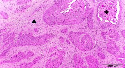
MedDocs Publishers
   Introduction                                                                Bacteriological examination of the pus collected inside the
                                                                           mandibular mass was performed by Blood Agar and MacConkey
    Squamous cell carcinoma (SCC) is a malignant tumor of epi-             agar over 48 h at 37 °C in aerobic and anaerobic atmosphere;
dermal origin well known in domestic animals, especially in cats           colonies of interest were characterized by morphology, catalase
where it’s the most prevalent oral neoplasm, generally found               and oxidase test, Gram staining and typing by API Biomerieux
at mandibular, maxillary or sublingual regions. As it happens              biochemical galleries. Tissue samples from the lesion were fixed
for other types of neoplasia, it’s reported sporadically in differ-        in 10% neutral buffered formalin and, after decalcification of the
ent wild animals, free-ranging or captive, as fish [1,2], birds [3],       ossified areas by Osteosoft (Merck, Darmstadt, Germany), par-
reptiles [4,5] and various classes of mammals: cetaceans such              affin-embedded and cut into 3 µm thickness sections for histo-
as Tursiops truncatus [6], marsupials such as Isoodon macrou-              logical and immunohistochemical examinations. Some sections
rus [7], rodents such as Agouti paca [8], rhinocerotids, such as           were stained with Haematoxylin and Eosin, additional sections
Rhinoceros unicornis [9], felids, such as Leopardus pardalis [10],         were subjected to immunohistochemistry using primary anti-
Panthera tigris [11], Panthera tigris altaica [12], Panthera leo           bodies specific for cytokeratin AE1/AE3 (1:200 dilution; Dako,
[13], Panthera pardus pardalis [14] different species of Lynx [15-         Santa Clara, California, USA) and for vimentin V9 (1:50 dilution;
17] and ruminants; among these, it has been observed in differ-            Dako, Santa Clara, California, USA); labelling with each antibody
ent species of cervidae, such as Cervus elaphus [18], Elaphurus            was detected by 3,3’-diaminobenzidine (DAB), counterstaining
davidianus [19], Odocoileus virgianianus [20], Cervus nippon               slides with Mayer’s Haemotoxylin.
pseudaxis [21] and Rangifer tarandus tarandus [22]. Aim of this
work is to describe the gross and microscopic findings observed               Bacteriological examination highlighted coryneform bacilli,
in the first case, to our best knowledge, of an oral squamous cell         partially hemolytic, Gram positive, catalase and oxidase nega-
carcinoma in a free-ranging roe deer (Capreolus capreolus).                tive, later identified as Trueperella pyogenes.
   Case details                                                                Histologically, a non-capsulated and ulcerated soft tissue
                                                                           neoplasia has been observed. Neoplastic cells were arranged in
   Subject of this report is an adult male of roe deer (it was             nests and anastomosing trabeculae, growing from oral mucosa
not possible to define exactly the age due to the abnormal con-            and associated with abundant stromal fibrous connective tissue
sumption of the teeth) found by the rangers in Aosta Valley                (desmoplasia), with initial extending into the mandibular bone
Region (North-west Italy), at 1100 m of altitude; due to cachexia          tissue. There was a widespread tendency to keratinization,
and an evident swelling at the right mandibular region without             without corneal pearls, but with single keratinized cells or wider
skin ulcers or fistulas (Fig. 1A), the animal was recovered in a           areas occupied by numerous squamous keratinocytes; dissocia-
wildlife recovery center and subsequently euthanized for the               tion and degeneration of squamous neoplastic cells resulted in
poor conditions and prognosis. At the necropsy, a globular mass            cysts, lined with a single layer of keratinizing epithelial cells and
of about 15 cm in diameter, involving the intermediate portion             filled with large number of acantholytic keratinocytes, often in-
of the right mandibular body (Fig. 1B), was detected; the neo-             vaded by neutrophils, with an extensive pseudo-glandular pat-
formation contained various pus collections, associated with               tern (Figure 2).
vegetables fragments, draining in the oral cavity at the level of
the first molar and causing luxation and loosing of the other
teeth of the same dental arch (Fig. 1C). No other pathological
findings were observed, apart from a conspicuous infestation by
intestinal Trichostrongilidae (the eggs were observed by parasi-
tological analysis with flotation in 100% zinc sulphate solution).

                                                                              Figure 2: Islands and cords of neoplastic epithelial cells, with
                                                                            diffuse desmoplastic reaction (triangle) and pseudo-glandular fea-
                                                                             tures with acantholytic keratinocytes (asterisk). HE.

                                                                               The cellular elements were characterized by abundant eo-
                                                                           sinophilic cytoplasm, oval nuclei with small clumped chroma-
                                                                           tin, high anisocytosis and anisokaryosis, with the presence of
                                                                           "giant" cells with huge nuclei, mitotic index of about 1 division
                                                                           per high-power (400x) field (Fig. 3); it were also observed large
   Figure 1: Male roe deer (Capreolus capreolus) showing cachex-           lytic areas with bacterial aggregates, superficial ulcers covered
 ia and an extensive enlargement at the right mandibular region;           by serocellular crusts with abundant neutrophils and numerous
 B - Large mass involving the intermediate portion of the right man-       protozoan cysts (Sarcocystis spp.) in the annexed muscle lay-
 dibular body; C-Note the loosing of teeth, vegetables fragments           ers.
 and ulcers at the surface of the neoformation (partially removed
  from mandibular body).

Journal of Veterinary Medicine and Animal Sciences                     2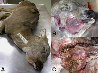
MedDocs Publishers
[28] and many cases of hepatocellular tumor, observed in Brit-
ain, probably associated with high levels of spruce needles, buds
and twig tips (rich in tannins and terpenes) in the diet [29].
Different hypothesis about the causes of the SCC in roe deer
can be advanced considering the various risk factors reported
for this type of tumor in people and animals, including sex (male
are more affected than females), age (cat and dogs of average
10-11 years old are most affected), chronic inflammations, ul-
traviolet light, papillomavirus infection, bracken fern ingestion,
carcinogens in tobacco, coal tar, soot, arsenic and smegma [30,
31]; further anamnestic information, unfortunately often un-
available in the case of free-living animals, would be necessary
to understand the exact pathogenesis of the neoplasia we have
described.
Figure 3: Marked cytoplasmic eosinophilia, anisocytosis
and anisokaryosis of neoplastic squamous cells. HE. Conclusion
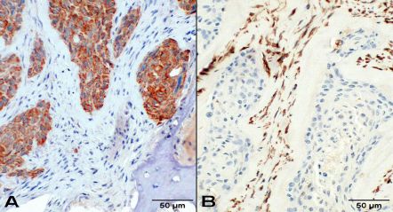
The cytoplasm of neoplastic cells was strongly positive for In conclusion, to our best knowledge based on the referenc-
cytokeratin (Fig. 4A) and negative for vimentin (Fig. 4B), last one es found, this is the first report of SCC in roe deer; adding this
well expressed by the cells of desmoplastic connective stroma. type of cancer to the list of tumors that can affect the alpine
wild ruminants contributes to the study and knowledge of neo-
No metastasis was found in vascular and lymphatics lumen, plastic processes in all wildlife.
in regional lymph nodes or in thoracic and abdominal organs.
References
Based on histological features, and particularly on the pseu-
1. Mawdesley-Thomas LE, Bucke D. Squamous cell carcinoma in a
do-glandular pattern expressing cysts filled with acantholytic
gudgeon (Gobio gobio, L.). Pathol Vet. 1967; 4: 484-489.
keratinocytes, we classified the neoplasia as acantholytic SCC.
2. Wildgoose WH. Papilloma and squamous cell carcinoma in koi
carp (Cyprinus carpio). Vet. Rec. 1992; 130: 153-157.
3. Ramis A, Gibert X, Majó N, Grifols J. Metastatic oral squamous
cell carcinoma in a Montagu’s harrier (Circus pigargus). J. Vet.
Diagn. Invest. 1999; 11: 191-194.
4. Anderson ET, Kennedy-Stoskopf S, Sandy JR, Dorn B, Boyette T,
et al. Squamous Cell Carcinoma with Vascular Invasion in a Dia-
mondback Rattlesnake (Crotalus adamanteus). J. Zoo Wildlife
Med. 2010; 41: 745-749.
5. Hill AG, Denis MM, Pyne M (2016) Squamous cell carcinoma
with hepatic metastasis in a saltwater crocodile (Crocodylus po-
rosus). Aust Vet J. 2016; 94: 83-86.
6. Ewing RY, Mignucci-Giannoni AA. A Poorly Differentiated Pulmo-
nary Squamous Cell Carcinoma in a Free-Ranging Atlantic Bottle-
nose Dolphin (Tursiops truncatus). J. Vet. Diagn. Invest. 2003;
Figure 4: Neoplastic cells are strongly positive for cytok-
15: 162-165.
eratin (A) and negative for vimentin (B), last one widely ex-
pressed by desmoplastic stroma. IHC. 7. Beck AP, Shima AL, Bennett MD, Johnson LK. Metastatic
Squamous Cell Carcinoma in a Northern Brown Bandicoot
(Isoodon macrourus). Vet Sci. 2017; 14: 4.
Discussion and conclusions
8. Luppi MM, Malta MC, Costa ME, Motta RO, Santos RL. Multi-
In roe deer (Capreolus capreolus) different types of neo- centric squamous cell carcinoma in a paca (Agouti paca) resem-
plasia are signaled; in a surveillance performed in Switzerland bling Bowen’s disease. J. Zoo Wildlife Med. 2008; 39: 244-247.
[23], the authors refers 32 cases of tumors, observed in lymph
9. Naik SN, Ishwad CS, Karawale MS, Wani MV. Squamous cell car-
nodes (lymphosarcoma, histiocytic sarcoma), head (spindle cell cinoma in an Indian rhinoceros. Vet Rec. 1986; 118: 590-591.
sarcoma, melanoma, ossifying fibroma, osteosarcoma, fibro-
sarcoma, round cell tumor, carcinoma of salivary gland), ovary 10. Leme MCM, Martins AMCRF, Bodini MES, Carvalho PR, Portu-
(carcinoma/teratoma, granulosa cell tumor), skin (fibroma, fib- gal MASC. Carcinoma de celulas escamosas em uma jaguatirica
rosarcoma, fibropapilloma), lung (carcinoma) and liver (cholan- (Leopardus pardalis). Arq Inst Biol. 2003; 70: 217-219.
giocarcinoma, hepatocellular carcinoma), kidney (renal adeno- 11. Kloft HM, Ramsay EC, Sula MM. Neoplasia in Captive Panthera
carcinoma, adenoma) other than some neoplasia defined as Species. J Comp Pathol. 2019; 166: 35-44
undetermined. A similar study in Sweden [24] showed 19 cases
of tumors, overlapping to those observed in Switzerland a part 12. de Oliveira AR, de Carvalho TF, Arenales A, Tinoco HP, Coelho
CM, et al. Mandibular squamous cell carcinoma in a captive Si-
two cases of hemangiosarcoma and one of rhabdomyosarcoma.
berian tiger (Panthera tigris altaica). Braz. J. Vet. Path. 2018; 11:
Other single reports refer the same type of some neoplasms
97- 101.
observed in Switzerland and Sweden, in other words adenoma
[25], teratoma [26], lymphosarcoma [27], viral fibropapillomas 13. Mwase M, Mumba C, Square D, Kawarai S, Madarame H. Cu-
Journal of Veterinary Medicine and Animal Sciences 3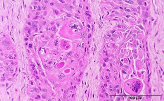
MedDocs Publishers
      taneous squamous cell carcinoma presenting as a wound with                23.   Pewsner M, Origgi FC, Frey J, Ryser-Degiorgis MP. Assessing Fifty
      discharging sinus tracts in a wild African lion (Panthera leo). J               Years of General Health Surveillance of Roe Deer in Switzerland:
      Comp Pathol. 2013; 149: 520-523.                                                A Retrospective Analysis of Necropsy Reports. PLoS One. 2017;
                                                                                      19; 12: e0170338.
14.   Napier JE, Lund MS, Armstrong DL, McAloose. A retrospective
      study of morbidity and mortality in the North American Amur               24.   Aguirre AA, Bröjer C, Mörner T. Descriptive epidemiology of roe
      leopard (Panthera pardus orientalis) population in zoologic insti-              deer mortality in Sweden. J. Wildlife Dis. 1999; 35: 753-762.
      tution from 1992 to 2014. J. Zoo Wildlife Med. 2018; 49: 70-78.
                                                                                25.   Craig WA. Adenoma in a British roe deer (Capreolus capreolus).
15.   Altamura G, Eleni C, Meoli R, Cardeti G, Friedrich KG, et al.                   Vet. Rec. 1979; 104: 214-215.
      Tongue squamous cell carcinoma in a European lynx (Lynx lynx):
      papillomavirus infection and histologic analysis. Vet Sci. 2011; 5:       26.   Barlow AM, Couper D. Cutaneous teratoma in a wild roe deer
      1-6.                                                                            (Capreolus capreolus) in the UK. Vet. Rec. 2006; 159: 211-212.

16.   Gunson DE, Klein LV, Reid CF. Gingival squamous cell carcinoma            27.   Woodford M. Lymphosarcoma in a wild roe deer. Vet Rec. 1966;
      in a Canadian lynx. J Am Vet Med Assoc.1978; 173: 1228-1230.                    79: 74.

17.   Sladakovic I, Burnum A, Blas-Machado U, Kelly LS, Garner BC,              28.   Rajský D, Rajský M, Garaj P, Kropil R, Ivan M, et al. Emergence
      et al. Mandibular Squamous Cell Carcinoma in a Bobcat (Lynx                     and expansion of roe deer (Capreolus capreolus) fibropapilloma-
      rufus). J. Zoo Wildlife Med. 2016; 47: 370-373.                                 tosis in Slovakia. Eur J Wildlife Res. 2016; 62: 43-49.

18.   Ulrich R, Teifke JP, Voigt U, Seehusen F. Oral squamous cell car-         29.   de Jong CB, van Wieren SE, Gill RM, Munro R. Relationship be-
      cinoma in a red deer (Cervus elaphus). J. Wildlife Dis. 2014: 50:               tween diet and liver carcinomas in roe deer in Kielder. Forest
      113-116.                                                                        and Galloway Forest. Vet Rec. 2004; 155: 197-200.

19.   Agrimi U, Morelli L, Di Guardo G. Squamous cell carcinoma of              30.   Brown CC, Baker DC, Barker IK. Alimentary system. In: Pathol-
      the skin in a Père David’s deer (Elaphurus davidianus). J. Wildlife             ogy of domestic animals Jubb, Kennedy, and Palmer’s, 5th Edit.,
      Dis. 1993; 4: 616-617.                                                          Maxie MG, Elsevier, Edinburgh, Great Britain. 2007; 3-296.

20.   Stroud RK, Amundson TE. Squamous cell carcinoma in a free-                31.   Goldschmidt MH, Shofer FS. Skin Tumors of the dog and cat. 1th
      ranging white-tailed deer (Odocoileus virginianus). J. Wildlife                 Edit., Butterworth-Heinemann, Oxford. 1992: 38-40.
      Dis.1983; 19: 162-164.

21.   Ensley PK, Janssen DL, Anderson MP. Squamous cell carcinoma
      in an Indochina sika deer. J Am Vet Med Assoc. 1980; 177: 932.

22.   Gonzalez-Alonso-Alegre EM, Rodriguez-Alvaro A, Martinez-
      Nevado E, Martinez-de-Merlo EM, Sanchez-Maldonado B. Con-
      junctival squamous cell carcinoma in a reindeer (Rangifer taran-
      dus tarandus). Vet Ophthalmol. 2013: Suppl 1: 113-116.

Journal of Veterinary Medicine and Animal Sciences                          4

You can also read