GEOGRAPHIC AND GENOMIC DISTRIBUTION OF SARS-COV-2 MUTATIONS - PREPRINTS.ORG
←
→
Page content transcription
If your browser does not render page correctly, please read the page content below
Preprints (www.preprints.org) | NOT PEER-REVIEWED | Posted: 30 April 2020                  doi:10.20944/preprints202004.0529.v1

              Geographic and Genomic Distribution of
                     SARS-CoV-2 mutations
                                             Daniele Mercatelli and Federico M. Giorgi*
          Department of Pharmacy and Biotechnology, University of Bologna, Via Selmi 3, 40126, Bologna, Italy
          * Corresponding author email: federico.giorgi@unibo.it
          ORCID 0000-0003-3228-0580 (Daniele Mercatelli)
          ORCID 0000-0002-7325-9908 (Federico M. Giorgi)

          Abstract
          The novel respiratory disease COVID-19 has reached the status of worldwide pandemic and large efforts
          are currently being undertaken in molecularly characterizing the virus causing it, SARS-CoV-2. The genomic
          variability of SARS-CoV-2 specimens scattered across the globe can underly geographically specific
          etiological effects. In the present study, we gather the 10,014 SARS-CoV-2 complete genomes currently
          available thanks to the collection endeavor of the GISAID consortium and thousands of contributing
          laboratories. We analyze and annotate all SARS-CoV-2 mutations compared with the reference Wuhan
          genome NC_045512.2. Our analysis shows the prevalence of single nucleotide transitions as the major
          mutational type across the world. There exist at least three clades characterized by geographic and genomic
          specificity. In particular, the clade G, prevalent in Europe, carries a D614G mutation in the Spike protein,
          which is responsible for the initial interaction of the virus with the host human cell. Our analysis may drive
          local modulation of antiviral strategies based on the molecular specificities of this novel virus.

          Keywords
          SARS-CoV-2; genomics coronavirus; COVID-19 evolution

          Abbreviations
          AA: aminoacid
          COVID-19: Coronavirus Disease 2019
          GISAID: Global Initiative on Sharing All Influenza Data
          Indel: insertion/deletion event
          NSP: non-structural protein
          ORF: open reading frame
          S: SARS-CoV-2 spike protein
          SARS-CoV-2: Severe Acute Respiratory Syndrome, Coronavirus 2
          SNP: single nucleotide polymorphism

          Introduction
          Initially reported in mid-December 2019 in the Chinese city of Wuhan, the newly emerged severe acute
          respiratory syndrome virus (SARS-CoV-2) is a single-stranded RNA beta-coronavirus with a very compact
          29,903 nucleotides-long genome. This virus causes a serious disease known as Coronavirus Disease 2019
          (COVID-19), which has spread in over 210 countries in less than four months, counting more than 2,5
          million confirmed cases and almost 180,000 deaths reported worldwide as of April 22, 2020 (source: World
          Health Organization). A difference in case fatality rates across countries was observed, possibly due to a
          diverse demographic composition and the type of measures that have been taken in different countries to
          limit viral spreading [1]. According to data from the public database of the Global Initiative on Sharing All

            © 2020 by the author(s). Distributed under a Creative Commons CC BY license.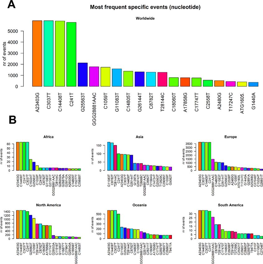
Preprints (www.preprints.org) | NOT PEER-REVIEWED | Posted: 30 April 2020               doi:10.20944/preprints202004.0529.v1

          Influenza Data (GISAID), three major clades of SARS-CoV-2 can be identified [2], that have been
          subsequently named as clade G (variant of the spike protein S-D614G), clade V (variant of the ORF3a coding
          protein NS3-G251), and clade S (variant ORF8-L84S). However, as more complete sequences become
          available, the need to define specific geographic distributions of virus variants becomes of practical
          importance to define clinical and political strategies at the local level. Despite several reports having
          confirmed a relatively low variability of SARS-CoV-2 genomes [3,4], it is still unclear if different fatality rates
          disease spreading speed in different countries may be the consequence of clade’s differences in virulence,
          as discussed by a recent commentary comparing different strains in the USA [5]. It is therefore possible that
          more insights into the pathogenesis and virulence of this virus may come from comparative genomic
          analysis linked to epidemiologic data coming from different countries.
          Genetic variance analyses must now play a crucial role in expanding knowledge on this new virus to adopt
          measures to contain its outbreak. Complete viral genome sequences have been made rapidly publicly
          available to the research community and have recently surpassed the 10,000 units, thanks to the worldwide
          effort of scientists and to the GISAID consortium. This data avalanche will result in an unprecedently rapid
          effort to analyze data to understand genome diversity [6,7], to hypothesize targetable targets for drug
          repositioning [8,9] and to develop prevention strategies [10]. In the present study, we performed the largest
          comparative study so far by analyzing more than 10,000 complete SARS-CoV-2 genomes. We will report all
          mutations and stratify them genomically and geographically, also highlighting insurgence of sub-clades and
          genomic highly variable spots. These finding may be extremely useful to design and think about the efficacy
          of measures that have been taken on a regional basis to limit SARS-CoV-2 spreading.

          Methods
          10,014 SARS-CoV-2 genomic sequences were downloaded from GISAID (Supplementary File 1) on April 20,
          2020. Only viruses affecting human hosts were selected, excluding low coverage sequences and incomplete
          (
Preprints (www.preprints.org) | NOT PEER-REVIEWED | Posted: 30 April 2020 doi:10.20944/preprints202004.0529.v1
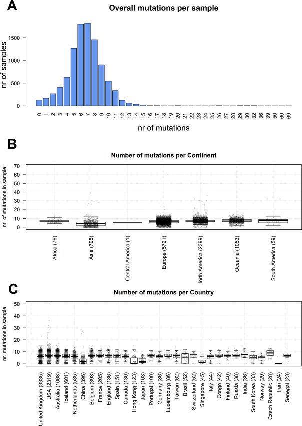
Figure 1. (A) distribution of number of mutational events for all SARS-CoV-2 genome samples analyzed. (B)
Distributions of number of mutations for each sample, stratified per continent. The main boxplot rectangles are drawn
between the 1st and 3rd quartile, with the median value indicated as a thick line. Boxplot whiskers fall on the closest
point to the 1st/3rd quartile + 1.5 interquartile range as described in the R boxplot() function. The number in brackets
after the continent name indicates the number of sequenced genomes. (C) As in B, with stratification performed
country-wise, using the 30 countries with the highest number of sequenced genomes.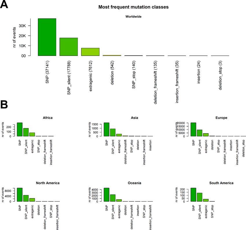
Preprints (www.preprints.org) | NOT PEER-REVIEWED | Posted: 30 April 2020                doi:10.20944/preprints202004.0529.v1

          We analyzed the nature of each mutation, highlighting a prevalence of single-nucleotide polymorphisms
          (SNPs) over short insertion/deletion events (indels) (Figure 2 A and Supplementary File 5). Worldwide, we
          observed 39,036 aminoacid(aa)-changing SNP events, with less than half silent SNPs (19,629). Short in-
          frame deletions (3x deletions reducing the viral protein length without introducing stop codons) are the
          next largest class (542 total events), followed by 154 SNPs introducing a stop codon. We observe only 135
          frameshift deletions, 35 frameshift insertions, 24 in-frame deletions (inserting multiples of 3 nucleotides)
          and 3 deletions introducing a stop codon in the mutated frame. Overall, 7806 mutations were located
          outside gene regions, prevalently in the untranslated regions (UTRs) of SARS-CoV-2 genome.
          The distribution of these events is largely uniform across all continents (Figure 2 B).

          Figure 2. (A) Worldwide distribution of SARS-CoV-2 mutation classes. “SNP”, “deletion” and “insertion” terms without
          further specifications are intended as frameshift-preserving aa-changing events. (B) Distribution of mutation classes
          in continents.

          We then classified the SARS-CoV-2 mutations according to their type, observing a prevalence of SNP
          transitions (purine->purine and pyrimidine->pyrimidine) over SNP transversions (purine->pyrimidine and
          vice versa). The most common event, both worldwide and continent-wise, is by far the C>T transition,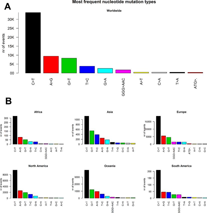
Preprints (www.preprints.org) | NOT PEER-REVIEWED | Posted: 30 April 2020 doi:10.20944/preprints202004.0529.v1
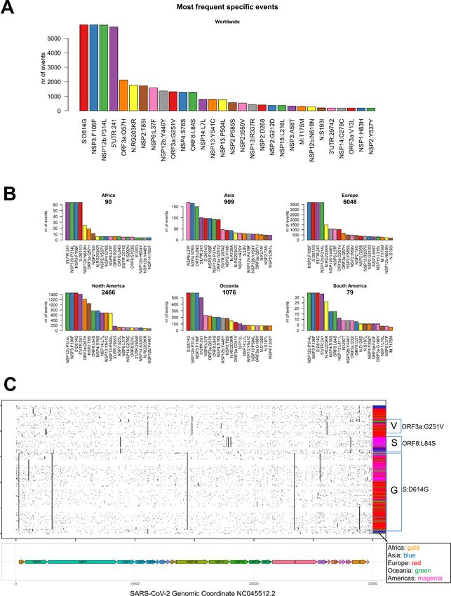
accounting with roughly 50% of all observed worldwide viral mutations (Figure 3 A), followed by the A>G
transition. The most common transversion, G>T, is the third most common event, with 8348 occurrences
(Supplementary File 5). The most common indel, the deletion of the ATG codon, is the 10 th most common
event, with a total of 412 occurrences. The G>T transversion is however the second most common event
overall in Asia and Australia, while the G>A transversion is the second most common event in South America
(Figure 3B).
Figure 3. (A) Worldwide and (B) continent-stratified distribution of SARS-CoV-2 mutation types. Colors are assigned
randomly but preserved across panels to facilitate tracking of identical types across continents.
We went into higher detail and analyzed the effects of each mutation on the protein sequences of SARS-CoV-
2. Our results in this case start to address major differences across continents. The most prevalent mutation
in sequenced genomes worldwide is a transversion affecting the 23,403rd nucleotide adenosine (Figure 4 APreprints (www.preprints.org) | NOT PEER-REVIEWED | Posted: 30 April 2020 doi:10.20944/preprints202004.0529.v1
and Supplementary File 5), transformed into a guanosine (A23403G), defining the so-called G-clade of
SARS-CoV-2 genomes, prevalent in Europe (where overall the highest sequencing effort has been
undertaken, and therefore the highest number of samples), Oceania, South America and Africa (Figure 4 B).
This mutation causes a D614G (aspartate to glycine in protein position 614) aa-change of the Spike (S)
protein, which is responsible for the initial entry of the virus in the cell via the ACE2 human receptor [13].
The most frequent mutation in Asia is G11083T, causing a L37F aa-change in Non-structural protein 6
(Table 1).
Figure 4. (A) Worldwide and (B) continent-stratified distribution of SARS-CoV-2 most frequent specific events,
annotated as nucleotide coordinates over the reference genome NC_045512.2. Colors are assigned randomly but
preserved across panels to facilitate tracking of identical types across continents.Preprints (www.preprints.org) | NOT PEER-REVIEWED | Posted: 30 April 2020 doi:10.20944/preprints202004.0529.v1
Figure 5. (A) Worldwide and (B) continent-stratified distribution of SARS-CoV-2 most frequent specific events,
annotated protein changes using the format protein:mutation. Colors are assigned randomly but preserved across
panels to facilitate tracking of identical types across continents. (C) Dot mat showing as X-axis the 29,903 nucleotide
positions (sorted from left, 5’ to right, 3’) of SARS-CoV-2, and as Y axis the 10,014 genomes analyzed in this study. ThePreprints (www.preprints.org) | NOT PEER-REVIEWED | Posted: 30 April 2020 doi:10.20944/preprints202004.0529.v1
genomic sequences were clustered using the “complete” clustering algorithm. Coding sequence regions are shown at
the bottom of panel B. To the right of the plot, we assigned a color to each sample according to the continent of origin.
Further right, we manually annotated the groups according to the known GISAID clades (V, S and G) and the mutations
that named them. The ORF8, where the S-clade T28144C/ORF8:L84S mutation is located, is situated directly to the left
of the N gene.
We summarized the 20 most frequent events in Table 1. Apart from the already cited S:D614G mutation in
the Spike protein, we observed a silent mutation (F106F) in NSP6, a proline-to-leucine in NSP12b (P314L)
and a mutation in the 5’UTR at genomic coordinates 241 (Figure 5 A). All these four mutations are prevalent
in Europe, Africa, Americas and Oceania (Figure 5 B), and almost always co-occurring in the sequenced
genomes (Figure 5 C), determining the strongest signature for the G clade of SARS-CoV-2 genomes. The
5’UTR:241 mutations seems slightly less frequent than the other three G-clade events, but this could be due
to the lower coverage of genomic tails in sequencing experiments.
The second most represented clade, V, is characterized and named by a mutation in ORF3a, G251V (Table
1 and Figure 5). We detected that this mutation is often (>95% of the times) co-occurring with the silent
mutation at genomic location C14805T and by the more frequent V-clade associated NSP6-L37F. Clade V
seems prevalent in a subset of European sequences (Figure 5). We identified a V-subclade, characterized by
the three prevalent V variants, plus two extra aa-changing SNPs, both affecting NSP2: P585S and I559V
(Table 1).
Finally, we observed a clade most represented in genomes sequenced in Asia and Americas, dubbed “S” by
the GISAID consortium due to a L84S mutation in the ORF8, already identified in early February, 2020 [4].
The L84S mutation is often co-occurring with a silent mutation in the NSP4 gene, C8782T, and by other less
frequent events in NSP13 and NSP14 (Table 1).
Our analysis also highlighted a curious event in the Nucleocapsid gene (N) of some genomes characterized
by the S:D614G mutation and therefore belonging to the G clade. A succession of three nucleotides with
genomic coordinates 28881, 28882 and 28883 is affected by almost-always (99.7%) co-occurring events,
transforming the triplet GGG into AAC and causing two aminoacids of the N protein to change (RG203KR).
This trinucleotide Nucleocapsid event defines a distinct G-subclade of the SARS-CoV-2 population. Two
mutations on the NSP2, G212D and the deletion of Aspartate (D) 268 (Table 1), are not associated
specifically to any of the three main clades, but instead we observe amongst the samples more similar to
the original reference (Figure 5C, top group). In particular, the D268 deletion is observed in a minor part of
S clade samples.
Finally, we include a distribution of the most common mutations (annotated as protein events) in the 25
countries with the highest number of sequenced genomes (Supplementary File 6). We noticed that all
European countries share very similar profiles with each other. In Asia, we could however observe a marked
difference between Japan (where the NSP6:L37F mutation prevails), China (where the ORF8:L84S and
NSP4:S76S events are the most common) and Hong Kong (marked by ORF3a:G251V events).
Discussion
Our analysis confirms a low mutation rate of the virus, with an average of 6.7 mutations per sample with
respect to the reference SARS-CoV-2 genome sequences. However, the existing mutations allow to group
the samples into three distinct clades, G, S and V, characterized by a collection of specific mutations. The
clades can be further characterized by most recent mutations and will likely be split even further in the
future.
While the aa-changing SNPs are the most prevalent mutational events, we observed also silent SNPs and
extragenic (mostly 5’UTR) SNPs. The silent events may not determine an immediate effect on the protein
sequences, but they have repercussions as they may change the codon usage and translation efficiency. InPreprints (www.preprints.org) | NOT PEER-REVIEWED | Posted: 30 April 2020 doi:10.20944/preprints202004.0529.v1
the case of the 5’UTR SNPs, mutations may affect the transcription and replication rates of the virus, or the
folding of the genomic ssRNA, processes that have been only recently and only partially elucidated [14].
The early studies currently performed on SARS-CoV-2 transcriptome dynamics may also suggest
mechanisms for mutation onset, which our study shows being prevalently single-nucleotide transitions.
This phenomenon can be associated to defective efficiency of the viral RNA-depedent RNA polymerase or,
as recently suggested, by mechanisms of RNA editing triggered by the host cell as a defense mechanism
[15]. Whatever the origin, SARS-CoV-2 tends to retain its genomic integrity across propagation, with almost
no reported large indels across sequenced genomes (the largest reported being a unique 80-nucleotide
deletion in ORF7a, in Arizona sample EPI_ISL_424669 – Supplementary Files 1 and 4).
Table 1. The 20 most frequent mutation events observed in sequenced SARS-CoV-2 genomes.
Genomic Effect on Nr of
Class Genomic Region Clade
Coordinate protein/UTR samples
A23403G S:D614G 5935 AA-changing SNP Spike protein G
Non-Structural protein 3 (predicted
C3037T NSP3:F106F 5933 silent SNP G
phosphoesterase)
Non-Structural protein 12, post-ribosomal
C14408T NSP12b:P314L 5908 AA-changing SNP G
frameshift (RNA-dependent RNA polymerase)
C241T 5'UTR:241 5787 5'UTR SNP 5' UnTranslated Region G
G25563T ORF3a:Q57H 2126 AA-changing SNP ORF3a protein G
GGG28881AAC N:RG203KR 1773 AA-changing SNP triplet Nucleocapsid protein G
C1059T NSP2:T85I 1731 AA-changing SNP Non-Structural protein 2 G
Non-Structural protein 6 (transmembrane
G11083T NSP6:L37F 1585 AA-changing SNP V
protein)
Non-Structural protein 12, post-ribosomal
C14805T NSP12b:Y446Y 1372 silent SNP V
frameshift (RNA-dependent RNA polymerase)
G26144T ORF3a:G251V 1312 AA-changing SNP ORF3a protein V
C8782T NSP4:S76S 1289 silent SNP Non-Structural protein 4 S
T28144C ORF8:L84S 1276 AA-changing SNP ORF8 protein S
C18060T NSP14:L7L 796 silent SNP Non-Structural protein 14 (3'-to-5' exonuclease) S
A17858G NSP13:Y541C 786 AA-changing SNP Non-Structural protein 13 S
C17747T NSP13:P504L 765 AA-changing SNP Non-Structural protein 13 S
C2558T NSP2:P585S 564 AA-changing SNP Non-Structural protein 2 V
A2480G NSP2:I559V 524 AA-changing SNP Non-Structural protein 2 V
T17247C NSP13:R337R 447 silent SNP Non-Structural protein 13 S
ATG1605del NSP2:D268 406 deletion Non-Structural protein 2
G1440A NSP2:G212D 370 AA-changing SNP Non-Structural protein 2
Further studies combining genomic details with epidemiological information and clinical features of COVID-
19 patients may be extremely useful to identify strategies and therapies that can help to reduce the burden
of this disease. One important effect of mapping mutations is the development of antiviral therapies
targeting specific regions. For example, the development of protein-based and RNA-based vaccines based
on the SARS-CoV-2 Spike region [16] will have to take into account the observed diversity of the Spike
protein, affected by a mutation D614G in the G clade, which are the most common viruses observed in
European samples.Preprints (www.preprints.org) | NOT PEER-REVIEWED | Posted: 30 April 2020 doi:10.20944/preprints202004.0529.v1
Supplementary Material Legends
Supplementary File 1: GISAID acknowledgment table reporting the geographic origin and contributions
of all genomes analyzed in this study.
Supplementary File 2: annotation of NC_045512.2 SARS-CoV-2 Wuhan genome sequence (GFF3 format).
Supplementary File 3: bash/R scripts used to generate and annotate genome variants.
Supplementary File 4: full annotation of all mutations identified by this study. Columns are described here.
Sample: GISAID sample id; refpos: position in the NC_045512.2 reference genome; refvar: nucleotide
composition of the reference at refpos coordinate (a “.” Indicates an insertion); qvar: variant in the query
sample (a “.” indicates a deletion); qlength: length of the query genome (reference genome is always 29,903
nucleotides long); region: region annotated in the event position (coding sequence, intergenic or UTR);
variant: either a protein change (shown as aminoacid code) or the genomic position (if the event affects a
noncoding region); varclass: variant class (as in Figure 2); annotation: full name of the protein coded by the
affected region (if coding).
Supplementary File 5: table indicating the number of events associated to all mutation classes and most
frequent mutation types and specific events.
Supplementary File 6: country-stratified distribution of SARS-CoV-2 most frequent specific events,
annotated protein changes using the format protein:mutation.
Author Contributions
F.M.G. designed the study. F.M.G. and D.M. performed research, analyzed data and wrote the manuscript.
Acknowledgments
The authors wish to thank Michele Morgante, Davide Scaglione and Luca Triboli for the fruitful discussions.
Conflict of Interest
The authors declare no conflict of interest.
Funding
This work was supported by the Italian Ministry of University and Research, Montalcini Grant 2016.
References
1. Dowd, J.B.; Andriano, L.; Brazel, D.M.; Rotondi, V.; Block, P.; Ding, X.; Liu, Y.; Mills, M.C. Demographic
science aids in understanding the spread and fatality rates of COVID-19. Proc. Natl. Acad. Sci. 2020,
202004911, doi:10.1073/pnas.2004911117.
2. Forster, P.; Forster, L.; Renfrew, C.; Forster, M. Phylogenetic network analysis of SARS-CoV-2 genomes.
Proc. Natl. Acad. Sci. 2020, 202004999, doi:10.1073/pnas.2004999117.
3. Lu, R.; Zhao, X.; Li, J.; Niu, P.; Yang, B.; Wu, H.; Wang, W.; Song, H.; Huang, B.; Zhu, N.; et al. Genomic
characterisation and epidemiology of 2019 novel coronavirus: implications for virus origins and
receptor binding. The Lancet 2020, S0140673620302518, doi:10.1016/S0140-6736(20)30251-8.
4. Ceraolo, C.; Giorgi, F.M. Genomic variance of the 2019-nCoV coronavirus. J. Med. Virol. 2020, 92, 522–
528, doi:10.1002/jmv.25700.Preprints (www.preprints.org) | NOT PEER-REVIEWED | Posted: 30 April 2020 doi:10.20944/preprints202004.0529.v1
5. Brufsky, A. Distinct Viral Clades of SARS-CoV-2: Implications for Modeling of Viral Spread. J. Med. Virol.
2020, jmv.25902, doi:10.1002/jmv.25902.
6. Andersen, K.G.; Rambaut, A.; Lipkin, W.I.; Holmes, E.C.; Garry, R.F. The proximal origin of SARS-CoV-2.
Nat. Med. 2020, 26, 450–452, doi:10.1038/s41591-020-0820-9.
7. Shen, Z.; Xiao, Y.; Kang, L.; Ma, W.; Shi, L.; Zhang, L.; Zhou, Z.; Yang, J.; Zhong, J.; Yang, D.; et al. Genomic
diversity of SARS-CoV-2 in Coronavirus Disease 2019 patients. Clin. Infect. Dis. 2020, ciaa203,
doi:10.1093/cid/ciaa203.
8. Wu, C.; Liu, Y.; Yang, Y.; Zhang, P.; Zhong, W.; Wang, Y.; Wang, Q.; Xu, Y.; Li, M.; Li, X.; et al. Analysis of
therapeutic targets for SARS-CoV-2 and discovery of potential drugs by computational methods. Acta
Pharm. Sin. B 2020, S2211383520302999, doi:10.1016/j.apsb.2020.02.008.
9. Zhou, Y.; Hou, Y.; Shen, J.; Huang, Y.; Martin, W.; Cheng, F. Network-based drug repurposing for novel
coronavirus 2019-nCoV/SARS-CoV-2. Cell Discov. 2020, 6, 14, doi:10.1038/s41421-020-0153-3.
10. Zhao, S.; Chen, H. Modeling the epidemic dynamics and control of COVID-19 outbreak in China. Quant.
Biol. 2020, 8, 11–19, doi:10.1007/s40484-020-0199-0.
11. Coronaviridae Study Group of the International Committee on Taxonomy of Viruses The species Severe
acute respiratory syndrome-related coronavirus: classifying 2019-nCoV and naming it SARS-CoV-2.
Nat. Microbiol. 2020, 5, 536–544, doi:10.1038/s41564-020-0695-z.
12. Delcher, A.L. Fast algorithms for large-scale genome alignment and comparison. Nucleic Acids Res. 2002,
30, 2478–2483, doi:10.1093/nar/30.11.2478.
13. Guzzi, P.H.; Mercatelli, D.; Ceraolo, C.; Giorgi, F.M. Master Regulator Analysis of the SARS-CoV-2/Human
Interactome. J. Clin. Med. 2020, 9, 982, doi:10.3390/jcm9040982.
14. Kim, D.; Joo-Yeon, L.; Jeong-Sun, Y.; Jun Won, K.; Narry, K.; Hyeshik, C. The architecture of SARS-CoV-2
transcriptome. Cell In press, doi:https://doi.org/10.1016/j.cell.2020.04.011.
15. Milewska, A.; Kindler, E.; Vkovski, P.; Zeglen, S.; Ochman, M.; Thiel, V.; Rajfur, Z.; Pyrc, K. APOBEC3-
mediated restriction of RNA virus replication. Sci. Rep. 2018, 8, 5960, doi:10.1038/s41598-018-24448-
2.
16. Callaway, E. Coronavirus vaccines: five key questions as trials begin. Nature 2020, 579, 481–481,
doi:10.1038/d41586-020-00798-8.You can also read