Purification and characterization of angiotensin converting enzyme-inhibitory derived from crocodile blood hydrolysates - SciELO
←
→
Page content transcription
If your browser does not render page correctly, please read the page content below
a
ISSN 0101-2061 (Print)
Food Science and Technology ISSN 1678-457X (Online)
DDOI: https://doi.org/10.1590/fst.08318
Purification and characterization of angiotensin converting enzyme-inhibitory derived
from crocodile blood hydrolysates
Arnon NGO-SON1, Somporn KATEKAEW1*
Abstract
Various enzyme types were used to hydrolyze crocodile blood peptides showing an Angiotensin I-converting enzyme (ACE)
inhibitory activity. Alcalase hydrolysates (ALH) and Protease G6 hydrolysates (PG6H) showed the highest degree of hydrolysis
(PACE-I from crocodile blood hydrolysates
2.2 Crocodile blood hydrolysates 2.5 Purification of ACE-inhibitory peptides
The crude crocodile blood extract (CBE) was pre-incubated The separation and purification of a protein hydrolysate
at 52.5 °C, prior to adjusting the pH to 7.0. The enzymatic sample was performed with a chromatography column containing
hydrolysis of CBE was varied in different proteases including 40 mL CM Sepharose cation-exchange resin (BIO RAD BioLogic
Alcalase, Flavourzyme, Protease G6, Protease GN and Protamex LP, USA). Before packing the column, the resin was pretreated
(Table 1). The enzyme-to-substrate (E/S) ratio was set to 1:1 w/w with 2 M NaCl and thoroughly washed with deionized water to
for each enzyme with a reaction time of 12 hours under a eliminate interstitial solution. The hydrolysate solution was then
temperature‑controlled water bath (TAITEC-EX Thermo minder, loaded. The unbound fraction was washed off the resin with excess
Saitama, Japan). The mixture reactions were stopped by boiling 50 mM Tris-HCl buffer (pH 7.0) at a flow rate of 0.5 mL/min,
water for 30 min and cooled immediately in an ice bath. Then while the bound fractions were eluted by 2 M NaCl dissolved in
reactions were centrifuged at 10,000 x g for 20 min (Hettich 50 mM Tris-HCl buffer (pH 7.0). Elution curves were obtained by
ROTINA 380R, German). The supernatants as protein hydrolysates measuring absorbance at 220 nm using a UV detector. The fractions
were collected and stored at -4 °C until used for analysis. that showed high ACE-inhibitory activity were collected and
then concentrated using a speed vacuum concentrator (Savant
2.3 Degree of hydrolysis Instrument, U.S.A) for 5 h. The most active fraction was separated
by RP-HPLC. Purified fractions from ion exchange chromatography
The degree of hydrolysis (DH) was analysed by the method were filtered with a 0.22 μm filter. The filtered fraction (4 mL)
using o- phthaldialdehyde (OPA) according to Nielsen et al. was loaded onto a SunFire C18 Prep column (10×150 mm,
(2001). The DH (%) was calculated by their equation and used 10 μm, Waters Co., Milford, MA, USA) with mobile phases 0.1%
the htot value ~7.6. (v/v) trifluoroacetic acid (TFA) in deionized water (A) and 0.1%
(v/v) TFA in 60% acetonitrile (B). A linear gradient at flow rate
2.4 Angiotensin I-converting enzyme inhibitory (ACE-I) of 1 mL/min: 15 min at 93% A; 5-60 min at 7-45%B; 65-68 min
activity at 55-45%B; 68-75 min at 30-70%B; 75-80 min at 100%B; and
80‑90 min at 93-7%B was performed. The UV absorbance of the
The method used was a HPLC method based on the assay eluent was monitored at 220 nm. The fractions were concentrated
modified from Lahogue et al. (2010). The substrate HHL using a speed vacuum concentrator for 5 h to further evaluate
(Hippuryl-His-Leu) was dissolved (5 mM) in 0.1 M sodium the ACE-I activity and continually rechromatographed in the
borate buffer (pH 8.3) containing 0.3 M NaCl. The assay was same RP‑HPLC column expect that different gradient elution
performed by mixing 50 µL of substrate solution with 20 µL of condition. The mobile phase was water as eluent A and 100%
each protein hydrolysate (or borate buffer as control). After 10 min acetonitrile as eluent B. The gradient applied was eluted by eluent
of incubation at 37 ˚C, 10 µL of ACE solution (100 mU/mL) A for 10 min with the following eluent B concentrations: 0-5 min
were added and the sample was further incubated at 37 °C for 100-0% (v/v), 5-40 min, 75-25% (v/v), 50-65 min 0-100% (v/v)
30 min. The reaction was stopped by the addition of 100 µL of and then 65‑70 min, 100-0% (v/v).
1 M HCl and the solution was filtered through a 0.22 µm nylon
syringe filter before being analyzed by reversed-phase HPLC.
2.6 Identification of ACE-inhibitory peptides
The HPLC analysis was performed on a Jupiter C18 column
(4.6×250 mm), particle size 5 µm (Phenomenex, U.S.A) with a The molecular weight and amino acid sequence of the target
Varian chromatographic system and analyses were detected at peptides were determined by using a liquid chromatography coupled
the wavelength of λ = 228 nm. The mobile phase comprised an to mass spectrometry (LC-MS/MS) analysis and performed on
isocratic system consisting of 12.5% (v/v) HPLC-grade acetonitrile nano-LC (Easy-nLC II, Bruker Daltonics) directly connected
in deionized water, and its pH was adjusted to 3.0 by adding to a mass spectrometer hybrid quadrupole-time-of-flight
glacial acetic acid (Pegg et al., 2007). The column was eluted (MicrOTOF-Q II, Bruker Daltonics) with captive spray ionization
at a flow rate of 1 mL/min with an isocratic elution system for (Bruker Daltonics). Three microliters (~1000 ng) of sample
16 min. The IC50 value of ACE-I activity versus protein in the volume were injected and separated by an analytical column at
concentration range of 5-30 µg/mL was determined. flow rate of 300 nL/min. The gradient mobile phases consisted
Table 1. Details of commercial enzymes from manufacturers.
Temp. range
Enzymes pH range Source Type of proteinase Preferential specificity Enz. Conc.
(˚C)
Protease G6 1
55-70 7.0-10.0 Bacillus lichenformis Alkaline serine endopeptidase Broad specificity mainly 174,000 Du/g
hydrophobic-COOH
Alcalase 2.4 L 2 55-70 6.5-8.5 Bacillus lichenformis Endoprotease Broad specificity mainly 0.7 AU-A/g
hydrophobic-COOH
Protease GN 1 40-60 6.0-8.0 Bacillus amyloliquefaciens Metallo neutral endopeptidase Leu, Phe-NH3 & others 480 AU-A/g
Flavourzyme 2 50-55 5.5-7.5 Aspergillus oryzae Endoprotease/ exopeptidase Arg, Lys-COOH 150 LAPU/g
Protamex 2 35-60 5.5-7.5 Bacillus protease complex Endoprotease/ exopeptidase - 1.5 AU/g
1
Data from Genencor International, Inc., USA; 2 Data from Novozymes A/S, Denmark.
2 2/6 Food Sci. Technol, Campinas, Ahead of Print, 2019Ngo-Son; Katekaew
of (A) water containing 0.1% formic acid and (B) acetonitrile compositions and therefore, the ACE-inhibitory activity
containing 0.1% formic acid. The gradient elution mode was (Wu et al., 2009).
as follows: 0-20 min, 30%-60% B; 20-30 min, 60%-80% B.
The inhibition rate of CBE hydrolysates towards ACE
The instrument was operated with positive ions using a range
involved in the regulation of blood pressure (Li et al., 2015) were
of 50-3000 m/z and capillary voltage of the ion source, flow rate
determine, All hydrolysates at 10 µg/mL showing different DH,
of dry gas, and dry temperature were set to 1500 V, 3.0 L/min,
the CBE hydrolyzed by Protease G6 (PG6H) had significantly
and 160 °C, respectively.
higher ACE-I activity (94.22%) than other hydrolysates. On the
The MS/MS measurements were based on collision induced other hand, CBE hydrolyzed by Alcalase (ALH) and Protamex
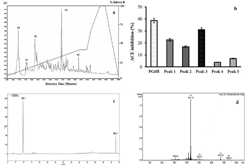
dissociation (CID). The precursor ions were selected in the (PTH) showed very low antihypertensive activity (PACE-I from crocodile blood hydrolysates tyrosine, lysine, leucine, isoleucine, and valine residues in the hydrophobic amino acids with a total percentage of 44.4% and C-terminal of peptides (Guang & Phillips, 2009; Ngo et al., 2014; 55.5% for LL-9 (Leu-Pro-Glu-Ser-Val-His-Leu-Asp-Lys) and VL-9 Iwaniak et al., 2014). In addition, the hydrophobic ratio of RC1 (Val-Leu-Ser-Thr-Ser-Phe-Pro-Pro-Lys) peptides, respectively. and RC2 peptides showed strong hydrophobic ratios of 60 and This result suggests that the peptides effectively interacted with 83%, respectively. This result is consistent with a previous result the ACE active site, and maybe explain the stronger inhibition that the amino acid composition of purified peptides consist of activity of VL-9 (Mirzaei et al., 2017). In this study, the ratio of Figure 1. Elution profile of the Protease G6 hydrolysate separated on a CM-Sepharose cation exchange column (a). The ACE inhibitory activity of peptide fractions (b). Figure 2. Preparative RP-HPLC chromatograms of unbound fractions isolated from CM- sepharose Fast Flow (a). The ACE inhibitory activity of five fractions (b). Rechromatogram of peak 3 from Preparative RP-HPLC (c). Identification of the molecular mass and amino acid sequence of the peptide from RC1 using LC-MS/MS (d). 4 4/6 Food Sci. Technol, Campinas, Ahead of Print, 2019
Ngo-Son; Katekaew

hydrophobic amino acids is higher than in the previous studies               Collins, R., & MacMahon, S. (1994). Blood pressure, antihypertensive
indicating two peptides can effectively inhibit the ACE activity.              drug treatment and the risks of stroke and of coronary heart disease.
                                                                               British Medical Bulletin, 50(2), 272-298. http://dx.doi.org/10.1093/
    Furthermore, our results showed that the two peptides did                  oxfordjournals.bmb.a072892. PMid:8205459.
not match the sequence database. These results might show that               Deng, H., Zheng, J., Zhang, F., Wang, Y., & Kan, J. (2014). Isolation of
there are two novel ACE-inhibitory peptides from crocodile                     angiotensin I-converting enzyme inhibitor from pepsin hydrolysate
blood hydrolysates. However, further studies are required to                   of porcine hemoglobin. European Food Research and Technology,
investigate their ACE-inhibitory activity (in vivo) and their                  239(6), 933-940. http://dx.doi.org/10.1007/s00217-014-2290-0.
synthetic analogues in order to determine the structure needed               Ghassem, M., Babji, A. S., Said, M., Mahmoodani, F., & Arihara, K.
for mechanical inhibition of ACE activity.                                     (2014). Angiotensin I–converting enzyme inhibitory peptides from
                                                                               snakehead fish sarcoplasmic protein hydrolysate. Journal of Food
                                                                               Biochemistry, 38(2), 140-149. http://dx.doi.org/10.1111/jfbc.12031.
4 Conclusion
                                                                             Guang, C., & Phillips, R. D. (2009). Plant food-derived angiotensin
    ACE-inhibitory protein hydrolysates were successfully produced             I converting enzyme inhibitory peptides. Journal of Agricultural
from crocodile blood by Protease G6 (PG6H). The ACE‑inhibitor                  and Food Chemistry, 57(12), 5113-5120. http://dx.doi.org/10.1021/
peptides were efficiently purified from crude PG6H through                     jf900494d. PMid:19449887.
multi-step chromatographic purification comprising of ion                    Hyun, C. K., & Shin, H. K. (2000). Utilization of bovine blood plasma
exchange chromatography followed by two‑step RP-HPLC, and                      proteins for the production of angiotensin I converting enzyme
their sequences were analyzed by LC–MS/MS. This work could                     inhibitory peptides. Process Biochemistry, 36(1-2), 65-71. http://
applied to produce antihypertensive peptides from low-cost                     dx.doi.org/10.1016/S0032-9592(00)00176-X.
blood proteins and in an environmental sustainable manner.                   Iwaniak, A., Minkiewicz, P., & Darewicz, M. (2014). Food-originating
Furthermore, PG6H could potentially be used to formulate                        ACE inhibitors, including antihypertensive peptides, as preventive
therapeutic food products for the prevention or treatment of                    food components in blood pressure reduction. Comprehensive
hypertension.                                                                   Reviews in Food Science and Food Safety, 13(2), 114-134. http://
                                                                                dx.doi.org/10.1111/1541-4337.12051.
                                                                             Jandaruang, J., Siritapetawee, J., Thumanu, K., Songsiriritthigul, C.,
Acknowledgements                                                                Krittanai, C., Daduang, S., Dhiravisit, A., & Thammasirirak, S.
    This study was funded by the Higher Education Research                      (2012). The effects of temperature and pH on secondary structure
                                                                                and antioxidant activity of Crocodylus siamensis hemoglobin. The
Promotion and National Research University Project of Thailand,
                                                                                Protein Journal, 31(1), 43-50. http://dx.doi.org/10.1007/s10930-
Office of the Higher Education Commission, through the Food                     011-9372-7. PMid:22101803.
and Functional Food Research Cluster of Khon Kaen University
                                                                             Kommanee, J., Phosri, S., Daduang, S., Temsiripong, Y., et al (2014).
(Grant No. F-2553-M-24). Purification and characterization of                  Comparisons of anti-inflammatory activity of crocodile (Crocodylus
ACE inhibitory peptides from crocodile blood using LC-MS/MS                    siamensis) blood extract. Warasan Khana Witthayasat Maha
was provided by the Research Instrument Center (RIC-KKU),                      Witthayalai Chiang Mai, 41, 627-634.
Khon Kaen University, Thailand.                                              Ktari, N., Fakhfakh, N., Balti, R., Ben Khaled, H., Nasri, M., & Bougatef,
                                                                                A. (2013). Effect of degree of hydrolysis and protease type on the
References                                                                      antioxidant activity of protein hydrolysates from Cuttlefish (Sepia
                                                                                officinalis) by-products. Journal of Aquatic Food Product Technology,
Agostoni, A., & Cicardi, M. (2001). Drug-induced angioedema                     22(5), 436-448. http://dx.doi.org/10.1080/10498850.2012.658961.
  without urticaria. Drug Safety, 24(8), 599-606. http://dx.doi.
                                                                             Lahogue, V., Réhel, K., Taupin, L., Haras, D., & Allaume, P. (2010). A
  org/10.2165/00002018-200124080-00004. PMid:11480492.
                                                                               HPLC-UV method for the determination of angiotensin I-converting
Bah, C. S. F., Bekhit, A. E. D. A., Carne, A., & McConnell, M. A. (2013).      enzyme (ACE) inhibitory activity. Food Chemistry, 118(3), 870-875.
  Slaughterhouse blood: an emerging source of bioactive compounds.             http://dx.doi.org/10.1016/j.foodchem.2009.05.080.
  Comprehensive Reviews in Food Science and Food Safety, 12(3), 314-         Lalor, F., & Wall, P. G. (2011). Health claims regulations. British Food
  331. http://dx.doi.org/10.1111/1541-4337.12013.                               Journal, 113(2), 298-313. http://dx.doi.org/10.1108/00070701111105358.
Barzideh, Z., Latiff, A. A., Gan, C. Y., Abedin, M. Z., & Alias, A.          Li, Y., Sadiq, F. A., Liu, T., Chen, J., & He, G. (2015). Purification
   K. (2014). ACE inhibitory and antioxidant activities of collagen              and identification of novel peptides with inhibitory effect against
   hydrolysates from the Ribbon Jellyfish (Chrysaora sp.). Food Technology       angiotensin I-converting enzyme and optimization of process
   and Biotechnology, 52(4), 495-504. http://dx.doi.org/10.17113/                conditions in milk fermented with the yeast Kluyveromyces marxianus.
   ftb.52.04.14.3641. PMid:27904323.                                             Journal of Functional Foods, 16, 278-288. http://dx.doi.org/10.1016/j.
Chen, J., Wang, Y., Ye, R., Wu, Y., & Xia, W. (2013). Comparison of              jff.2015.04.043.
  analytical methods to assay inhibitors of angiotensin I-converting         Maraming, P., Maijaroen, S., Klaynongsruang, S., Boonsiri, P., Daduang,
  enzyme. Food Chemistry, 141(4), 3329-3334. http://dx.doi.org/10.1016/j.      S., Chung, J. G., & Daduang, J. (2018). Antitumor ability of KT2
  foodchem.2013.06.048. PMid:23993489.                                         peptide derived from leukocyte peptide of Crocodile against human
Cheung, I. W. Y., & Li-Chan, E. C. Y. (2010). Angiotensin-I-converting         HCT116 colon cancer xenografts. In Vivo, 32(5), 1137-1144. http://
  enzyme inhibitory activity and bitterness of enzymatically-produced          dx.doi.org/10.21873/invivo.11356. PMid:30150436.
  hydrolysates of shrimp (Pandalopsis dispar) processing byproducts          Mirzaei, M., Mirdamadi, S., Ehsani, M. R., & Aminlari, M. (2017).
  investigated by Taguchi design. Food Chemistry, 122(4), 1003-1012.           Production of antioxidant and ACE-inhibitory peptides from
  http://dx.doi.org/10.1016/j.foodchem.2010.03.057.                            Kluyveromyces marxianus protein hydrolysates: Purification and

Food Sci. Technol, Campinas,      Ahead of Print, 2019                                                                                           5/6 5
ACE-I from crocodile blood hydrolysates
molecular docking. Journal of Food and Drug Analysis. http://dx.doi. – a short report. Polish Journal of Food and Nutrition Sciences, 57,
org/10.1016/j.jfda.2017.07.008. PMid:29567240. 447-450.
Ngo, D. H., Ryu, B., & Kim, S. K. (2014). Active peptides from skate Ryan, J. T., Ross, R. P., Bolton, D., Fitzgerald, G. F., & Stanton, C. (2011).
(Okamejei kenojei) skin gelatin diminish angiotensin-I converting Bioactive peptides from muscle sources: meat and fish. Nutrients,
enzyme activity and intracellular free radical-mediated oxidation. 3(9), 765-791. http://dx.doi.org/10.3390/nu3090765. PMid:22254123.
Food Chemistry, 143, 246-255. http://dx.doi.org/10.1016/j.
Sivaperumal, P., Kamala, K., Natarajan, E., & Dilipan, E. (2013).
foodchem.2013.07.067. PMid:24054237.
Antimicrobial peptide from crab haemolymph of Ocypoda macrocera
Nielsen, P. M., Petersen, D., & Dambmann, C. (2001). Improved method (Maline Edwards 1852) with reference to antioxidant: A case study.
for determining food protein degree of hydrolysis. Journal of Food International Journal of Pharmacy and Pharmaceutical Sciences, 5,
Science, 66(5), 642-646. http://dx.doi.org/10.1111/j.1365-2621.2001. 719-727.
tb04614.x.
Srihongthong, S., Pakdeesuwan, A., Daduang, S., Araki, T., Dhiravisit,
Pakdeesuwan, A., Araki, T., Daduang, S., Payoungkiattikun, W.,
A., & Thammasirirak, S. (2012). Complete amino acid sequence
Jangpromma, N., & Klaynongsruang, S. (2017). In vivo wound
of globin chains and biological activity of fragmented crocodile
healing activity of Crocodile (Crocodylus siamensis) hemoglobin
and evaluation of antibacterial and antioxidant properties of hemoglobin (Crocodylus siamensis). The Protein Journal, 31(6), 466-
hemoglobin and hemoglobin hydrolysate. Journal of Microbiology 476. http://dx.doi.org/10.1007/s10930-012-9424-7. PMid:22648692.
and Biotechnology, 27(1), 26-35. http://dx.doi.org/10.4014/ Wu, J., Aluko, R. E., & Muir, A. D. (2009). Production of angiotensin
jmb.1603.03046. PMid:27713216. I-converting enzyme inhibitory peptides from defatted canola
Patathananone, S., Thammasirirak, S., Daduang, J., Chung, J. G., meal. Bioresource Technology, 100(21), 5283-5287. http://dx.doi.
Temsiripong, Y., & Daduang, S. (2015). Bioactive compounds from org/10.1016/j.biortech.2009.03.090. PMid:19570676.
crocodile (Crocodylus siamensis) white blood cells induced apoptotic Xie, L., Tong, W., Yu, D., Xu, J., Li, J., & Gao, C. (2012). Bovine serum
cell death in hela cells. Environmental Toxicology, 31(8), 986-997. albumin nanoparticles modified with multilayers and aptamers for
http://dx.doi.org/10.1002/tox.22108. PMid:25691005. pH-responsive and targeted anti-cancer drug delivery. Journal of
Pegg, R. B., Rybarczyk, A., & Amarowicz, R. (2007). Determination of Materials Chemistry, 22(13), 6053-6060. http://dx.doi.org/10.1039/
hippuric acid by RP-HPLC using two different analytical columns c2jm16831f.
6 6/6 Food Sci. Technol, Campinas, Ahead of Print, 2019You can also read