Kinematics of healthy American Pit Bull Terrier dogs
←
→
Page content transcription
If your browser does not render page correctly, please read the page content below
Original Paper                                                      Veterinarni Medicina, 66, 2021 (01): 8–16

                                                                   https://doi.org/10.17221/178/2019-VETMED

Kinematics of healthy American Pit Bull
Terrier dogs
Michelle Campano de Souza1, Jessica Ragazzi Calesso2*,
Bruna Cenci1, Mauro Jose Lahm Cardoso1, Felipe Arruda Moura3,
Rafael Fagnani4
1
  Department of Veterinary Medicine, State University of Londrina, Londrina, PR, Brazil
2
  Department of Clinic and Surgery Veterinary, Federal University of Minas Gerais, Belo
  Horizonte, MG, Brazil
3
  Sport Sciences Department, Laboratory of Applied Biomechanics, State University of Londrina,
  Londrina, PR, Brazil
4
  Department of Veterinary Medicine, University of Northern Paraná, Londrina, PR, Brazil
*Corresponding author: jessicacalesso@gmail.com

Citation: De Souza MC, Calesso JR, Cenci B, Cardoso MJL, Moura FA, Fagnani R (2021): Kinematics of healthy American
Pit Bull Terrier dogs. Vet Med-Czech 66, 8–16.

Abstract: A visual clinical gait analysis is useful, however, it may overlook small, but important, details about
the movement, as well as differences between the normal and pathological locomotion. The branch of mechanics
that describes the spatial and temporal components of motion is called kinematics, providing quantitative data
regarding linear and angular motion. The objective of this study was to establish kinematic gait data of healthy
American Pit Bull Terriers and to contribute to the understanding of the locomotion. We evaluated the articular
and pelvic angles, and the spatiotemporal variables for walking and trotting from eleven dogs with no previous
history of joint and musculoskeletal diseases. Twenty reflective markers were positioned at the anatomical points
of interest. The animals walked and trotted in a linear space, led by the same researcher. The kinematic data were
collected through optoelectronic cameras and analysed by motion analysis software. The movements analysed
during the gait phases were the flexion, extension, range of motion (ROM), angle at the moment of the support
phase, stride length and velocity. Comparing the angles between walking and trotting, there were more expressive
differences for the pelvic limb joints. There was no difference between the left and right sides at all of the joint
angles of the pelvic limbs during walking and trotting. Therefore, the movement of the pelvic limb is symmetrical
in both trotting and walking. Our results present reference values for healthy American Pit Bull Terriers, having
clinical relevance for studies of dogs with musculoskeletal diseases.

Keywords: canine gait; joint angles; kinetics; musculoskeletal system

  A kinematic evaluation is the study that describes       is the support of two limbs, with diagonal pairs
the spatial and temporal components of motion,             (DeCamp 1997). A clinical gait analysis (visual ob-
and involves the position, velocity, and accelera-         servation) is useful, but not very precise, whereas
tion of a body, regardless of the forces that cause        a kinematical quantitative analysis covers a more
movement (Hamill and Knutzen 2008).                        efficient evaluation, calculating distance variables
  The cycle of movement when walking includes              and angular variables (Hamill and Knutzen 2008;
the support of two or three limbs, and at trot, there      Gustas et al. 2013).

8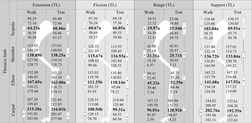
Original Paper                                                  Veterinarni Medicina, 66, 2021 (01): 8–16

                                                               https://doi.org/10.17221/178/2019-VETMED

  The motion of a kinematic analysis may reveal de-     Data collect
tails about the movement, as well as the differences
between a normal and pathological locomotion.             The study was conducted at the Laboratory of
  The American Pit Bull Terrier is a large breed        Applied Biomechanics in the Department of Sports
used as a companion dog as well as a working dog.       Sciences at the State University of Londrina. Each
This breed, like other large breeds, may present        dog had twenty spherical reflective markers (1.5 cm
with musculoskeletal changes such as hip dyspla-        in diameter), positioned on the skin (3M adhesive
sia, elbow dysplasia, shoulder subluxation, biceps      tape) over the spine of the scapula (acromion),
tenosynovitis, ruptured cruciate ligaments, among       major tubercle of the humerus, lateral epicon-
other disorders such as cauda equina syndrome and       dyle of the humerus, fifth metacarpal, iliac crest,
obesity, which also promote locomotion alterations      major femoral trochanter, femoral condyle, lat-
(Bach et al. 2015).                                     eral malleolus and fifth metatarsal head, on both
  Pain, muscle weakness, and an abnormal range          sides (Figure 1A). These markers were analysed
of motion (ROM) are some of the important factors       in two moments, walking and trotting, always
that may affect the gait. In dogs, the effect of pain   guided by the same individual, in a seven-metre
on the gait causes a decrease in the support phase      linear path. The acquisition of the three-dimen-
and less contact with the floor. Weakness affects the   sional kinematics was performed by six cameras
movement, increases or decreases the joint when         (Optitrack ®; Leyard, Oregon, USA), with a sam-
the muscle usually contracts. Compensation usual-       pling frequency of 240 Hz.
ly occurs in other joints (DeCamp 1997). Thus, the        Ten attempts were recorded for each dog, and
biomechanics can provide important information          the cycles of motion were selected in which all the
in order to quantify normal and abnormal move-          markers could be identified. The system cre-
ments, including the function of the limbs and the      ated three-dimensional coordinates, in which the
body as a whole (Angle et al. 2012).                    X axis represented the craniocaudal displacement,
  Therefore, this study aimed at establishing ki-       the Y axis represented the medium-lateral and the
nematic data on the gait of clinically healthy dogs     Z axis represented the vertical one.
of the American Pit Bull Terrier breed and to con-        The markers were reconstructed using Motive
tribute to the understanding of the locomotion.         Body v1.8.0 software (Leyard, Oregon, USA). The
                                                        three-dimensional coordinates of each of the mark-
                                                        ers were smoothed by a third-order Butterworth
MATERIAL AND METHODS                                    low-pass digital filter with a 6 Hz cut-off frequency,
                                                        defined after the spectral analysis. Matlab® software
Animals                                                 was used to implement the kinematics equations.
                                                        The absolute articular angles of flexion and exten-
  We studied eleven American Pit Bull Terrier           sion were evaluated for the joints of the scapula
dogs, selected from tutors and the Hoffmann Pit         and the iliac crest.
kennel, located in Londrina, Paraná, Brazil.              For the joints of the shoulder, elbow, carpal, hip
  Four of the animals were females and seven were       joint, knee and tarsus, the relative angles were
males, aged between 2 and 6 years, and without          quantified (Figure 1B).
any previous history of joint or musculoskeletal          The variables identified were the maximum and
diseases.                                               minimum angles of each joint, the range of motion
  All the animals were clinically evaluated (gen-       (defined as the subtraction between the maximum
eral physical and orthopaedic examination), and         and minimum values) and the angle at the first mo-
a body condition score (BCS) between 4 and 6 was        ment of the support phase. The spatiotemporal
established by direct inspection and palpation, de-     variables quantified were:
scribed by Laflamme (1997). Dogs with changes,            a) the stride length (defined as the distance trav-
such as lameness and joint pain, would have been        elled by the dog during the movement cycle, con-
excluded from the study.                                sidering the three-dimensional average coordinates
  This study was approved by the Ethics Committee       between the points of the scapula and crest), nor-
of the State University of Londrina (Protocol No.       malised by the dog height and
6321.2016.68).                                            b) stride speed.

9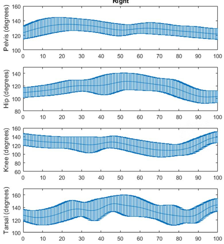
Original Paper                                                           Veterinarni Medicina, 66, 2021 (01): 8–16

                                                                        https://doi.org/10.17221/178/2019-VETMED

     Figure 1

     (A)                               (B)

Figure 1. Reflective markers at the anatomical points of interest
Source: Personal archive (A); Done et al. (2010) adapted (B)
Joint angles analysed: 1: Scapula; 2: Shoulder; 3: Elbow; 4: Carpal; 5: Metacarpal; 6: Iliac crest; 7: Hip (coxal); 8: Knee;
9: Tarsus; 10: Metatarsus

Statistical analysis                                             Comparing the angles of the thoracic limb, we
                                                               found that the scapula ROM was greater P < 0.05
   The analysed variables did not present normality            during the trot when compared to the walk. The
by the Lilliefors test, P < 0.05. Thus, the data were          extension angles and the shoulder support phase
evaluated by non-parametric tests and the descrip-             were greater (P < 0.05) during the walk compared
tive statistics were presented in medians and quar-            to the trot. However, the shoulder ROM was greater
tiles. On the other hand, the comparison between               (P < 0.05) in the trot in relation to the walk. The
the angles observed in the right limbs and in the              elbow flexion angle was bigger (P < 0.05) during
left limbs was performed by the Mann-Whitney                   the walk when compared to the trot. The exten-
U test (P < 0.05) at two moments: during walking               sion angles and of the carpal ROM were greater
and during a trot. All the analyses were performed             (P < 0.05) during the trot when compared to the
in the Statistica v13.0 program (Statsoft, Brazil).            walk. In turn, the flexion angle of the carpus was
                                                               higher (P < 0.05) during the walk (Figure 2).
                                                                 When comparing the walk and trot angles of the
RESULTS                                                        pelvic limbs, the differences in the angles between
                                                               the walk and the trot were more expressive, with
  In the evaluation of the spatiotemporal values, the          alterations in most of the analysed anatomical re-
average length of the stride was 1.41 ± 0.12 m/m               gions, except for the extension and support angles
with an average speed of 1.17 ± 0.17 m/s during                of the knees.
walking. The length for the stride during trot-                  For the iliac crest joint, the extension, flexion
ting was 1.98 ± 0.22 m/m with a mean velocity                  and support angles were higher (P < 0.05) during
of 2.04 ± 0.33 m/s. There was less variability in              the trot. However, the ROM angle was higher dur-
the length and mean velocity of the gait during the            ing the walk. For the hip (coxal) joint, the angles
walk, indicating a more homogenous pattern when                of extension, ROM and support were higher during
compared to the length and average speed of the                the walk. However, the flexion angle was higher
gait during the trot.                                          during the trot. The knee flexion was greater dur-

10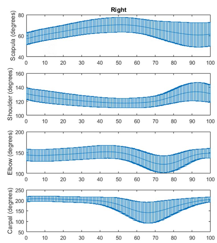
Original Paper Veterinarni Medicina, 66, 2021 (01): 8–16
https://doi.org/10.17221/178/2019-VETMED
Figure 2
Figure 2. Medians, quartiles and minimum and maximum values of the extension, flexion, ROM and support angles
of the thoracic limb (TL) during walking and during trotting. The medians with the same letters did not differ in the
Wilcoxon test, at P < 0.05
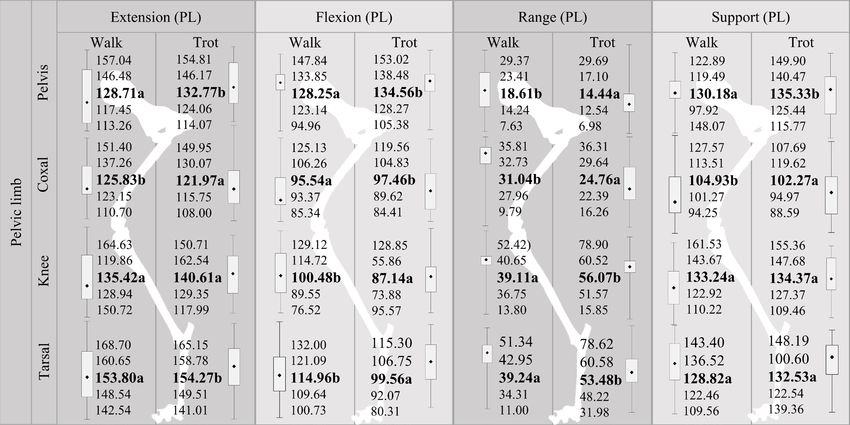
ing the walk, while the ROM was greater in the when compared to the right one (Table 1). There
trot. For the tarsus, the extension and ROM angles was no difference between the left and right sides
were higher in the trot, while the flexion angle was at all of the angles in all the regions of the pelvic
higher during the walk (Figure 3). limbs.
The angles of the right and left thoracic limbs When comparing the angles of the right and left
were compared during the walk. In the scapula, thoracic limbs during trotting, in the support angle,
only the ROM was greater (P < 0.05) in the left limb the angle of the right shoulder was higher when
Figure 3
Figure 3. Medians, quartiles and minimum and maximum values of the extension, flexion, ROM and support angles
of the pelvic limb (PL) during walking and during trotting. The medians with the same letters did not differ in the
Wilcoxon test, at P < 0.05
11
Original Paper                                                               Veterinarni Medicina, 66, 2021 (01): 8–16

                                                                         https://doi.org/10.17221/178/2019-VETMED

Table 1. Medians (med), quartiles (Q25–Q75) and minimum (min) and maximum (max) angles of extension, flexion,
amplitude and support of the right and left thoracic joints during walking

                                       Scapula                Shoulder                   Elbow               Carpal
                                   R             L        R              L           R           L       R            L
                        med       65.03a     60.31a    143.95a      138.45a       152.03a    153.45a   205.77a    213.28a
                        min       50.50      54.22     120.73       121.02        136.21     146.05    197.01     191.01
Angle of extension      max       88.29      81.21     153.55       144.99        175.44     169.91    236.30     243.85
                        Q25       61.83      56.05     128.90       125.91        143.71     149.52    200.40     199.83
                        Q75       70.35      72.16     147.16       140.65        169.89     165.74    210.75     217.55
                        med       60.67a     54.05a    116.74a      112.03a       116.29a    110.82a   106.98a    114.64a
                        min       47.60      39.27     107.39        99.86         91.03      93.96     91.80      76.70
Angle of flexion        max       87.56      81.04     125.23       126.53        151.86     152.03    228.51     197.17
                        Q25       52.39      50.46     113.45       104.51        100.68     105.11     95.97     104.33
                        Q75       75.49      76.24     119.12       122.04        123.31     116.50    134.46     124.91
                        med       17.33a     21.06b     23.77a       20.21a        43.33a     46.44a   100.41a     96.01a
                        min       12.51      14.86       6.57        17.33          3.24       7.37      2.54       9.63
ROM                     max       30.51      27.48      36.88        28.13         59.41      56.92    114.78     167.15
                        Q25       15.62      19.39      18.24        18.46         38.80      41.49     77.10      92.42
                        Q75       20.46      23.38      28.77        24.29         52.68      49.96    107.69      99.82
                        med       56.79a     56.97a    134.40a      129.06a       143.98a    144.52a   203.31a    202.48a
                        min       51.24      41.54     119.82       116.81        124.27     125.51    193.16     182.11
Support phase           max       68.34      78.17     144.02       144.99        164.05     165.23    234.82     215.14
                        Q25       54.95      50.68     122.54       122.18        130.58     136.42    197.57     193.73
                        Q75       61.54      60.23     136.72       135.30        157.77     158.88    208.54     209.24
L = left; R = right; ROM = range of motion; a,bThe results showed a significant difference

Table 2. Medians (med), quartiles (Q25–Q75) and minimum (min) and maximum (max) angles of extension, flexion,
amplitude and support of the right and left thoracic joints during trotting

                                       Scapula                Shoulder                   Elbow               Carpal
                                   R             L        R              L           R           L       R            L
                        med       65.89a     59.89a    144.05a      135.53a       149.87a    155.19a   215.70a    224.59a
                        min       47.37      50.04     129.05       118.73        132.23     126.31    205.94     203.47
Angle of extension      max       85.40      81.60     157.01       151.95        174.51     165.85    283.74     277.11
                        Q25       54.67      54.25     134.27       128.59        141.71     145.37    211.32     218.15
                        Q75       70.83      77.32     148.30       141.78        171.92     162.28    247.24     235.74
                        med       61.11a     63.85a    113.19a      113.52a       117.38a    109.73a    91.74a    102.88a
                        min       47.04      39.86     103.84       103.91         78.21      89.20     66.60      71.17
Angle of flexion        max       84.18      82.66     126.51       119.42        135.46     123.94    193.46     219.94
                        Q25       49.50      49.57     109.44       109.45         86.24     100.56     83.69      84.77
                        Q75       77.91      76.67     119.03       116.68        124.71     113.50    130.95     116.62
                        med       24.09a     23.23a     27.10a       21.69a        53.46a     51.48a   122.34a    120.91a
                        min       16.85      17.75      18.89         5.21         10.85       7.24     28.81      18.23
ROM                     max       29.75      29.20      45.91        37.96         65.73      59.80    203.74     205.94
                        Q25       20.28      19.83      24.88        18.58         41.68      39.25     87.78     100.37
                        Q75       26.12      26.49      29.27        34.93         56.77      53.98    139.56     133.88
                        med       51.77a     52.32a    137.63b      133.24a       149.87a    147.15a   187.25a    204.29b
                        min       47.04      43.47     124.39       118.73        119.10     120.20    179.02     179.32
Support phase           max       63.67      63.85     157.01       150.73        169.32     156.09    237.61     219.94
                        Q25       48.70      50.08     132.98       128.59        137.04     137.47    184.49     198.13
                        Q75       58.65      57.67     143.49       135.15        164.25     153.31    191.63     206.49

L = left; R = right; ROM = range of motion; a,bThe results showed a significant difference

12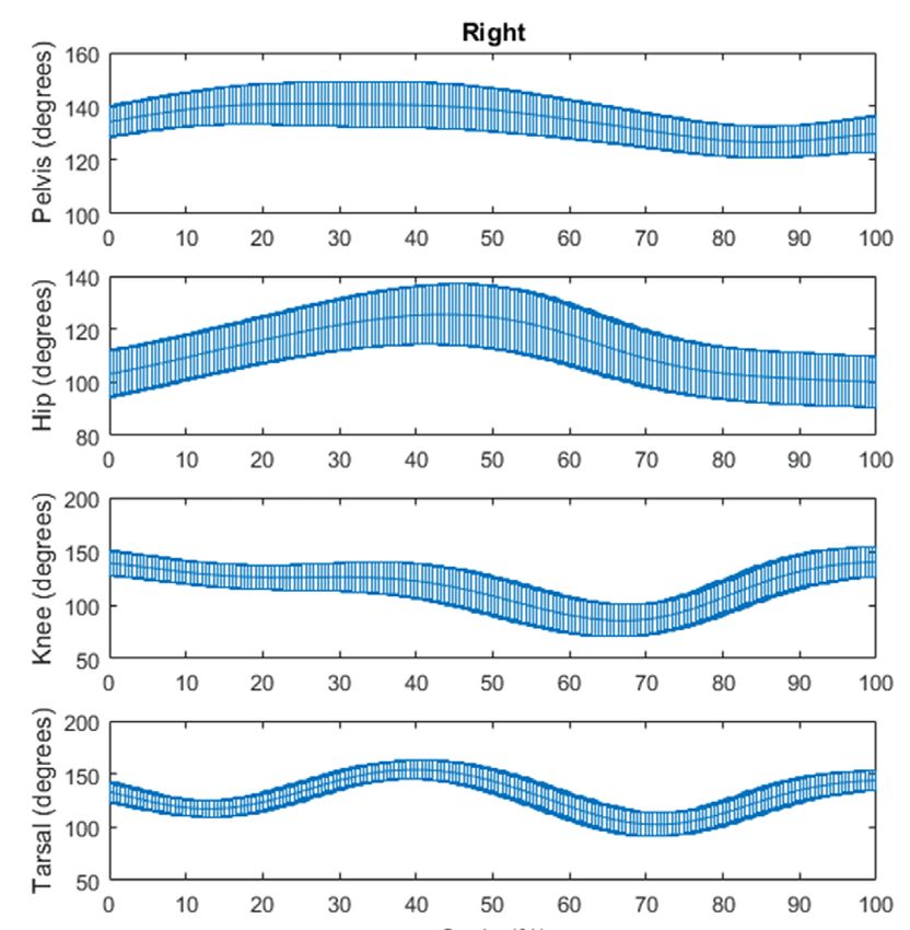
Original Paper                                                     Veterinarni Medicina, 66, 2021 (01): 8–16

                                                                   https://doi.org/10.17221/178/2019-VETMED

compared to the left, while the angle of support             In the joint movement of the limbs of the dogs,
of the left carpus was higher (Table 2). There was         the maximum angular values occurred during the
no difference between the sides at all of the angles       extension and the minimum angular values occurred
in all the regions of the pelvic limbs.                    during the maximum flexion (Figures 4, 5, 6 and 7).

                        Right                                                         Left
Figure 4

                      Cycle (%)                                                     Cycle (%)
Figure 4. Joint movement of the thoracic limbs of the dogs studied during walking

                        Right                                                         Left
Figure 5

                      Cycle (%)                                                     Cycle (%)
Figure 5. Joint movement of the pelvic limbs of the dogs studied during walking

13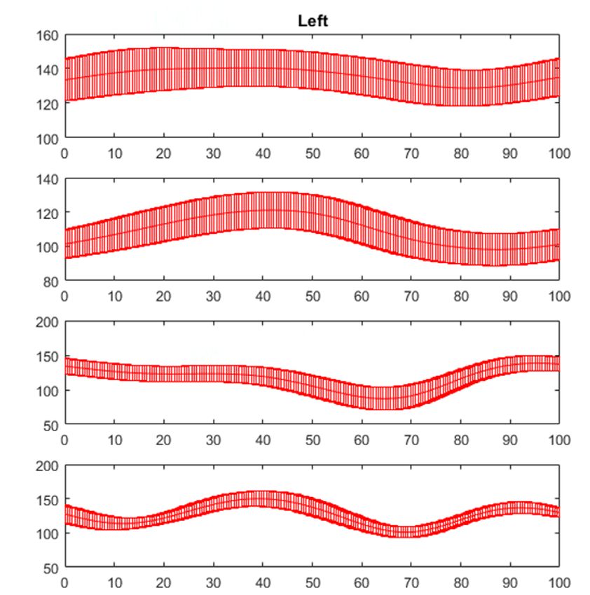
Original Paper                                                      Veterinarni Medicina, 66, 2021 (01): 8–16

                                                                    https://doi.org/10.17221/178/2019-VETMED

                        Right                                                          Left
Figure 6

                      Cycle (%)                                                      Cycle (%)
Figure 6. Joint movement of the thoracic limbs of the dogs studied during trotting
                        Right                                                          Left
Figure 7

                       Cycle (%)                                                     Cycle (%)
Figure 7. Joint movement of the pelvic limbs of the dogs studied during trotting

DISCUSSION                                                  had no previous history of joint or musculoskeletal
                                                            diseases, were of both sexes, being the criteria similar
  The objective of this study was to establish the kine-    to those described by Angle et al. (2012).
matic gait data of healthy American Pit Bull Terriers         The American Pit Bull Terrier was selected be-
and to contribute to the understanding of the loco-         cause it is large in size, which is common in pre-
motion. The selected dogs were clinically healthy and       senting with musculoskeletal changes that result

14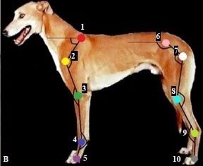
Original Paper                                                       Veterinarni Medicina, 66, 2021 (01): 8–16

                                                                    https://doi.org/10.17221/178/2019-VETMED

in locomotion alterations (Bach et al. 2015). In ad-         of motion for the carpal joint (323 degrees). However,
dition, the criteria for being shorthaired dogs (fa-         the difference in the ROM values between the stud-
vouring marker fixation), the ease of manipulation           ies is noteworthy. The authors did not standardise
of the animals, being docile to humans and the               a specific breed, which may justify the differences
lack of kinematic studies related to the breed were          between the studies.
also taken into account.                                       Kopec et al. (2018) compared the kinematics of
  A kinematic gait analysis in healthy dogs has              healthy dogs, of different breeds, with an average
been addressed by some authors (Jarvis et al. 2013;          body weight of 22.3 ± 1.0 kg, during trotting. They
Silva et al. 2014; Kopec et al. 2018). However, none         found that the elbow joint reached a maximum ex-
of them addressed the gait in healthy American Pit           tension at the end of the support phase, that is, im-
Bull Terriers. Agostinho et al. (2011) suggest that          mediately before toe-off, with the mean values of the
each breed must have a specific database.                    ROM and maximum extension for the elbow joint
  Most of the kinematic studies used a gait tread-           being 65.81 ± 7.48 degrees and 132.03 ± 11.49 de-
mill with standard values between 1.80 and 2.22 m/s          grees, respectively. An increase in the trotting speed
(Poy et al. 2000), which makes the spatio-temporal           can increase the ROM for the joints of the thoracic
values found in our study unprecedented for the              limbs (Kopec et al. 2018). The dogs in the present
American Pit Bull Terrier breed, without the use             study trotted at their natural pace, at a speed that
of a treadmill. A recent study (Jarvis et al. 2013)          seemed comfortable. Rodrigues (2011) evaluated the
evaluated healthy dogs of different large breeds,            gait of six Golden Retriever dogs, affected by mus-
observing a mean length of 1.95 ± 0.28 m/m, and              cular dystrophy, during walking. The mean flexion
a velocity of 2.4 m/s in the trot. These values are          of the articular angle of the carpus was 122.60 ±
similar to the ones presented in our study.                  11.45 degrees, and was 149.90 ± 19.35 degrees in the
  Comparing the joint angles of the pelvic limbs,            extension. The mean flexion for the hip joint was
there was no difference between the right and left           121.40 ± 11.86 degrees and the mean increased
sides in all the regions, both for walking and trot-         to 150.50 ± 20.17 degrees in the extension. For the
ting, corroborating the results of other studies com-        knee joint, the mean flexion was 121.20 ± 12.46 de-
paring the extension and flexion angles (Gillette and        grees and was 146 ± 18.62 degrees for the extension.
Zebas 1999). Therefore, the movement of the pelvic           Compared to our study, a remarkable difference was
limb was symmetrical, a relevant fact for kinematic          observed between the values of carpus (extension),
studies of dogs with musculoskeletal diseases.               hip joint (flexion and extension), and knee (flex-
  Comparative studies between Labrador Retrievers            ion) joints. This difference may be due to the inflic-
and Greyhound dogs, reported significant differenc-          tion (muscular dystrophy) and/or being of different
es in the hip biomechanics. The ROM of the pelvic            breeds. In view of the above, more kinematic studies
limbs and the stride length of the Greyhound were            are necessary in dogs, standardising the breeds and
greater than the values of the Labrador Retriever,           conditions that may alter the gait to clarify how the
which indicates that the body factors specific to each       conformation and structure can affect the function.
breed influence the animal’s movement (Hottinger               Nielsen et al. (2003) performed a gait analysis us-
et al. 1996; Bertram et al. 2000).                           ing a two-dimensional and three-dimensional sys-
  In our study, the mean values for the carpal joints        tem of thoracic limbs of healthy dogs and stated
were 191.4 ± 13.7 degrees (right side) and 201.7 ±           that there are notable differences between these
10.3 degrees (left side). For the iliac crest joints, they   analyses, such as the angular time-series, because
were 134.2 ± 5.6 degrees (right side) and 133.4 ±            they are totally different technologies. To the best
12.1 degrees (left side). The degrees we reported are        of our knowledge, our study is the first that evalu-
different from a previous study (Jarvis et al. 2013),        ated the three-dimensional movement of American
which reported 211.6 ± 10 degrees (carpal joint) and         Pit Bull Terriers.
112.1 ± 10.3 degrees (crest joint). The greatest value         Various research studies have suggested that the
in the range of motion was found for the carpus joint,       variability in the conformation and size can affect
both at walk (right: 79.6 degrees, left: 90.9 degrees)       the dogs’ kinematics. It is believed that a study
and at a trot (right: 120.2 degrees, left: 110.2 de-         including multiple breeds of various sizes leads
grees), corroborating the information from with              to a wide variation in the results and limits the abil-
Jarvis et al. (2013), who also reported a greater range      ity to detect the expressive characteristics, since the

15
Original Paper Veterinarni Medicina, 66, 2021 (01): 8–16
https://doi.org/10.17221/178/2019-VETMED
pattern of locomotion can change between breeds Done SH, Goody PC, Evans SA, Stickland NC Atlas Col-
(Carr et al. 2013; Miqueleto et al. 2013; Kopec et al. orido de Anatomia Veterinária do Cão e do Gato [Color
2018). In the present study, the biometric data of the atlas of veterinary anatomy]. 2nd ed. Rio de Janeiro: Else-
dogs were not measured, but a single breed was stan- vier; 2010. 544 p. Portuguese.
dardised, in order to minimise any breed variability. Gillette RL, Zebas CJ. A two-dimensional analysis of limb
Therefore, we believe that this study can contrib- symmetry in the trot of Labrador retrievers. J Am Anim
ute to establish normality data for gait variables Hosp Assoc. 1999 Nov-Dec;35(6):515-20.
in healthy American Pit Bull Terriers and be use- Gustas P, Pettersson K, Honkavaara S, Lagersted AS, Bys-
ful for the rehabilitation of affected dogs. However, trom. Kinematic and temporospatial assessment of ha-
additional studies using other dogs with the same bituation of Labrador retrievers to treadmill trotting.
morphological characteristics and the same proto- Vet J.2013 Dec;198(Suppl1):114-9.
col would be important to determine the viability Hamill J, Knutzen KM. Bases biomecânicas do movimento
of the method (Miqueleto et al. 2013). humano [Biomechanical bases of human movement].
In conclusion it can be stated, that kinematic 2nd ed. Barueri, Brasil: Manole; 2008. p. 3-30. Portuguese.
studies of locomotion may be useful to characterise Hottiinger HA, DeCamp CEN, Olivier B, Hauptman JG,
the gait of healthy dogs and evaluate the progression Soutas-Little R. Noninvasive kinematics analysis of the
of clinical or surgical treatments. In this study, the walk in healthy largebreed dogs. Am J Vet Res. 1996 Mar;
movement of the pelvic limb is symmetrical in both 57(3):381-8.
the trotting and walking phases, which is relevant Jarvis SL, Worley DR, Hogy SM, Hill AE, Haussler K, Reiser
for the study of dogs with musculoskeletal diseases. RF. Kinematic and kinetic analysis of dogs during trotting
after amputation of a thoracic limb. Am J Vet Res. 2013
Sept;74(9):1155-63.
Conflict of interest Kopec NL, Williams JM, Tabor GF. Kinematic analysis of the
thoracic limb of healthy dogs during descending stair and
The authors declare no conflict of interest. ramp exercises. Am J Vet Res. 2018 Jan;79(1):33-41.
Laflamme DP. Development and validation of a body con-
dition score system for dogs: A clinical tool. Canine Pract.
REFERENCES 1997 Jun;22(3):10-5.
Miqueleto NSML, Rahal SC, Agostinho FS, Siqueira EGM,
Agostinho FS, Rahal SC, Miqueleto NS, Verdugo MR, Ina- Araujo FAP, El-Warrak AO. Kinematic analysis in healthy
massu LR, El-Warrak AO. Kinematic analysis of Labrador and hip-dysplastic German Shepherd dogs. Vet J. 2013
Retrievers and Rottweilers trotting on a treadmill. Vet Feb;195(2):210-5.
Comp Orthop Traumatol. 2011 Feb;24(3):185-91. Nielsen C, Stover SM, Schulz KS, Hubbard M, Hawkins.
Angle TC, Gillette RL, Weimar WH. Kinematic analysis Two-dimensional link segment model of the forelimb
of maximal movement initiation in Greyhounds. Aust of dogs at a walk. Am J Vet Res. 2013 May;64(5):609-17.
Vet J. 2012 Mar;90(3):60-8. Poy NSJ, DeCamp CE, Bennett RL, Hauptman JG. Addi-
Bach M, Villanova Junior JA, Tasqueti UI, Pimpao CT, Prado tional kinematic variables to describe differences in the
AMB, Michellotto Junior PV. Estudo retrospectivo de caes trot between clinically normal dogs and dogs with hip
portadores de ruptura do ligamento cruzado cranial: 32 ca- dysplasia. Am J Vet Res. 2000 Aug;61(8):974-8.
sos (2006 a 2012) [A retrospective study of dogs with cra- Rodrigues EAF. Validacao de modelo biomecanico de mar-
nial cruciate ligament rupture: 32 cases (2006–2012). Semin cha para uso em testes pre-clinico com celulas tronco
Cienc Agrar. 2015 May-Jun;36(3):1409-18. Portuguese. [Validation of a biomechanical gait model for use in pre-
Bertram JEA, Lee DV, Case HN, Todhunter RJ. Comparison clinical stem cell tests] [dissertation]. São Paulo, Brasil:
of the trotting gaits of Labrador retriever and Greyhound. Universidade de São Paulo; 2011. p. 1-96. Portuguese.
Am J Vet Res. 2000 Aug;61(7):832-8. Silva GCA, Cardoso MT, Gaiad TP, Brolio MP, Oliveira VC,
Carr JG, Millis DL, Weng HY. Exercises in canine physical Assis Neto A, Martins DS, Ambrosio CE. Kinematic gait
rehabilitation: Range of motion of the forelimb during stair analyses in healthy Golden Retrievers. Pesq Vet Bras.
and ramp ascent. J Small Anim Pract. 2013;54(8):409-13. 2014 Dec;34(12):1265-70.
DeCamp CE. Kinetic and kinematic gait analysis and the
assessment of lameness in the dog. Vet Clin North Am Received: December 16, 2019
Small Anim Pract.1997 Jul;27(4):825-41. Accepted: October 20, 2020
16You can also read