The Possible Relation between Pityriasis Alba and Intestinal Parasitic Infestation Among Children in Tikrit City, A Case Control Study
←
→
Page content transcription
If your browser does not render page correctly, please read the page content below
864    Indian Journal of Forensic Medicine & Toxicology, January-March 2021, Vol. 15, No. 1

   The Possible Relation between Pityriasis Alba and Intestinal
   Parasitic Infestation Among Children in Tikrit City, A Case
                          Control Study

                                     Asaf Kamal Asaad Taha1, Mazin Hamid Ayyash2
       1
           Scholar Researcher, M.B.Ch.B, Salahuldin General Hospital, Iraq, 2FICMS, Higher Diploma in Laser,
                  Assistant Professor in Dermatoveneriology, Tikrit University College of Medicine, Iraq

                                                             Abstract
      Background: There is a solid belief stated that the appearance of hypopigmented patches on children
      face is a sign of their helminthic or parasitic intestinal infestation despite the constant health education
      about the absence of such relation. Pityriasis Alba (PA) is common benign skin lesion characterized by
      fine scaly hypopigmented patches (HP) or macules (1) mostly in the face and upper body parts. (2-4) PA
      may be atopic dermatitis related or endemic PA. PA occurs mostly in 3-16 years old children and accounts
      of 5% of pediatric population worldwide. Prevalence rate in Iraq is high (38.2%). No specific cause of
      PA has been identified. Diagnosis of PA depends on the clinical picture and treatment may implicate
      sunscreen and topical corticosteroids despite its self-limiting privilege. Helminthiasis (worm infestation)
      is the hosting of parasitic worms after invading humans and other animals necessarily to complete their
      lifecycle. Enterobius vermicularis is the most common helminthic intestinal infestation among children in
      Iraq. The main complaint of helminthiasis is the perianal itching, especially at bed time. The study aims to
      determine the relation of intestinal helminths with PA. Study Design: This prospective and a case-control
      study had consisted of 43 males (53.5%) and females (46.5%) children of 5-15 years of age presented with
      HP. Results and Discussion: 4 (9.3%) cases presented with Enterobius vermicularis given antihelminthic
      drugs and had their infestation eradicated completely after a week. 39 (90.7%) children presented with no
      infestation, regarded as control group, and so given placebo treatment. Number and size of HP varied after
      the 6 weeks of treatment. Although cases shown more numerous (6.0 ± 2.9) HP and larger size (120.0 ± 72.2)
      than control group (4.9 ± 3.6 and 90.0 ± 58.3 respectively), but the difference was not significant for both
      groups (cases and control) and between them for both HP number and sizes (p>0.6). Conclusion: There is
      no enough prove of the relationship between children intestinal parasitic infestation and the appearance of
      hypopigmented patches.

      Keywords: pityriasis alba, erythema streptogenes, pityriasis streptogenes, impetigo furfuracea, pityriasis
      simplex, parasites, Enterobius vermicularis, pinworm, threadworm, seat worm, nematode, roundworm.

                       Introduction                                       1. Atopic Dermatitis Related PA, is mostly related
                                                                      with postinflammatory hypopigmentation. Most of
     Pityriasis Alba (PA) is common benign skin lesion or
                                                                      the patients were cases or have a history of atopy, and
dermatological disorder characterized by the appearance
                                                                      PA in this case regarded as an atopic dermatitis minor
of fine scaly hypopigmented patches (HP) or macules (1)
                                                                      manifestation.
that are most commonly seen in the face, neck, shoulders,
trunk, (2) and to a lesser extent, in extremities(2-4). There             2. Endemic PA, Endemic PA is usually occurring
are two main (typical) types of pityriasis alba and two               among infants to children of low socioeconomic
atypical types:                                                       condition. (6)

                                                                          3. Pigmented Pityriasis Alba, mostly described
                                                                      as a central darker patch surrounded by a lighter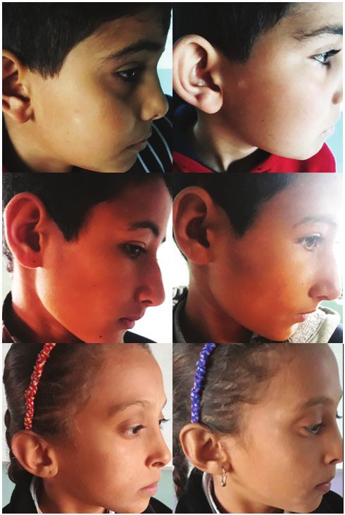
Indian Journal of Forensic Medicine & Toxicology, January-March 2021, Vol. 15, No. 1   865

colored scaly zone of PA. Pigmented PA lesion have a                Telling the patients and/or their parents about the
bluish hue in the center that is surrounded by a halo of       PA benign nature and self-limited course is mandatory.
hypopigmentation.                                              They should be informed about its slow resolution that
                                                               may exceed a one year(1). Patients can be informed to
    4. Extensive        PA       (Progressive    Macular
                                                               follow some lifestyle modification with support use
Hypomelanosis). The lower part of the trunk is the
                                                               of sunscreens, skin moisturizers, and skin hygiene.
most common site of involvement symmetrically and
                                                               Topical steroids (low-potency) appear to be more widely
usually in a relapsing attitude. Extensive PA patches are
                                                               prescribed.
widespread and have more persistent course. Lesions
aren’t favoring the face. Females are more often affected           Intestinal Parasites:
than male. (7-8)
                                                                    According to the given hypothesis, this study
     There is no gender difference for the disease. There      emphasizes on helminthic infestation. Among the
is a global distribution of PA, though its prevalence may      most common modes of transmitting these organisms
differ among different countries. The most prevalent           is through contaminated water, food, soil, as well
countries were Iraq (38.2%), India (31%), Mali (20%),          as contact. Helminthiasis (worm infestation) is the
and Egypt (18%); while lower prevalence was in                 hosting of parasitic worms after invading humans and
developed countries like United states (5%) and Hong           other animals necessarily to complete their lifecycle
Kong (1%)(9). PA occurs more predominantly in children         and either causing clinical manifestation or hide as an
between the ages of 3-16 years. PA is noninfectious and        asymptomatic carrier status. In third world countries,
there is no identified peculiar aetiology. Many cases          including Iraq, the majority of helminths infections are
with PA are presented with iron and copper deficiency.         associated with poor sanitary facilities, indiscriminate
Possible triggering factors that may cause PA are              disposal of human waste, inadequacy and lack of quality
deficiency of vitamins & calcium, temperature variations,      drinking water. It is also potentiated by poverty and
humidity, excessive sunlight exposure, frequent bathing,       low socioeconomic status. Enterobius vermicularis
usage ofharsh detergents and soaps, dry and itching            or the so-called pinworm, threadworm, or seatworm,
skin, hypopigmentation, worms and parasites, stress,           is a nematode (roundworm) that is common in human
deficiency of copper and atopic diseases and/or a family       children transmitted by feco-oral route. It hosts humans
history of eczema(2-3). The diagnosis in most cases of         only(11).
pityriasis alba is straightforward and depending on
                                                                    Night perianal itching grew the suspicion of the
the clinical picture(3). PA most frequently seen as 2 or
                                                               worm infestation. Eggs can be recovered using the
3 macules or patches in different stages. Vargas et al
                                                               “Scotch Tape” technique in the morning before a bowel
described 3 stages of PA:
                                                               movement. Transparent Scotch Tape is applied directly
    1. The early stage, also called papular                    to the perianal area, and then placed on a microscope
erythematous stage, begins as faint pink to red elevated       slide for examination. Eggs are football shaped and
bordered round to oval macule or patch that may last for       have an outer shell. Infectious larvae are often visible
weeks. In most cases this erythematous stage may run           inside the egg. The small adult worms may be seen in
unnoticed.                                                     a stool test (ova and parasites). Because the eggs are
                                                               lightweight and highly infectious, it is important for bed
    2. The next intermediate stage, papular
                                                               linens, towels, and clothing to be washed in hot water to
hypochromic stage, or follicular pityriasis alba. The
                                                               prevent reinfection(5).
patch is converted into a smooth scaly layer.
                                                                                Patients and Methods
     3. The final stage, the smooth hypochromic
stage presented as a visible, round and hypopigmented              This was a prospective and a case-control study
macule with mostly well-defined borders. In this stage,        conducted in the department of dermatology of
the patient usually or his/her parents will seek medical       Salahuldin-General Hospital in Tikrit city, Salahuldin
assistance(10).                                                province, Iraq. The study was conducted during the
866 Indian Journal of Forensic Medicine & Toxicology, January-March 2021, Vol. 15, No. 1
period from Nov 2019 to May 2020. The study targeted visit. After a one week another contact with the patient
children aged between 5 and 15 years. A total of 43 to evaluate the state of helminthic eradication. After 6
children were enrolled in the study. The intestinal week patients were contacted to observe the changes in
parasite infested attendant children presented with skin skin hypopigmentation. Clinical efficiency of parasite
hypopigmented patches were regarded as the case group. elimination was evaluated one week after the completion
The non-infested children with PA were the control of antiparasitic therapy. A positive clinical response
group. All participants passed a medical examination included: complete HP disappearance and reduction
by a dermatologist and consolidated by taking opinions of intensity, size and/or hue of HP. A negative clinical
of two or more dermatologists. Three separated (2- response included: the absence of visual changes of HP
days apart) stool samples were taken for parasitological or enlargement of size and hue of HP(13).
examination from all the participants. Additionally,
stool samples and sticker tapes were taken from PA Results
children with parasitic infestation a week after treatment The forty-three studied children ages were ranged
completion to confirm parasite elimination. No local from 5 to 15 years. Their mean age was 9.2 years (Table
drug applications, sunblock, or soothing agents were 1). It was consisted of 23 (53.5%) males and 20 (46.5%)
given for HP. Therapy was given to PA patients infected females. Twenty-eight of them were living in urban
with Enterobius vermicularis with mebendazole (a single districts (65.1%) whereas 15 were living in rural areas
100 mg chew tablet that can be repeated 3 weeks later if (34.9%). The HP presented in different numbers, sizes,
infestation had not been eradicated) (12); while parasite and various body regions (Table 2). Four (9.3%) children
free PA children were given placebo made of starch pills were shown to have parasitic intestinal infestation, while
of the same size and shape of the mebendazole tablet, 39 (90.7%) children with HP were free of infestation.
and given to the control group in the same amount All infesting parasites were the helminths Enterobius
and frequency. Both treatment and placebo were well vermicularis that were symptomatic and had been
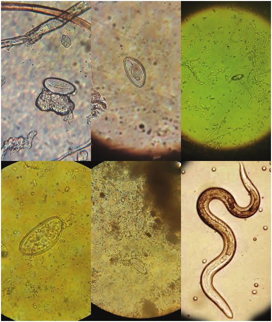
tolerated, and no side effects were complained. All isolated and observed microscopically (Figure 1).
patients were examined for the pityriasis alba at the first
Table 1. Demography of the studied sample.
Range 5-15
Age (years)
Mean ± SD 9.2 ± 2.6
Males 23 (53.5%)
Gender Number (%)
Females 20 (46.5%)
Urban 28 (65.1%)
Residence Number (%)
Rural 15 (34.9%)
SD: Standard Deviation
Table 2. Hypopigmented patch distribution and sun exposure.
Hypopigmented Patch Range Mean ± SD
Number 1-18 5.0 ± 3.5
Sum of size (mm) 20-325 92.8 ± 59.3
Average size (mm) 12-50 20.7 ± 6.5
Duration (months) 1-36 10.7 ± 8.7
Sun exposure (hour/day) 1-8 4.5 ± 2.4
Family members 1-4 Median = 2
SD: Standard DeviationIndian Journal of Forensic Medicine & Toxicology, January-March 2021, Vol. 15, No. 1 867
HP distributed in different parts of the body. Ninety-three percent of children had HP in the cheeks and to a lesser
extent in forehead and neck 70% and 67% respectively. This study had shown HP presented in shoulders in 19%,
upper extremities in 33%, and in the trunk in 9% of cases.
Figure 1. Ova and a helminth of Enterobius vermicularis as shown under light microscope in the positive
infested studied cases.
None of cases observed in the study had their HP eliminated completely after 6 weeks of treatment (Figure 2).
The study shown 14 (32.56%) children of fixed size HP after this period and the rest were underwent change in their
HP size as shown in Table 3.
Table 3. Destiny of HP change after six weeks of treatment or placebo.
HP Size Change Infested Non-infested Total
Disappear 0% 0% 0%
Reduced 75% 46.15% 48.84%
Fixed 0% 35.90% 32.56%
Enlarge 25% 17.95% 18.60%
HP: hypopigmented patch868 Indian Journal of Forensic Medicine & Toxicology, January-March 2021, Vol. 15, No. 1
Figure 2. Children with HP at first visit (left) and 6 weeks after treatment (right), there is no marked
changes in the size, number, or distribution of PA.
Cases present with an average of 6 hypopigmented patches in the first visit as well as in six weeks after treatment,
while control group shown HP average number of 4.9 in the first visit and 4.8 six weeks after placebo (Table 4). The
HP size in infested and control groups were summarized in Table 4.Indian Journal of Forensic Medicine & Toxicology, January-March 2021, Vol. 15, No. 1 869
Table 4. Hypopigmented patch number and size in infested compared to non-infested children in the first
visit (V1) and six weeks after treatment (V2).
Infested Non-infested Total
HP p-value
(Mean ± SD) (Mean ± SD) (Mean ± SD)
V1 6.0 ± 2.9 4.9 ± 3.6 5.0 ± 3.5 0.7642
V2 6.0 ± 3.6 4.8 ± 4.1 4.9 ± 4.0 0.7718
Number
change 0.0 ± 2.8 -0.1 ± 1.9 -0.1 ± 2.0 0.9602
p-value 1 0.976
sum (mm) in V1 120.0 ± 72.2 90.0 ± 58.3 92.8 ± 59.3 0.61
sum (mm) in V2 111.3 ± 89.2 86.8 ± 78.9 89.1 ± 79.0 0.7566
Size
change (mm) -8.8 ± 69.8 -3.2 ± 39.7 -3.7 ± 42.2 0.8886
p-value 0.9044 0.9522
HP: hypopigmented patch, SD: Standard Deviation, p: probability (p>0.05: not significant), mm: millimeter.
Discussion Similar to what found by Vinod et al (14) and
Toychiev et al (16), the majority of children in this study
In Iraq, there is a common belief among society
had HP in the head especially in cheeks (93%), forehead
regarding the presence of white patches in the face of
(70%), and in the neck (67%). (14, 16)
the child as a sign of intestinal infestation.
The study shown those children with intestinal
Lower than a tenth (9.3%) of children with
infestation had slightly more numerous HP compared
hypopigmented patch complaining of intestinal parasitic
with control group (mean = 6.0 and 4.9 respectively)
infestation is not enough evidence as a causation for the
(Table 4). This difference was not statistically significant
majority (90.7%) of HP incorporated in the study were
(p=0.7642); nor significant was the difference between
found to be free infestation. A near finding was obtained
the number of HP observed after 6 weeks of taking
by Vinod et al (2012) whom microscopic examination of
antihelminthic vs placebo treatment (p=0.7718).
the stool shown ova presented in 15.5% of the sample,
This was agreeing with Vinod et al(14) whom found a
and concluded no significant relationship between
similar non-significant relation; but disagreeing with
intestinal parasitic infestation and HP(14). Unlikely,
both Toychiev et al and Osipova. This study shown no
Osipova (2017) founded 43.4% out of 30 children with
significant change in number of HP after 6 weeks of
HP had helminthic intestinal infestation and concluded
the treatment or placebo for both intestinally infested
a positive significant relationship between infestation
children (p=1) and control group (p=0.976). This was
and HP(15). Three out of the four infested children in this
disagreeing with what was found by Toychiev et al
study were living in rural area. This may be due to the
whom declared that 33.3% of children with enterobiasis
lower hygiene, lower sanitation, habitual longer contact
had complete disappearance of their HP after 6 weeks of
with soil, or eating unclean vegetables or fruits.
treatment and had their parasitic infestation eliminated.
It is also disagreeing with Osipova whom founded 69.2%870 Indian Journal of Forensic Medicine & Toxicology, January-March 2021, Vol. 15, No. 1
of cases underwent complete disappearance of their HP 3. Bolognia J, Julie V, Schaffer A, Lorenzo C.
after 6 weeks of antihelminthic treatment (compared to Dermatology. 4th ed. Canada: Elsevier; 2018: 420,
0% in this study (Table 3). 442-3.
4. Griffiths W, Christopher E, Jonathan B, tanya
In this study, children with intestinal helminthic B, Robert C, Daniel C. Rook’s Textbook of
infestation shown larger HP size (measured by mean Dermatology. 9th ed. UK: John Wiley & Sons, Ltd;
of the summation of diameters of patches for each 2016: 86, 110, 811.
child) compared with control group (120 and 90
5. Stefan R. Jawetz, Melnick, & Adelberg’s Medical
mm respectively) (Table 4). This difference was not Microbiology. 28th ed. New York: McGraw-Hill
statistically significant (p=0.61). Similar insignificant Education; 2019: 117-20.
difference was found between case vs control after
6. Ruiz Maldonado R. Hypomelanotic Conditions of
treatment and placebo (p=0.7566). Although there was the Newborn and Infant. Dermatologic Clinics.
some decline in the HP size of the group of cases after 2017; 3(25): 373–382.
treatment (averaged 8.8 mm reduction), it was also
7. Plensdorf S, Livieratos M, Dada N. Pigmentation
statistically not significant (p=0.9044). A lesser (3.2mm)
Disorders: Diagnosis and Management. Am Fam
(but also insignificant p=0.9522) decline in the average Physician. 2017 Dec; 12(96): 797-804.
of HP size of control group after 6 weeks of placebo
8. Fenner J, Silverberg N. Skin diseases associated
intake. This decline was observed in 21 (48.84%) (i.e.
with atopic dermatitis. Clin. Dermatol. 2018 Sep -
nearly a half) of the total 43 observations. These finding Oct; 5(36): 631-640.
were disagreeing with Toychiev et al whom founded
9. Vanderhooft S, Francis J, Pagon R. Prevalence of
20.3% of patients had reduced HP size significantly.
hypopigmented macules in a healthy population. J.
Pediatr. 2016; 3(129): 355–361.
Conclusions
10. Lee D, Kang J, Kim S, Seo J, Sung H, Hwang S. A
1. Despite the familiarity of the relation between Case of Extensive Pityriasis Alba. Ann Dermatol.
intestinal helminthic infestation and HP among the 2018 Sep; 3(20): 146-8.
society, there is no significant relationship between the
11. Jogfdujpot F, Hotez P, Brindley P, Bethony J,
two. King C, Pearce E, et al. Helminth infections: the.
great neglected tropical diseases. Africa (Lond.).
2. Despite the frequently observed cases of
2018;(118): 1311-21.
intestinal infestation attending pediatrics consultation as
well as children with HP attending dermatology’s one; 12. Kiri A, Rebecca B, Alison B. British National
there is no prove stating the former as an aetiology of the Formulary (BNF). 76th ed. London: BMJ Group
and Pharmaceutical Press; 2019: 788-9.
latter.
13. Shalini SL. MSD MANUAL P. RS, K. JL, L. RB,
Conflict of Interest: None R M, editors. Kenilworth, NJ, USA: Merck Sharp
& Dohme Corp., a subsidiary of Merck & Co., Inc.;
Funding: Self 2020: 49-52.
Ethical Clearance: Not required 14. Vinod S, Singh G, Dash K, Grover S. Clinico
epidemiological study of pityriasis alba. Indian J.
References Dermatol. Venereol. Leprol. 2012; 5(68): 338-340.
1. Miazek N, Michalek I, Pawlowska-Kisiel M, 15. Osipova S. Possible association of intestinal
Olszewska M, Rudnicka L. Pityriasis Alba-- parasitic diseases with hypopigmentation. J
Common Disease, Enigmatic Entity: Up-to-Date Bacteriol Parasitol. 2017 Sep; 8(4): 55.
Review of the Literature. Pediatr Dermatol. 2018 16. Toychiev A, Mirzoeva M, Davis N, Islamova J,
Jan; 8(4): 441-5. Osipova S. Pityriasis alba: Possible associations
2. William DJ, Misha AR, Dirk ME. Andrews’ with intestinal helminths. Int J Clin. 2019 October;:
Diseases of the Skin. 13th ed. New York: Elsevier e13441.
Inc.; 2020: 87, 110-1.You can also read