Effect of ozone with or without nano-hydroxyapatite paste on chemically induced intact carious lesions around orthodontic brackets (In Vitro Study)
←
→
Page content transcription
If your browser does not render page correctly, please read the page content below
IOSR Journal of Dental and Medical Sciences (IOSR-JDMS)
e-ISSN: 2279-0853, p-ISSN: 2279-0861.Volume 18, Issue 9 Ser.6 (September. 2019), PP 70-75
www.iosrjournals.org

    Effect of ozone with or without nano-hydroxyapatite paste on
    chemically induced intact carious lesions around orthodontic
                      brackets (In Vitro Study)
               Mariam M. Mowafy1; Sayed M. Ghorab2; Shaza M. Hammad3
                                     1
                             Faculty of Dentistry, Mansoura University, Egypt.
2
 Lecturer in Department of Dental Biomaterials, Faculty of Dentistry, Mansoura University, Mansoura, Egypt.
 3
   Professor and Head of Department of Orthodontics, Faculty of Dentistry, Mansoura University, Mansoura,
                                                   Egypt.
                                Corresponding Author: Mowafy M. Mariam

Abstract
Introduction: To evaluate the effect of ozonated water with and without nano-hydroxyapatite (NHA)
paste on remineralizing artificially created initial enamel caries around orthodontic brackets using
polarized light microscopy and Vickers microhardness tester
Material and Methods: A total of 120 extracted human premolar sound teeth were collected and coded from 1
to 120. Metallic brackets were bonded to the buccal surface of the teeth, then specimens were divided into 4
groups following different remineralization regimen: ozonated water, ozonated water + 10 % NHA paste, 10 %
NHA paste alone, and control (saliva). The surface microhardness (SMH) was measured using HVS-50 Digital
Display Vickers microhardness tester at baseline. The specimens were subjected to demineralizing solution to
create initial enamel caries and enamel SMH was recorded. The specimens were subjected to pH cycling model
then enamel SMH was recorded. The results were validated qualitatively using polarized light microscope
(PLM). The results were analyzed using repeated measures, one-way ANOVA with post hoc multiple
comparisons at α =0.05 level.
Results: A statistically significant difference was found between all treatment groups and control one
(P
Effect of Ozone with or Without Nano-Hydroxyapatite Paste on Chemically Induced Intact Carious
structure.[7]Ebadifar et al.[8] reported that toothpaste containing NHA showed a higher remineralizing effect
than toothpaste containing fluoride.
Yet, there are inadequate studies in the literature about the reversing action of ozone on incipient caries
with intact enamel surface. Considering that, the purpose of our study was to assess the effect of ozonated water,
with and without NHA addition, on reversing initial enamel caries around orthodontic brackets.
II. Materials And Methods
The calculated sample size of the study was 120 specimens at 5% level of significance and 80 %
power. A total of 120 caries-free human premolars extracted for orthodontic purposes were used in this in-vitro
study. Teeth with caries, cracks, pitting, hypo-mineralization, fluorosis or previous treatment with chemical
agents were excluded. Teeth were evaluated under dental microscope with x40 magnification
(M320 F12, Leica Microsystems, Wetzlar, Germany) for gross irregularities, cracks or hypoplastic areas. All
teeth were polished with fluoride-free prophypaste (i-Faste, i-dental, Lithuania) and rubber cup, washed with tap
water and stored in distilled water at room temperature until time of use. The root portion of each tooth was
embedded in a cylindrical block of acrylic resin using specific molds to facilitate their utilization. Metallic Roth
brackets (Morelli Ltd, SP, Brazil) with 0.022 inch slot were bonded to the buccal surface of the teeth using
Transbond XT (3 M Unitek, Monrovia, Calif, USA) according to manufacturer's instructions.
Blocks were numbered randomly from 1 to 120. Then, they were allocated into 4 equal groups (n=30)
based on their remineralization protocol: Group 1- ozonated water, Group 2- ozonated water and finger-brushed
with tooth paste containing 10% NHA, Group 3- finger-brushed with tooth paste[9] containing 10% NHA only,
Group 4- saliva (control group). Three of these groups were experimental (groups 1-3) and one of them was a
control group.
The specimen surfaces were dried and the surface microhardess (SMH) at baseline was measured using
Vickers micro-hardness tester prior to any acid exposure.
All the specimens were immersed in demineralizing solution (2mM CaCl2, 2mM NaH2PO4, 50 mM
CH3COOH at pH 4.4)[10] for 4 days to resemble an active area of demineralization leading to formation of
artificial caries.[11]
The specimen surfaces were dried and the SMH was re-measured using Vickers microhardness tester
after initial caries induction.
pH cycling model:
The pH cycling model was settled upon to mimic the oral cavity regarding the progressive course of
alternating demineralization and remineralization.[11] All specimens in the experimental groups were exposed
to the pH cycling model for a 28 day period. Each of the specimens was immersed in the demineralizing
solution for a 3-hour period daily. Then, each specimen was treated with its corresponding remineralizing agent
for a 1-minute period. Next, all the specimens were immersed in the remineralizing solution (1.5mM CaCl2,
0.9mM NaH2PO4, 0.15M KClin 0.1 m Tris buffer at pH 7)[10] for a period of 21 hours. This was followed-up
with treatment of the samples again with the corresponding remineralizing agent for 1 min. The remineralizing
solution was replenished every 48 hours and the demineralizing agent replenished every 5 days. Ozonated water
delivered 2 minutes daily, divided, to the corresponding group. NHA paste was applied onto the specimen using
the index finger twice daily. On daily basis, the teeth in the control group were subjected to the demineralizing
solution for 3 hours followed up by immersion in artificial saliva that was replaced every 2 days.
To prevent the compromise of the solutions reaching the saturation threshold, fresh solutions were
added according to the guidelines set by Featherstone and Zero (1992).[12]
After the execution of the pH cycling steps, all the specimens' SMH were re-assessed using Vickers hardness
test.
Microhardness test procedure
The specimens' SMH were assessed using Digital Display Vickers Micro-hardness Tester (Model
HVS-50, LaizhouHuayin Testing Instrument Co., Ltd. China). A 200g load was applied with a Vickers diamond
indenter to the surface of the specimen for 20 seconds. Three indentations were placed on the surface of each
specimen at a distance of at least 0.5 mm away from the adjacent indentations. With the help of a 20x objective
lens, the lengths of the indentations' diagonals were measured by built-in scaled microscope. Then, SMH values
were calculated from Vickers values using the following equation:
HV=1.854 P/d2
where, HV is Vickers hardness value in Kgf/mm2, P is the load in Kgf and d is the length of the indentation's
diagonals in mm.[13]
DOI: 10.9790/0853-1809067075 www.iosrjournals.org 71 | PageEffect of Ozone with or Without Nano-Hydroxyapatite Paste on Chemically Induced Intact Carious
Polarizing Light Microscope
Considering the evaluation of the depth of remineralization, representative specimens' sections from all
the four groups were longitudinally sectioned down to an approximate thickness of 150μm. Firstly, the specimen
was sectioned to a thickness of 300 to 400μm with a flexible diamond disc (DFS-Diamon, Germany). The
sections were further ground to the required thickness with descending grits of carborundum (600-1100
grit).Each section was cleaned with deionized water and air-dried and then was aligned longitudinally and fixed
on the slide using DPX mounting medium. Each section obtained was analyzed for depth of the lesion under
PLM (Orthoplan, Leitz, Wetzlar, Germany) with digital camera (Leica MC190 HD, Germany). The lesion depth
was measured from the surface of the tooth to the maximum depth using the Leica Application Suite software
(Leica LAS EZ v3.0).
III. Statistical Analysis
The collected data were coded, processed and analyzed using Statistical Package of Social Science
(SPSS) program for windows (version 16). Quantitative continuous data were presented in mean and standard
deviation (SD).
One way Analysis of variance (ANOVA) test was used for testing significance of means between
different groups while repeated measures ANOVA test was used for testing means within each group (at least
one pairwise difference). If the p-value < 0.05, we reject null hypothesis (H0) and conclude that there is a
significant difference between at least one pair of means. Bonferroni post hoc test was performed to test where
the pairwise differences are.
IV. Results
The descriptive statistics of the SMH of the studied groups at baseline, after demineralization and after
remineralization by the corresponding remineralizing regimen are presented in Table I. According to the
ANOVA, there were significant differences between the SMH values in-between groups comparison after
remineralization (pEffect of Ozone with or Without Nano-Hydroxyapatite Paste on Chemically Induced Intact Carious
a Repeated measures ANOVA
b Post hoc comparison
c One-way ANOVA
Table 1 The descriptive statistics of the SMH of the studied groups at different levels of mineralization
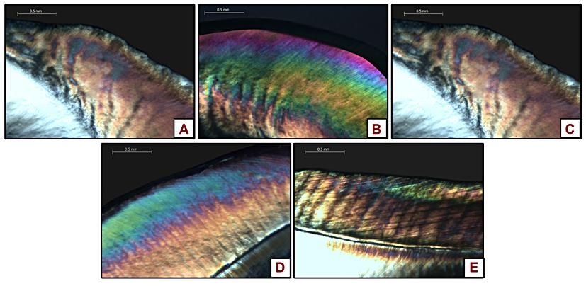
Figure 1 The photomicrographs taken under PLM (A), demineralized enamel (B), treated with ozone alone (C),
treated with ozone+ NHA (D), treated with NHA alone (E), saliva(control)
Figure 2 Graph representing the average depth of remineralization of the representative specimen of each group
as seen under PLM
V. Discussion
Enamel demineralization can form rapidly because of the high and continuous challenge of plaque
when associated with orthodontic appliances. Demineralization is a reversible process; hence, the partially
demineralized HA crystals in teeth can grow to their original size if they are exposed to favorable remineralizing
conditions.[14]
The aim of this study was to evaluate the potential remineralization effect of ozonated water against
demineralized enamel around orthodontic brackets. In the same manner, the combined treatment of ozonated
water and NHA was put to investigation. The enamel surface changes were assessed using Vickers
microhardness tester and PLM.
Ozone therapy has a wide range of dental applications owing to its exceptional properties such as anti-
microbial, anti-inflammatory, immune-stimulating, anti-hypoxic, detoxicating and biosynthetic actions.[15,16]
Nonetheless it has gained its popularity in dental practice for non-invasive, effective dental care meeting the
demands of the patients.[17]
The results of this study indicated that all of the treatment procedures used in this study showed
significant remineralization of artificially created enamel caries. The specimens in all the experimental groups
had experienced a decrease in microhardness after demineralization, excluding any disparity, and had
rehardened after remineralization.
The specimens treated with ozonated water have shown the highest SMH values with a mean of
293.20±26.8 Kgf/mm2. This can be interpreted in various ways such as the bactericidal effect of ozone on
DOI: 10.9790/0853-1809067075 www.iosrjournals.org 73 | PageEffect of Ozone with or Without Nano-Hydroxyapatite Paste on Chemically Induced Intact Carious
cariogenic bacteria[18] or its oxidizing ability to eliminate the proteins in demineralized enamel promoting
lesion remineralization through facilitating the diffusion of remineralizing agents.[19]
In accordance with our results, Yamayoshi and Tatsumi[20] revealed that ozone was a strong
oxidizing agent to the microorganisms' cell wall and cytoplasmic membranes. Moreover, Baysan et al.[21] have
assessed the antimicrobial effect of ozone on oral microorganisms in vitro giving significant reduction in the
count of microorganism cells. The results of Sadatullah et al.[18] have rationalized our study revealing that the
exposure to the ozonated water for 30 sec reduced the total bacterial population of the 24-hour plaque by 45.3%.
On the other hand, Baysan and Beighton[22] examined ozone ability to kill microorganisms related to
non-cavitatedocclusal caries. Their results were contradictory to the current study’s results, where Baysan and
Beighton's results revealed failure to significantly reduce bacterial count in infected dentine found under
demineralized enamel. More recently, Tahmassebi et al.[6] investigated the effect of ozone on the progress of
induced caries-like lesions on enamel showing that ozone treatment alone had no effect on protecting enamel
against demineralization or promoting remineralization.
Atabek and Oztas[19] have revealed that ozone application, whether applied alone or with a
remineralizing solution, is an effective method for caries reversal. He B et al.[23] and Huang S et al.[24]
undergone studies that showed significant improvement in SMH post-treatment with NHA toothpaste. Increase
in mineral content after treatment with NHA paste was evident in the study of Tschoppe et al [25] while no
significance was found to be in the study of Itthagarun et al.[26]
The significant increase in SMH in the NHA-treated group establishes that this material re-hardens the
softened enamel by gradual deposition of the mineral that precipitates and nucleates in the dark zone of
demineralization thereby offering complete biomimetic regeneration of the lost enamel crystallites.[27]
Moreover, Enan and Hammad have revealed that there is a direct proportionality between NHA concentration
and the rate of enamel remineralization, up to a certain limit.[28]
Surprisingly, the enamel SMH of the control group has been restored beyond the baseline level but
insignificantly (p>0.05). This result is in harmony with the opinion of Featherstone[29] confirming that with
good oral hygiene remineralization may be established. This may be due to the bio-available mineral
components readily found in saliva.
The dark zone representing the remineralization depth of each representative specimen may be
interpreted by possible filling of the micro-pores on demineralized enamel as revealed by Hannig et al.[30]and
Roveri et al.[31] This would enhance the remineralizing effect and boost the mechanical properties of the tooth
assuring the results of micro-hardness revealed by this study.
Withal, the investigators should apprehend the constraints of this method which cannot completely
simulate the intraoral conditions, thus the proposal of the in vitro results for clinical use should be within limits.
Moreover, the pH cycling model may not thoroughly simulate the intraoral settings because some changes exist
in pH variations based on the patient's hygienic means, nutritional habits, fluoride consumption and salivary
composition and quality.[32]
To conclude, ozone therapy is found to be an effective method in reversing demineralized enamel.
Moreover, ozone significantly enhanced the remineralizing ability of NHA compared to NHA paste alone. In
spite of the promising results of our study, clinical research is needed to substantiate the mechanism of action of
ozone. These results should be confirmed clinically to counteract any limitations of our in vitro study.
References
[1]. Nascimento P, Fernandes M, Figueiredo F, et al. Fluoride-releasing materials to prevent white spot lesions around orthodontic
brackets: a systematic review. Braz Dent J. 2016;27(1):101-107.
[2]. Julien KC, Buschang PH, Campbell PM. Prevalence of white spot lesion formation during orthodontic treatment. Angle Orthod.
2013;83(4):641-647.
[3]. Kumar A, Bhagawati S, Tyagi P, et al. Current interpretations and scientific rationale of the ozone usage in dentistry: A systematic
review of literature. Eur J Gen Dent. 2014;3(3):175-180.
[4]. Huth KC, Paschos E, Brand K, et al. Effect of ozone on non-cavitated fissure carious lesions in permanent molars. A controlled
prospective clinical study. Am J Dent. 2005;18(4):223-228.
[5]. Ximenes M, Cardoso M, Astorga F, et al. Antimicrobial activity of ozone and NaF-chlorhexidine on early childhood caries. Braz
Oral Res. 2017;31.
[6]. Tahmassebi J, Chrysafi N, Duggal M. The effect of ozone on progression or regression of artificial caries-like enamel lesions in
vitro. J Dent. 2014;42(2):167-174.
[7]. Li L, Pan H, Tao J, et al. Repair of enamel by using hydroxyapatite nanoparticles as the building blocks. J Mater Chem.
2008;18(34):4079-4084.
[8]. Ebadifar A, Nomani M, Fatemi SA. Effect of nano-hydroxyapatite toothpaste on microhardness ofartificial carious lesions created
on extracted teeth. J Dent Res Dent Clin Dent Prospects. 2017;11(1):14-17.
[9]. Samuel S, Dorai S, Khatri S, et al. Effect of ozone to remineralize initial enamel caries: in situ study. Clin Oral Investig.
2016;20(5):1109-1113.
[10]. Stookey GK. The Featherstone laboratory pH cycling model: a prospective, multi-site validation exercise. Am J Dent.
2011;24(5):322-328.
[11]. Shetty S, Hegde MN, Bopanna TP. Enamel remineralization assessment after treatment with three different remineralizing agents
using surface microhardness: An in vitro study. J Conserv Dent. 2014;17(1):49-52.
DOI: 10.9790/0853-1809067075 www.iosrjournals.org 74 | PageEffect of Ozone with or Without Nano-Hydroxyapatite Paste on Chemically Induced Intact Carious
[12]. Featherstone J, Zero D. An in situ model for simultaneous assessment of inhibition of demineralization and enhancement of
remineralization. J Dent Res. 1992;71:804-810.
[13]. Badr S, Ibrahim MA. Protective effect of three different fluoride pretreatments on artificially induced dental erosion in primary and
permanent teeth. J Am Sci. 2010;6(6):442-451.
[14]. Neel EAA, Aljabo A, Strange A, et al. Demineralization–remineralization dynamics in teeth and bone. Int J Nanomed.
2016;11:4743-4763.
[15]. Baysan A, Lynch E. The use of ozone in dentistry and medicine. Part 2. Ozone and root caries. Prim Dent Care. 2006;13(1):37-41.
[16]. Almaz ME, Sönmez IŞ. Ozone therapy in the management and prevention of caries. J Formos Med Assoc. 2015;114(1):3-11.
[17]. Bhatia V, Bhatia G. Ozone and its Clinical Applications-A Boon for Advanced Dentistry. Indian J Cont Dent. 2013;1(1):83-87.
[18]. Sadatullah S, Mohamed NH, Razak FA. The antimicrobial effect of 0.1 ppm ozonated water on 24-hour plaque microorganisms in
situ. Braz Oral Res. 2012;26(2):126-131.
[19]. Atabek D, Oztas N. Effectiveness of ozone with or without the additional use of remineralizing solution on non-cavitated fissure
carious lesions in permanent molars. Eur J Dent. 2011;5(4):393-399.
[20]. Yamayoshi T, Tatsumi N. Microbicidal effects of ozone solution on methicillin-resistant Staphylococcus aureus. Drugs Exp Clin
Res. 1993;19(2):59-64.
[21]. Baysan A, Whiley R, Lynch E. Antimicrobial effect of a novel ozone–generating device on micro–organisms associated with
primary root carious lesions in vitro. Caries Res. 2000;34(6):498-501.
[22]. Baysan A, Beighton D. Assessment of the ozone-mediated killing of bacteria in infected dentine associated with non-cavitated
occlusal carious lesions. Caries Res. 2007;41(5):337-341.
[23]. He B, Huang S, Jing J, et al. Measurement of hydroxyapatite density and Knoop hardness in sound human enamel and a
correlational analysis between them. Clin Oral Investig. 2010;55(2):134-141.
[24]. Huang S, Gao S, Yu H. Effect of nano-hydroxyapatite concentration on remineralization of initial enamel lesion in vitro. Biomed
Mater. 2009;4(3):034104.
[25]. Tschoppe P, Zandim DL, Martus P, et al. Enamel and dentine remineralization by nano-hydroxyapatite toothpastes. J Dent.
2011;39(6):430-437.
[26]. Itthagarun A, King NM, Cheung Y. The effect of nano-hydroxyapatite toothpaste on artificial enamel carious lesion progression: an
in-vitro pH-cycling study. Hong Kong Dent J. 2010;7(2):61-66.
[27]. Jeong S, Jang S, Kim KN, et al., editors. Remineralization potential of new toothpaste containing nano-hydroxyapatite. Key
Engineering Materials; 2006: Trans Tech Publ.
[28]. Enan ET, Hammad SM. Microleakage under orthodontic bands cemented with nano-hydroxyapatite-modified glass ionomer: An in
vivo study. Angle Orthod. 2013;83(6):981-986.
[29]. Featherstone JD. Caries prevention and reversal based on the caries balance. Pediatr Dent. 2006;28(2):128-132.
[30]. Hannig M, Hannig C. Nanomaterials in preventive dentistry. Nat Nanotechnol. 2010;5:565-569.
[31]. Roveri N, Battistella E, Bianchi CL, et al. Surface enamel remineralization: biomimetic apatite nanocrystals and fluoride ions
different effects. J Nanomater. 2009;8:1-9.
[32]. Malekafzali B, Ekrami M, Mirfasihi A, et al. Remineralizing Effect of child formula dentifrices on artificial enamel caries using a
pH cycling model. J Dent (Tehran). 2015;12(1):11-17.
Mowafy M. Mariam. “Effect of ozone with or without nano-hydroxyapatite paste on
chemically induced intact carious lesions around orthodontic brackets (In Vitro Study).”
IOSR Journal of Dental and Medical Sciences (IOSR-JDMS), vol. 18, no. 9, 2019, pp 70-75.
DOI: 10.9790/0853-1809067075 www.iosrjournals.org 75 | PageYou can also read