Morphometric aspects of the foramen magnum and the orbit in Brazilian dry skulls
←
→
Page content transcription
If your browser does not render page correctly, please read the page content below

Available online at www.ijmrhs.com

                                                                           International Journal of Medical Research &
                                      ISSN No: 2319-5886                         Health Sciences, 2016, 5, 4:34-42

      Morphometric aspects of the foramen magnum and the orbit in
                         Brazilian dry skulls
         *Lucas A. S. Pires1, Álvaro R. Teixeira1, Tulio F. O. Leite2, Marcio A. Babinski
                                    and Carlos A. A. Chagas1
 1
     Morphology Department, Biomedical Institute, Fluminense Federal University, Niterói, Rio de
                                         Janeiro, Brazil
       2
         General Surgery Department, Santa Casa Hospital, Ribeirão Preto, São Paulo, Brazil
                             *Corresponding e-mail:lucaspires@id.uff.br
_____________________________________________________________________________________________

ABSTRACT

Morphometric analysis of crania structures are of great significance to anatomists, forensic doctors,
anthropologists, and surgeons. We performed a morphometric study regarding the foramen magnum and the bony
orbit on the right side of the cranium in 77 skulls with the purpose of identifying a correlation between these
measures, as they are often employed alone to identify the genre and race of a skeleton with no other remains, since
the cranium is a structure that can resist fire, explosions, and mutilations. The foramen magnum receives special
attention, as it is located in a region together with many strong muscles and ligaments. The measures were taken
with a sliding digital caliper. Our results showed that the foramen magnum had a mean anteroposterior diameter of
34.23±2.54 mm, and the mean transverse diameter was 28.62±2.83 mm. The most common shape for the foramen
magnum was oval. The mean right orbital height was 32.89±2.45 mm, and the mean right orbital breadth was
37.15±2.68 mm. There was a weak to moderate correlation between these measures. Furthermore, the foramen
magnum and the orbit are regions of surgical and clinical significance, thus requiring knowledge regarding the
morphometric aspects of such areas, since they can often suffer morphological changes due to a number of diseases
and they undergo surgical procedures in order to treat these conditions.

Keywords: Anatomy, Anthropology, Foramen Magnum, Orbit, Skull
_____________________________________________________________________________________________

                                                 INTRODUCTION

Morphometric analysis of the cranium is often employed with the purpose of identifying the age, stature, ethnicity,
sex, and even the species of a damaged skeleton. These measures of the skull are crucial to forensic investigations
and anthropologic exams, and they are also relevant to clinical, surgical and anatomical studies [1-3].

The orbit is a cavity shaped like a pyramid with its apex located posteriorly and the base anteriorly located at the
viscerocranium. It is formed by the orbital process of the frontal bone, lesser wing of the sphenoid bone, orbital plate
and frontal process of the maxilla, zygomatic bone, orbital plate of the palatine bone, lacrimal bone, and the orbital
lamina of the ethmoid bone. The orbit contain the eye and associated muscles, the lacrimal glands, the ophthalmic
artery and veins, the ophthalmic nerve, the optic (II), oculomotor (III), trochlear (IV), and the abducens (VI) nerves.
The orbital characters vary amongst different races and ethnic groups, also, it is known to possess sexual
dimorphism, and it is anthropologically relevant in race definition [4-10].

The foramen magnum is an aperture located at the base of the skull. It is formed by the four portions of the occipital
bone (two lateral, one squamous, and one basal) and can present different shapes. Furthermore, it is pathway to
structures that crosses the head and neck like the cerebellum, medulla oblongata, meninges, vertebral arteries, and
the spinal branch of the accessory nerve (XI) [2-4,11-14].

                                                          34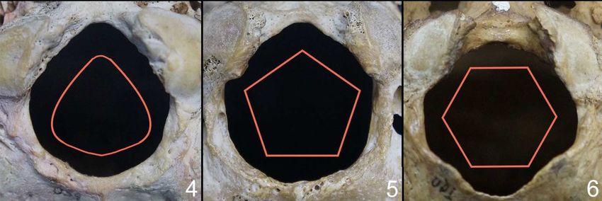
Pires et al                                Int J Med Res Health Sci. 2016, 5(4):34-42
______________________________________________________________________________
Concerning its site, measures of the occipital region seems to be an alternative to determinate certain characteristics
of cadaveric remains in cases where human fragments are greatly damaged by insults (fire, explosions, and
multilations), as the basicranium is protected by large and strong tissues, such as muscle, tendons and ligaments
[2,15,16].

Other areas of the skull such as nasal aperture, zygomatic extension, malar size, supraorbital ridge, chin, nuchal
crest, mastoid process size, nasal size, mandibular symphysis, occipital condyles, forehead shape, and palate size
were morphometrically studied in order to increase the number of parameters used to identify the gender, ethnicity,
race, and by consequence, the accuracy (when those methods are combined), which could lead to a decrease in the
usage of more sophisticated and expensive methods, such as DNA analysis [2,8,15-17]. The orbit is an important
factor as well in answering questions regarding the sex and ethnicity of a cadaver, which means that, it is also a
region of the skull that possess sexual dimorphism characteristics: orbit pertaining to female skulls are usually
rounder, and orbits belonging to male skulls generally have a square format [18,19].

Measures of the foramen magnum are relevant in cases of achondroplasia (as there is a high risk of spinal cord
stenosis in the base of the skull), Arnold-Chiari malformation (downward herniation of the cerebellar tonsils),
foramen magnum meningioma, plagiocephaly, basilar invagination, and others cranial deformities [3,16,18,20-22].

Morphological knowledge of the orbit is essential, since the orbit is a region exposed to several types of surgical
procedures (ophthalmological, maxillary surgeries and reconstructive cosmetic surgeries of face), moreover, the
orbit seems to be an important factor during ultrasound evaluation to diagnose diseaseas such as Down syndrome
[7,9,10,23-26].

The present study is an attempt to research the foramen magnum and its relation to the craniometric measures of the
orbit in Brazilian skulls, with the purpose of verifying any type of morphometric connection between the
neurocranium and viscerocranium. Studies about the FM and the orbit morphometry in Brazilians are scarce. Since
Brazil has a mixed population, we find that it is necessary to determine a reference value regarding the
morphometric aspects of the FM and the bony orbit in order to help anthropologists, forensic doctors, surgeons,
physicians, and anatomists identify morphological variants of this structure.

                                         MATERIALS AND METHODS

We performed a morphometric analysis of the foramen magnum and the right side orbit in 77 skulls pertaining to the
Morphology Department of the Fluminense Federal University. All skulls were from unknown sex and belonged to
adults of age above 18 years. They had great condition overall, skulls with a damaged or broken foramen magnum
and orbits were excluded from this study.

The measures were taken using a sliding digital caliper (0.01 mm), each measurement was repeated in order to avoid
any errors. The criterion for the antero-posterior diameter (APD) of the foramen magnum was the distance between
the basion and opisthion; the transverse diameter (TD) criterion was the distance between the points of maximum
curvature of the foramen magnum lateral margins (Fig. 1). The area of the foramen magnum was calculated using
the Radinsky’s formula (A = ¼ x pi x TD x APD).

The right orbital breadth (ROB) was the distance between the maximum breadth of the orbit from the
maxillofrontale to the middle of the lateral orbital border (ectoconchion) and the right orbital height (ROH) was the
maximum internal height of the orbit perpendicular to its breadth (Fig. 2). We also calculated the orbital index (OI)
of the right orbit and the FM index (FMI): (orbital height/ orbital breadth) x 100; (transverse diameter/
anteroposterior diameter) x 100, respectively.

The foramen magnum shape was classified in 5 categories: oval, round, tetragonal, pentagonal, and hexagonal (Fig.
3).

Statistical analysis was performed using GraphPad Prism 6 software, the collected data were analyzed using the
paired, two-tailed student’s t-test (p >0.05 was considered significant), the Pearson’s correlation method, and
D'Agostino& Pearson omnibus normality test. Descriptive statistics (mean, standard deviation, median, minimum
and maximum values) were also analyzed. All pictures were taken with a Sony Alpha ILCE-3000K (20.1
Megapixels).

                                                         35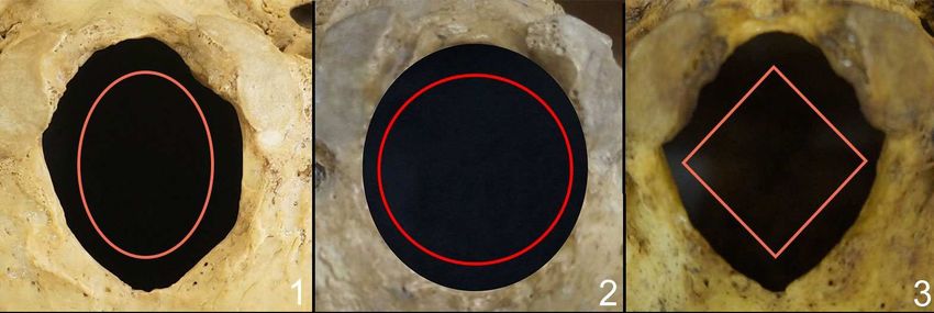
Pires et al                                Int J Med Res Health Sci. 2016, 5(4):34-42
______________________________________________________________________________
                                                       RESULTS

The FM had a mean anteroposterior diameter (MAPD) of 34.23 mm, with a standard deviation (SD) of 2.54 mm,
and the mean transverse diameter (MTD) was 28.62 mm, with a SD of 2.83 mm. The APD median was 34.29 mm
and the TD median was 28.15 mm, the maximum value (MaxV) for the APD and TD was 39.72 mm and 36.01 mm,
respectively, and the minimum value (MinV) was 26.90 mm for the APD and 22.67 mm for the TD (Table 1). The
most common shape for the FM was oval (Table 2).

The mean ROH was 32.89 mm, with a SD of 2.45 mm, and the mean ROB was 37.15 mm, with a SD of 2.68 mm,
the ROH and ROB median was 32.56 mm and 37.02 mm, respectively. The MinV and MaxV for the ROH were
28.06 mm and 39.15 mm. The MinV and MaxV for the ROB were 29.45 mm and 45.05 mm (Table 3).

The mean area of the foramen magnum was 772.4 mm with a SD of 116.7, and the mean foramen magnum index
(FMI) was 83.75 mm with a SD of 7.23 mm. The mean OI was 88.72 mm with a SD of 6.89 (Table 4).

The D'Agostino& Pearson omnibus normality test in all measures taken was considered normal (p =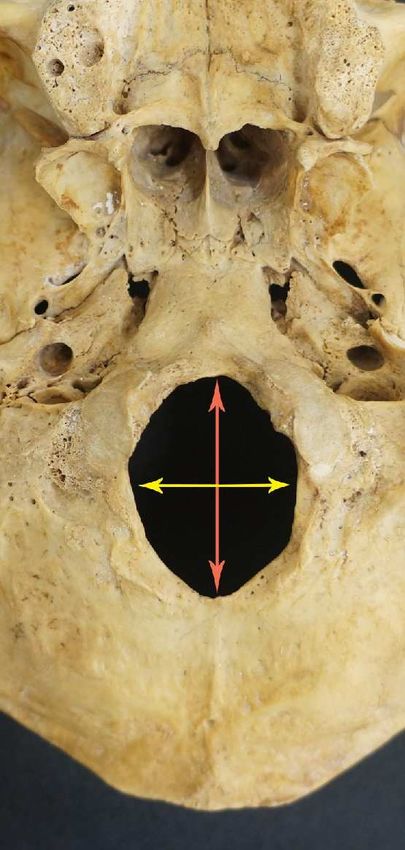
Pires et al                                Int J Med Res Health Sci. 2016, 5(4):34-42
______________________________________________________________________________
was 25.2±2.4 mm in Indians, and the most common shape of the FM was round. Osunwoke et al. (2012) [30]
reported that the MAPD was 36.11±0.24 mm and the MTD was 26.65±0.24 mm in Nigerians subjects, the MAPD
was higher than in our study, although the MTD was lower. Tubbs et al. (2012) [28] found that the MAPD was 31
mm and the MTD was 27 mm, they also reported that there was no significant difference between sexes, in contrast
to other authors. Santhosh et al. (2013) [13] reported the MAPD and MTD in a male Indian population were 34.37
mm and 28.98 mm, respectively, and in women 33.80 mm and 27.60 mm, respectively. Natsis et al. (2013) [31]
found that the MAPD was 35.53±3.06 mm and the MTD was 30.31±2.79 mm in a Greek population, also, the FM
most common shape was “two semi-circles”. Mehta et al. (2014) results showed that the MFMI was 82.5
mm±6.780, in an Indian population. Also in Indians, Ganapathy et al. (2014) [3] found that the MAPD was 33.9±2.3
mm and the MTD was 28.7±2.5 mm, although the most common shape of the FM was oval, in accordance to our
study. Patel and Mehta (2014) [21] reported that the MAPD was 33.7 mm and the MTD was 28.29 mm in an Indian
population from South Gujarat. Other studies of the FM are depicted in Table 5.

Even though studies reported that the FM measures are higher in male subjects than in females [15,33,34], Kamath
et al. (2015) [16] stated that the FM measures may overlap and should not be used by itself for the determination of
genre, as other authors found that there was no sexual dimorphic characteristics regarding the FM [1,17]. It was
reported that the size of the FM in patients with achondroplasia and other neurological problems was reported to be
smaller in all ages, and it was stated that the basichondrocranium of fetuses with hindbrain malformations (such as
the Arnold-Chiari malformation) is shorter and smaller than the ones in normal fetuses [20,21,35,36], although
Furtado et al. (2010) [37] stated that there were no significant relation between the FM measures in pediatric patients
with Arnold-Chiari malformation (Type I) and patients without. This brings forth the need to study this
morphometric aspect using the same parameters for measurements.

Regarding the morphometric aspects of the orbit, we found relatively few studies that used the same anatomical
points that ours did to measure the bony orbit. Ji et al. (2010) [23] observed the orbit measures through a CT-scan in
Chinese subjects, and their results revealed that the mean ROH was 33.28±1.58 mm, and the mean ROB was 38±94
mm, also, their research revealed that there wasn’t a statistically significant difference of the OH between male and
female patients. Ukoha et al. (2011) [38] found that the ROB was 36.03±0.37 mm and the ROH was 31.90±0.70
mm, stating that there were no statistically significance difference between the right and left sides. Fetouh and
Mandour (2014) [25] stated that in a male Egyptian population, the mean ROH was 35.83±1.23 mm, the mean ROB
was 43.62±1.13 mm, and the mean OI was 82.20±2.97 mm, and in females, the mean ROH was 35.53±0.95 mm, the
mean ROB was 42.75±1.35 mm, and the OI was 84.13±3.76 mm. Elzaki et al. (2015) [8] found that the ROB was
34.10±1.76 mm and the ROH was 37.90±2.57 mm, they also found that the OB was slightly higher in man than in
woman, but the OH was similar. Many results showed that the males orbit area was significantly larger than in
females [23-25], although there is some divergence whether the symmetry of the orbit: some authors agree that there
is a significant difference between right orbit and left orbit [23,25], while other authors found no statistically
significant difference between them [24,38]. Other morphometric studies regarding the orbit are depicted in Table 6.

Kanchan et al. (2014) [18], in a study similar to ours, compared the FM with the orbit, and they reported (using the
Pearson’s correlation test) that there was a statistically significant correlation between the OI and FMI, in
discordance with our results. They also reported that the FMI in South Indians was 79.70±6.98 mm, and the OI was
84.23±6.64, less than our study showed.

It is important to notice that other relationships with the foramen magnum were observed in other studies, like the
carotid foramina [39] (Schaefer 1999), in which it was stated that the correlation was statistically significant, and
jugular foramen (Osunwoke et al. 2012) [30] in which they stated that 95% of skulls with a larger FM had a larger
jugular foramen. With the comparison between measurements belonging to different ethnicities from other
populations of the world, one could verify if there is indeed a correlation to a particular population group, or even,
discern the many different types of races that compose a particular population [32].

Study of the shape and size of the FM is crucial to determine pathological changes caused by diseases such as:
achondroplasia, occipital vertebra, basilar invagination, condylar hypoplasia, and atlas assimilation, Jeune’s
asphyxiating, thoracic dystrophy, Marchesani’s syndrome, foramen magnum meningioma, Arnold-Chiari
malformation, and plagiocephaly [3,16,18,20-22,28]. Those diseases can cause compression of the structures that
traverses the FM and produce symptoms like respiratory complications, lower cranial nerve dysfunctions, upper and
lower extremity paresis, hypo or hypertonia, hyperreflexia or clonus, and general delay during motor development
can appear [28]. Testut and Latarjet (1977) [11] stated that the difficulty of bony resection during surgery is directly
proportionate to the size of the FM.

                                                          37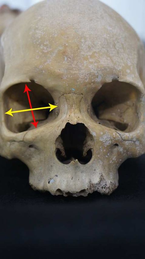
Pires et al                                Int J Med Res Health Sci. 2016, 5(4):34-42
______________________________________________________________________________
Evaluation of the morphometric aspect of the orbit seems to be relevant in cases of pathological disorders and
traumatic injuries, in order to estimate craniofacial asymmetry, the severity of the injury, and to discuss possible
complications during preoperative planning, as the orbit is exposed to several types of surgical procedures (optic
nerve decompression, vascular ligation, exenteration, excision of the lacrimal gland), requiring anatomical
knowledge of the region [9,10,23-26]. It is also stated that tumors confined within the periorbita in the anterior two
thirds of the orbit can be approached extracranially, although tumors located in the apical area, medially to the optic
nerve, often require a transcranial approach [9,26].

                        TABLE 1 DESCRIPTIVE STATISTICS OF THE FORAMEN MAGNUM (mm)

                                              Mean      SD     MinV       MaxV       Median
                                      APD     34.23     2.54   26.90      39.72      34.29
                                      TD      28.62     2.83   22.67      36.01      28.15

                                    TABLE 2 - SHAPES OF THE FORAMEN MAGNUM

                                                               N (77)     %
                                               Oval            41         53.24%
                                               Round           19         24.67%
                                               Tetragonal      13         16.88%
                                               Egg-Shaped      2          2.36%
                                               Pentagonal      1          1.29%
                                               Hexagonal       1          1.29%

                                TABLE 3 DESCRIPTIVE STATISTICS OF THE ORBIT (mm)

                                             Mean      SD      MinV      MaxV      Median
                                       OH    32.89     2.45    28.06     39.15     32.65
                                       OB    37.15     2.68    29.45     45.05     37.02

                      TABLE 4 INDEX AND AREA OF THE FORAMEN MAGNUM AND ORBIT (mm)

                                                      Mean Index       SD     Mean Area       SD
                                Foramen Magnum        83.75            7.23   772.4           116.7
                                Orbit                 88.72            6.89   -               -

                             TABLE 5 - Foramen Magnum Studies in Different Populations (mm)

                   Population/Race          Author                             N      MAPD            MTD
                   Indians                  Chethan et al., 2012               53     31±2.4          25.2±2.4
                   Indians                  Biswas et al., 2015                53     34.02±1.79      28.10±2.16
                   Indians                  Ganapathy et al., 2014             100    33.9±2.3        28.7±2.5
                   M Indians                Kamath et al., 2015                41     33.21±3.25      26.92±2.52
                   F Indians                Kamath et al., 2015                31     30.99±3.49      25.45±2.31
                   M Indians                Kanchan et al., 2013               69     34.51±2.77      27.36±2.09
                   F Indians                Kanchan et al., 2013               49     33.60±2.63      26.74±2.36
                   Indians                  Mehta et al., 2014                 100    34.32           28.37
                   Indians                  Patel and Mehta, 2014              100    33.70           28.29
                   M Indians                Santhosh et al., 2013              63     34.37±2.38      28.98±2.22
                   F Indians                Santhosh et al., 2013              38     33.80±2.56      27.60±2.67
                   M Indians                Shanthi and Lokanadham, 2013       66     37.1±3.3        32.0±3.1
                   F Indians                Shanthi and Lokanadham, 2013       34     33.8±3.8        30.4±3.0
                   M Indians                Shepur et al., 2014                175    33.40±2.60      28.50±2.20
                   F Indians                Shepur et al., 2014                175    33.10 ±2.70     27.30 ±2.00
                   M Turkish                Murshed et al., 2013               57     37.2±3.43       31.6±2.99
                   F Turkish                Murshed et al., 2013               53     34.6±3.16       29.3 ± 2.19
                   M Iranians               Uthman et al., 2012                43     34.9±2          29.5±2.5
                   F Iranians               Uthman et al., 2012                45     32.9±2          27.3±2.2
                   Caucasians               Tubbs et al., 2012                 72     31              27
                   Nigerians                Osunwoke et al., 2012              120    36.11±2.60      29.56±2.60
                   Greeks                   Natsis et al., 2013                143    35.53±3.06      30.31±2.79
                   Central Western Europe   Gruber et al., 2009                111    36.6±2.8        31.1±2.7
                   M Brazilians             Manoel et al., 2009                139    35.7±0.29       30.3±0.20
                   F Brazilians             Manoel et al., 2009                76     35.1±0.33       29.4±0.23
                   M Brazilians             Suazo et al., 2009                 144    36.5±2.6        30.6±2.5
                   F Brazilians             Suazo et al., 2009                 71     35.6±2.5        29.5±1.9
                                                     *M = Male, F = Female

                                                               38
Pires et al Int J Med Res Health Sci. 2016, 5(4):34-42
______________________________________________________________________________
TABLE 6 - Orbit Studies in Different Populations (Right Orbit)
Population/Race Author N MOH MOB
Indians Gosavi et al., 2014 128 32.31±2.52 39.46±2.57
Indians Biswas et al., 2015 53 32.1±2.3 36.6±3.3
Indians Kumar and Nagar, 2014 68 33.47±1.56 42.06±1.68
Sudanese Elzaki et al., 2015 110 37.90±2.57 34.10±1.76
M Egyptians Fetouh and Mandour, 2014 30 35.83±1.23 43.62±1.13
F Egyptians Fetouh and Mandour, 2014 22 35.53±0.95 42.75±1.35
Chinese Ji et al., 2015 64 33.45±1.63 39.10±1.83
Nigerians Ukoha et al., 2011 70 31.90±0.70 36.03±0.37
*M = Male, F = Female
Fig. 1 FORAMEN MAGNUM MEASUREMENTS
*Foramen magnum measured parameters.Red arrow = antero-posterior diameter (APD), yellow arrow = transverse diameter (TD)
39Pires et al Int J Med Res Health Sci. 2016, 5(4):34-42
______________________________________________________________________________
Fig. 2 ORBIT MEASUREMENTS
*Measured parameters of the bony orbit. Red arrow = orbital height (OH), yellow arrow = orbital breadth (OB)
Fig. 3 SHAPES OF THE FORAMEN MAGNUM FOUND IN OUR STUDY
*Shapes of the foramen magnum. 1 = oval shape; 2 = round shape; 3 = tetragonal shape; 4 = egg shaped; 5 = pentagonal shape; 6 = hexagonal
shape
40Pires et al Int J Med Res Health Sci. 2016, 5(4):34-42
______________________________________________________________________________
CONCLUSION
Even though morphometric studies of the FM and the orbit are relatively frequent, factors such as different
classification of the FM shape, different methods of taking the measurements, different methods of statistical
analysis, and miscegenation can make the comparison difficult or impossible.
We assessed the clinical and surgical significance of the foramen magnum and the orbit, furthermore, we were able
to establish a morphometric parameter for both structures, thus enhancing anatomic and anthropological knowledge
regarding Brazilian skulls. Furthermore, our data presented a base in which the investigation of the quantitative
morphology of the foramen magnum development in Brazilian skulls.
Our statistical analysis showed a moderate correlation between these structures could indicate a morphometric
relation between the neurocranium and the viscerocranium. Since the orbit is an anatomic area used to determine the
sex and the ethnicity, its correlation with the foramen magnum can be an important factor for forensic medicine.
Acknowledgements
None. Also, the Authors declare no conflicts of interest whatsoever.
REFERENCES
[1] Gruber P, Henneberg M, Böni T, Rühli FJ. Variability of Human Foramen Magnum Size.Anat Rec.
2009;292:1719.
[2] Manoel C, Prado FB, Caria PHF, Groppo FC. Morphometric analysis of the foramen magnum in human skulls of
brazilian individuals: its relation to gender. Braz J Morphol Sci. 2009;26(2):104-8.
[3] Ganapathy A, Sadeesh T, Rao S. Morphometric analysis of foramen magnum in adult human skulls and CT
images. Int J Cur Res Rev. 2014;6(20):11-5.
[4] Gardner E, Gray DJ, O'Rahilly R. Anatomy; a regional study of human structure. 4th ed. Philadelphia: Saunders;
1975.
[5] Berger AJ, Kahn D. Growth and Development of the Orbit. Oral Maxillofac Surg Clin North Am.
2012;24(4):545-55.
[6] Xing S, Gibbon V, Clarke R, Liu W. Geometric morphometric analyses of orbit shape in Asian, African, and
European human populations. Anthropological Science. 2012;121(1):1-11.
[7] Gosavi SN, Jadhav SD, Zambre BR. A Study of Orbital Morphometry in Indian Dry Skulls. Asian J Biomed
Pharm Sci. 2014;4(29):23-5.
[8] Elzaki MM, Ayad CE, Hassan HA, Abdalla EA. Cranio-OrbitoZygomatic Normative Measurements In Adult
Sudanese: CT Based Study. GloAdv Res J Med Med Sci. 2015;4(11):477-84.
[9] Nitek S, Bakoń L, Sharifi M, Rysz M, Chmielik LP, Sadowska-Krawczenko I. Morphometry of the Orbit in
East-European Population Based on Three-Dimensional CT Reconstruction. Advances in Anatomy. 2015;2015.
[10] Biswas S, Chowdhuri S, Das A, Mukhopadhyay PP. Observations on Symmetry and Sexual Dimorphism from
Morphometrics of Foramen Magnum and OrbitsIn Adult Bengali Population. J Indian Acad Forensic Med.
2015;37(4):346-51.
[11] Testut L, Latarjet A. Tratado de Anatomia Humana. Barcelona: Salvat; 1977.
[12] Murshed KA, Çiçekcibaşi AE, Tuncer I. Morphometric Evaluation of the Foramen Magnum and Variations in
its Shape: A Study on Computerized Tomographic Images of Normal Adults. Turk J Med Sci. 2003;33:301-6.
[13] Santhosh CS, Vishwanathan KG, Ashok G, Siddesh RC, Tejas J. Morphometry of the Foramen Magnum: An
Important Tool in Sex Determination. Research and Reviews: Journal of Medical and Health Sciences.
2013;2(4):88-91.
[14] Shanthi CH, Lokanadham S. Morphometric Study on Foramen Magnum of Human Skulls. Medicine Science.
2013;2(4):792-8.
[15] Gapert R, Black S, Last J. Sex Determination from the Occipital Condyle: Discriminant Function Analysis in an
Eighteenth and Nineteenth Century British Sample. Am J PhysAnthropol. 2009;138:384-94.
[16] Kamath VG, Asif M, Shetty R, Avadhani R. Binary Logistic Regression Analysis of Foramen Magnum
Dimensions for Sex Determination. Anat Res Int. 2015;2015.
[17] Kanchan T, Gupta A, Krishan K. Craniometric Analysis of Foramen Magnum for Estimation of Sex.
International Journal of Medical, Health, Biomedical and Pharmaceutical Engineering. 2013;7(7):111-3.
[18] Kanchan T, Krishan K, Gupta A, Acharya J. A Study of Cranial Variations Based on Craniometric Indices in a
South Indian Population. J Craniofac Surg. 2014;25:1645-9.
[19] Kumar A, Nagar M. Morphometry of the Orbital Region: "Beauty is bought by judgement of the eyes". Int J
Anat Res. 2014;2(3):566-70.
41Pires et al Int J Med Res Health Sci. 2016, 5(4):34-42
______________________________________________________________________________
[20] Leikola J, Haapamäki V, Karppinen A, Koljonen V, Hukki J, Valanne L, et al. Morphometric comparison of
foramen magnum in non-syndromiccraniosynostosis patients with or without Chiari I malformation. ActaNeurochir.
2012;154:1809-13.
[21] Patel R, Mehta CD. Morphometric study of Foramen Magnum at the base of human skull in South
Gujarat.Journal of Dental and Medical Sciences. 2014;13(6 Ver. IV):23-5.
[22] Ridder T, Anderson RCE, Hankinson TC. Ventral Decompression in Chiari Malformation, Basilar Invagination,
and Related Disorders.NeurosurgClin N Am. 2015;26:571-8.
[23] Ji Y, Qian Z, Dong Y, Zhou H, Fan X. Quantitative morphometry of the orbit in Chinese adults based on a
three-dimensional reconstruction method. J Anat. 2010;217(5):501-6.
[24] Rossi AC, AzevedoFHdS, Freire AR, Groppo FC, Júnior ED, Caria PHF, et al. Orbital aperture morphometry in
Brazilian population by postero-anterior Caldwell radiographs. J Forensic Leg Med. 2012;19(8):470-3.
[25] Fetouh FA, Mandour D. Morphometric analysis of the orbit in adult Egyptian skulls and its surgical relevance.
Eur J Anat. 2014;18(4):303-15.
[26] Kumar SS, Gnanagurudasan E. Morphometry of Bony Orbit Related to Gender in Dry Adult Skulls of South
Indian Population. International Journal of Health Sciences & Research (IJHSR). 2015;5(9).
[27] Chethan P, Prakash KG, Murlimanju BV, Prashanth KU, Prabhu LV, Saralaya VV, et al. Morphological
Analysis and Morphometry of the Foramen Magnum: An Anatomical Investigation. Turk Neurosurg.
2012;22(4):416-9.
[28] Tubbs RS, Griessenauer CJ, Loukas M, Shoja MM, Cohen-Gadol AA. Morphometric Analysis of the Foramen
Magnum: An Anatomic Study. Neurosurgery. 2012;66(2):385-8.
[29] Suazo G, Russo P, Zavando M, Smith R. Sexual Dimorphism in the Foramen Magnum Dimensions. Int J
Morphol. 2009;27(1):21-3.
[30] Osunwoke EA, Oladipo GS, Gwunireama IU, Ngaokere JO.Morphometric analysis of the foramen magnum and
jugular foramen in adult skulls in southern Nigerian population.Am J SciInd Red. 2012;3(6):446-8.
[31] Natsis K, Piagkou M, Skotsimara G, Piagkos G, Skandalakis P. A morphometric anatomical and comparative
study of the foramen magnum region in a Greek population.SurgRadiol Anat. 2013;35:925-34.
[32] Mehta M, Saini V, Nath S, Patel MN, Menon SK. CT scan images to determine the origin from craniofacial
indices for Gujarati population. Journal of Forensic Radiology and Imaging. 2014;2(2):64-71.
[33] Uthman AT, Al-Rawi NH, Al-Timimi JF. Evaluation of foramen magnum in gender determination using helical
CT scanning.Dentomaxillofac Rad. 2012;41:197-202.
[34] Marin-Padilla M, Marin-Padilla T. Morphogenesis of experimentally induced Arnold-Chiari malformation. J
Neurol Sci. 1981;50:29-55.
[35] Catalina-Herrera CJ. Study of the anatomic metric values of the foramen magnum and its relation to sex.Acta
Anat. 1987;130(4):344-7.
[36] Furtado SV, Thakre DJ, Venkatesh PK, Reddy K, Hedge AS. Morphometric analysis of foramen magnum
dimensions and intracranial volume in pediatricChiari I malformation.ActaNeurochir. 2010;152:221-7.
[37] Ukoha U, Egwu O, Okafor I, Ogugua P, Onwudinjo O, Udemezue O. Orbital dimensions of adult male
nigerians: a direct measurement study using dry skulls. Int J Biol Med Res. 2011;2(3):688-90.
[38] Schaefer MS. Brief Communication: Foramen Magnum–Carotid Foramina Relationship: Is It Useful for
Species Designation? Am J PhysAnthropol. 1999;110(4):467-71.
42You can also read